virus infection
Hugo R. Rosen
J Clin Invest.
2013;
123(10)
:4121-4130.
https://doi.org/10.1172/JCI67714
.
Since the discovery of hepatitis C virus (HCV) by molecular cloning almost a quarter of a
century ago, unprecedented at the time because the virus had never been grown in cell
culture or detected serologically, there have been impressive strides in many facets of our
understanding of the natural history of the disease, the viral life cycle, the pathogenesis, and
antiviral therapy. It is apparent that the virus has developed multiple strategies to evade
immune surveillance and eradication. This Review covers what we currently understand of
the temporal and spatial immunological changes within the human innate and adaptive host
immune responses that ultimately determine the outcomes of HCV infection.
Review
Find the latest version:
Review
The magnitude of the clinical problem
Hepatitis C virus (HCV) infection represents the most common blood-borne viral infection without a vaccine, and an estimated 3% of the world’s population is infected (i.e., approximately 180 million individuals) (1). Only a minority (~20%) of individuals exposed to HCV can spontaneously clear the infection, and most infected patients remain undiagnosed (2). HCV-related liver fail-ure is a leading cause of cirrhosis and liver cancer and is a primary indication for liver transplantation (3, 4). There have been extra-ordinary advances in HCV treatment in the last two decades, and the current standard of care involves pegylated IFN, ribavirin, and as of May 2011, a protease inhibitor targeting genotype 1 (either boceprevir or telaprevir) (1). Complicated regimens, drug toxici-ties, and costs remain significant hurdles for many patients, and triple therapy may not be available for the majority of HCV-in-fected patients (5). Further, approximately one-third of treated patients fail to experience a sustained virologic response and therefore remain at risk for disease progression, with the propor-tion being even higher in prior nonresponders and others, all of whom comprise the difficult-to-treat patient groups (6). However, improved treatments are on the horizon, and in the near future, all-oral regimens not requiring IFN and given for shorter treat-ment durations will become a reality (7).
The virus and the innate hepatocyte response
First cloned in 1989 (8), hepatitis C is an enveloped, posi-tive-stranded RNA hepacivirus that is approximately 9.6 kb in length. Following binding to cell surface proteins and entry by receptor-mediated endocytosis (reviewed in refs. 9, 10), HCV translation and replication begin in the cytosol. Pattern recogni-tion receptors (PRRs) play major roles in the recognirecogni-tion of HCV RNA, such as retinoic-inducible gene I (RIG-I), which serves as a cytoplasmic viral sensor. In addition, the PRR toll-like receptor 3 (TLR-3) recognizes extracellular double-stranded RNAs (dsRNAs) generated from virus released from an infected cell and subse-quently relocalizes to the endosome. A single-point mutation in RIG-I and a lack of TLR-3 expression in the human hepatocellular carcinoma–derived cell line Huh-7.5 and its derivatives contribute to a 50-fold greater permissiveness for HCV replication (11, 12).
In 2005, the cloning of Japanese fulminant hepatitis (JFH-1) — an HCV genotype 2a isolate with exceptional efficiency at viral genome replication that does not require adaptive mutations (13–15) — combined with expression in Huh-7.5–derived cells, for the first time allowed the production of workable titers of infec-tious virus in culture, overcoming a major obstacle that had hith-erto hindered the development of antiviral agents (9).
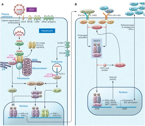
Innate recognition of HCV in hepatocytes occurs through dsRNA sensor protein kinase R (PKR), RIG-I, and TLR-3 (Figure 1A). PKR binds to the HCV internal ribosomal entry site (IRES) as early as 2 hours after infection and prior to the interaction with RIG-I; both pathways result in the recruitment of mitochondrial antiviral signaling (MAVS, also known as CARDIF/IPS-1/VISA) and tumor necrosis factor receptor–associated factor 3 (TRAF3) (16). PKR preferentially induces IFN-stimulated genes (ISGs) including the ubiquitin-like modifier ISG15 that negatively regulates RIG-I ubiq-uitylation. ISG15 induction inhibits the ability of RIG-I to recruit MAVS and TRAF3, and thereby may lead to a net proviral effect (16, 17). The latter is supported by recent data indicating that pharma-cological PKR inhibition decreases HCV replication and increases IFN induction (18). RIG-I binds the polyuridine motif of the HCV genome 3′ nontranslated region, i.e., HCV pathogen–associated molecular patterns (PAMPs), leading to the recruitment of a sig-naling complex that activates transcription factors and the pro-duction of type I and III IFNs as well as proinflammatory cytokines (refs. 19, 20, and Figure 1A). The signals driving this response are relayed through MAVS localized within both mitochondria and peroxisomes (21). The ER contains a specialized domain, the mitochondrial-associated membrane (MAM), which physically links the ER to mitochondria and has been implicated in NLRP3 inflammasome signaling (21). Once sufficient viral proteins have accumulated in the cytosol, HCV uses its multifunctional NS3/4A protease, essential for HCV replication, to target the MAM-an-chored synapse, cleaving MAVS from the MAM (but not from the mitochondria) and ablating RIG-I–mediated innate immune signaling (21). Another independent signaling pathway involves the binding of activated TLR-3 to the adaptor TRIF (Toll/inter-leukin-1 receptor domain–containing adapter–inducing IFN-β), which can also be cleaved by NS3/4A (19). Thus, the NS3 serine protease inhibitors that are part of the current triple-therapy reg-imen inhibit replication but would also be expected to restore innate responses within hepatocytes. Signaling from either MAVS
Emerging concepts in immunity
to hepatitis C virus infection
Hugo R. Rosen
Division of Gastroenterology and Hepatology, Departments of Medicine and Immunology, University of Colorado and Denver VA, Aurora, Colorado, USA.
Since the discovery of hepatitis C virus (HCV) by molecular cloning almost a quarter of a century ago,
unprece-dented at the time because the virus had never been grown in cell culture or detected serologically, there have been
impressive strides in many facets of our understanding of the natural history of the disease, the viral life cycle, the
pathogenesis, and antiviral therapy. It is apparent that the virus has developed multiple strategies to evade immune
surveillance and eradication. This Review covers what we currently understand of the temporal and spatial
immu-nological changes within the human innate and adaptive host immune responses that ultimately determine the
outcomes of HCV infection.
Conflict of interest: The author has declared that no conflict of interest exists.
or TRIF leads to the activation of various transcription factors, which in turn induce the production of type I and type III IFNs (via IFN regulatory factors [IRFs]), as well as proinflammatory cytokines and chemokines (via NF-κB and AP-1).
Type III IFNs, which consist of four IFN-λs, are antiviral cytokines that display type I IFN-like (IFN-α/β–like) antiviral activity (22, 23), but are structurally and genetically closer to the
[image:3.585.42.543.76.512.2]members of IL-10 family of cytokines. Type III IFNs signal through a heterodimeric receptor composed of IL-28Rα (also known as IFN-λR1) and the IL-10 receptor β chain (IL-10Rβ) (ref. 24 and Figure 1). In contrast to the nearly ubiquitous expression of the IFN-α receptor (IFN-αR), IL-28Rα has been found primarily on epithelial cells and dendritic cells (DCs) (24). Following both in vitro and in vivo HCV infection, type III IFNs are upregulated at
Figure 1
review
the mRNA and protein levels to an even greater extent than type I IFNs (25, 26). IFNs activate the JAK/STAT pathway, culminating in the induction of hundreds of ISGs encoding effector proteins that include ISG56, IFITM1, viperin, and 2′–5′ oligoadenylate synthase (OAS1), which restrict HCV infection within hepatocytes (27, 28), as well as PKR and IRF-7. Moreover, the promoters of early ISGs can be stimulated directly by IRF-3 (16). The relative requirement for IRF-3, IRF-7, or both vary according to cell and IFN type (29). Notably, type III IFNs, in addition to inducing well-known ISGs, activate a distinct set of genes in primary human hepatocytes from the type I IFNs (including those involved in chemotaxis and anti-gen presentation) with different kinetics of induction, suggesting divergent signaling pathways following receptor engagement (25).
Multiple proteins expressed by HCV have evolved important interactions with host cell proteins that benefit the viral cycle either directly or indirectly by disarming antiviral responses (reviewed in refs. 10, 30, 31). In addition to actions of the NS3/4A protease described above, for example, HCV core protein inter-feres with STAT signaling (via the induction of SOCS1/3) and may contribute to IFN resistance by diminished binding of ISGF3 to nuclear IFN–stimulated response elements (ISREs) (Figure 1B and ref. 19). An additional negative feedback loop is exemplified by USP18, a protease that decreases ISGylation of cellular pro-teins, thereby attenuating tyrosine phosphorylation of STAT1 and the expression of ISGs, decreasing the antiviral activity of IFNs (32). Paradoxically, higher hepatic ISG expression is predic-tive of a nonresponse to IFN-α treatment (33, 34), and a number of mechanisms have been proposed. The induction of type IIIs as the predominant antiviral pathway and driver of ISG induction (25, 29) may render hepatocytes refractory to further type I IFN action, conceptually supported by the observation that blocking type III IFN enhances the antiviral activity of exogenous IFN-α (ref. 25 and Figure 1B).
Association of genetic variation in IFN-λ genes and HCV recovery
One of the most ground-breaking discoveries in the area of HCV host response within the past five years has been that SNPs in chromosome 19 within or near the IFNL3 gene (encoding IFN-λ3, also known as IL-28B) are highly predictive of both antiviral suc-cess and spontaneous recovery in untreated patients (35, 36). As described above, in humans, four genes encode the members of the type III IFN family, i.e., IFN-λ1 (IFNL1, also known as IL29), IFN-λ2 (IFNL2 or IL28A), IFN-λ3 (IFNL3), and IFN-λ4 (IFNL4). Among these, IFN-λ3 is the most potent in inhibiting JFH-1 rep-lication (25, 37). A genome-wide association study that examined the frequencies of about 600,000 SNPs demonstrated that patients homozygous for the C allele at the rs12979860 SNP of IFNL3 had a 2-fold greater chance of cure with pegylated IFN and ribavirin compared with those with the TT genotype (35). The advanta-geous allele is more frequently found in mixed–European descent and Asian populations relative to African-Americans, and much of the race-related impairment in response to antiviral therapy is explained by population differences in the incidence of IFNL3
genotypes. Subsequent reports have confirmed that the IFNL3
genotype is the most important baseline predictor of sustained virologic response (SVR) after treatment with pegylated IFN plus ribavirin in patients with genotype 1 infection (38). The effect of the IFNL3 genotype is evident within the first 48 hours following treatment initiation, indicating that IFN-λ3 somehow primes the
host response to HCV, decreasing the threshold for virologic con-trol with treatment (24). The favorable IFNL3 variant is associated with lower ISG expression in pretreatment liver biopsies (39), and exogenous IFN-α induces a rapid antiviral state (40). Interestingly, serum levels of IFN-γ–induced protein 10 (IP-10 or CXCL10), a well-characterized marker of HCV (41) that may antagonize T cell recruitment in chronic infection (42), enhance the predictive value of the IFNL3 genotype in patients receiving dual therapy (43). In addition, the dinucleotide ss469415590 variant of IFNL4 (44), a newly identified gene upstream of IFNL3, provides an even greater prediction of impaired viral kinetics among African-Americans than rs12979860 variants of IFNL3 (44). Recent data suggest that while IFN-λ4 induces weak expression of ISGs that could provide an antiviral response to lower HCV load, it uniquely induces genes involved in HCV-related liver injury (e.g., chemokine CCL5 and the proto-oncogene FOS) and also reduces the responsiveness to type I and type III IFNs that are required for efficient HCV clearance (44).
The multicellular immune response to HCV
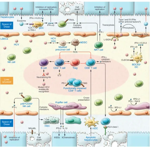
DCs. The orchestration of diverse, multifunctional cell types fol-lowing HCV infection (Figure 2) ultimately governs the outcome of infection, and these cell types represent potential targets for pharmacologic and immunotherapeutic approaches (31). DCs play crucial roles in innate pathogen sensing as well as in the initia-tion of adaptive immunity (45). Several major subsets of DCs have been identified, including plasmacytoid DCs (pDCs) and myeloid DCs (mDCs). pDCs are the main producer of type I IFNs, synthe-sizing up to 109 IFN molecules per cell within 12 hours after
acti-vation (46). IFN production by pDCs requires cell-to-cell contact and is proportional to the number of HCV-infected hepatocytes; further, IFN production is mediated by TLR7 activation indepen-dently of HCV RNA replication within pDCs (47). Recently, HCV RNA–containing exosomes produced by infected hepatocytes have been shown to transfer their RNA to pDCs that subsequently respond by secreting IFN-α (48); moreover, HCV subgenomic rep-licon cells that replicate viral RNA without producing infectious virus particles can also trigger type I IFNs. Recent work from our group (49) has demonstrated that intracellular sensing of the HCV PAMP by pDCs leads to robust types I and III IFN production and is mediated by signaling through RIG-I, challenging the dogma that TLR, and not RLR, signaling within pDC predominates (50). Furthermore, conditioned medium from HCV PAMP–transfected human pDCs greatly inhibits HCV replication and induces STAT1 and IRF9 within Huh 7.5.1 cells. Although these collective data implicate sentinel roles for pDCs in HCV sensing, further work is necessary to understand the characteristics of pDCs that favor acti-vation, which occurs in only a small subset, and the mechanisms that lead to their depletion or functional impairment in chronic infection, as described in some but not all studies (48, 51–54). In this regard, the ligation of C-type lectin immunoreceptors, blood DC antigen 2 (BDCA2), and DC immunoreceptor (DCIR) on pDCs by HCV E2 glycoprotein antagonizes the production of IFNs (55), and the relative expression of these receptors likely affects the ability of pDCs to respond to HCV within the hepatic microenvironment. Another C-type lectin, CLEC9A, is expressed by a distinct population of DCs, blood DC antigen 3 (BDCA3)+,
Figure 2
review
favorable response also correlates with greater type III IFN pro-duction than the less favorable IFNL3 genotypes, shedding some light on the puzzling lack of association between IFNL3 SNPs and hepatic expression of IFN-λs (57). Although hepatocytes com-prise most of the mRNA message from liver biopsies, recent data suggest that there may be cell-specific effects on expression, and BDCA3+ DCs and pDCs are potent sources of IFN-λs (56). This
concept is further supported by the recent demonstration that donor and recipient IFNL3 polymorphisms differentially affect HCV outcomes following liver transplantation (58).
NK cell phenotype and function associated with HCV infection outcomes. NK cells constitute an early host defense against viral pathogens (59–61), eliminating virus-infected cells both directly via cytolytic mechanisms and indirectly by secreting cytokines such as IFN-γ (62, 63). Although NK cells have been classically viewed as innate immune cells, their effects can extend into periods of adaptive immunity, and hepatic NK cells demonstrate adaptive immu-nity to structurally diverse antigens (64, 65). NK cell activity is stringently controlled by activating and inhibitory NK receptors (NKRs). NKRs include the predominantly inhibitory killer Ig–like receptors (KIRs), C-type lectin-like receptors of the NKG2 fam-ily comprising inhibitory (NKG2A) and activatory (NKG2C/D) isoforms, as well as the natural cytotoxicity receptors (NCRs) such as NKp30 (also known as NCR3), NKp44 (also known as NCR2), and NKp46 (also known as NCR1) that deliver activating signals (66–68). Emerging data indicate that NK cells play central roles in every stage of HCV infection, from the protection against infection in injection drug users (IDUs) to the prediction of antiviral success or failure with IFN-based therapies.
By convention, CD56+ NK cells have been assigned to
func-tional categories on the basis of the relative cell surface density of CD56; namely, CD56dimCD16+ NK cells, predominant in
periph-eral blood, display potent cytolytic activity, whereas the poorly cytolytic CD56bright NK cells enriched in tissue are responsible
for cytokine production, although recent data call into question this simple classification (69). IDUs repeatedly exposed to HCV infection but who remain uninfected have proportionally higher circulating frequencies of CD56dim mature effector NK cells (70).
The balance between positive and negative signals from activating and inhibitory cell surface receptors is likely to result in NK cell functional changes (71). In our IDU cohort, there was a correlation between the frequency of NK cells expressing the activating recep-tor NKp30 and in vitro lymphokine-activated killing. NKp30, which is induced by IL-2, is most highly expressed on the NK and NKT cells of exposed, uninfected subjects (70). The NKp46 recep-tor is considered the major human natural cytotoxicity receprecep-tor involved in NK cell–mediated killing (72, 73) and is more highly expressed on the NK cells of women and people of mixed Euro-pean descent (i.e., populations known to demonstrate higher rates of spontaneous resolution of HCV infection) (74–76). NKp46 ligand expression is induced on hepatocytes following HCV infec-tion (76), although whether this represents a specific HCV compo-nent or an unspecific stress response is not yet clear. TLR stimula-tion of purified NKp46hi is associated with increased transcription
of cytotoxicity-related genes as compared with NKp46lo
counter-parts. Two recent studies (76, 77) with further discussion in an editorial (78) highlight that NKp46hi NK cells (or ligation with
an agonist NKp46 antibody) have increased anti-HCV activity in vitro, a process mediated by IFN-γ; accordingly, intrahepatic accu-mulation of NKp46hi NK cells is inversely correlated with HCV
RNA levels (77). An inverse correlation also exists with the stage of fibrosis and ex vivo intrahepatic NKp46hi frequencies; in vitro,
blockade of NKp46 reduces NK cell–mediated killing of human hepatic stellate cells (HSCs), implicating potentially important antifibrotic roles (77) for this population of NK cells (Figure 2).
NK cells are rapidly activated by cytokine stimulation, and thus, studies have examined whether the response to anti-HCV therapy (the major component of which is IFN-α) would be predicted by the phenotype and function of NK cells. Pretreatment expansion of CD56– NK cells functionally skewed toward MIP-1β production
(rather than IFN-γ) is a predictor of failure to pegylated IFN-α and ribavirin (79). Patients with impaired viral kinetics following initi-ation of antiviral therapy have NK cells with higher expression lev-els of inhibitory receptors (80), and a multivariate model based on two inhibitory receptors (NKG2A and CD158e) is highly predictive of SVR. Pegylated IFN-α transcriptionally upregulates ISGs and TNF-related apoptosis–inducing ligand (TRAIL) in NK cells. HCV also enhances the susceptibility of primary human hepatocytes to TRAIL-mediated killing via increased expression of the death receptors DR4 and DR5 (81). The ex vivo expression of TRAIL and CD107a on NK cells increases as early as hours following the ini-tiation of antiviral therapy (82, 83), correlates with the induction of phosphorylated STAT1 levels (84), and is associated with early virologic response. Thus, on aggregate, these data identify NK cell markers associated with protection from early infection and pre-dictors of response to IFN-based therapy. Moreover, NK cells can demonstrate regulatory and reciprocal interactions with B and T cells, DCs, macrophages, and endothelial cells (ECs), thus func-tioning to amplify or attenuate immune responses (60).
Liver-resident macrophages and ECs. Kupffer cells (KCs) constitute the first macrophage population with which pathogens, bacterial endotoxins, and microbial debris derived from the gastrointestinal tract come into contact, and together with the liver sinusoidal ECs (LSECs), make up the hepatic reticuloendothelial system (ref. 85 and Figure 2). ISG upregulation within KCs is a strong positive and independent predictor of subsequent response to antiviral therapy with pegylated IFN and ribavirin, whereas hepatocyte ISG expression was associated with nonresponse (86), underscoring the importance of examining different patterns of cellular activation. HCV enters KCs (but not undifferentiated monocytes) via a phago-cytic uptake that is independent of productive infection, leading to the induction of IFN-β–dependent innate immune responses through RIG-I/MAVS, as well as inflammatory IL-1β–dependent signaling through TLR7/MyD88-dependent and NLRP3 inflam-masome pathways (87). The induction of proinflammatory genes may lead to the recruitment of immune cells to the liver and aug-ment the processes that result in liver fibrosis and cirrhosis.
and the development of viral persistence (89, 91, 92). These effects are largely mediated through phosphoinositide 3-kinase (PI3K), indicating shared regulation between MyD88- and TRIF-depen-dent TLR signaling pathways (89). Galectin-9 (Gal-9), another important inhibitory ligand (93), is expressed robustly by KCs and circulates at high levels in HCV-infected patients (94). Gal-9 binds to T cell Ig and mucin domain–containing molecule 3 (TIM3), inducing apoptosis of HCV-specific CD8+ T cells and expanding
Tregs (94). Further, KC-derived Gal-9 mediates T cell dysfunction and predicts poor prognosis in patients with hepatitis B–related hepatocellular carcinoma (95). Gal-9 has pleiotropic roles that include the inhibition and regulation of NK cell functions that may impact viral persistence (96).
LSECs function to clear waste molecules that have entered the circulation (97) and comprise approximately 50% of nonparenchy-mal cells in the liver. Although long recognized as central to the induction of cytotoxic CD8+ T cell (CTL) tolerance and apoptosis
(98, 99), an understanding of the roles in antiviral responses of LSECs, which express a wide array of PRRs (100), is just begin-ning to emerge. Their strategic anatomic position (Figure 2) places LSECs as the first cells in contact with blood flow in the sinus-oids (101). Expression of the C-type lectin L-SIGN by LSECs was postulated to mediate capture of HCV particles and transcytosis of the virus across the endothelial barrier, thereby
concentrat-ing infectious particles and facilitatconcentrat-ing their direct contact with hepatocytes (102). Moreover, LSECs are highly efficient scavengers that pinocytose particles less than 0.2 μm in size, which typically encompasses virus-sized particles (103). The recent demonstration that LSECs, rather than KCs, clear the bulk of blood-borne human adenovirus underscores their importance during the viremic phase of any natural viral infection (103). Even if LSECs do not sus-tain active replication, viral sensing and its downstream effector responses, including exosomal transfer of ISG products (ref. 104 and Figure 2), may impact other processes within the hepatic microenvironment and warrant further investigation.
[image:7.585.45.286.78.453.2]B cells and humoral immunity. Whether the humoral response plays an important role in controlling HCV infection remains contro-versial (105, 106); indeed, the fact that hypogammaglobulinemic humans can spontaneously eradicate HCV (107) may suggest that antibody responses are dispensable. Studies showing a lack of association between neutralizing antibodies (NAbs) and viral clearance were confounded by poorly defined viral inoculum and heterogeneous patient populations. However, in a study of healthy young women infected with identical, single-source viral inocu-lum, spontaneous resolvers demonstrated an early induction of NAb responses directed against HCV envelope glycoproteins, including broader cross-neutralization of heterologous strains, whereas chronically evolving subjects had delayed and low-titered NAb responses (108). The use of retroviral pseudoparticles bearing autologous HCV glycoproteins (HCVpp) from the initial inocu-lum allowed the precise examination of strain-specific neutraliza-tion. The late phase of chronic infection is accompanied by the induction of NAb responses as well as an increasing rate of enve-lope viral sequence evolution (106), indicative of ongoing humoral selection pressure. Viral escape may occur through several addi-tional mechanisms (108), including an impaired cross-neutral-izing activity that selects the outgrowth of viral variants and the interplay of HCV glycoproteins with HDL and the scavenger recep-tor SR-BI (Figure 1A), which can prevent the effect of NAbs and enhance cell entry of HCV and infection (109). Only a minority of patients with resolved HCV infection exhibit NAbs 10–17 years after viral clearance (108), which may underlie the absent or lim-ited protective immunity against reinfection (110). Moreover, human liver chimeric mice infused with HCV E2–specific human monoclonal antibodies directed against a conserved epitope are protected against heterologous HCV (111), raising the possibility
Figure 3
review
that future approaches using a combination of such NAbs and blocking receptors involved in viral entry (112) might demonstrate efficacy in high-risk populations (e.g., to prevent allograft reinfec-tion in patients who have undergone liver transplantareinfec-tion or in healthcare workers with needle-stick injuries).
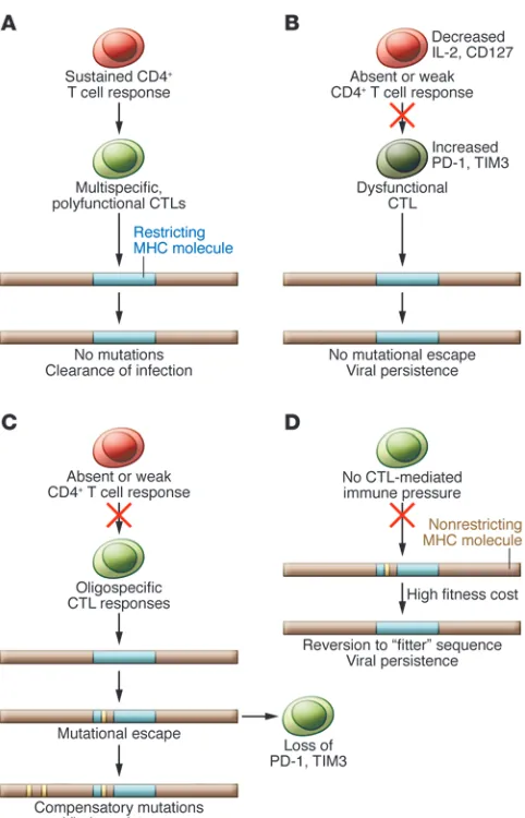
CTLs in HCV infection. CTLs are the primary effector cells that mediate viral clearance through the secretion of antiviral cytokines, which is about 100- to 1,000-fold more effective than cytolytic activ-ity (10) (113). In the absence of viral clearance, the display of HCV peptides persists on the surface of hepatocytes, and the continued presence of CTLs results in significant immune-mediated liver injury (31). Further, the secretion of inflammatory and profibrotic cytokines such as TGF-β activates stellate cells, the primary source of fibrosis, ultimately leading to progressive liver damage. Chim-panzee and human studies were highly instructive in understanding the distinguishing features of CTL responses that provide perma-nent viral control versus viral persistence (10, 114–118), includ-ing the recognition that CD4+ T cells are critical both for limiting
immune evasion and priming effector memory CTLs (119, 120). A complex interplay between the opposing forces of immune selection pressure and viral fitness costs drives mutations in HCV (Figure 3). The high rate of replication coupled with the lack of
proofreading ability of its polymerase provides the HCV genome with a means to escape HLA-restricted immune responses (10, 121). The ability of HCV to mutate at an amino acid residue is con-strained, however, by fitness costs, and compensatory mutations may be required to restore HCV replicative capacity (121–123).
Anergy occurs when T cells are initially primed improperly by sig-naling through the TCR in the absence of costimulatory or inflam-matory signals (124, 125), which results in a failure to develop proper function from the outset. In contrast, exhausted T cells are primed by antigen, costimulation, and inflammation. Although these T cells initially develop effector functions, prolonged, exces-sive stimulation leads to the progresexces-sive loss of function over time (124). Compelling evidence exists for both anergy and exhaustion in chronic HCV infection. Programmed cell death protein 1 (PD-1) is an immunoreceptor tyrosine–based inhibition motif–contain-ing (ITIM-containmotif–contain-ing) receptor expressed on activated T cells that mediates hyporesponsiveness. PD-1 is upregulated in chronic infection within the intrahepatic compartment (91, 126) and its ligand PD-L1 is expressed by a number of nonparenchymal cells in the liver including KCs, LSECs, and DCs, as well as IFN-exposed hepatocytes (10). Moreover, PD-1 expression on HCV-specific CTLs inversely correlates with early and sustained virologic response to combination IFN-based antiviral therapy (127) and is associated with impaired control of replication in vitro (128). In association with PD-1, the coexpression of multiple inhibitory receptors, including 2B4, CD160, KLRG1, and TIM3, identifies HCV-spe-cific CTLs with impaired proliferation and cytokine production and correlates with the absence of sequence variation within cog-nate epitopes, consistent with the necessity for ongoing antigen triggering (129, 130). In this regard (131), the frequency of dual TIM3posPD-1pos HCV–specific CTLs is predictive of viral persistence
in patients with acute HCV infection and is increased within the central memory subset (compared with the effector memory popu-lation) and within the hepatic compartment relative to the periph-eral compartment. Increased levels of TIM3 and PD-1 expression are associated with CTL dysfunction, with TIM3hi/PD-1hi CTLs
pro-ducing less IFN-γ, TNF-α, and CD107a than their TIM3lo/PD-1lo
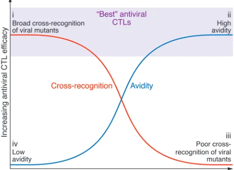
counterparts. Importantly, blockade of different coinhibitory path-ways may differentially enhance CTL effector functions. CTL avid-ity, also termed “functional avidity” (132, 133), refers to the overall sensitivity of the T cell response to antigen density. High-avidity CTLs are those that secrete substantial amounts of IFN-γ when stimulated with relevant APCs loaded with low concentrations of peptide. Considerable data in other model systems demonstrate the immense potential of high-avidity CTLs to preferentially control certain viral infections as well as tumors (134). In infections that can rapidly evolve escape mutations, such as with HCV, another potentially important attribute of CTLs is the ability to cross-recog-nize mutated epitopes (135–137). From a standpoint of the design of novel immunotherapeutic approaches, the use of TCRs derived from CTLs demonstrating relatively higher avidity and cross-reac-tivity would seem optimal (Figure 4). HCV TCR transduction of human T cells renders them responsive to HCV peptide–loaded hepatocytes (138) and inhibits viral replication in vitro (139).
Future directions
The past five years have seen a remarkable degree of progress in the understanding of HCV pathogenesis, and we are in the midst of an extraordinary phase of development with direct-acting antiviral agents that may cure the majority of patients undergoing
antivi-Figure 4
[image:8.585.45.284.88.262.2]1. Rosen HR. Clinical practice. Chronic hepatitis C infection. N Engl J Med. 2011;364(25):2429–2438. 2. Mitchell AE, Colvin HM, Palmer Beasley R.
Insti-tute of Medicine recommendations for the preven-tion and control of hepatitis B and C. Hepatology. 2010;51(3):729–733.
3. Davis GL, Albright JE, Cook SF, Rosenberg DM. Projecting future complications of chronic hep-atitis C in the United States. Liver Transpl. 2003; 9(4):331–338.
4. Biggins SW, et al. Projected future increase in aging hepatitis C virus-infected liver transplant candidates: a potential effect of hepatocellular car-cinoma. Liver Transpl. 2012;18(12):1471–1478. 5. Aronsohn A, Jensen D. Distributive justice and the
arrival of direct-acting antivirals: who should be first in line? Hepatology. 2011;53(6):1789–1791. 6. Dabbouseh NM, Jensen DM. Future therapies for
chronic hepatitis C. Nat Rev Gastroenterol Hepatol. 2013;10(5):268–276.
7. Gane EJ, et al. Nucleotide polymerase inhibitor sofosbuvir plus ribavirin for hepatitis C. N Engl J Med. 2013;368(1):34–44.
8. Choo QL, Kuo G, Weiner AJ, Overby LR, Brad-ley DW, Houghton M. Isolation of a cDNA clone derived from a blood-borne non-A, non-B viral hep-atitis genome. Science. 1989;244(4902):359–362. 9. Rice CM. New insights into HCV replication:
potential antiviral targets. Top Antivir Med. 2011; 19(3):117–120.
10. Rehermann B. Hepatitis C virus versus innate and adaptive immune responses: a tale of coevolution and coexistence. J Clin Invest. 2009;119(7):1745–1754. 11. Bartenschlager R, Pietschmann T. Efficient hepa-titis C virus cell culture system: what a difference the host cell makes. Proc Natl Acad Sci U S A. 2005; 102(28):9739–9740.
12. Li K, et al. Immune evasion by hepatitis C virus NS3/4A protease-mediated cleavage of the Toll-like receptor 3 adaptor protein TRIF. Proc Natl Acad Sci U S A. 2005;102(8):2992–2997.
13. Wakita T, et al. Production of infectious hepatitis C virus in tissue culture from a cloned viral genome.
Nat Med. 2005;11(7):791–796.
14. Zhong J, et al. Robust hepatitis C virus infection in vitro. Proc Natl Acad Sci U S A. 2005;102(26):9294–9299. 15. Lindenbach BD, et al. Complete replication of
hepatitis C virus in cell culture. Science. 2005;
309(5734):623–626.
16. Arnaud N, et al. Hepatitis C virus reveals a novel early control in acute immune response. PLoS Pathog. 2011;7(10):e1002289.
17. Kim MJ, Hwang SY, Imaizumi T, Yoo JY. Negative feedback regulation of RIG-I-mediated antiviral sig-naling by interferon-induced ISG15 conjugation.
J Virol. 2008;82(3):1474–1483.
18. Arnaud N, et al. Hepatitis C virus controls inter-feron production through PKR activation. PLoS One. 2010;5(5):e10575.
19. Horner SM, Gale M. Intracellular innate immune cascades and interferon defenses that control hepatitis C virus. J Interferon Cytokine Res. 2009; 29(9):489–498.
20. Saito T, Owen DM, Jiang F, Marcotrigiano J, Gale M. Innate immunity induced by composition-de-pendent RIG-I recognition of hepatitis C virus RNA. Nature. 2008;454(7203):523–527.
21. Horner SM, Liu HM, Park HS, Briley J, Gale M. Mitochondrial-associated endoplasmic reticulum membranes (MAM) form innate immune synapses and are targeted by hepatitis C virus. Proc Natl Acad Sci U S A. 2011;108(35):14590–14595.
22. Zhou Z, Hamming OJ, Ank N, Paludan SR, Nielsen AL, Hartmann R. Type III interferon (IFN) induces a type I IFN-like response in a restricted subset of cells through signaling pathways involving both the Jak-STAT pathway and the mitogen-activated protein kinases. J Virol. 2007;81(14):7749–7758. 23. Choubey D, Moudgil KD. Interferons in
autoim-mune and inflammatory diseases: regulation and roles. J Interferon Cytokine Res. 2011;31(12):857–865. 24. Balagopal A, Thomas DL, Thio CL. IL28B and the control of hepatitis C virus infection. Gastroenterol-ogy. 2010;139(6):1865–1876.
25. Thomas E, et al. HCV infection induces a unique hepatic innate immune response associated with robust production of type III interferons. Gas-troenterology. 2012;142(4):978–988.
26. Park H, et al. IL-29 is the dominant type III inter-feron produced by hepatocytes during acute hepatitis C virus infection. Hepatology. 2012; 56(6):2060–2070.
27. Helbig KJ, et al. The antiviral protein viperin inhib-its hepatitis C virus replication via interaction with nonstructural protein 5A. Hepatology. 2011; 54(5):1506–1517.
28. Raychoudhuri A, Shrivastava S, Steele R, Kim H, Ray R, Ray RB. ISG56 and IFITM1 proteins inhibit hepatitis C virus replication. J Virol. 2011; 85(24):12881–12889.
29. Osterlund PI, Pietila TE, Veckman V, Kotenko SV, Julkunen I. IFN regulatory factor family members dif-ferentially regulate the expression of type III IFN (IFN-lambda) genes. J Immunol. 2007;179(6):3434–3442. 30. Yamane D, McGivern DR, Masaki T, Lemon SM.
Liver injury and disease pathogenesis in chronic hepatitis C. Curr Top Microbiol Immunol. 2013; 369:263–288.
31. Mengshol JA, Golden-Mason L, Rosen HR. Mecha-nisms of disease: HCV-induced liver injury. Nat Clin Pract Gastroenterol Hepatol. 2007;4(11):622–634. 32. Randall G, et al. Silencing of USP18 potentiates
the antiviral activity of interferon against hep-atitis C virus infection. Gastroenterology. 2006; 131(5):1584–1591.
33. Sarasin-Filipowicz M, et al. Alpha interferon induces long-lasting refractoriness of JAK-STAT signaling in the mouse liver through induction of USP18/UBP43. Mol Cell Biol. 2009; 29(17):4841–4851.
34. Afdhal NH, et al. Hepatitis C pharmacogenetics: state of the art in 2010. Hepatology. 2011;53(1):336–345. 35. Ge D, et al. Genetic variation in IL28B predicts
hep-atitis C treatment-induced viral clearance. Nature. 2009;461(7262):399–401.
36. Thomas DL, et al. Genetic variation in IL28B and spontaneous clearance of hepatitis C virus. Nature. 2009;461(7265):798–801.
37. Marcello T, et al. Interferons alpha and lambda inhibit hepatitis C virus replication with distinct signal transduction and gene regulation kinetics.
Gastroenterology. 2006;131(6):1887–1898. 38. Thompson AJ, et al. Interleukin-28B
polymor-phism improves viral kinetics and is the strongest pretreatment predictor of sustained virologic response in genotype 1 hepatitis C virus. Gas-troenterology. 2010;139(1):120–129.e18.
39. Urban TJ, et al. IL28B genotype is associated with differential expression of intrahepatic interferon-stimulated genes in patients with chronic hepatitis C. Hepatology. 2010;52(6):1888–1896.
40. Sarasin-Filipowicz M, et al. Interferon signaling and treatment outcome in chronic hepatitis C. Proc Natl Acad Sci U S A. 2008;105(19):7034–7039.
ral therapy. However, as antiviral therapy will not be accessible for most patients in resource-limited regions, the development of a safe and effective HCV vaccine is paramount. HCV poses multi-ple important obstacles to vaccine development (140), including a considerable primary sequence divergence that is greater than HIV (141). In vaccinated animals, despite initial control, viral persistence is associated with T cell immune escape (142), higher viral mutation rates (143, 144), and expression of inhibitory recep-tors, which suggest that in the absence of rapid viral clearance, an environment for selective pressure or T cell exhaustion remains. Longitudinal liver biopsies revealed correlations between higher PD-1 expression and failure to control viremia, pointing to the possibility that strong induction and activation of CTLs may be counterproductive if they target promiscuous HCV epitopes that do not impact viral fitness (145). HCV is able to suppress the early innate immune response both within hepatocytes and innate lymphocytes, such as NK cells, and can attenuate or escape T cell responses. The nature and extent of immune restoration that occurs with regimens not including IFN-α, which is known to have antiproliferative effects (146), remain to be defined. Greater under-standing of the complex crosstalk between different cell types within the hepatic microenvironment may soon allow the
identi-fication of targeted approaches to address the sequelae of chronic infection, including fibrosis and the development of cancer.
Acknowledgments
I would like to thank Michael Gale and Stacy Horner (University of Washington) Emmanuel Thomas (University of Miami), Todd Allen (Massachusetts General Hospital/Broad Institute), Arash Grakoui (Emory University), Michael Nishimura (Loyola Uni-versity), Martha Alexander-Miller (Wake Forest School of Medi-cine), Stephen Polyak (University of Washington), Young Hahn (University of Virginia), Robert Thimme (University of Freiburg), and members of my research laboratory for their helpful discus-sions. I would also like to acknowledge support from NIH grants RO1DK60590, RO1DK071560, U19 AI1066328, R21AI103361, R56AI100991, K24AI083742, and R01HD075549,and from VA Merit Review grants.
review
41. Beinhardt S, et al. Serum level of IP-10 increases predictive value of IL28B polymorphisms for spontaneous clearance of acute HCV infection.
Gastroenterology. 2012;142(1):78–85.e2.
42. Casrouge A, et al. Evidence for an antagonist form of the chemokine CXCL10 in patients chronically infected with HCV. J Clin Invest. 2011;121(1):308–317. 43. Darling JM, et al. Quantitation of pretreatment
serum interferon-gamma-inducible protein-10 improves the predictive value of an IL28B gene polymorphism for hepatitis C treatment response.
Hepatology. 2011;53(1):14–22.
44. Prokunina-Olsson L, et al. A variant upstream of IFNL3 (IL28B) creating a new interferon gene IFNL4 is associated with impaired clearance of hepatitis C virus. Nat Genet. 2013;45(2):164–171. 45. Lande R, Gilliet M. Plasmacytoid dendritic cells: key
players in the initiation and regulation of immune responses. Ann N Y Acad Sci. 2010;1183:89–103. 46. Hagberg N, et al. IFN-alpha production by
plas-macytoid dendritic cells stimulated with RNA-containing immune complexes is promoted by NK cells via MIP-1beta and LFA-1. J Immunol. 2011; 186(9):5085–5094.
47. Takahashi K, et al. Plasmacytoid dendritic cells sense hepatitis C virus-infected cells, produce inter-feron, and inhibit infection. Proc Natl Acad Sci U S A. 2010;107(16):7431–7436.
48. Dreux M, et al. Short-range exosomal transfer of viral RNA from infected cells to plasmacytoid dendritic cells triggers innate immunity. Cell Host Microbe. 2012;12(4):558–570.
49. Stone AE, et al. Hepatitis C virus pathogen asso-ciated molecular pattern (PAMP) triggers produc-tion of lambda-interferons by human plasmacytoid dendritic cells. PLoS Pathog. 2013;9(4):e1003316. 50. Takeuchi O, Akira S. MDA5/RIG-I and virus
recog-nition. Curr Opin Immunol. 2008;20(1):17–22. 51. Sagan SM, Sarnow P. Plasmacytoid dendritic cells
as guardians in hepatitis C virus-infected liver. Proc Natl Acad Sci U S A. 2010;107(17):7625–7626. 52. Wertheimer AM, Bakke A, Rosen HR. Direct
enu-meration and functional assessment of circulating dendritic cells in patients with liver disease. Hepa-tology. 2004;40(2):335–345.
53. Liang H, et al. Differential effects of hepatitis C virus JFH1 on human myeloid and plasmacytoid dendritic cells. J Virol. 2009;83(11):5693–5707. 54. Shiina M, Rehermann B. Cell culture-produced
hepatitis C virus impairs plasmacytoid dendritic cell function. Hepatology. 2008;47(2):385–395. 55. Florentin J, et al. HCV glycoprotein E2 is a novel
BDCA-2 ligand and acts as an inhibitor of IFN pro-duction by plasmacytoid dendritic cells. Blood. 2012; 120(23):4544–4551.
56. Yoshio S, et al. Human blood dendritic cell antigen 3 (BDCA3) dendritic cells are a potent producer of interferon-lambda in response to hepatitis C virus.
Hepatology. 2013;57(5):1705–1715.
57. Honda M, et al. Hepatic ISG expression is associ-ated with genetic variation in interleukin 28B and the outcome of IFN therapy for chronic hepatitis C.
Gastroenterology. 2010;139(2):499–509.
58. Biggins SW, et al. Differential effects of donor and recipient IL28B and DDX58 SNPs on severity of HCV after liver transplantation. J Hepatol. 2013; 58(5):969–976.
59. Biron CA. Initial and innate responses to viral infections--pattern setting in immunity or disease.
Curr Opin Microbiol. 1999;2(4):374–381.
60. Vivier E, et al. Innate or adaptive immunity? The example of natural killer cells. Science. 2011; 331(6013):44–49.
61. Jost S, Altfeld M. Control of human viral infections by natural killer cells. Annu Rev Immunol. 2013; 31:163–194.
62. Biron CA, Brossay L. NK cells and NKT cells in innate defense against viral infections. Curr Opin
Immunol. 2001;13(4):458–464.
63. Cooper MA, Fehniger TA, Caligiuri MA. The biol-ogy of human natural killer-cell subsets. Trends Immunol. 2001;22(11):633–640.
64. Paust S, et al. Critical role for the chemokine recep-tor CXCR6 in NK cell-mediated antigen-specific memory of haptens and viruses. Nat Immunol. 2010; 11(12):1127–1135.
65. Jiang X, Chen Y, Peng H, Tian Z. Memory NK cells: why do they reside in the liver? Cell Mol Immunol. 2013;10(3):196–201.
66. Moretta A, et al. Activating receptors and corecep-tors involved in human natural killer cell-mediated cytolysis. Annu Rev Immunol. 2001;19:197–223. 67. Lanier LL. NK cell recognition. Annu Rev Immunol.
2005;23:225–274.
68. Moretta A, Biassoni R, Bottino C, Mingari MC, Moretta L. Natural cytotoxicity receptors that trig-ger human NK-cell-mediated cytolysis. Immunol Today. 2000;21(5):228–234.
69. De Maria A, Bozzano F, Cantoni C, Moretta L. Revisiting human natural killer cell subset func-tion revealed cytolytic CD56(dim)CD16+ NK cells as rapid producers of abundant IFN-γ on activa-tion. Proc Natl Acad Sci U S A. 2011;108(2):728–732. 70. Golden-Mason L, Cox AL, Randall JA, Cheng L, Rosen HR. Increased natural killer cell cytotoxicity and NKp30 expression protects against hepatitis C virus infection in high-risk individuals and inhibits repli-cation in vitro. Hepatology. 2010;52(5):1581–1589. 71. Golden-Mason L, Rosen HR. Natural killer cells:
primary target for hepatitis C virus immune eva-sion strategies? Liver Transpl. 2006;12(3):363–372. 72. Sivori S, et al. NKp46 is the major triggering
recep-tor involved in the natural cytotoxicity of fresh or cultured human NK cells. Correlation between surface density of NKp46 and natural cytotoxicity against autologous, allogeneic or xenogeneic target cells. Eur J Immunol. 1999;29(5):1656–1666. 73. Mandelboim O, et al. Recognition of
haemag-glutinins on virus-infected cells by NKp46 acti-vates lysis by human NK cells. Nature. 2001; 409(6823):1055–1060.
74. Thomas DL, et al. The natural history of hepatitis C virus infection: host, viral, and environmental factors. JAMA. 2000;284(4):450–456.
75. Rosen HR, et al. Selective decrease in hepatitis C virus-specific immunity among African Americans and outcome of antiviral therapy. Hepatology. 2007; 46(2):350–358.
76. Golden-Mason L, Stone AE, Bambha KM, Cheng L, Rosen HR. Race- and gender-related variation in NKp46 expression associated with differential anti-HCV immunity. Hepatology. 2012;56(4):1214–1222. 77. Kramer B, et al. Natural killer p46High expression defines a natural killer cell subset that is poten-tially involved in control of hepatitis C virus repli-cation and modulation of liver fibrosis. Hepatology. 2012;56(4):1201–1213.
78. Heeg M, Thimme R. NK cells and hepatitis C: NKp46 expression linked to antiviral and antifi-brotic activity. Hepatology. 2012;56(4):1197–1200. 79. Gonzalez VD, et al. Expansion of functionally
skewed CD56-negative NK cells in chronic hepa-titis C virus infection: correlation with outcome of pegylated IFN-alpha and ribavirin treatment.
J Immunol. 2009;183(10):6612–6618.
80. Golden-Mason L, et al. Natural killer inhibitory recep-tor expression associated with treatment failure and interleukin-28B genotype in patients with chronic hepatitis C. Hepatology. 2011;54(5):1559–1569. 81. Lan L, et al. Hepatitis C virus infection sensitizes
human hepatocytes to TRAIL-induced apoptosis in a caspase 9-dependent manner. J Immunol. 2008; 181(7):4926–4935.
82. Stegmann KA, et al. Interferon-alpha-induced TRAIL on natural killer cells is associated with con-trol of hepatitis C virus infection. Gastroenterology.
2010;138(5):1885–1897.
83. Ahlenstiel G, et al. Early changes in natural killer cell function indicate virologic response to inter-feron therapy for hepatitis C. Gastroenterology. 2011; 141(4):1231–1239.
84. Edlich B, et al. Early changes in interferon signal-ing define natural killer cell response and refrac-toriness to interferon-based therapy of hepatitis C patients. Hepatology. 2012;55(1):39–48.
85. Vollmar B, Menger MD. The hepatic microcircu-lation: mechanistic contributions and therapeutic targets in liver injury and repair. Physiol Rev. 2009; 89(4):1269–1339.
86. Chen L, et al. Cell-type specific gene expression signature in liver underlies response to interferon therapy in chronic hepatitis C infection. Gas-troenterology. 2010;138(3):1123–1123.
87. Negash AA, et al. IL-1beta production through the NLRP3 inflammasome by hepatic macro-phages links hepatitis C virus infection with liver inflammation and disease. PLoS Pathog. 2013; 9(4):e1003330.
88. Dolganiuc A, et al. Viral and host factors induce macrophage activation and loss of toll-like recep-tor tolerance in chronic HCV infection. Gastroenter-ology. 2007;133(5):1627–1636.
89. Tu Z, Pierce RH, Kurtis J, Kuroki Y, Crispe IN, Orloff MS. Hepatitis C virus core protein subverts the antiviral activities of human Kupffer cells. Gas-troenterology. 2010;138(1):305–314.
90. Sen GC, Sarkar SN. Transcriptional signaling by double-stranded RNA: role of TLR3. Cytokine Growth Factor Rev. 2005;16(1):1–14.
91. Golden-Mason L, Palmer B, Klarquist J, Mengshol JA, Castelblanco N, Rosen HR. Upregulation of PD-1 expression on circulating and intrahepatic hepatitis C virus-specific CD8+ T cells associated with reversible immune dysfunction. J Virol. 2007; 81(17):9249–9258.
92. Kassel R, Cruise MW, Iezzoni JC, Taylor NA, Pruett TL, Hahn YS. Chronically inflamed livers up-regu-late expression of inhibitory B7 family members.
Hepatology. 2009;50(5):1625–1637.
93. Anderson AC, et al. Promotion of tissue inflammation by the immune receptor Tim-3 expressed on innate immune cells. Science. 2007;318(5853):1141–1143. 94. Mengshol JA, et al. A crucial role for Kupffer
cell-de-rived galectin-9 in regulation of T cell immunity in hepatitis C infection. PLoS One. 2010;5(3):e9504. 95. Li H, et al. Tim-3/galectin-9 signaling pathway
mediates T cell dysfunction and predicts poor prog-nosis in patients with HBV-associated hepatocellu-lar carcinoma. Hepatology. 2012;56(4):1342–1351. 96. Golden-Mason L, et al. Galectin-9 functionally
impairs natural killer (NK) cells in humans and mice. J Virol. 2013;87(9):4835–4845.
97. Seternes T, Sorensen K, Smedsrod B. Scavenger endothelial cells of vertebrates: a nonperipheral leukocyte system for high-capacity elimination of waste macromolecules. Proc Natl Acad Sci U S A. 2002; 99(11):7594–7597.
98. Karrar A, Broome U, Uzunel M, Qureshi AR, Sumitran-Holgersson S. Human liver sinusoidal endothelial cells induce apoptosis in activated T cells: a role in tolerance induction. Gut. 2007; 56(2):243–252.
99. Tiegs G, Lohse AW. Immune tolerance: what is unique about the liver. J Autoimmun. 2010;34(1):1–6. 100. Wu J, et al. Toll-like receptor-induced innate immune responses in non-parenchymal liver cells are cell type-specific. Immunology. 2010;129(3):363–374. 101. Huebert RC, et al. Immortalized liver endothelial
cells: a cell culture model for studies of motility and angiogenesis. Lab Invest. 2010;90(12):1770–1781. 102. Cormier EG, et al. L-SIGN (CD209L) and
103. Ganesan LP, Mohanty S, Kim J, Clark KR, Robin-son JM, AnderRobin-son CL. Rapid and efficient clearance of blood-borne virus by liver sinusoidal endothe-lium. PLoS Pathog. 2011;7(9):e1002281.
104. Li J, et al. Exosomes mediate the cell-to-cell trans-mission of IFN-α-induced antiviral activity. Nat Immunol. 2013;14(8):793–803.
105. Rehermann B. Cellular immune response to the hep-atitis C virus. J Viral Hepat. 1999;6(suppl 1):31–35. 106. Liu L, Fisher BE, Dowd KA, Astemborski J, Cox AL,
Ray SC. Acceleration of hepatitis C virus envelope evolution in humans is consistent with progres-sive humoral immune selection during the transi-tion from acute to chronic infectransi-tion. J Virol. 2010; 84(10):5067–5077.
107. Christie JM, et al. Clinical outcome of hypogamma-globulinaemic patients following outbreak of acute hepatitis C: 2 year follow up. Clin Exp Immunol. 1997;110(1):4–8.
108. Pestka JM, et al. Rapid induction of virus-neutraliz-ing antibodies and viral clearance in a svirus-neutraliz-ingle-source outbreak of hepatitis C. Proc Natl Acad Sci U S A. 2007; 104(14):6025–6030.
109. Bartosch B, et al. An interplay between hypervari-able region 1 of the hepatitis C virus E2 glycopro-tein, the scavenger receptor BI, and high-density lipoprotein promotes both enhancement of infec-tion and protecinfec-tion against neutralizing antibodies.
J Virol. 2005;79(13):8217–8229.
110. Lai ME, et al. Hepatitis C virus in multiple episodes of acute hepatitis in polytransfused thalassaemic children. Lancet. 1994;343(8894):388–390. 111. Bukh J. Animal models for the study of hepatitis C
virus infection and related liver disease. Gastroenter-ology. 2012;142(6):1279–1287.e3.
112. Lacek K, et al. Novel human SR-BI antibodies pre-vent infection and dissemination of HCV in vitro and in humanized mice. J Hepatol. 2012;57(1):17–23. 113. Jo J, et al. Analysis of CD8+ T-cell-mediated inhibi-tion of hepatitis C virus replicainhibi-tion using a novel immunological model. Gastroenterology. 2009; 136(4):1391–1401.
114. Lechner F, et al. Analysis of successful immune responses in persons infected with hepatitis C virus. J Exp Med. 2000;191(9):1499–1512. 115. Smyk-Pearson S, Tester IA, Lezotte D, Sasaki AW,
Lewinsohn DM, Rosen HR. Differential antigenic hierarchy associated with spontaneous recovery from hepatitis C virus infection: implications for vaccine design. J Infect Dis. 2006;194(4):454–463. 116. Tester I, et al. Immune evasion versus recovery after
acute hepatitis C virus infection from a shared source. J Exp Med. 2005;201(11):1725–1731. 117. Bowen DG, Walker CM. Adaptive immune
responses in acute and chronic hepatitis C virus infection. Nature. 2005;436(7053):946–952. 118. Bowen DG, Walker CM. Mutational escape from
CD8+ T cell immunity: HCV evolution, from chim-panzees to man. J Exp Med. 2005;201(11):1709–1714. 119. Grakoui A, et al. HCV persistence and immune eva-sion in the absence of memory T cell help. Science.
2003;302(5645):659–662.
120. Smyk-Pearson S, et al. Spontaneous recovery in acute human hepatitis C virus infection: functional T-cell thresholds and relative importance of CD4 help. J Virol. 2008;82(4):1827–1837.
121. Oniangue-Ndza C, et al. Compensatory mutations restore the replication defects caused by cytotoxic T lymphocyte escape mutations in hepatitis C virus polymerase. J Virol. 2011;85(22):11883–11890. 122. Neumann-Haefelin C, et al. Human leukocyte
anti-gen B27 selects for rare escape mutations that sig-nificantly impair hepatitis C virus replication and require compensatory mutations. Hepatology. 2011; 54(4):1157–1166.
123. Fitzmaurice K, et al. Molecular footprints reveal the impact of the protective HLA-A*03 allele in hepati-tis C virus infection. Gut. 2011;60(11):1563–1571. 124. Virgin HW, Wherry EJ, Ahmed R. Redefining
chronic viral infection. Cell. 2009;138(1):30–50. 125. Rosen HR. Transplantation immunology: what
the clinician needs to know for immunotherapy.
Gastroenterology. 2008;134(6):1789–1801. 126. Radziewicz H, et al. Liver-infiltrating lymphocytes
in chronic human hepatitis C virus infection display an exhausted phenotype with high levels of PD-1 and low levels of CD127 expression. J Virol. 2007; 81(6):2545–2553.
127. Golden-Mason L, Klarquist J, Wahed AS, Rosen HR. Cutting edge: programmed death-1 expression is increased on immunocytes in chronic hepatitis C virus and predicts failure of response to antivi-ral therapy: race-dependent differences. J Immunol. 2008;180(6):3637–3641.
128. Seigel B, Bengsch B, Lohmann V, Bartenschlager R, Blum HE, Thimme R. Factors that determine the antiviral efficacy of HCV-specific CD8(+) T cells ex vivo. Gastroenterology. 2013;144(2):426–436. 129. Bengsch B, et al. Coexpression of PD-1, 2B4, CD160
and KLRG1 on exhausted HCV-specific CD8+ T cells is linked to antigen recognition and T cell dif-ferentiation. PLoS Pathog. 2010;6(6):e1000947. 130. Wood NA, Linn ML, Bowen DG. Exhausted or just
sleeping: awakening virus-specific responses in chronic hepatitis C virus infection. Hepatology. 2011; 54(5):1879–1882.
131. McMahan RH, et al. Tim-3 expression on PD-1+ HCV-specific human CTLs is associated with viral persistence, and its blockade restores hepatocyte-directed in vitro cytotoxicity. J Clin Invest. 2010; 120(12):4546–4557.
132. Snyder JT, Alexander-Miller MA, Berzofskyl JA, Belyakov IM. Molecular mechanisms and biologi-cal significance of CTL avidity. Curr HIV Res. 2003; 1(3):287–294.
133. Slifka MK, Whitton JL. Functional avidity matura-tion of CD8(+) T cells without selecmatura-tion of higher affinity TCR. Nat Immunol. 2001;2(8):711–717. 134. Sedlik C, et al. In vivo induction of a high-avidity,
high-frequency cytotoxic T-lymphocyte response is associated with antiviral protective immunity.
J Virol. 2000;74(13):5769–5775.
135. Lee JK, et al. T cell cross-reactivity and conforma-tional changes during TCR engagement. J Exp Med. 2004;200(11):1455–1466.
136. Yerly D, et al. Increased cytotoxic T-lymphocyte epitope variant cross-recognition and functional avidity are associated with hepatitis C virus clear-ance. J Virol. 2008;82(6):3147–3153.
137. Dazert E, et al. Loss of viral fitness and cross-rec-ognition by CD8+ T cells limit HCV escape from a protective HLA-B27-restricted human immune response. J Clin Invest. 2009;119(2):376–386. 138. Zhang Y, et al. Transduction of human T cells with
a novel T-cell receptor confers anti-HCV reactivity.
PLoS Pathog. 2010;6(7):e1001018.
139. Pasetto A, et al. TCR-redirected human T cells inhibit hepatitis C virus replication: hepatotoxic potential is linked to antigen specificity and func-tional avidity. J Immunol. 2012;189(9):4510–4519. 140. Houghton M. Prospects for prophylactic and
ther-apeutic vaccines against the hepatitis C viruses.
Immunol Rev. 2011;239(1):99–108.
141. Gottwein JM, et al. Development and characteriza-tion of hepatitis C virus genotype 1-7 cell culture systems: role of CD81 and scavenger receptor class B type I and effect of antiviral drugs. Hepatology. 2009; 49(2):364–377.
142. Folgori A, et al. A T-cell HCV vaccine eliciting effec-tive immunity against heterologous virus challenge in chimpanzees. Nat Med. 2006;12(2):190–197. 143. Zubkova I, et al. T-cell vaccines that elicit effective
immune responses against HCV in chimpanzees may create greater immune pressure for viral muta-tion. Vaccine. 2009;27(19):2594–2602.
144. Feinstone SM, Hu DJ, Major ME. Prospects for pro-phylactic and therapeutic vaccines against hepatitis C virus. Clin Infect Dis. 2012;55(suppl 1):S25–S32. 145. Rollier CS, et al. Vaccine-induced early control of
hepatitis C virus infection in chimpanzees fails to impact on hepatic PD-1 and chronicity. Hepatology. 2007;45(3):602–613.
146. Burton JR, et al. Prospective analysis of effector and regulatory CD4+ T cells in chronic HCV patients undergoing combination antiviral therapy. J Hepa-tol. 2008;49(3):329–338.
147. Gale M, Foy EM. Evasion of intracellular host defence by hepatitis C virus. Nature. 2005;436(7053):939–945. 148. Golden-Mason L, et al. Loss of IL-7 receptor
α-chain (CD127) expression in acute HCV infection associated with viral persistence. Hepatology. 2006; 44(5):1098–1109.
149. Shin EC, et al. The frequency of CD127(+) hepatitis C virus (HCV)-specific T cells but not the expres-sion of exhaustion markers predicts the outcome of acute HCV infection. J Virol. 2013;87(8):4772–4777. 150. Tacke RS, et al. Myeloid suppressor cells induced by hepatitis C virus suppress T-cell responses through the production of reactive oxygen species. Hepatol-ogy. 2012;55(2):343–353.