RESEARCH
Possible involvement of normalized
Pin1 expression level and AMPK activation
in the molecular mechanisms underlying renal
protective effects of SGLT2 inhibitors in mice
Masa‑Ki Inoue
1, Yasuka Matsunaga
3, Yusuke Nakatsu
1, Takeshi Yamamotoya
1, Koji Ueda
1, Akifumi Kushiyama
2,
Hideyuki Sakoda
4, Midori Fujishiro
5, Hiraku Ono
6, Misaki Iwashita
7, Tomomi Sano
7, Fusanori Nishimura
7,
Kenichi Morii
8, Kensuke Sasaki
8, Takao Masaki
8and Tomoichiro Asano
1*Abstract
Background: Recently, clinical studies have shown the protective effects of sodium glucose co‑transporter2 (SGLT2) inhibitors against progression of diabetic nephropathy, but the underlying molecular mechanisms remain unclear. Methods: Diabetic mice were prepared by injecting nicotinamide and streptozotocin, followed by high‑sucrose diet feeding (NA/STZ/Suc mice). The SGLT2 inhibitor canagliflozin was administered as a 0.03% (w/w) mixture in the diet for 4 weeks. Then, various parameters and effects of canagliflozin on diabetic nephropathy were investigated.
Results: Canagliflozin administration to NA/STZ/Suc mice normalized hyperglycemia as well as elevated renal mRNA of collagen 1a1, 1a2, CTGF, TNFα and MCP‑1. Microscopic observation revealed reduced fibrotic deposition in the kidneys of canagliflozin‑treated NA/STZ/Suc mice. Interestingly, the protein level of Pin1, reportedly involved in the inflammation and fibrosis affecting several tissues, was markedly increased in the NA/STZ/Suc mouse kidney, but this was normalized with canagliflozin treatment. The cells showing increased Pin1 expression in the kidney were mainly mesangial cells, along with podocytes, based on immunohistochemical analysis. Furthermore, it was revealed that canagliflozin induced AMP‑activated kinase (AMPK) activation concentration‑dependently in CRL1927 mesangial as well as THP‑1 macrophage cell lines. AMPK activation was speculated to suppress mesangial cell proliferation and exert anti‑inflammatory effects in hematopoietic cells.
Conclusion: Therefore, we can reasonably suggest that normalized Pin1 expression and AMPK activation contribute to the molecular mechanisms underlying SGLT2 inhibitor‑induced suppression of diabetic nephropathy, possibly at least in part by reducing inflammation and fibrotic change.
Keywords: Diabetes mellitus, Nephropathy, SGLT2 inhibitor, Canagliflozin, AMPK, Pin1
© The Author(s) 2019. This article is distributed under the terms of the Creative Commons Attribution 4.0 International License
(http://creat iveco mmons .org/licen ses/by/4.0/), which permits unrestricted use, distribution, and reproduction in any medium,
provided you give appropriate credit to the original author(s) and the source, provide a link to the Creative Commons license, and indicate if changes were made. The Creative Commons Public Domain Dedication waiver (http://creat iveco mmons .org/
publi cdoma in/zero/1.0/) applies to the data made available in this article, unless otherwise stated.
Open Access
*Correspondence: tasano@hiroshima‑u.ac.jp
1 Department of Medical Science, Graduate School of Medicine, University of Hiroshima, 1‑2‑3 Kasumi, Minami‑ku, Hiroshima City, Hiroshima 734‑8551, Japan
Background
Recently, treatment objectives for diabetes mellitus have been advanced in accordance with the emergence of novel and potent hypoglycemic agents, which have made normalization of hyperglycemia easier than ever. Never-theless, the most important and ultimate goals of diabetes treatment, the suppression of various diabetic complica-tions and eventual extension of the life span with pre-served quality of life, remain unchanged. Nephropathy, one of the three typical complications related to diabetes mellitus, is the most common cause of renal failure and can lead to the need for dialysis therapy [1]. Restriction of protein intake and administration of agents blocking the action of angiotensin II, in addition to the normalization of hyperglycemia, can markedly delay the progression of diabetic nephropathy [2, 3]. Further measures are still, however, necessary to reduce the incidence of progress-ing to diabetic nephropathy severe enough to require dialysis therapy.
Sodium glucose co-transporter2 (SGLT2) inhibitors are unique anti-diabetic drugs, since their mechanism of action involves excretion of excessive blood glucose into urine [4]. In the early period after their introduction, there was considerable concern regarding harmful effects on the kidney, since the estimated glomerular filtration rate (eGFR) is temporally reduced soon after the initia-tion of SGLT2 inhibitor administrainitia-tion [5]. However, to date, many clinical studies have shown SGLT2 inhibi-tors to block the progression of diabetic nephropathy in the long-term [6–10]. Treatment with canagliflozin was shown to be associated with decreased albuminuria and long-term preservation of eGFR [11]. Similar renal pro-tective effects were reported in a clinical study using another SGLT2 inhibitor, dapagliflozin [12]. In addition, the potential molecular mechanisms for SGLT2 inhibi-tion-mediated reno-protection were shown based on the in vitro findings using human proximal tubular cell lines treated with empagliflozin and canagliflozin [13]. Such renal protective effects of SGLT2 inhibitors were also observed in diabetic rodent models [14]. However, the molecular mechanisms underlying the favorable effects of SGLT2 inhibitors on the kidney have not been fully elucidated, though hypotheses have been put forward. One hypothesis is that an increased sodium concentra-tion in tubular fluid causes opposite changes in single-nephron GFR via a tubuloglomerular feedback response. Another is that prevention of fibrosis and impaired proxi-mal tubular functioning, by reducing glucose re-absorp-tion and its accompanying ATP consumpre-absorp-tion, mediated by SGLT2 inhibitors contributes to the preservation of glomerular function. The effects exerted by blocking glu-cose re-absorption on the renin–angiotensin system and erythropoietin production also are possible mechanisms.
Taken together, these observations suggest that multi-ple independent mechanisms may contribute to SGLT2 inhibitor-induced renal protective effects.
Prolyl isomerase Pin1 associates with the motif con-taining pSer-Pro or pThr-Pro, and isomerizes the pro-line residue in target proteins, thereby regulating their functions. To date, we and other research groups have revealed Pin1 to be highly involved in the development of metabolic syndromes, such as obesity, fatty liver and hypertension [15, 16]. Interestingly, Pin1 expressions are also known to be markedly upregulated, depending on nutrient conditions. However, the roles of Pin1 in the kidney have yet to be clarified.
Herein, we present the novel proposal that Pin1 and AMPK are involved in the protective action of SGLT2 inhibitors against diabetic nephropathy development.
Methods
Animals, diets and canagliflozin treatment protocols
Eight-week old C57BL/6J mice were purchased from CLEA Japan. After acclimation, both nicotinamide (120 mg/kg) and streptozotocin (100 mg/kg) were injected intraperitoneally into mice to induce relatively mild damage to pancreatic β cells. After 1 week, treated mice were randomly divided into two groups and were fed a high sucrose diet (HSD) with or without the SGLT2 inhibitor canagliflozin mixed, at 0.03% (w/w), into the diet. Control mice were fed a normal diet (ND) and all groups had free access to water and food.
All animals were handled in accordance with the guide-lines for the care and use of experimental animals pub-lished by Hiroshima University.
Reagent
Canagliflozin was provided by Mitsubishi Tanabe Pharma Corporation (Osaka, Japan). Streptozotocin and AICAR were purchased from Wako (Osaka, japan). Nicotina-mide was purchased from SIGMA (St. Lewis, MO, USA). 2-Deoxyglucose (2-DG) was purchased from Tokyo Chemical Industries (Tokyo, Japan). Compound C was purchased from Calbiochem (CAS 866405-64-3). The cell culture reagents RPMI and DMEM were purchased from Nissui (Tokyo, Japan). Fetal bovine serum (FBS) was obtained from Biosera (Kansas City, MO, USA).
Antibodies were obtained from Cell Signaling Tech-nology (pACC-11818S, ACC-3662S, pAMPK-4188S, AMPK-5832S, Cyclin D1-2922S, p-p70-9234S, p-70-2708S), R&D (PDGFRβ) and Santa Cruz (actin sc-47778 F1417, Pin-1sc46660 B0917).
Immunohistochemistry
glomeruli. For immunostaining of Pin1 or PDGFRβ, deparaffinized sections were treated as follows. Briefly, slides were incubated with 0.1% Triton solution for 5 min and then heated in 10 mM citrate (pH = 6.0) to activate the antigens. After being washed, the sections were incu-bated with anti-Pin1 or anti-PDGFRβ antibody at 4 °C overnight. After being washed with phosphate buffered saline (PBS), the slides were stained with a commercial diaminobenzidine staining kit.
Cell culture
CRL1927 cells were maintained in Dulbecco’s modified Eagle’s medium (DMEM) containing 10% FBS at 37 °C in 5% CO2 in air. THP-1 cells were maintained in RPMI 1640 containing 10% FBS at 37 °C in 5% CO2 in air. For Pin1 knockdown, CRL1927 cells were treated with Nega-tive siRNA or Pin1 siRNA, using RNAiMAX (Thermo Fisher Scientific, Tokyo, Japan). The sequences of siRNA are as follows.
Pin1-1: AGU AUU UAU UGU UCC UAA ATT Pin1-2: CAG UAU UUA UUG UUC CUA ATT.
MTT assay
CRL1927 cells were cultivated at a concentration of 3 × 10⁴ cells/ml on collagen-coated 24-well plates. After being left overnight, canagliflozin was added to the cul-ture medium at the indicated concentrations for 24 h. The cells were then incubated with DMEM containing 10% 3-(4,5-dimethylthiazol-2-yl)-2,5-diphenytletetrazolium bromide (MTT) for 4 h and absorbance was measured at 540 nm.
ADP/ATP ratio assay
The ADP/ATP ratio was determined using an Enzy-Light™ ADP/ATP ratio assay kit (BioAssay Systems, Hayward, CA, USA). CRL1927 cells were cultivated at a concentration of 3 × 10⁴ cells/ml in collagen-coated 24-well plates. After being left overnight, the samples were treated with canagliflozin (0, 2, 5 and 10 μM) for 1 h. The ADP/ATP ratio assay was then performed and luminosity was measured.
Propidium iodide assay
Cultivated CRL1927 cells were treated with 100 μM canagliflozin or 100 μM H2O2. After 24 h, 10 μg/ml pro-pidium iodide was added to the cells, which were then incubated for 1 h allowing the penetration of PI into the dead cells. After being washed with PBS, the cells were observed under a fluorescence microscope.
Immunoblotting
Kidneys were homogenized in lysis buffer contain-ing 50 mM Tris–HCl (pH = 7.4), 150 mM NaCl, 1 mM
ethylenediaminetetraacetic acid, 1% Triton X-100, 1 mM NaF, 1 mM Na3VO4, and 1 mM phenylmethylsulfonyl fluo-ride. The lysates were incubated on ice for 30 min and then centrifuged at 15,000 rpm for 30 min at 4 °C. After adjust-ing the protein concentrations, the supernatants were mixed and boiled with sample buffer. Proteins from cell lines were directly solubilized with sample buffer (0.4M Tris-HCl, 8% SDS, 20% glycerol, 10% ME, 0.2% BPB). Proteins were separated by SDS-PAGE and then trans-ferred to PVDF membranes. After being blocked with 3% bovine serum albumin, membranes were incubated with primary antibody (1:2000) for 1 h at room temperature (RT). Next, the membranes were washed with PBS three times and then reacted with secondary antibodies (1:4000) for 1 h at RT. After being washed, proteins were detected, using Supersignal West Pico Substrate (Thermo Scientific, Waltham, MA, USA) or ImmunoStar LD (Wako).
Real‑time PCR
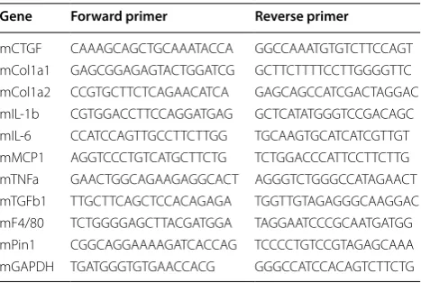
Total RNA from cells or tissues was isolated using Sep-asol reagent (Nacalai Tesque, Kyoto, Japan). First-strand cDNA was obtained using a Verso cDNA Synthesis Kit (Thermo Scientific), according to the kit instructions. This kit contains a reagent designed to exclude genomic DNA contamination. Real-time PCR was performed using the CFX96 real-time PCR system (Bio-Rad, Hercu-les, CA, USA) with KAPA SYBR Green. For experiments employing THP-1, the cells were pre-treated with cana-gliflozin for 30 min and then stimulated with 10 ng/ml of lipopolysaccharide (LPS) for 6 h. The primers used are shown in Table 1.
Statistical analysis
The results were analyzed using EZR (Saitama Medical Center, Jichi Medical University, Saitama, Japan) [17]. Values are presented as mean ± SEM. Statistical signifi-cance was calculated employing Student’s unpaired t-test
Table 1 The list of primer sequences
Gene Forward primer Reverse primer
when comparing two groups, and one-way ANOVA fol-lowed by the post hoc Tukey’s test for multiple compari-sons. In this study, we considered P < 0.05 to indicate a statistically significant difference.
Results
Treatment with canagliflozin suppressed diabetic nephropathy development and reduced the expressions of genes related to fibrosis and inflammation
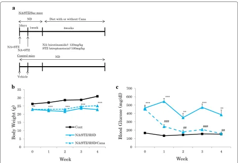
We created type 2 diabetic model mice by injecting both streptozotocin (120 mg/kg) and nicotinamide (100 mg/ kg), to cause mild pancreatic β cell dysfunction, followed by HSD feeding to induce insulin resistance. These dia-betic mice (NA/STZ/Suc mice) were randomly divided into two groups and treated with or without canagliflo-zin, respectively (Fig. 1a). Hyperglycemia was normalized in the former group. The body weights of NA/STZ/Suc mice were significantly lower than those of the controls because of diabetes mellitus. Treatment with canagli-flozin did not affect body weight (Fig. 1b), but markedly normalized hyperglycemia in the NA/STZ/Suc mice (Fig. 1c).
Four weeks after the initiation of canagliflozin admin-istration, the mice were sacrificed and the microscopic findings of the kidney with AZAN staining were com-pared among the groups. Microscopic observation revealed glomerular fibrosis in the kidney to be increased in the NA/STZ/Suc mice, as compared with the controls. Canagliflozin-treated NA/STZ/Suc mice generally had less collagen deposition than untreated NA/STZ/Suc mice (Fig. 2a).
To biochemically confirm the microscopic changes in collagen depositions observed in the kidneys of NA/STZ/ Suc diabetic mice and their attenuation in the SGLT2 inhibitor treated mice, mRNA levels of collagen 1a1 and 1a2 were measured. NA/STZ/Suc mice showed mark-edly up-regulated collagen 1a1 and 1a2, both of which were normalized by canagliflozin treatment (Fig. 2b). In addition, although administration of canagliflozin had no impact on TGFβ expression levels, CTGF expression was reduced. Similarly, mRNA levels of F4/80, a marker of infiltrating macrophages, and those of inflammatory cytokines such as TNFα and MCP-1, were also elevated in NA/STZ/Suc mice. These elevations were partially
NA (nicotinamide): 120mg/kg STZ (streptozotocin):100mg/kg
Vehicle
a
NA+STZ NA+STZ
2days 1week
Diet with or without Cana NA/STZ/Suc mice
ND
Control mice ND
4weeks
0 5 10 15 20 25 30 35
0 1 2 3 4
Body
We
ight (g
)
Week
Cont
NA/STZ/HSD
NA/STZ/HSD/Cana
b
0 100 200 300 400 500 600 700
0 1 2 3 4
Blood Glucose (mg/dl
)
Week
c
*** *** *** ***
** ***
***
***
###
###
** **
##
##
normalized by canagliflozin treatment. On the whole, these results suggest canagliflozin to exert suppressive effects against inflammation and fibrosis in the kidney (Fig. 2c).
Pin1 protein expression was elevated in the kidneys of diabetic model mice and was normalized by canagliflozin treatment
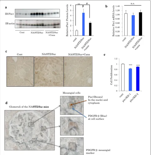
Previous reports have indicated the involvement of Pin1 in the formation of fibrosis in diverse tissue types [18– 20]. In addition, we previously reported Pin1 expression to be regulated by nutrient conditions [15]. Therefore, we investigated the Pin1 expression levels in the kid-neys of normal and NA/STZ/Suc mice with and without canagliflozin treatment. Importantly, it was clearly dem-onstrated that renal Pin1 protein levels were markedly increased in the NA/STZ/Suc mice as compared with the normal mice (Fig. 3a). On the other hand, there was no change in the Pin1 mRNA expression level (Fig. 3b). Canagliflozin-treated NA/STZ/Suc mice showed reduced Pin1 protein levels, which as were closer to those of
normal mice, as compared with non-treated NA/STZ/ Suc mice. The Pin1 expression level in the normal mouse kidney is reportedly higher in the tubule than in the glo-merulus, observations confirmed in this study (left panel of Fig. 3c) [19]. However, immunochemical analysis using anti-Pin1 antibody revealed Pin1 expression in the NA/ STZ/Suc mouse kidneys to be elevated mainly in the glomerulus, though also slightly in the tubules (Fig. 3c). Magnified images of double staining with anti-Pin1 and anti-PDGFR-β, specific mesangial cell markers, revealed the cells with increased Pin1 expression in the diabetic condition included mesangial cells (Fig. 3d). Moreover, treatment of CRL1927 cells with Pin1 siRNA slightly, but significantly, suppressed cell proliferation (Fig. 3e).
Canagliflozin activated AMPK in mesangial cells
It was recently reported that canagliflozin activates AMPK, exerting a direct effect in hepatocytes and ves-sel cells [21, 22]. In addition, increased Pin1 expres-sion reportedly leads to suppressed AMPK activation via the association of Pin1 with the γ subunit of AMPK
a
ContNA/STZ/Suc
NA/STZ/Suc+Cana
b
c
##
** * #
n.s
** #
** n.s **
#
** ** #
0 0.5 1 1.5 2 2.5 3
Collagen 1a1 mRNA
levels
0 0.5 1 1.5 2 2.5
TGFb mRNA
levels
0 0.2 0.4 0.6 0.81 1.2 1.4 1.6 1.82
Collagen 1a2 mRNA
levels
0 2 4 6 8 10 12 14
MC
P-1 mRNA
levels
0 0.2 0.4 0.6 0.8 1 1.2
CTGF mRNA
level
s
0 0.51 1.52 2.53 3.54 4.5
TNFa mRNA
levels
0 0.5 1 1.5 2 2.5
F4/80 mRNA
levels
Fig. 2 The expressions of fibrosis and inflammation markers were elevated in the NA/STZ/Suc mice, and were normalized by canagliflozin treatment. a AZAN staining of kidney sections. Scale bars: 50 μm. b Relative mRNA levels of fibrotic markers in the kidneys were investigated. (n = 5).
[23]. Since AMPK impacts numerous cellular functions including suppression of cell proliferation and inflam-matory cytokine expressions, we examined the effects of canagliflozin on AMPK phosphorylation in the mesangial CRL1927 and the macrophage THP-1 cell lines.
In the in vivo experiments, only a slight increase in AMPK phosphorylation in whole cell lysates of mouse kidney after venous injection of canagliflozin into tails (200 mg/kg) was detected, possibly because the kid-ney contains a wide range of cells including many not
c
d
Cont NA/STZ/Suc NA/STZ/Suc+Cana
a
IB:Pin1
IB:actin
Cont NA/STZ/Suc NA/STZ/Suc
+Cana 0
1 2 3 4 5 6
RelativePin1 Protein Levels
** #
Glomeruli of the NA/STZ/Suc mice
Pin1(Brown) In the nuclei and cytoplasm
PDGFR-β(Blue)
at cell surface Mesangial cells
PDGFR-β: mesangial marker
b
n.s.0.00 0.20 0.40 0.60 0.80 1.00 1.20 1.40
Relative Pin1 mRNA
levels
*** ***
e
Cell Proliferatio
n
0 0.2 0.4 0.6 0.8 1 1.2
a
b
c
d
e
g
f
h
responsive to canagliflozin (Fig. 4a). In the in vitro exper-iments, AMPK and ACC were concentration-depend-ently phosphorylated in CRL1927 cells in response to canagliflozin, with no alteration in the protein amount of AMPK (Fig. 4b) [24]. The ADP/ATP ratio in mesan-gial cells was increased, canagliflozin-concentration dependently (Fig. 4c). The CRL1927 cell proliferation at the time point of 24 h after the addition of canagliflozin was also significantly suppressed (Fig. 4d). Addition of an AMPK activator, either 2-DG or AICAR, also decreased cell growth. Moreover, anti-proliferative effects exerted by canagliflozin were partially blocked by the AMPK inhibitor, compound C, suggesting the anti-proliferative effects of canagliflozin to be mediated, at least partially, by AMPK activation.
In contrast, numbers of apoptotic CRL1927 cells were unchanged with canagliflozin treatment, while being increased with H2O2 treatment (Fig. 4e). In addition, the levels of expression of the Cyclin D1 and phospho
p70S6K proteins, involved in regulating cell proliferation and chemokine expression, were significantly decreased in canagliflozin-treated CRL1927 mesangial cells (Fig. 4f).
Canagliflozin activated AMPK and suppressed cytokine expressions in THP‑1 macrophages
Canagliflozin-induced AMPK and ACC phosphoryla-tions were also clearly observed in THP-1 macrophages at canagliflozin concentrations of no less than 2 μM and were concentration-dependent (Fig. 5a). Canagliflozin suppressed the LPS-induced increases in mRNA expres-sion levels of IL-1β, IL-6, and MCP-1, but did not signifi-cantly alter TNFα, in THP-1 cells (Fig. 5b).
Discussion
Recent reports have revealed that SGLT2 inhibitors exert renal protective effects in diabetic rodent models [14]. Consistent with previous reports, our results also showed
b
0 2 5 10 IB:pACC
IB:tACC
IB:pAMPK
IB:tAMPK
IB:actin
Cana (µM)
a
0 50 100 150 200 250
IL-1b
mRNA
levels
0 20 40 60 80 100 120 140
IL-6 mRNA
levels
0 10 20 30 40 50 60
MCP-1 mRNA
levels
0 5 10 15 20 25 30 35 40 45
TNFa
mRNA
levels
** **
** n.s.
1hr
c
canagliflozin to improve the features of diabetic nephrop-athy. However, details of the underlying mechanisms remain controversial and several hypotheses have been put forward. In this study, we obtained data suggesting contributions of Pin1 and AMPK to the renal protective effects of SGLT2 inhibitors. Herein, we newly revealed that Pin1 protein in the mesangial cells of the kidney is also upregulated in the hyperglycemic state of NA/STZ/ Suc mice, although there appeared some other unidenti-fied cells with increased Pin1 expression. However, the expression levels of Pin1 mRNA did not differ among the three groups. We speculate that the changes in Pin1 tein expression levels in the kidneys are regulated by pro-tein degradations.
Pin1 has been recognized as being involved in the pathogenesis of several diseases, including cancers and Alzheimer’s disease [25, 26]. In addition, previous reports described Pin1 as playing a critical role in the fibrotic processes of bleomycin-induced interstitial pneumonia [27], non-alcoholic hepatosteatosis [26] and phosphate-induced nephritis [20], and that all of these actions were blocked in Pin1 deficient mice. In terms of the kidney, macrophage infiltration and extracellular matrix accu-mulation in the interstitium, after feeding of a high phos-phate diet, were reportedly suppressed in Pin1 null mice [20]. Unfortunately, to our knowledge, there is no murine model expressing CRE specifically in mesangial cells, and thus generation of mesangial cell-specific Pin1 KO mice was not feasible. Nevertheless, the report showing sup-pressed renal fibrosis in Pin1 KO mice fed a high-phos-phate diet suggests that increased Pin1 in diabetic mice might also be involved in diabetic kidney impairment, making at least a limited contribution, and also that nor-malization of the Pin1 expression level by the treatment with an SGLT2 inhibitor is involved in its protective effect.
In the rat kidney, SGLT2 was reported to be expressed in proximal tubular epithelial cells and mesangial cells [28]. Canagliflozin inhibited high-glucose-induced acti-vation of the protein kinase C (PKC)-NAD(P)H oxidase pathway and increased reactive oxygen species produc-tion in mesangial cells [29]. Canagliflozin also normalized the expression of TGF-β1, a key cytokine that mediates extracellular matrix accumulation and glomerular expan-sion in diabetes, and the expresexpan-sion of fibronectin, a pre-dominant matrix protein in glomerular expansion in the mesangial cells of diabetes models [29]. Although cana-gliflozin did not decrease the mRNA expression level of TGF-β1 in the whole kidney in our study, the possibil-ity that TGF-β1 expression was suppressed only in the mesangial cells expressing SGLT2 cannot be ruled out.
Pin1 PPIase activity was reportedly enhanced in human pulmonary eosinophils treated with a PKC-α agonist
[30]. On the other hand, Pin1 inhibits PKC-α degradation by the proteasome. Therefore, PKC-α might be located upstream from Pin1 activation, while Pin1 raises cyto-solic levels of PKC-α. In addition, Pin1 reportedly pro-motes the stability of TGF-β1 mRNA, which would drive fibroblast proliferation and extracellular matrix deposi-tion. Taken together, these observations allow us to spec-ulate that increased Pin1 in mesangial cells is involved in fibrosis via PKC and TGF-β1.
Another interesting finding is that canagliflozin induces AMPK activation in mouse kidneys, mesangial CRL1927 cells and THP-1 macrophages. This effect is likely to be physiological, since it is observed at canagliflozin concen-trations of at least 2 μM and is concentration-dependent. Canagliflozin-induced AMPK activation was previously reported in the liver and vessel cells [21, 22]. It is possi-ble that the canagliflozin-induced AMPK activation takes place only in a limited number of cell types such as mesan-gial cells, since renal cells are highly diverse. Pin1 strongly suppresses AMPK activation via its association with the γ subunit of AMPK without altering ATP levels [23]. On the other hand, the AMPK activation data obtained in this study by administering canagliflozin represent an acute in vitro response, mediated by direct inhibition of mitochondrial complex I by canagliflozin. AMPK activa-tion reportedly induced numerous acactiva-tions leading to the production of ATP and to reduced energy consumption. AMPK reportedly inactivates the mTOR pathway, thereby suppressing its downstream p70S6K and cyclin D1 [31]. As a result, cell cycle or protein production is attenuated by AMPK activation, in good agreement with our present data. Thus, it is possible that canagliflozin-induced AMPK activation contributes to the suppression of mesangial cell proliferation. In addition, activation of AMPK report-edly suppresses NF-κB and thereby reduces inflamma-tory cytokine expressions [32], which would eventually contribute to the tissue protective effects. Furthermore, increased Pin1 expression reportedly suppresses AMPK activation via the association of Pin1 with the γ subunit of AMPK. In the mesangial cells of diabetic mice, increased Pin1 might inhibit AMPK and decreased Pin1 by cana-gliflozin might activated AMPK, accounting for the con-tribution of canagliflozin to the amelioration of diabetic nephropathy. This is the first report, to our knowledge, to raise the possibility of Pin1 and AMPK involvement in the SGLT2 inhibitor-mediated protection against the devel-opment of diabetic kidney disease.
we consider correction of hyperglycemia by canagliflozin to be involved in the protective effects observed in this study. In addition, we revealed that canagliflozin activates AMPK, and suppresses both cell proliferation and inflammation, independently of SGLT2 inhibition. We assume that uni-dentified mechanisms are also involved in the reno-protec-tive effects exerted by canagliflozin. Further studies will be required to clarify this issue.
Conclusions
It may be reasonable to consider normalized Pin1 expres-sion and AMPK activation to be at least partially respon-sible for the molecular mechanisms underlying SGLT2 inhibitor-induced suppression of diabetic nephropathy.
Abbreviations
2DG: 2‑deoxy‑d‑glucose; ACC : acetyl‑CoA carboxylase; ADP: adenosine
diphosphate; AICAR : 5‑aminoimidazole‑4‑carboxamide‑1‑β‑d‑ribofuranoside;
AMPK: AMP‑activated protein kinase; ATP: adenosine triphosphate; Cana: cana‑ gliflozin; C.C.: compound C; CTGF: connective tissue growth factor; DMEM: Dulbecco’s modified Eagle’s medium; eGFR: estimated glomerular filtration rate; HSD: high sucrose diet; KO: knock out; LPS: lipopolysaccharide; MCP‑1: monocyte chemoattractant protein 1; mTOR: mammalian target of rapamycin; MTT: 3‑(4,5‑dimethylthiazol‑2‑yl)‑2,5‑diphenytletetrazolium bromide; ND: normal diet; NF‑kB: nuclear factor‑kappa B; PBS: phosphate buffered saline; PCR: polymerase chain reaction; PDGFRβ: platelet derived growth factor receptor‑beta; Pin1: peptidylprolyl cis/trans isomerase, NIMA‑interacting 1; PMSF: phenylmethylsulfonyl fluoride; PVDF: poly vinylidene di‑fluoride; RPMI: Roswell Park Memorial Institute Medium; RT: room temperature; Ser: serine; SGLT2: sodium glucose co‑transporter2; STZ: streptozotocin; TGF: transforming growth factor; Thr: threonine; TNFα: tumor necrosis factor‑α.
Acknowledgements
A part of this study was carried out at the Analysis Center of Life Science, Natu‑ ral Science Center for Basic Research and Development, Hiroshima University.
Authors’ contributions
MKI, YN and TA conceived and designed the experiments. MKI, YN, TY, YM, KU, AK, KM and KS performed the experiments. MKI, YN, YM, HS, MF, HO, MI, TS, FN and TA analyzed the data. MKI, YN, TY, and TA wrote the manuscript. All authors read and approved the final manuscript.
Funding
This study was partly supported by Grant‑in‑Aid for Scientific Research (B) (To T.A), and also by Grant‑in‑Aid for Scientific Research (B) (To T.A.), Japan Foundation for Applied Enzymology (To Y. N.) and Mitsubishi Tanabe Pharma Corporation (Osaka, Japan).
Availability of data and materials Not applicable.
Ethics approval and consent to participate
All animals in this study were handled in accordance with the guidelines for the care and use of experimental animals published by Hiroshima University.
Consent for publication Not applicable.
Competing interests
The authors declare that they have no competing interests.
Author details
1 Department of Medical Science, Graduate School of Medicine, University of Hiroshima, 1‑2‑3 Kasumi, Minami‑ku, Hiroshima City, Hiroshima 734‑8551, Japan. 2 Division of Diabetes and Metabolism, The Institute for Adult Diseases,
Asahi Life Foundation, Chuo‑ku, Tokyo 103‑0002, Japan. 3 Center for Trans‑ lational Research in Infection & Inflammation, School of Medicine, Tulane University, 6823 St. Charles Avenue, New Orleans, LA 70118, USA. 4 Division of Neurology, Respirology, Endocrinology and Metabolism, Department of Internal Medicine, Faculty of Medicine, University of Miyazaki, 5200 Kihara, Kiyotake, Miyazaki 889‑1692, Japan. 5 Division of Diabetes and Metabolic Dis‑ eases, Nihon University School of Medicine, Itabashi, Tokyo 173‑8610, Japan. 6 Department of Clinical Cell Biology, Graduate School of Medicine, Chiba Uni‑ versity, 1‑8‑1 Inohana, Chuo‑ku, Chiba City, Chiba 260‑8670, Japan. 7 Section of Periodontology, Division of Oral Rehabilitation, Faculty of Dental Science, Kyushu University, Fukuoka, 3‑1‑1 Maidashi, Higashi‑ku, Fukuoka City, Fukuoka 812‑0054, Japan. 8 Department of Nephrology, Hiroshima University Hospital, 1‑2‑3 Kasumi, Minami‑ku, Hiroshima City, Hiroshima 734‑8551, Japan.
Received: 21 April 2019 Accepted: 8 July 2019
References
1. Collins AJ, Foley RN, Gilbertson DT, Chen SC. United States Renal Data System public health surveillance of chronic kidney disease and end‑ stage renal disease Kidney. Int Suppl. 2015;5(1):2–7.
2. Banki NF, Ver A, Wagner LJ, Vannay A, Degrell P, Prokai A, Gellai R, Lenart L, Szakal DN, Kenesei E, Rosta K, Reusz G, Szabo AJ, Tulassay T, Baylis C, Fekete A. Aldosterone antagonists in monotherapy are protective against streptozotocin‑induced diabetic nephropathy in rats. PLoS ONE. 2012;7(6):e39938.
3. Zhang F, Liu H, Liu D, Liu Y, Li H, Tan X, Liu F, Peng Y, Zhang H. Effects of RAAS inhibitors in patients with kidney disease. Curr Hypertens Rep. 2017;19(9):72.
4. Kakinuma H, Oi T, Hashimoto‑Tsuchiya Y, Arai M, Kawakita Y, Fukasawa Y, Iida I, Hagima N, Takeuchi H, Chino Y, Asami J, Okumura‑Kitajima L, Io F, Yamamoto D, Miyata N, Takahashi T, Uchida S, Yamamoto K. (1S)‑1,5‑anhy‑ dro‑1‑[5‑(4‑ethoxybenzyl)‑2‑methoxy‑4‑methylphenyl]‑1‑thio‑d‑glucitol
(TS‑071) is a potent, selective sodium‑dependent glucose cotrans‑ porter 2 (SGLT2) inhibitor for type 2 diabetestreatment. J Med Chem. 2010;53(8):3247–61.
5. Feng C, Wu M, Chen Z, Yu X, Nie Z, Zhao Y, Bao B. Effect of SGLT2 inhibitor on renal function in patients with type 2 diabetes mellitus: a systematic review and meta‑analysis of randomized controlled trials. Int Urol Neph‑ rol. 2019. https ://doi.org/10.1007/s1125 5‑019‑02112 ‑6.
6. Neal B, Perkovic V, Mahaffey KW, de Zeeuw D, Fulcher G, Erondu N, Shaw W, Law G, Desai M, Matthews DR, CANVAS Program Collaborative Group. Canagliflozin and cardiovascular and renal events in type 2 diabetes. N Engl J Med. 2017;377(7):644–57.
7. Panchapakesan U, Pegg K, Gross S, Komala MG, Mudaliar H, Forbes J, Pollock C, Mather A. Effects of SGLT2 inhibition in human kidney proximal tubular cells—renoprotection in diabetic nephropathy? PLoS ONE. 2013;8(2):e54442.
8. Heerspink HJ, Desai M, Jardine M, Balis D, Meininger G, Perkovic V. Cana‑ gliflozin slows progression of renal function decline independently of glycemic effects. J Am Soc Nephrol. 2017;28(1):368–75.
9. Wanner C, Inzucchi SE, Lachin JM, Fitchett D, von Eynatten M, Mattheus M, Johansen OE, Woerle HJ, Broedl UC, Zinman B; EMPA‑REG OUTCOME investigators. Empagliflozin and progression of kidney disease in type 2 diabetes. N Engl J Med. 2016;375(4):323–34.
10. Perkovic V, Jardine MJ, Neal B, Bompoint S, Heerspink HJL, Charytan DM, Edwards R, Agarwal R, Bakris G, Bull S, Cannon CP, Capuano G, Chu PL, de Zeeuw D, Greene T, Levin A, Pollock C, Wheeler DC, Yavin Y, Zhang H, Zinman B, Meininger G, Brenner BM, Mahaffey KW; CREDENCE Trial Investigators. Canagliflozin and renal outcomes in type 2 diabetes and nephropathy. N Engl J Med. 2019. https ://doi.org/10.1056/NEJMo a1811 744.
11. Yale JF, Bakris G, Cariou B, Nieto J, David‑Neto E, Yue D, Wajs E, Figueroa K, Jiang J, Law G, Usiskin K, Meininger G, DIA3004 Study Group. Efficacy and safety of canagliflozin over 52 weeks in patients with type 2 diabetes mellitus and chronic kidney disease. Diabetes Obes Metab. 2014;16(10):1016–27.
•fast, convenient online submission
•
thorough peer review by experienced researchers in your field
• rapid publication on acceptance
• support for research data, including large and complex data types
•
gold Open Access which fosters wider collaboration and increased citations maximum visibility for your research: over 100M website views per year
•
At BMC, research is always in progress.
Learn more biomedcentral.com/submissions
Ready to submit your research? Choose BMC and benefit from: that dapagliflozin reduces weight and blood pressure but does not
improve glycemic control. Kidney Int. 2014;85(4):962–71.
13. Pirklbauer M, Schupart R, Fuchs L, Staudinger P, Corazza U, Sallaberger S, Leierer J, Mayer G, Schramek H. Unraveling reno‑protective effects of SGLT2 inhibition in human proximal tubular cells. Am J Physiol Renal Physiol. 2019;316(3):F449–62.
14. Wang XX, Levi J, Luo Y, Myakala K, Herman‑Edelstein M, Qiu L, Wang D, Peng Y, Grenz A, Lucia S, Dobrinskikh E, D’Agati VD, Koepsell H, Kopp JB, Rosenberg AZ, Levi M. SGLT2 protein expression is increased in human diabetic nephropathy: SGLT2 PROTEIN INHIBITION DECREASES RENAL LIPID ACCUMULATION, INFLAMMATION, AND THE DEVELOPMENT OF NEPHROPATHY IN DIABETIC MICE. J Biol Chem. 2017;292(13):5335–48. 15. Nakatsu Y, Mori K, Matsunaga Y, Yamamotoya T, Ueda K, Inoue Y, Mitsu‑
zaki‑Miyoshi K, Sakoda H, Fujishiro M, Yamaguchi S, Kushiyama A, Ono H, Ishihara H, Asano T. The prolyl isomerase Pin1 increases β‑cell prolifera‑ tion and enhances insulin secretion. J Biol Chem. 2017;292(28):11886–95. 16. Nakatsu Y, Sakoda H, Kushiyama A, Zhang J, Ono H, Fujishiro M, Kikuchi T, Fukushima T, Yoneda M, Ohno H, Horike N, Kanna M, Tsuchiya Y, Kamata H, Nishimura F, Isobe T, Ogihara T, Katagiri H, Oka Y, Takahashi S, Kurihara H, Uchida T, Asano T. Peptidyl‑prolyl cis/trans isomerase NIMA‑interact‑ ing 1 associates with insulin receptor substrate‑1 and enhances insu‑ lin actions and adipogenesis. J Biol Chem. 2011;286(23):20812–22. 17. Kanda Y. Investigation of the freely available easy‑to‑use software ‘EZR’ for
medical statistics. Bone Marrow Transplantat. 2013;48:452–58. 18. Yang JW, Hien TT, Lim SC, Jun DW, Choi HS, Yoon JH, Cho IJ, Kang KW.
Pin1 induction in the fibrotic liver and its roles in TGF‑β1 expression and Smad2/3 phosphorylation. J Hepatol. 2014;60(6):1235–41.
19. Shen ZJ, Hu J, Shiizaki K, Kuro‑o M, Malter JS. Phosphate‑induced renal fibrosis requires the prolyl isomerase Pin1. PLoS ONE. 2016;11(2):e0150093.
20. Liu X, Liang E, Song X, Du Z, Zhang Y, Zhao Y. Inhibition of Pin1 allevi‑ ates myocardial fibrosis and dysfunction in STZ‑induced diabetic mice. Biochem Biophys Res Commun. 2016;479(1):109–15.
21. Mancini SJ, Boyd D, Katwan OJ, Strembitska A, Almabrouk TA, Kennedy S, Palmer TM, Salt IP. Canagliflozin inhibits interleukin‑1β‑stimulated cytokine and chemokine secretion in vascular endothelial cells by AMP‑ activated protein kinase‑dependent and ‑independent mechanisms. Sci Rep. 2018;8(1):5276.
22. Hawley SA, Ford RJ, Smith BK, Gowans GJ, Mancini SJ, Pitt RD, Day EA, Salt IP, Steinberg GR, Hardie DG. The Na+/Glucose cotransporter inhibitor canagliflozin activates AMPK by inhibiting mitochondrial function and increasing cellular AMP levels. Diabetes. 2016;65(9):2784–94. 23. Nakatsu Y, Iwashita M, Sakoda H, Ono H, Nagata K, Matsunaga Y, Fuku‑
shima T, Fujishiro M, Kushiyama A, Kamata H, Takahashi S, Katagiri H,
Honda H, Kiyonari H, Uchida T, Asano T. Prolyl isomerase Pin1 negatively regulates AMP‑activated protein kinase (AMPK) by associating with the CBS domain in the γ subunit. J Biol Chem. 2015;290(40):24255–66. 24. Dyck JR, Kudo N, Barr AJ, Davies SP, Hardie DG, Lopaschuk GD. Phospho‑
rylation control of cardiac acetyl‑CoA carboxylase by cAMP‑dependent protein kinase and 5′‑AMP activated protein kinase. Eur J Biochem. 1999;262(1):184–90.
25. Nakatsu Y, Matsunaga Y, Yamamotoya T, Ueda K, Inoue Y, Mori K, Sakoda H, Fujishiro M, Ono H, Kushiyama A, Asano T. Physiological and pathogenic roles of prolyl isomerase Pin1 in metabolic regulations via multiple signal transduction pathway modulations. Int J Mol Sci. 2016;17(9):1495. 26. Nakatsu Y, Otani Y, Sakoda H, Zhang J, Guo Y, Okubo H, Kushiyama A,
Fujishiro M, Kikuch T, Fukushima T, Ohno H, Tsuchiya Y, Kamata H, Naga‑ machi A, Inaba T, Nishimura F, Katagiri H, Takahashi S, Kurihara H, Uchida T, Asano T. Role of Pin1 protein in the pathogenesis of nonalcoholic steatohepatitis in a rodent model. J Biol Chem. 2012;287(53):44526–35. 27. Shen ZJ, Braun RK, Hu J, Xie Q, Chu H, Love RB, Stodola LA, Rosenthal LA,
Szakaly RJ, Sorkness RL, Malter JS. Pin1 protein regulates Smad protein signaling and pulmonary fibrosis. J Biol Chem. 2012;287(28):23294–305. 28. Wakisaka M, Nagao T, Yoshinari M. Sodium glucose cotransporter 2
(SGLT2) plays as a physiological glucose sensor and regulates cellular contractility in rat mesangial cells. PLoS ONE. 2016;11(3):e0151585. 29. Maki T, Maeno S, Maeda Y, Yamato M, Sonoda N, Ogawa Y, Wakisaka M,
Inoguchi T. Amelioration of diabetic nephropathy by SGLT2 inhibitors independent of its glucose‑lowering effect: a possible role of SGLT2 in mesangial cells. Sci Rep. 2019;9(1):4703.
30. Shen ZJ, Esnault S, Rosenthal LA, Szakaly RJ, Sorkness RL, Westmark PR, Sandor M, Malter JS. Pin1 regulates TGF‑beta1 production by activated human and murine eosinophils and contributes to allergic lung fibrosis. J Clin Investig. 2008;118(2):479–90.
31. Inoki K, Zhu T, Guan KL. TSC2 mediates cellular energy response to con‑ trol cell growth and survival. Cell. 2003;115(5):577–90.
32. Yang Z, Kahn BB, Shi H, Xue BZ. Macrophage α1 AMP‑activated protein kinase (α1AMPK) antagonizes fatty acid‑induced inflammation through SIRT1. J Biol Chem. 2010;285(25):19051–9.
Publisher’s Note