SPECTROSCOPIC STUDIES OF THE CONFORMATIONAL
STABILITY AND LIGAND BINDING PROPERTIES OF
CALMODULIN
Laura M asino
A thesis submitted in partial fulfilment o f the requirements o f the University o f London for the degree o f Doctor o f Philosophy
June 2000
D h ision o f Physical Biochemistry Department o f Biochemistry N aional Institute for Medical Research University College London
Mi l Hill Gower Street
ProQuest Number: U642493
All rights reserved
INFORMATION TO ALL USERS
The quality of this reproduction is dependent upon the quality of the copy submitted.
In the unlikely event that the author did not send a complete manuscript and there are missing pages, these will be noted. Also, if material had to be removed,
a note will indicate the deletion.
uest.
ProQuest U642493
Published by ProQuest LLC(2015). Copyright of the Dissertation is held by the Author.
All rights reserved.
This work is protected against unauthorized copying under Title 17, United States Code. Microform Edition © ProQuest LLC.
ProQuest LLC
789 East Eisenhower Parkway P.O. Box 1346
ABSTRACT
In this work the conformational stability o f calmodulin (CaM) and its tryptic fragments has been investigated using optical spectroscopic techniques such as far-UV circular dichroism, fluorescence, and absorption spectroscopy. CaM is a ubiquitous eukaryotic calcium binding protein that consists o f two structurally similar globular domains connected by a flexible linker, and each containing a pair o f helix-loop-helix calcium binding motifs.
The results o f chemical and thermal dénaturation experiments show that the stability o f CaM and its isolated domains in the absence o f calcium is relatively low and is strongly dependent on ionic strength and temperature. This raises the question o f how apo-CaM is stabilised in vivo at resting calcium concentrations. In the presence o f ligands such as calcium, magnesium, and target peptides, the stability o f CaM is greatly enhanced, as predicted by the ligand binding theory. The extent o f the stabilising effect depends on the free ligand concentration and on the affinity o f the native and denatured states o f CaM for the ligand. The interactions o f CaM with magnesium have been investigated and the results show that magnesium competes with calcium for the EF- hand sites and reduces the affinity o f Ca^^-CaM for targets. As a consequence, magnesium amplifies the intrinsic differences in affinity o f the N- and C-domains for calcium and for target sequences. Thus, CaM is extremely sensitive to general environmental conditions as well as to specific ligand interactions. Chemical dénaturation studies also show that the behaviour o f GuHCl is very complex, owing to its ionic strength contribution and to competition with Ca^^-binding. Therefore urea may be more appropriate in the study o f the stability o f calmodulin and o f proteins that bind metal ions or that are sensitive to changes in the ionic strength o f the solution.
ACKNOWLEDGEMENTS
I would like to thank my supervisor Dr. Peter Bayley for giving me the opportunity to work on this project and for helping me to fulfil my wish to undertake scientific research. I express my gratitude to him for his guidance, for the enthusiasm he instilled in me for biophysical studies, for constructive discussions about my data, and for advice about future work.
I am also extremely grateful to Dr. Stephen Martin, who introduced me step by step to the experimental methods o f this laboratory and passed on to me some o f his experience with incredible patience and constant attention. I thank him for his support and for being there at any time to answer my questions and to give me help and suggestions about my project. I also thank him for kindly allowing me to use his data analysis programs.
I thank Peter Browne for his help in protein preparation and for the purification o f fragment T rlC , and Dr. Jens Kleinjung for his help with the HPLC technique. Special thanks go to Daniela Romano and Muriel Erent for their friendship, their encouragement, and for sharing with me all the good times as well as the difficult moments o f the years spent here.
TABLE OF CONTENTS
T itle... 1
A bstract...3
Acknowledgements...4
Table o f contents... 5
List o f Figures...10
List o f Tables... 13
Abbreviations...15
CHAPTER 1 - Introduction... 16
1.1 Calmodulin... 16
1.1.1 The structure o f calmodulin... 17
1.1.2 Calcium binding...20
1.1.3 Specificity o f calmodulin for calcium... 22
1.1.4 Magnesium binding...23
1.2 Interaction o f calmodulin with targets... 25
1.2.1 Sequence motifs for calmodulin recognition...26
1.2.2 Structures o f calmodulin-target peptide complexes... 27
1.2.3 Models for the mechanism o f target activation... 29
1.2.4 Calcium-independent interactions with targets...30
1.3 Mutational studies o f calm odulin...31
1.4 Protein folding and stability... 34
1.4.1 Protein folding...34
1.4.2 Kinetic studies o f protein folding... 37
1.4.3 Methods o f dénaturation and data analysis... 38
1.4.4 Stability o f calm odulin... 42
C H A P T E R 2 - M aterials an d m ethods... 45
2.1 M aterials... 45
2.1.1 Chemicals... 45
2.1.2 Preparation o f Ca^^-free buffer... 46
2.1.3 Purification o f WT calmodulin... 46
2.1.4 Preparation o f Ca^'^-free calmodulin... ... 47
2.1.5 Preparation o f calmodulin fragments...47
2.1.6 Preparation o f calmodulin m utants... 48
2.1.7 Peptides... 49
2.2 Experimental techniques...49
2.2.1 Absorption spectroscopy... 49
2.2.2 Fluorescence spectroscopy... 50
2.2.3 Circular dichroism spectroscopy... 50
2.2.4 Stopped flow ...51
2.3 Theory and experimental procedures... 52
2.3.1 Calcium binding to calm odulin...52
2.3.1.1 Stoichiometric and intrinsic binding constants... 52
2.3.1.2 Calculating concentrations... 56
2.3.1.3 Magnesium/calcium competition... 60
2.3.2 Determination o f chelator affinities... 63
2.3.3 Determination o f stoichiometric association constants for calcium binding using chromophoric calcium chelators... 65
2.3.4 Determination o f peptide affinities by direct fluorometric titration 68 2.3.5 Determination o f peptide affinities using a competition assay... 70
2.3.6 Chemical and thermal dénaturation...73
2.3.6.1 Chemical and thermal dénaturation - simple two-state unfolding... 74
2.3.6.2 Chemical and thermal dénaturation - biphasic unfolding curves... 78
2.3.7 Effects o f ligand binding on stability... 83
CHAPTER 3 - Chemical and thermal dénaturation... 87
3.1 Introduction...87
3.2 Materials and methods... 88
3.3 Results... 89
3.3.1 The optical properties o f calmodulin...89
3.3.2 Chemical dénaturation o f apo-proteins... 92
3.3.3 Chemical dénaturation o f calcium-saturated proteins... 97
3.3.3.1 Isolated domains... 97
3.3.3.2 Intact CaM ... 102
3.3.3.3 Stabilisation by calcium... 109
3.3.4 Thermal dénaturation... I l l 3.3.5 Chemical dénaturation o f the apo-CaM-FFFu co m p lex ...116
3.3.6 Kinetics o f unfolding and refolding o f Tr2C and intact calm odulin 119 3.4 D iscussion...124
3.4.1 Chemical and thermal dénaturation o f calmodulin and its dom ains 124 3.4.2 Inter-domain interactions... 126
CHAPTER 4 - Effects o f ionic strength and pH on stability and C a^-binding... 129
4.1 Introduction... 129
4.2 Materials and methods... 130
4.3 Results...131
4.3.1 Effects o f ionic strength on the stability o f the apo-proteins... 131
4.3.2 Effects o f GuHCl on Ca^'^-binding... 139
4.3.3 Kinetics o f Ca^^ - dissociation... 142
4.3.4 Effects o f p H ...142
4.4 Discussion...147
CHAPTER 5 - Stability and Ca^-binding properties of the p-sheet
mutants of calm odulin... 152
5.1 Introduction...152
5.2 Materials and m ethods... 157
5.3 Results... 157
5.3.1 Urea-unfolding o f the apo-proteins...157
5.3.2 Urea-unfolding o f the holo-proteins... 162
5.3.3 Calcium binding properties... 171
5.4 D iscussion... 174
5.4.1 Urea-unfolding o f the apo-proteins... 174
5.4.2 Urea-unfolding o f the holo-proteins... 175
5.4.3 Calcium binding properties... 176
CHAPTER 6 - Interactions o f magnesium with calmodulin... 180
6.1 Introduction... 180
6.2 Materials and m ethods... 181
6.3 R esults... 183
6.3.1 Conformational effects o f Mg^^... 183
6.3.2 Direct determination o f Mg^"*" affinities... 186
6.3.3 Urea-induced dénaturation o f Mg^^-CaM... 188
6.3.4 Competition between Ca^^ and Mg^^... 192
6.3.5 Effect o f Mg^^ on the interaction o f calmodulin with N M 2... 194
6.4 Discussion... 198
6.4.1 Binding o f Mg^^ to calmodulin: effects on conformation and stability... 198
6.4.2 Effects o f Mg^"^ on the binding o f Ca^^ and o f target sequences 199 6.4.2.1 Dependence o f NM2 affinity on Ca^^ concentration...201
C H A PT E R ? - General conclusions...207
7.1 Thermodynamic stability o f the apo-proteins...207
7.2 Ligand binding... 209
7.2.1 Calcium... 210
7.2.2 M agnesium... 211
7.2.3 Target sequences... 212
7.3 Interdomain interactions...215
Bibliography... 218
LIST OF FIGURES
CHAPTER 1 - Introduction
1.1 Solution structure o f calcium-free calmodulin... 19
CHAPTER 2 - Materials and methods 2.1 Schematic representation o f Ca^^ binding to CaM ... 53
2.2A Ca^^ binding profiles for CaM and its isolated fragm ents... 57
2.2B Concentrations o f stoichiometric species o f CaM, T rlC , and Tr2C as a function o f free calcium concentration... 57
2.3 Schematic representation o f Ca^^ and Mg^"^ binding to a calmodulin domain...62
CHAPTER 3 - Chemical and thermal dénaturation 3.1 Absorption and Tyr-138 fluorescence spectra o f CaM ...90
3.2 Far- and near-UV CD spectra o f CaM ... 91
3.3 Urea-induced dénaturation o f the apo-proteins...93
3.4 GuHCl-induced dénaturation o f the apo-proteins...96
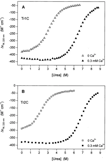
3.5 Urea-induced dénaturation o f T rlC and Tr2C at 0 and 0.3 mM Ca^^ 99 3.6 GuHCl-induced dénaturation o f T rlC and Tr2C at 0, 1, 10 and 100 mM Ca^^... 100
3.7 Urea- and GuHCl-induced dénaturation o f CaM at different Ca^^ concentrations monitored using far-UV C D ... 103
3.8 Urea- and GuHCl-induced dénaturation o f CaM at different Ca^^ concentrations monitored using Tyr-138 fluorescence... 104
3.9 Urea- and GuHCl-induced dénaturation o f CaM at 0 and 1 mM Ca^^ monitored using Tyr-138 absorption... 105
3.10 Urea- and GuHCl-induced dénaturation profiles for holo-CaM compared with the sum o f the curves for the isolated fragnients... 106
3.12 Thermal dénaturation o f apo-Tr 1C, apo-Tr2C, apo-CaM
and holo-CaM monitored using far-UV C D ... 113
3.13 Thermal dénaturation o f apo-CaM monitored using Tyr-138 fluorescence and absorption... 114
3.14A Fraction o f unfolded protein for the C-domain o f apo-CaM in the presence o f FFFu... 117
3.14B Determination o f the kj for FFFu using competition with W F F u...117
3.15 Kinetics o f refolding and calcium binding o f Tr2C in the presence o f urea... 122
CHAPTER 4 - Effects of ionic strength and pH on stability and Ca^-binding 4.1 Urea-induced dénaturation o f apo-Tr 1C and apo-Tr2C at different KCl concentrations... 132
4.2A Urea-induced dénaturation o f apo-CaM at different KCl concentrations... 133
4.2B KCl titration o f apo-CaM ... 133
4.3 KCl titration o f holo-CaM ...134
4.4 KCl titrations o f apo-CaM in the presence o f 2 and 5 M urea... 137
4.5A Thermal dénaturation o f apo-CaM in 0 and 100 mM K C l... 138
4.5B Ca^^ titrations o f 5-nitro BAPTA in the absence and in the presence o f Tr2C...138
4.6 pH titrations o f apo-CaM and CaM in 1 mM Ca^^ ... 144
4.7 Urea-induced and thermal dénaturation o f CaM at pH 6.5...146
CHAPTER 5 - Stability and Ca^-binding properties o f the p-sheet mutants o f calmodulin 5.1 Solution structure o f calcium-free CaM: position o f the hydrophobic residues at position 8 in each calcium binding loop...154
5.2 Urea-induced dénaturation o f the CaM mutants I27G, I63G, V136P, V136G, IlOOG, and V I 36A in the absence o f Ca^^... 160
5.3 A Fractions o f unfolded protein for apo-Tr 1C, the apo-N-domain o f WT CaM, and the apo-N-domain o f V 136A... 161
5.3B Fractions o f unfolded protein for apo-Tr2C, the apo-C-domain
o f WT CaM, and the apo-C-domain o f I27G ... 161 5.4 Urea-induced dénaturation o f 11OOG and V 136A at different
Ca^^ concentrations... 163 5.5 Urea-induced dénaturation o f I27G and 163G at different
Ca^^ concentrations... 165 5.6 Ca^^ titration o f V 136G monitored using far-UV C D ...168 5.7 Ca^^ titration o f V136G monitored using absorption spectroscopy... 169 5.8 Ca^^ titration o f apo-V 13 6P and o f WT holo-CaM monitored
using far-UV C D ... 170 5.9A Ca^^ titrations o f WT CaM, 163G, IlOOG, and V136G monitored
using Tyr-138 fluorescence... 173 5.9B Kinetics o f calcium dissociation from the C-domain o f I27G ... 173
CHAPTER 6 - Interactions of magnesium with calmodulin
6.1 Absorption and Tyr-138 fluorescence spectra o f CaM under
different solvent conditions...184 6.2 Far- and near-UV CD spectra o f CaM under different solvent
conditions... 185 6.3 Mg^^ titrations o f apo-CaM monitored using Tyr-138 fluorescence... 186 6.4 Urea-induced dénaturation o f T rlC , Tr2C and CaM at 10 mM M gC li 190 6.5 Ca^^ titrations o f 5,5’-dibromo-BAPTA in the presence o f CaM
at different MgCl] concentrations... 193 6.6 CaM titration o f NM2 in the presence o f 100 pM Ca^^... 196
CHAPTER 7 - General conclusions
7.1 Fractions o f unfolded protein for the N- and C-terminal domains
o f apo-CaM, Ca^^-CaM and Mg^^-CaM... 213
Appendix 1 - Calmodulin structures and calcium coordination in an EF-hand
A. 1 Structures of free CaM, saturated CaM, and
LIST OF TABLES
CHAPTER 1 - Introduction
1.1 Macroscopic Ca^^-binding constants for CaM and its tryptic fragm ents.... 22
CHAPTER 3 - Chemical and thermal dénaturation
3.1 Urea-unfolding o f apo-Tr 1C, apo-Tr2C, and apo-CaM ... 94 3.2 GuHCl-unfolding o f apo-Tr 1C, apo-Tr2C, and apo-CaM ... 97 3.3 Urea- and GuHCl-unfolding o f holo-TrlC and holo-Tr2C... 101 3.4 Urea- and GuHCl-unfolding o f holo-CaM monitored by far-UV C D 107 3.5 Urea- and GuHCl-unfolding o f holo-CaM monitored
by Tyr-138 fluorescence and Tyr-138 absorption...108 3.6 Thermal unfolding o f apo-Tr 1C, apo-Tr2C, and apo-CaM ... 115 3.7 Urea-unfolding o f apo-CaM in the presence o f FFFu... 118 3.8 Fast and slow observed rates expressed in terms o f the
intrinsic rate constants... 121
CHAPTER 4 - Effects of ionic strength and pH on stability and
Ca^-binding
4.1 Effects o f KCl on urea-unfolding o f apo-Tr 1C, apo-Tr2C,
and apo-CaM, monitored using far-UV C D ... 135 4.2 Effects o f KCl on thermal unfolding o f apo-CaM, monitored
using Tyr-138 fluorescence...139 4.3 Calcium binding constants for 5-nitro BAPTA and the tryptic
fragments o f CaM ...140 4.4 Kinetics o f Ca^^-dissociation from the C-domain o f C aM ...143
CHAPTER 5 - Stability and Ca^-binding properties o f the p-sheet
mutants o f calmodulin
5.1 Amino acid sequences for the four binding loop regions
o f Drosophila melanogaster calmodulin... 153 5.2 Effects o f mutations on secondary structure: far-UV CD intensities 155 5.3 Macroscopic Ca^^-binding constants for WT CaM and the six
p-sbeet m utants... 156 5.4 Urea-unfolding o f the non-mutated apo-domain in the P-sbeet m utants 159 5.5 Urea-unfolding o f the P-sbeet mutants in the presence o f Ca^"^... 166
CHAPTER 6 - Interactions of magnesium with calmodulin
6.1 Average stoichiometric association constants for the binding o f Ca^"^ and Mg^^ to the C- and N-domains o f CaM,
determined by direct titration...187 6.2 Effect o f 10 mM MgCl2 on urea-unfolding o f T rlC , Tr2C, and C aM 191
6.3 Apparent stoichiometric binding constants for Ca^^ in the
presence o f Mg^^... 194 6.4 Observed dissociation constants for the binding o f NM2 to CaM
in the absence and in the presence o f 5 mM Mg^^... 197 6.5 Apparent stoichiometric binding constants for Ca^^ in the
presence o f Mg^^...200 6.6 Calculated dissociation constants for the binding o f NM2 to CaM
ABBREVIATIONS
5,5’-Br2BAPTA
5-nitro BAPTA
Apo-CaM CaM CD EDTA EGTA
Hepes Holo-CaM MES Quin2
sk-MLCK sm-MLCK TNS T rlC Tr2C Tris
5,5’ -dibromo-1,2-bis(2-aminophenoxy)ethane-A, N ,N ’,N tetraacetic acid
5 -nitro-1,2-bis(2-aminophenoxy)ethane-iV, N ,N ’,N ’-tetraacetic acid
calcium free calmodulin
Drosophila melanogaster calmodulin circular dichroism
ethylenediaminetetraacetic acid
ethylene glycol-bis(p-aminoethyl ether)N,N,N’,N ’-tetraacetic acid
A-(2-hydroxyethyl)piperazine-A^-(2-ethane sulphonic acid) calcium saturated calmodulin
2-(A-morpholino)ethanesulphonic acid
2-[[2-bis(carboxymethyl)amino-5-methylphenoxy]methyl]-6-methoxy-8-bis-(carboxymethyl)aminoquinoline
skeletal muscle myosin light chain kinase smooth muscle myosin light chain kinase 2-(p-toluidino)naphthalene-6-sulfonic acid
N-terminal tryptic fragment o f calmodulin (residues 1-75) C-terminal tryptic fragment o f calmodulin (residues 78-148) tris(hydroxymethyl)methylamine
CHAPTER 1
INTRODUCTION
1.1 Calmodulin
1.1.1 The structure o f calmodulin
CaM is a small (Mr = 16,700 D) acidic protein which comprises 148 amino acids. At neutral pH, the total number o f negatively charged residues is 38, whereas that o f positively charged residues is 14. Although the distribution o f charges on the surface o f the protein is different for Ca^^-ffee and Ca^^-saturated CaM, the overall charge o f the protein is clearly negative for both forms. The isoelectric point (pi) is approximately 4.1 (Coyne et al., 1985; Wandosell et al., 1986).
The crystal structure o f Ca^^-CaM was first solved by Babu et al. (1985) at 3.0 Â, subsequently solved by Kretsinger et al. (1986) at 1.9 Â, and further refined by other groups (Babu et al., 1988; Chattopadhyaya et al., 1992). The crystal form o f Ca^^-CaM is a dumbbell-shaped molecule, approximately 65 Â long, composed o f two structurally similar globular domains (N-terminal and C-terminal), each containing a pair o f helix- loop-helix calcium binding motifs (EF-hands). The two domains are separated by an interconnecting a-helix o f approximately eight turns. The two calcium binding loops within each domain are connected by a short antiparallel p-sheet which involves hydrogen bonding between the hydrophobic residues at position 8 in each loop (Ile-27 and Ile-63 in the N-terminal domain, and He-100 and Val-136 in the C-terminal domain). Both domains o f Ca^^-CaM have a large, solvent-exposed hydrophobic surface which is composed o f a number o f residues clustered around a central core formed by the short p-sheet structure. Each surface has a deep hydrophobic cavity in the centre and is surrounded by a polar rim which is rich in negatively charged residues. Among the hydrophobic amino acids which become exposed upon calcium binding, the occurrence o f Met residues is particularly high. O f the nine Met residues present in CaM, four are in each o f the solvent exposed hydrophobic surfaces, contributing 46% o f the total accessible surface area o f the hydrophobic patches (O'Neil and DeGrado, 1990).
The NM R structures o f Ca^^-CaM are closely similar to the crystal structures; however, they showed that residues 78-81, corresponding to the middle section o f the central helix, adopt a non-helical conformation in solution and have a high degree o f mobility (Ikura et al., 1990; Ikura et al., 1991; Barbato et al., 1992). Thus, the central region acts as a flexible linker between the two domains, allowing them to adopt a variety o f positions with respect to each other and to bind CaM targets. The average
conformation o f the molecule in solution is also more compact than the elongated crystal form.
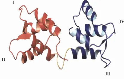
Upon calcium binding, CaM undergoes a conformational transition that increases its affinity for various target proteins. The mechanism o f activation o f CaM by calcium has been intensively investigated but the details remained unclear until the solution structure o f Ca^^-free CaM (apo-CaM) was solved. Before then, m ost o f the speculations about the Ca^^-induced conformational changes o f CaM were based on the model proposed by Strynadka and James (1988), which predicted the structure o f apo- CaM based on the crystal structure o f the highly homologous (51% sequence identity) protein troponin C. In 1995, the NM R solution structures o f apo-CaM and o f its C- terminal domain (Figure 1.1) allowed a direct comparison o f the apo- and Ca^^-forms o f the protein (Finn et al., 1995; Kuboniwa et al., 1995; Zhang et al., 1995a). They showed that the secondary structure o f apo-CaM (in its folded form) is closely similar to that o f Ca^^-CaM and the main differences between the two forms are in helix packing. Upon Ca^^ binding, the two helices in each EF-hand, which are nearly antiparallel in the absence o f calcium, shift to a more perpendicular orientation. This results in a more extended conformation and in the exposure o f the hydrophobic patch observed on the surface o f each domain o f Ca^^-CaM, which drives the binding o f target molecules. Circular dichroism (CD) measurements also indicate that Ca^^ binding induces an increase in the helical content o f the protein (Martin and Bayley, 1986). The deep hydrophobic cavities observed in the centre o f the Ca^^-CaM domains are completely absent in apo-CaM. However, even in the absence o f calcium, both the C- and the N-terminal domains contain a cluster o f hydrophobic residues, which in the N-domain is partially surrounded by acidic residues. The resulting surface in each apo- domain is thus negatively charged but partially hydrophobic. Owing to the tight helix packing o f apo-CaM and to the exposure o f the hydrophobic pockets in Ca^^-CaM, their structures are often referred to as the “closed” and “open” conformations, respectively.
II
I V
III
Figure 1.1 Solution structure of calcium-free calmodulin. The N-domain is shown in red, the C-domain in blue, and the flexible central linker which connects the two domains is shown in yellow. Numbers I to IV indicate the four calcium binding loops. The figure was prepared using the coordinates deposited by Kuboniwa et al. (1995) (PDB entry code ICFC).
1986), and calcium binding (Linse et aL, 1991a; Martin et al., 1996) properties o f intact CaM are well represented by a summation o f those o f its tryptic fragments. Therefore it has been concluded that the two domains o f CaM behave as two independent units. This observation has also been confirmed by the NM R solution structures o f holo-CaM (Ikura et al., 1991) and apo-CaM (Zhang et al., 1995a), in which no contacts between the two domains were detected. However, optical spectroscopy and scanning microcalorimetry studies on the stability and the calcium binding properties o f CaM mutants have suggested that the two domains may interact (Martin et al., 1992; Mukherjea et al., 1996; Protasevich et al., 1997). Evidence that the presence o f one domain does affect the calcium-binding properties o f the other has also been reported for WT CaM in proteolytic footprinting studies (Sorensen and Shea, 1998) and for mutants o f the related protein, troponin C (Moncrieffe et al., 1999).
1.1.2 Calcium binding
The EF-hand is a strongly conserved structural element widely distributed in the family o f calcium binding proteins, which includes CaM, troponin C, parvalbumin, oncomodulin, calbindin, S I00 proteins, and myosin light chains. The name EF-hand was introduced by R. H. Kretsinger to describe the structure o f a calcium binding site involving helices E and F o f parvalbumin (Kretsinger and Nockolds, 1973). This m otif comprises a helix-loop-helix sequence o f 29 residues and two EF-hand sites are usually associated in the same protein to yield a highly cooperative calcium binding system. The calcium binding functions are contained largely in the loop sequence, which consists o f twelve consecutive residues (Falke et al., 1994). The calcium ion is coordinated by seven oxygen ligands, five o f which are provided by carboxylate side- chains, one by a backbone carbonyl, and one by a water molecule. The six amino acids in the loop which are involved in calcium coordination are in positions 1, 3, 5, 7, 9 and 12. The residue in position 12 is strongly conserved and in nearly all known EF-hands is a glutamate, which contributes both its carboxyl oxygens to Ca^^ coordination. Two non-coordinating positions within the loop are also conserved: Gly at position 6 and the hydrophobic residue at position 8, which is predominantly He or Val.
orders o f magnitude, from ~ 10^ M'^ in parvalbumin to ~ 10^ M'^ in the low affinity sites o f CaM (Davis, 1992). One o f the future challenges is to identify the features o f the EF-hand m otif that determine the affinity for calcium; at present, it is not possible to predict the affinity o f a particular site even when the three-dimensional structure is known.
CaM contains a pair o f EF-hands in each globular domain. Ca^"^ binding sites III and IV in the C-terminal domain have higher affinity than sites I and II in the N- terminal domain (Thulin et al., 1984; Martin et al., 1985) and positive cooperativity is observed between the two sites in each domain (Linse et al., 1991a). Studies on the Ca^^ binding properties o f CaM and its tryptic fragments T rlC and Tr2C (corresponding to the N- and C-terminal domains o f CaM, respectively) have also shown that the isolated domains o f CaM retain the Ca^^ binding properties that they have in the intact molecule and that there is no cooperativity between the two globular domains in intact CaM (Linse et al., 1991a). The phenomenon o f positive cooperativity in Ca^^ binding is widespread in EF-hand domains containing a pair o f interacting sites. Positive cooperativity is particularly important for Ca^^-sensor proteins such as CaM, because it allows the protein to exert its regulatory effect over a narrow range o f calcium concentration. The intracellular [Ca^^] is ~ 0.1 pM under resting conditions and increases to 1-10 pM upon stimulation (Berridge et al., 1998). At present, the mechanism which underlies positive cooperativity within EF-hand sites is not understood. It has been suggested that the short p-sheet structure which links two neighbouring calcium binding loops back to back may play an important role in cooperativity (Falke et al., 1994).
The stoichiometric (or macroscopic) Ca^^ binding constants for CaM can be accurately determined using the chromophoric Ca^^ chelator method (Linse et al., 1991a) or flow dialysis (Porumb, 1994). The values obtained using these methods are in reasonable agreement and are in the range 10"^ to 10^ M '\ The stoichiometric Ca^^ binding constants determined for CaM and its tryptic fragments using the chromophoric chelator 5,5’-Br2BAPTA are listed in Table 1.1 (the chromophoric Ca^^ chelator method is described in Section 2.3.3 , Chapter 2).
T ab le 1.1 Macroscopic Ca^’^-binding constants for CaM and its tryptic fragments
Protein log(Ki) log(K2) log(Ks)
log(K4)
log(KiK2)log(KsK4)
W T C aM 5.33 6.32 4.33 5.33 11.65 9.67
T rlC 4.62 5.17 9.78
Tr2C 5.32 6.21 11.53
^ From Bayley et al. (1996). TrlC and Tr2C correspond to the isolated N- and C-terminal
domains, respectively. Measurements were made at 20 °C in 25 mM Tris, pH 8.0, 100 mM
KCl.
1.1.3 Specificity of calmodulin for calcium
In spite o f the important role that Ca^^-CaM plays in the regulation o f a wide variety o f cellular events, CaM shows a surprisingly broad specificity for binding metal ions. Many divalent as well as trivalent cations have been shown to interact with CaM (Klee, 1988; Ouyang and Vogel, 1998). They can be divided into two classes: those which, like Ca^^, induce the conformational change that allows CaM to bind and activate targets and those which interact with CaM but do not induce the same conformational change and thus do not result in activation o f target proteins. Among the divalent cations, many, including Cd^^, Zn^"^, Pb^^, Hg^^, and Mn^^, bind to CaM with relatively high affinity and are able to promote activation o f target enzymes, but, when present at high concentrations inhibit CaM activity (Klee, 1988; Ozawa et al.,
1999). The inhibition could be a result o f binding at non-specific sites on CaM. However, it is unlikely that the binding o f these metals to CaM is significant under physiological conditions, since their intracellular concentration is well below that needed for their interaction with CaM.
M'*) is much higher than that for Ca^^; it has been proposed that the ability o f lanthanides to replace Ca^^ in a number o f proteins arises from the similar ionic size and coordinating properties o f these ions (Martin and Richardson, 1979). Unlike Ca^^, Tb^^ binds sites I and II (N-terminal domain) with higher affinity than sites III and IV (C-terminal domain).
The specificity o f EF-hands for Ca^^ and, when appropriate, Mg^"^ can arise from charge or size selectivity (Falke et al., 1994). Charge selectivity is controlled by the net negative charge surrounding the ion binding site. If the cation bound to the site does not have sufficient positive charge density, electrostatic repulsion between the negatively charged residues in the loop prevents close packing o f the coordinating array, thus destabilising the binding site. This is why monovalent ions such as and N a \ which are the highest in concentration among the various metal ions inside the cell, are excluded from binding to EF-hand sites (Linse et al., 1991a; Linse et al., 1991b). Size selectivity is based on constraints imposed by the coordination number (Ca^^ is the only divalent ion which prefers seven-fold coordination) and by the size o f the binding cavity (Falke et al., 1994).
1.1.4 Magnesium binding
Mg^^ is the most abundant divalent ion in mammalian cells and its intracellular and extracellular concentrations are kept nearly constant at 0.5-2 mM in most multicellular organisms (Ebel and Gunther, 1980). Mg^^ concentration is thus 100 to 10,000-fold in excess over cytosolic calcium concentration (~ 0.1 to 1-10 p.M, see above). Several studies have shown that CaM binds Mg^"^, although the reported binding constants vary considerably, ranging from 10^ to 10"^ M'^ (Haiech et al., 1981; Tsai et al., 1987; for reviews see Cox et al., 1984, and Klee, 1988). The affinity o f CaM for Mg^^ is much lower than that for Ca^^ and spectroscopic and structural studies have shown that the binding o f Mg^"^ does not induce the major conformational changes caused by Ca^^ binding (Seamon, 1980; Drabikowski et al., 1982; Klee, 1988; Ohki et al., 1997). Recent NM R studies on the N-terminal domain o f CaM have also indicated that the overall conformation o f Mg^^-bound T rlC is similar to the “closed” apo-form, as opposed to the “open” Ca^^-bound conformation (Malmendal et al., 1998; Malmendal et al., 1999b).
The mechanism o f binding o f to CaM is still a matter o f debate and controversies concerning the number o f sites and the existence (or not) o f direct competition between Ca^"^ and Mg^^ still exist. The generally accepted model is that Ca^^ and Mg^^ share the same binding sites on both domains, thus competing directly with each other (Haiech et al., 1981; Tsai et al., 1987; Ohki et al., 1997). Crystal and NM R structures o f Mg^^ bound to the EF-hand proteins parvalbumin and calbindin have confirmed this model, showing that Mg^"^ and Ca^^ can compete for the same sites (Declercq et al., 1991; Andersson et al., 1997). There is also strong evidence that Mg^^ binds more strongly to the N-terminal domain o f CaM than to the C-terminal domain, showing opposite domain preference to Ca^^. However, flow dialysis and microcalorimetry studies have suggested that Mg^"^ binds to four sites which are distinct from the EF-hand Ca^^ binding sites (Milos et al., 1986; Gilli et al., 1998). According to this interpretation, Mg^"^ does not act as a direct competitor for Ca^^ binding, but rather as an allosteric effector.
Mg^^ has a strong preference for 6-fold coordination in an octahedral symmetry, as opposed to Ca^"^, which has less stringent demands on the number o f coordinating oxygen ligands (often 6-8) (Falke et al., 1994). This property o f Mg^^ binding, together with its smaller ionic radius, probably forms the structural basis o f the discrimination o f Ca^^ binding proteins against Mg^"^. The crystal structures o f Mg^"*" bound to EF-hand proteins have shown that the 6-fold coordination o f Mg^"^ is achieved in a similar way to the 7-fold coordination o f Ca^^ (Declercq et al., 1991; Andersson et al., 1997). In parvalbumin, this switch in the coordination number is enabled by the glutamate side chain at position 12 o f the EF-hand loop, which provides bidentate coordination o f Ca^^, but rotates to yield monodentate coordination when the site is occupied by Mg^^. In the calbindin Dgk EF-hand, the Glu at position 12 is not used for direct ligation o f Mg^^. An NM R study o f the N-terminal domain o f CaM suggests a similar coordination for CaM also, without ligation to the residue in the twelfth loop position (Malmendal et al., 1998). Mutational studies o f CaM have shown that the Glu at position 12 plays an important role in the conformational change induced by Ca^^ binding (Martin et al., 1992; Maune et al., 1992a; Evenas et al., 1997). This binding mode could thus explain why Mg^"^ does not induce the major structural rearrangements that occur upon Ca^^ binding.
target molecules does not occur upon binding o f Mg^"^. This is probably why the Mg^^- CaM complex, unlike those formed with other divalent cations such as Zn^^, Cd^^, Pb^^, Sr^^, and Ba^^, binds with low affinity to the target peptide corresponding to the CaM binding domain o f skeletal muscle myosin light chain kinase (sk-MLCK) (Ouyang and Vogel, 1998; Ozawa et ah, 1999). The possible role o f Mg^"^ in target recognition by Ca^^-CaM has been investigated by NM R (Ohki et ah, 1993; Ohki et al., 1997). The results have been taken to indicate that the presence o f Mg^^ decreases the affinity o f Ca^^-CaM for different target peptides, and these authors suggest that Mg^^ could act as an inhibitor o f the formation o f active Ca^^-CaM-target complexes.
1.2 Interaction of calmodulin with targets
An increase in intracellular Ca^^ concentration promotes interaction o f CaM with a number o f different enzymes, including protein kinases and phosphatases, NAD kinase, adenylate cyclase, nitric oxide synthase, phosphodiesterase, ion channels, calcium pumps, and cytoskeletal proteins. The mechanism by which CaM recognises and binds such a wide range o f target proteins is particularly interesting, because, although no single consensus sequence for binding CaM exists, the complexes formed are highly specific and o f high affinity (Crivici and Ikura, 1995). Many o f the known target enzymes are regulated by a form o f intrasteric inhibition, in which substrate binding is prevented by a pseudo-substrate inhibitory domain o f the enzyme. Upon an increase o f intracellular calcium concentration, the interaction o f Ca^^-CaM with the enzyme induces a conformational change that relieves the auto-inhibition, enabling the target protein to become functionally active (Crivici and Ikura, 1995). The interaction o f CaM with its targets has been difficult to study at the molecular level because o f the large size and the structural complexity o f the target enzymes. Therefore, the information available about the structure o f these complexes and the different modes of recognition has been obtained from studies o f CaM interacting with model peptides and peptide fragments o f the calmodulin-binding domains o f several target proteins.
I.2.1 Sequence motifs for calmodulin recognition
The calmodulin-binding domains have little sequence homology, but in various targets they have been shown to be a short region o f 14-26 residues with the propensity to form a basic amphiphilic a-helix (BAA motif) (O'Neil and DeGrado, 1990). The characteristic feature o f a BAA m otif is the presence o f clusters o f basic and hydrophobic residues on opposing faces o f a helical region. Many o f the target peptides studied are unstructured in solution and adopt a helical conformation when the complex is formed. This was first observed in CD studies, which have shown that the helicity o f the complex is greater than that o f the sum o f the individual components (Klevit et al.,
1985).
Both the hydrophobic and the charged nature o f the target peptides seem to contribute to binding to CaM. In particular, the hydrophobic residues o f the peptide can interact with the hydrophobic surfaces exposed in Ca^^-CaM, and the positively charged residues o f the peptide can make specific salt bridges with the acidic residues surrounding the hydrophobic patches on each domain o f CaM. The high abundance o f Met residues on the hydrophobic surfaces o f Ca^^-CaM may also be a key feature o f target recognition and CaM ’s functional versatility, since the conformational flexibility o f Met side chains may allow CaM to adjust to the specific structural characteristics o f different peptides (O'Neil and DeGrado, 1990; Siivari et al., 1995; Yuan et al., 1998).
1.2.2 Structures o f calmodulin-target peptide complexes
The solution structure o f Ca^^-CaM complexed with a 26-residue peptide (M l3) corresponding to the CaM binding region o f sk-MLCK was solved in 1992 using multidimensional NM R (Ikura et al., 1992). The structure shows that the two domains o f CaM retain essentially the same conformation as that o f Ca^^-CaM. However, the central flexible region (residues 78-81) expands to include residues 74-82. The central linker thus allows the two domains to surround the peptide, which adopts an a-helical conformation. Therefore, the overall shape o f the complex Ca^^-CaM-peptide is compact. The peptide lies in a hydrophobic channel formed by the two domains of CaM and its N-terminal portion interacts predominantly with the C-domain o f CaM whereas its C-terminal portion interacts with the N-domain o f CaM. Two hydrophobic residues o f the peptide, Trp-4 and Phe-17, separated by 12 residues, seem to fit into the hydrophobic pockets o f Ca^"^-CaM, anchoring the peptide to the two domains o f the protein. As a result o f the formation o f the complex, both the hydrophobic surfaces of Ca^^-CaM and the sk-MLCK peptide are largely buried and most o f the interactions between them involve van der Waals contacts between side chains.
Crystal structures o f complexes o f calmodulin with target peptides from smooth muscle MLCK (sm-MLCK) and CaM kinase II indicate similar modes o f interaction (Meador et al., 1992; Meador et al., 1993). In the case o f CaM kinase II, the two hydrophobic residues which anchor the peptide to the protein (Leu-10 and Leu-19) are only separated by 8 residues. To accommodate this change, the central linker region o f Ca^^-CaM is further unwound (residues 73-83) to enable these interactions to form. Thus, the flexible linker acts as a hinge, allowing the two domains to fold around target peptides o f different length. Binding patterns similar to that o f the Ca^^-CaM-MLCK complexes have also been observed for peptides derived from various other target proteins, such as nitric oxide synthase and CaM kinase I (Zhang and Yuan, 1998). The peptides from these enzymes form amphiphilic a-helices and bind to Ca^^-CaM in an antiparallel orientation, with the N-terminal region o f the peptide interacting w ith the C-terminal domain o f CaM (Zhang et al., 1995b).
The structure o f CaM complexed with the MLCK peptides has been used as a model for the interaction o f CaM with its targets and to explain m uch o f the experimental data obtained for CaM-target complexes. However, other modes o f interaction have also been observed, which are substantially different. Some target
peptides, for example, may bind CaM in a reverse orientation or without adopting a helical conformation (Crivici and Ikura, 1995). Optical spectroscopic studies o f the interaction between the isolated CaM domains and peptides derived from sk-MLCK have shovm that, although binding o f Trp-4 o f the peptide to the C-domain o f CaM promotes the antiparallel orientation o f the peptide in the complex, binding with opposite peptide polarity is also possible (Barth et a l, 1998).
The model o f a single a-helical target also fails to represent the interaction o f CaM with proteins which contain two non-contiguous CaM binding domains. In the case o f phosphorylase kinase, two distinct regions in the regulatory domain o f the catalytic subunit (y) have been shown to bind Ca^^-CaM simultaneously (Dasgupta et al., 1989). Upon formation o f the complex, CaM remains in an extended conformation, and one peptide (Phk5) adopts a helical structure, whilst the other (PhkI3) is in a non helical, elongated conformation (Trewhella et al., 1990). Thus, the overall shape o f the complex is different from the collapsed structure o f CaM bound to the MLCK peptide. Ca^^-CaM has also been shown to interact in a similar way with caldesmon (CaD), a protein involved in the regulation o f smooth muscle contraction. Ca^^-CaM binds simultaneously to two non-contiguous segments o f CaD and adopts an extended conformation (Krueger et al., 2000).
peptide containing the BAA sequence binds only to Ca^^-CaM. It is proposed that changes in the intracellular Ca^^ concentration may regulate the transition between two different conformations o f the complex.
1.2.3 Models for the mechanism o f target activation
Calcium binding studies have shown that, in the presence o f target proteins and peptides, the apparent affinity o f CaM for calcium is greatly increased (Olwin and Storm, 1985; Yazawa et al., 1987; Yazawa et al., 1992; Peersen et al., 1997). This also means that the presence o f calcium enhances the affinity o f CaM for the target by the same factor. The extent o f this effect varies depending on the specific target studied and on environmental conditions. For example, in the case o f the WFP peptide from the CaM binding domain o f sk-MLCK, the affinity o f CaM for the peptide is increased by calcium by a factor o f - 10^ (Martin et al., 1996). Thus, the target activation properties o f CaM are finely tuned by the coupled equilibria and kinetics o f calcium and target binding, allowing CaM to satisfy the diverse requirements o f different signalling pathways.
An important characteristic o f the mechanism o f target binding is the differential affinity o f the interactions o f the N- and C-terminal domains with calcium and with target sequences. The role o f individual CaM domains in the activation o f target enzymes has been extensively investigated (for a review, see Klee, 1988). When isolated, the two domains can bind target peptides and proteins with reduced affinity, but apparently both domains have to interact with the target sequence in order to have efficient activation o f the enzyme. It has also been shown that, although the domains contain some common determinants for interactions with target molecules, they can make distinct contributions to target binding and activation (Newton et al., 1984; Persechini et al., 1994; Bayley et al., 1996; Barth et al., 1998). Spectroscopic studies o f CaM in the presence o f the peptide WFF have shown that in this complex the preferential interaction is between the C-domain o f CaM and the N-terminal portion o f WFF (Martin et al., 1996). Thus, the C-domain has higher affinity for calcium (see Table 1.1) and for this target sequence. Evidence for a similar binding mode has also been provided in the case o f several other target sequences, with the C-domain binding to the target with higher affinity than the N-domain.
On the basis o f these observations, a general mechanism for Ca^^-CaM-target interaction has been proposed, in which an intermediate CaM-enzyme complex is formed at resting calcium concentrations (Bayley et ah, 1996). In this complex, only the C-domain o f CaM is Ca^^-loaded and bound to the enzyme, whereas the N-domain o f CaM is Ca^^-ffee and not bound to the target. Under these conditions, the pseudo substrate sequence o f the enzyme is still bound to the catalytic domain and the enzyme is inactive. An increase in calcium concentration causes saturation o f the N-domain and the subsequent binding o f this domain to the enzyme, inducing its activation by displacing the inhibitory domain from the catalytic site. The mechanism proposed in this model allows a rapid response to transient increases o f calcium concentration and suggests a possible differential role for the two domains o f CaM in recognition and activation o f target proteins.
1.2.4 Calcium-independent interactions with targets
Structural analysis o f apo-CaM has shown that the C-terminal domain o f the protein adopts a “semi-open” conformation, as opposed to the “closed” conformation o f the apo-N-terminal domain (Swindells and Ikura, 1996). A similar “semi-open” conformation was previously observed in the crystal structure o f the essential light chain o f scallop myosin (Houdusse and Cohen, 1995). Therefore, Swindells and Ikura proposed that apo-CaM may interact with the IQ m otif o f its target proteins primarily through the C-terminal domain. This hypothesis agrees with the observations of Urbauer et al. (1995) on the complex o f apo-CaM with the neuromodulin peptide (see above).
A different group o f proteins which interact with CaM in a Ca^^-independent manner is that o f proteins containing CaM as a subunit that does not dissociate even at low Ca^^ concentration (Crivici and Ikura, 1995). An example is phosphorylase kinase (PhK), a multimeric enzyme complex with an (aPyô) 4 subunit composition (Dasgupta
et al., 1989). CaM is the ô subunit o f the complex and remains tightly associated with the complex at low Ca^^ concentration, but the activation o f the catalytic subunit is Ca^^-dependent.
1.3 M u ta tio n a l stu d ies o f calm od u lin
Site-directed mutagenesis has been widely employed in order to study structural and functional properties o f proteins and to examine the role o f individual residues in the conformational stability and function o f the protein. The mechanisms o f calcium binding and target recognition and activation by CaM have been extensively investigated in several mutational studies o f CaM and CaM-target complexes.
Many o f the mutagenesis studies aimed at understanding the factors contributing to CaM ’s calcium affinity and specificity have focused on the highly conserved bidentate Glu at position 12 o f the loop. In a series o f studies, this residue has been mutated to either Gin or Lys in all o f the four calcium binding loops o f CaM, thus generating single, double and quadruple mutants (Beckingham, 1991; Mukherjea et al., 1996). The proteins in which the Glu is replaced with a Gin are called the BQ series, and the proteins in which the Glu is replaced with a Lys are called the BK series. Calcium binding and calcium dissociation from these mutant proteins have been
studied by flow dialysis (Maune et al., 1992b), CD (Maune et al., 1992a), NMR (Starovasnik et al., 1992; Evenâs et al., 1997), and stopped flow (Martin et al., 1992). The results indicated that the mutations greatly decrease the calcium affinity o f the mutated site, effectively eliminating calcium binding at this site. The mutations also decrease the calcium affinity o f the other site in the same domain, with the greatest effects observed for mutations in the C-terminal domain. In addition, there is evidence that mutations in one domain affect calcium binding at the non-mutated domain, suggesting the existence o f interactions between the N- and C-domains o f these mutant proteins. These studies showed that the bidentate Glu plays an important structural role in the Ca^^-induced conformational changes o f CaM. This explains the observation that the mutations in the BQ and BK series decrease the ability o f CaM to bind and activate target enzymes (Gao et al., 1993; M ukheijea and Beckingham, 1993). However, spectroscopic studies have shown that the binding o f these mutant calmodulins to target peptides, although occurring with reduced affinity, restores the native structure o f the proteins, compensating for the structural deficiencies introduced by the mutations (Findlay et al., 1995b).
Similar results were also obtained by Haiech et al. (1991), who mutated the Glu at position 12 in sites II and IV to Ala. These proteins bind two calcium ions only, but the binding o f calcium to the mutated domain is fully recovered upon binding to a target sequence. Interestingly, it was shown that even a very conservative mutation, such as that o f Asp to Glu at position 5 in loop IV, can cause a dramatic decrease o f calcium affinity in the mutated site and in the paired, non-mutated site in the same domain (Wu and Reid, 1997). This mutation also induces a significant decrease in the affinity for the enzyme phosphodiesterase and a reduction in the regulatory activity. Other mutational studies (Tan et al., 1996) have addressed the coupling between calcium binding and the subsequent conformational transition by inserting disulfide bridges that lock the globular domains o f CaM in its Ca^'^-free conformation. Tan and coworkers showed that the inhibition o f the calcium-induced conformational changes results in a decrease o f the calcium affinity and in the loss o f ability to activate target enzymes.
Ca^^-CaM are critical for the interaction with targets. The flexibility and high polarisability o f the Met side chains allow the protein to accommodate targets o f different sizes and to interact favourably with the solvent in the absence o f targets. In addition to the Met-rich hydrophobic surfaces, the flexible linker region also plays an important role in CaM ’s functional versatility (Zhang & Yuan, 1998, and references therein). Studies o f mutations in the central region o f CaM have shown that it affects the relative orientation o f the two globular domains and that its length can be adjusted to interact with one CaM binding region or with two non-contiguous sequences in a target enzyme.
Other mutagenesis studies have addressed the question o f CaM ’s conformational stability and the relationship between stability and function. The effects o f single residue mutations in the hydrophobic core o f both domains o f CaM were studied by replacing the hydrophobic residue at position 8 in each calcium binding loop
with a Gly (Browne et al., 1997). The residue at this position contributes to the short p- sheet region which links the two loops in the same domain and forms the hydrophobic core o f the domain. The results indicated that the thermal stability o f the mutated domain is strongly reduced, but the binding o f Ca^^ and o f a target sequence compensates for the conformational deficiency induced by the mutation. Thus, it was shown that the stability o f CaM is very sensitive to individual mutations and that the P- structure between adjacent Ca^^-binding sites plays an important role in the protein’s stability.
Finally, the role o f electrostatic residues in the stability o f apo-SynCaM was investigated by mutating residues *^EEE*‘' to and ‘‘*DEE‘^‘’ to "*K K K ‘“ (Protasevich et al., 1997). The results showed that mutations o f these charged residues in the central helix and in the C-terminal domain o f apo-SynCaM alter the thermodynamic stability o f the protein and also affect the conformation o f the N- terminal domain, thus suggesting the existence o f interdomain interactions.
1.4 Protein folding and stability
1.4.1 Protein folding
Protein folding is the process in which an extended polypeptide chain acquires compact packing through the formation o f specific secondary and tertiary interactions, to give a functional three-dimensional structure. In recent years, a renewed interest in protein folding has emerged owing to the progress in molecular biology, which has provided access to thousands o f new sequences. The number o f possible conformations that an unfolded polypeptide chain could adopt is astronomically large and the time required for a random search o f all possible conformers is much longer than is feasible on a biological time scale. This is referred to as the Levinthal paradox, and its conclusion is that proteins do not fold by sampling the entire conformational space randomly, but through some directed process (Levinthal, 1968). The “protein folding problem”, then, refers to the questions how and why a protein folds in a specific native conformation and which stabilising interactions occur that prevent the protein from fluctuating between different conformations (Creighton, 1992). This problem has been addressed using two distinct approaches: one is the study o f the physical chemical principles that underlie protein stability and folding pathways, and the other is the prediction o f the three-dimensional structure o f a protein from its amino acid sequence (Honig, 1999).
After Levinthal’s argument led to a search o f defined pathways, the problem o f protein folding has been addressed by trying to identify and analyse metastable structures thought to be intermediate states along the folding pathway. Owing to the fast kinetics o f protein folding and to the transient nature o f the intermediate states, such pathways are difficult to study kinetically. However, advances in experimental kinetic techniques have made it possible to demonstrate the occurrence o f intermediates and to characterise their structural properties (for a review, see Clarke and Waltho,
On the basis o f these observations, many different models have been proposed for the mechanism o f folding (for reviews, see Fersht, 1997, and Yon, 1997). Among them, the “framework model” and the “diffusion-collision model” propose that local elements o f secondary structure could form as early folding steps and diffuse until they collide and associate to give the tertiary structure. In contrast, the “hydrophobic- collapse model” postulates that a protein would rapidly collapse around its hydrophobic side chains, to form an intermediate state from which secondary structure can then form. The classical “nucléation model”, together with the recent “nucleation- condensation model” proposed by Fersht (1997), suggests that some neighbouring residues form native secondary structure elements that act as a nucleus from which the native structure can rapidly propagate.
Although originating from different experimental and theoretical approaches, these models have similar features and, in general, three common stages in the process o f protein folding can be identified, based on data available from a variety o f proteins (Matthews, 1993). Initially the unfolded protein collapses to a more compact form containing hydrophobic surfaces and secondary structure elements stabilised mainly by hydrogen bonds. This species consists o f an ensemble o f conformations that are in dynamic equilibrium and may contain non-native structural elements. In vitro experiments have shown that this initial reaction normally occurs in less than 5 ms. The following phase involves further development o f secondary structure and the beginning o f specific tertiary interactions, with the formation o f subdomains and domains not yet properly docked. This stage may consist o f more than one kinetic step and occurs in the 5-1000 ms time range. The final stage corresponds to the formation o f non-covalent interactions throughout the protein, which result in a compact packing o f the interior o f the protein and in the active three dimensional structure.
The “classical view” o f protein folding is based on phenomenological kinetic models and on experimental techniques which probe the average behaviour o f proteins. According to this view, protein folding is a set o f mechanistically defined steps which proceed via a limited number o f intermediates and a well defined pathway (Shakhnovich, 1997). Recently, new statistical mechanics models and advances in the experimental methods have led to a “new view” o f the kinetics o f protein folding (Dill and Chan, 1997). This new view regards the macroscopic states (such as the native, unfolded or intermediate states) as statistical distributions o f individual chain conformations and replaces the pathway concept o f sequential events with the funnel
concept o f parallel events. According to this theory, proteins reach their native conformation by a progressive organisation o f an ensemble o f partly folded structures down a folding funnel. Folding is thus considered as a multi-pathway diffusion-like process in which a single stable macroscopic state (the native state) can be reached by multiple routes in conformational space.
However, the term “neo-classical view” has been recently coined (Pande et al., 1998), to emphasise the point that the use o f statistical mechanics ensembles and the results o f lattice models are not necessarily inconsistent with the classical pathway concept. Indeed, the picture emerging from recent experimental progress is remarkably consistent with older ideas, such as the formation o f secondary structure elements or the hydrophobic collapse o f the denatured state as early events in the folding process (Honig, 1999).
1.4.2 Kinetic studies of protein folding
The kinetics o f the unfolding and refolding reactions o f a variety o f proteins have been intensively studied with the aim o f characterising folding pathways, populated intermediates and transition states. Refolding or unfolding in vitro are initiated by rapidly transferring a protein from denaturing conditions to an environment in which the native conformation is favoured, or vice versa. This is usually achieved using a stopped-flow mixing device and these processes are often studied as functions o f dénaturant concentration. For a simple, one-step reaction (U <-> N) with no intermediates, the reciprocal relaxation time (x’^) is the sum o f the forward and reverse rate constants. These rate constants are thus measured at different dénaturant concentrations and then extrapolated to non-denaturing conditions in order to have the rates o f folding and unfolding in the absence o f dénaturant. As the concentration o f dénaturant is increased, unfolding becomes more rapid whilst refolding becomes slower (Clarke and Waltho, 1997).
The stopped-flow technique limits the experimental observations to timescales o f milliseconds and longer. However, many steps in the process o f protein folding occur on much faster timescales and are complete within the dead time o f rapid mixing devices (~ 1 ms). The study o f fast events in protein folding has only recently become possible with the development o f new time-resolved experimental methods, such as laser photolysis, optical electron transfer, and laser-induced temperature jum p (for reviews see Plaxco and Dobson, 1996; Eaton et al., 1997; Callender et al., 1998). These techniques are capable o f initiating and monitoring the fast events in protein folding with temporal resolution down to picoseconds. These new approaches have shown that sub-millisecond processes in protein folding include the collapse o f the polypeptide chain and the formation o f structural elements such as helices, p structure, turns, and loops. They have also provided evidence that the formation o f secondary and tertiary structures can take place on nanosecond and microsecond timescales, respectively. An upper limit on the rate o f protein folding has recently been proposed based on new experiments on the rate o f intrachain diffusion (Hagen et al., 1996; McCammon, 1996). Hagen and coworkers have suggested that the time required for a random-coil protein to collapse to a compact structure cannot be shorter than ~ 1 ps. As new fast kinetic methods are refined and become more generally available, the knowledge o f the folding
process improves and a detailed description o f the complex dynamics o f protein folding becomes possible.
1.4.3 Methods o f dénaturation and data analysis
The native structures o f proteins are stabilised by a combination o f hydrogen bonds, van der Waals interactions, electrostatic interactions and the hydrophobic interaction. However, under physiological conditions the folded structures are only marginally stable and can often be disrupted by changes in the environment, such as a rise in temperature, variation o f pH, increase in pressure or the addition o f denaturing agents. This process is called dénaturation, it is usually reversible and does not involve changes in the covalent structure o f the protein (Creighton, 1993).
Although the denatured state o f a protein has no definite structure, but only an averaged, fluctuating conformation that is very labile, in most cases it is far from being a random coil, defined as a state in which no interactions occur between side chains. In fact, there is evidence that even under extreme denaturing conditions the unfolded state can retain significant amounts o f secondary structure, some o f which involve non native interactions (Shortle, 1996). Moreover, denatured states obtained under various unfolding conditions can have different physical properties, although they are indistinguishable thermodynamically, having the same enthalpy and heat capacity (Creighton, 1993). In particular, it has been shown that the thermally unfolded forms o f several proteins have residual non-random structure, whereas proteins unfolded using chemical dénaturants, such as urea or guanidine hydrochloride (GuHCl), seem to approach more closely the random coil behaviour (Matthews, 1993, and references therein). Thus, GuHCl and urea seem to be more effective in disrupting the non- co valent interactions that define the native structure.
non polar surfaces more favourably than water does. As a consequence, urea and GuHCl increase the solubility o f polar and non polar molecules in proportion to their accessible surface areas. Thus, unfolding may occur because all o f the constituent parts o f a protein are more soluble in urea and GuHCl solutions than in water. These effects would indirectly perturb the folded conformation o f proteins through changes in the properties o f the solvent.
However, evidence has also been provided that dénaturants can interact directly with proteins. Crystallographic studies have shown that GuHCl and urea bind to ribonuclease A and dihydrofolate reductase, respectively, and that the two dénaturants have similar effects on the native structures o f the proteins (Dunbar et al., 1997). The results indicated that both GuHCl and urea form hydrogen bonds to the protein, but van der Waals interactions are also observed. In addition, the dénaturants interact with both the protein side chains and the backbone and it does not appear that any particular type o f amino acid is a preferential target for dénaturant binding. NM R studies have also shown that urea interacts directly with proteins (Dotsch et al., 1995). Finally, a recent calorimetry study has also suggested a model in which urea denatures proteins by decreasing the hydrophobic effect and by directly binding to the protein via hydrogen bonds (Zou et al., 1998).
For most small globular proteins, dénaturation has been shown to approach closely a two state unfolding mechanism, N <-> U, in which only the native (N) and the unfolded (U) states are significantly populated at equilibrium (Pace, 1986). The transition from N to U (or vice versa) is usually highly cooperative. The conformational stability o f a protein is defined as the difference in free energy between these two states under physiological conditions: AG = Gu - Gn. For most globular proteins AG is between 5 and 15 kcal/mol (Pace, 1990). It has been suggested that the relatively low stability o f the folded conformation o f these proteins could be an important factor contributing to their turnover in the cell, or could provide the basis o f flexibility in their function.
In chemical unfolding experiments, AG is measured over a range o f dénaturant concentration and the free energy o f unfolding in water (AG°) is then obtained by extrapolation. Four methods have been proposed for the analysis and interpretation o f chemical dénaturation data:


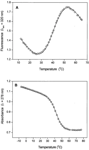
![Figure 3.14 (A) presence of 0, 40, and 100 pM FFFu (at low [KCl]). The curves were calculated using the data of Table with Fraction of unfolded protein for the C-domain of apo-CaM in the3.7](https://thumb-us.123doks.com/thumbv2/123dok_us/8612095.1401803/118.595.130.471.73.641/figure-presence-calculated-table-fraction-unfolded-protein-domain.webp)
