A C ase-B ased A ssista n t for D ia g n o sis
and A n a ly sis o f D y sm o rp h ic S y n d ro m es
C arl D a v id E v a n s
A dissertation subm itted in p artial fulfillment of the requirements for the degree of
D o c to r o f P h ilo s o p h y of the
U n iv e r s ity o f L o n d o n
D epartm ent of Com puter Science University College London
ProQuest Number: 10017424
All rights reserved
INFORMATION TO ALL USERS
The quality of this reproduction is dependent upon the quality of the copy submitted. In the unlikely event that the author did not send a complete manuscript and there are missing pages, these will be noted. Also, if material had to be removed,
a note will indicate the deletion.
uest.
ProQuest 10017424
Published by ProQuest LLC(2016). Copyright of the Dissertation is held by the Author. All rights reserved.
This work is protected against unauthorized copying under Title 17, United States Code. Microform Edition © ProQuest LLC.
ProQuest LLC
789 East Eisenhower Parkway P.O. Box 1346
A b stra ct
Dysmorphology is a field of medicine which has as one of its concerns the diagnosis of children bo m w ith m ultiple malformations. A p a tte rn of m alform ations recognised as occurring together and thought to be pathogenetically related is collectively czdled a dysmorphic 'syndrom e'. Abnormalities (dysmorphic features) m ay p ertain to clinical, ra diological, biochemical, histological or chromosomal defects. This diversity is reflected in the source of diagnostic expertise, which is relatively sparse and is provided by specialists &om varying disciplines such as clinical medicine, genetics and radiology.
Approximately 8 in 1000 of children are b o m w ith m ultiple m alform ations, and about h alf of these infants will be linked w ith a chromosomal disorder (diagnosed by performing
a karyotype). Of the rest, diagnosis (to recognised syndromes) is m ore difflcult and is a task performed by experienced specialists. Diagnosis is not always possible. A bout forty per cent of cases rem ain undiagnosed w ith respect to known disorders, and recognition of new syndromes is an im portant facet of dysmorphology. To assist such investigation, the physician has at hand reference sources such as joum als, syndrome compendia, and more recently, com puter databases. W hilst databases m ay assist diagnosis, the functions performed by specialists th a t invovle learning new syndromes have not been autom ated to any degree. This aspect provides the focus for this resesu’ch.
A ck n o w led g em en ts
I would like to thank a num ber of people at the departm ent of com puter science at UCL. I am particularly grateful to Elpida Keravnou and John W ashbrook for their guidcince and support throughout the period of this research. Also, I would also like to express thanks to Derek Long, Felicity Dams and Michael Luck for their invaluable assistance whilst preparing this thesis.
This research would not have been possible w ithout the help a num ber of m edical spe cialists who have found the tim e to assist me in my work. For this I am very grateful to Dr. Christine Hall Dr. J. A. M aat-K ient Professor David L. Rim oin and Professor Robin W inter
This research has been supported by funding from the Science and Engineering Research Council.
^ Great Ormond Street Hospital, London.
^On tem porary visit to the Institute of Child Health, London. ^ Cedars-Sinai Medical Center, UCLA.
C o n ten ts
1 Introduction to D ysm orp hology 14
1.1 Thesis O v e r v ie w ... 14
1.2 An Introduction to D ysm orphology... 16
1.3 Down Syndrome (Trisomy 21 S y n d ro m e)... 19
1.3.1 Diagnosticity and Occasional F e a t u r e s ... 21
1.3.2 A dditional Diagnostic I n f o r m a tio n ... 23
1.3.3 Etiology, Prognosis and Genetic C o u n s e lin g ... 24
1.4 Performance Tasks in D y sm o rp h o lo g y ... 25
1.4.1 D ia g n o s is ... 25
1.4.2 R esearch... 29
1.5 Conclusions and Goals of the R e s e a rc h ... 31
1.6 S u m m a r y ... 33
2 D ysm orphology, C om puter System s and A I 35 2.1 In tro d u c tio n ... 35
2.2 The London Dysmorphology D a ta b a s e ... 36
2.2.1 LDDB System G o a ls ... 37
2.2.2 A rchitecture ... 38
2.2.3 Mode of O p e ra tio n ... 39
2.2.4 D iscussion... 42
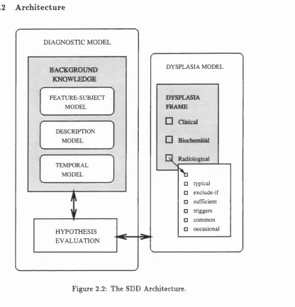
2.3 The Skeletal Dysplasia D iag n o stician ... 43
2.3.1 SDD System G o a ls ... 43
2.3.2 A rchitecture ... 44
2.3.3 Mode of O p e ra tio n ... 46
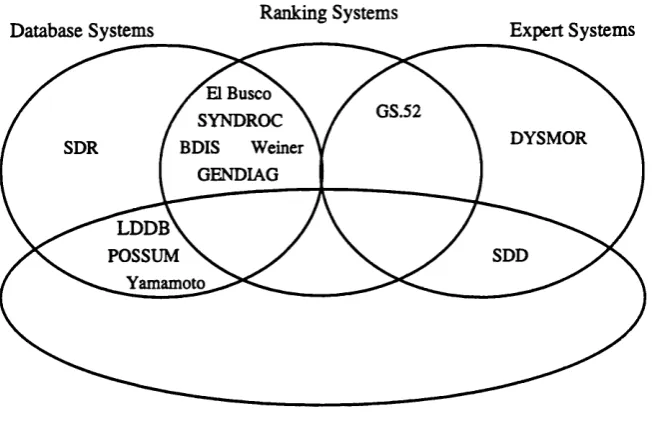
2.4 R elated W o r k ... 50
2.4.1 On-Line D a ta b a s e s ... 50
2.4.2 S D R ... 50
2.4.3 El Busca ... 51
2.4.4 S Y N D R O C ... 51
2.4.5 W ein er... 52
2.4.6 G E N D I A G ... 54
2.4.7 P r e u s ... 55
2.4.8 G S . 5 2 ... 56
2.4.9 D Y S M O R ... 57
2.4.10 Visual Reference S y s t e m s ... 57
2.4.11 D iscussion... 58
2.5 Modelling Procedures and Aspects of D y s m o rp h o lo g y ... 60
2.6 Case-Based Reasoning and L e a r n i n g ... 61
2.6.1 Introduction ... 61
2.6.2 A General CBR M o d e l... 63
2.7 Increm ental Concept F o rm a tio n ... 66
2.8 Conclusions and Philosophy of A p p ro ach ... 71
2.9 S u m m a r y ... 74
C a s e R e p r e s e n ta tio n a n d M e m o r y O r g a n is a tio n 75 3.1 In tro d u c tio n ... 75
3.2 System S o f t w a r e ... 77
3.3 Case R e p r e s e n ta tio n ... 78
3.3.1 Case S t r u c t u r e ... 80
3.3.2 Case G eneration and S t o r a g e ... 82
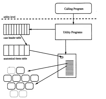
3.3.3 Generic Access and U tility F u n c tio n s ... 83
3.4 Memory O rganisation and In d e x in g ... 85
3.4.1 Indexing in D istributed Case Memory: An Extended Exam ple . . 87
3.4.2 D iscussion... 91
3.5 Case-Based Learning w ith D istributed M e m o r y ... 92
3.6 C o n c lu sio n s... 95
4 E xperim ents w ith a C ase-Based Learning A lgorithm 97
4.1 In tro d u c tio n ... 97
4.2 D iscrim ination N etw o rk s... 98
4.3 The UNIMEM A lg o r ith m ... 101
4.4 Experiments w ith U N IM E M ...105
4.4.1 Acrocephzdosyndactyly D ata S e t ... 105
4.4.2 Skeletal Dysplasias D ata Set ... 106
4.4.3 Chromosomal A bnorm ality Test C a s e s ...106
4.4.4 Results ... 110
4.5 D iscu ssio n... 122
4.6 A Critique of U N I M E M ... 125
4.7 C o n c lu sio n s...127
4.8 S u m m a r y ... 129
5 Integrating D om ain K now ledge w ith in th e CBL M odel 131 5.1 In tro d u c tio n ...131
5.2 The LDDB Model of Diagnostic Significance... 132
5.3 The W-UNTMEM A l g o r i t h m ... 133
5.4 R e s u lts ... 138
5.5 D iscu ssio n ...147
5.6 C o n clu sio n s...149
5.7 S u m m a r y ... 152
6 A G eneral T heory o f Sim ilarity 154 6.1 In tro d u c tio n ... 154
6.2 Tversky’s Theory of S i m i l a r i t y ...155
6.2.1 The C ontrast M o d e l ... 157
6.2.2 A sym metry and F o c u s ... 157
6.2.3 Similarity versus D iffe re n c e ... 158
6.2.4 Similarity in C o n te x t... 158
6.2.5 The Diagnosticity P r in c i p l e ... 159
6.2.6 D iscussion...159
6.3 Integrating the Contrast Model w ithin C B L ...162
6.3.2 The C-UNIMEM Algorithm 6.3.3 Results ... 6.3.4 D iscussion... 6.4 A C ritique of the CBL M o d e l... 6.5 CBR and General Simileirity Assessment 6.6 C o n c lu sio n s ... 6.7 S u m m a r y ...
164 166 175 183 187 190 192
7 A C B R A ssistan t for D iagnosis and A nalysis o f D ysm orphic Syndrom es 194
7.1 In tro d u c tio n ... 194
7.2 A CBR A ssistant Model and P r o c e d u r e ... 197
7.3 Ranking Hypotheses w ith the C ontrast M o d e l... 199
7.3.1 Experim ents w ith the Ranking M o d e l ... 201
7.3.2 D iscussion...204
7.4 Case-Based Learning w ith the Case-Based A ssistant M o d e l ... 208
7.4.1 Interactive CBL P r o c e d u r e ... 210
7.4.2 D iscussion...213
7.4.3 A dditional F u n c tio n a lity ...215
7.5 A dditional U tilitie s ... 216
7.5.1 The Indexing M o d e l ... 216
7.5.2 Editing the Diagnostic M o d e l...217
7.6 C o n c lu sio n s ... 217
7.7 S u m m a r y ... 218
8 C onclusions and Future W ork 220 8.1 In tro d u c tio n ...220
8.2 Relationship to other w o rk ... 221
8.2.1 C om puter Systems in D y s m o rp h o lo g y ... 221
8.2.2 CBR Models in M e d i c i n e ... 222
8.3 C ontributions to R e s e a r c h ... 224
8.3.1 C ontribution to Dysmorphology ... 224
8.3.2 C ontribution to Case-Based R easo n in g ... 226
8.4 C ritical A n a l y s i s ... 231
8.4.2 The CBR Assistant M o d e l...232
8.5 Future Work ... 233
A Term s and Procedures in D ysm orphology 235 A .l Terms of A natom ical D e v e lo p m e n t... 235
A .1.1 Individual A lterations of Form or S t r u c t u r e ... 235
A .1.2 General T erm inology...237
A .1.3 P attern s of Morphologic Defects ...237
A.2 General Principles of D ysm orphology... 239
A .2.1 Nonspecificity of Individual D efects...239
A.2.2 Variance in E x p ressio n ... 239
A.2.3 H etero g en eity ...239
A.2.4 E tio lo g y ... 240
A 3 Diagnostic Procedures and Investigations ... 240
A.3.1 H i s t o r y ... 240
A.3.2 Basic I n v e s tig a tio n s ...240
A.3.3 The Diagnostic P r o c e d u r e ...241
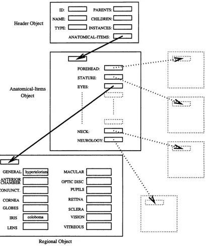
B CLOS C onstructs for LD D B R epresen tation o f Eye A nom alies 244 B .l D e s c r ip tio n ... 244
B.2 Eyes Object D e c la ra tio n ...245
B.3 Eyes Object G eneration M a c r o ...246
C Case Ordering C onsiderations for th e C -U N IM E M A lgorithm 247 C .l In tro d u c tio n ... 247
C.2 D iscu ssio n ...247
List o f F igures
1.1 Dysmorphology task analysis diagram ... 26
2.1 The LDDB A rchitecture... 38
2.2 The SDD A rchitecture... 44
2.3 The diagnostic cycle of SDD... 47
2.4 Syndrome com puter systems by category... 59
2.5 The general CBR m odel... 65
2.6 A concept hierzirchy in dysmorphology... 68
3.1 The four levels of syndrome nom enclature... 76
3.2 Software components of the general CBR architecture... 78
3.3 Object case structure... 79
3.4 Different representations of a case co-exist... 81
3.5 Object structure is hidden from calling procedures via the utility program s. 84 3.6 D istributed storage of a case... 86
3.7 Object retrieval functions... 89
3.8 Retrieval of syndromes w ith (eyes iris coloboma), (neck general abnorm al) and (neurology general abnorm al)... 90
3.9 Hierarchical linkage of header objects... 94
4.1 A sample discrim ination hierarchy... 99
4.2 The top level UNIMEM algorithm ...102
4.3 The m ain control functions in UNIM EM ...103
4.4 Idiogram of the num ber 5 chromosome...108 4.5 Network A .l: Concept hierarchy generated by the Acrocephalosyndactyly
4.6 Network S .l: Concept hierarchy generated by the skeletal dysplasias d a ta
set... 112
4.7 Mean GAF achieved by UNIM EM... 115
4.8 Network C5.1: Concept hierarchy generated by cases w ith a duplication in the num ber 5 chromosome (77 cases)...119
4.9 Network C2.1: Concept hierarchy generated by cases w ith a deletion in the num ber 2 chromosome (72 cases)...120
4.10 Diagnostic search w ith Network A ,1... 124
5.1 Modified (weighted) UNIMEM algorithm ...137
5.2 Network A.2: Revised concept hierarchy generated by the Acrocephalosyn dactyly d ata set... 142
5.3 Network S.2: Revised concept hierarchy generated by the skeletal dys plasias d ata set... 143
5.4 Network C5.2: Revised concept hierarchy generated by cases w ith a du plication in the number 5 chromosome...144
5.5 Network C2.2: Revised concept hierarchy generated by cases w ith a dele tion in the num ber 2 chromosome... 145
5.6 Com parative m ean GAF achieved by UNTMEM and W-UNTMEM... 146
5.7 A general CBL s y s t e m ... 150
6.1 Top level C-UNTMEM algorithm and generalise function...165
6.2 Evaluation contrast param eters: Acrocephalosyndactyly group... 166
6.3 Generalisation contrast param eters: Acrocephalosyndactyly group...167
6.4 Network A 3: Concept hierarchy generated by the Acrocephalosyndactyly d ata set...167
6.5 Evaluation contrast param eters: skeletal dysplasias group... 169
6.6 Generalisation contrast param eters: skeletal dysplasias group...170
6.7 Network S 3: Concept hierarchy generated by the skeletal dysplasias d a ta set... 170
6.8 Com parative m ean GAF achieved by UNTMEM, W-UNTMEM and C-UNIM EM... 171
6.10 Network C2.3: Concept hierarchy generated by cases with a deletion in
the num ber 2 chromosome...174
6.11 Network W .l: Concept nodes formed by Clinician A and Clinician B w ith SDR cases... ITT 6.12 Network H .l: Concept nodes formed by Clinician C w ith SDR cases. . . . 1T9 6.13 Mean GAF for Networks S .l, S.2, S.3, W .l and H . l ...181
T.l A general CBR model for dysmorphology... 198
T.2 Ranking contrast param eters... 201
T.3 A pplication of CBL to localised sections of the case-base...214
A .l Phenotype analysis in dysmorphology...238
C .l Network R . l ... 249
C.2 Network R .2 ... 250
C.3 Network R . 3 ... 251
List o f T ables
1.1 Main abnorm alities of Down syndrome... 20
2.1 Clinicsd regions represented by LDDB... 39
2.2 LDDB representation of eye abnorm alities... 40
2.3 Syndromes returned by feature set (07.06.03 AND 15.01 AND 32.01). . . 41
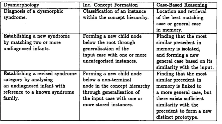
2.4 Com parative procedures of dysmorphology, CBR zmd increm ental concept form ation... 71
3.1 Number of stored objects per clinical region for 1885 LDDB syndromes. . 88
4.1 Five objects defined by attributes; colour, shape, size and orientation. . . 100
4.2 Acrocephalosyndactyly and Acrofacial Dysostoses d a ta set...107
4.3 Symbolic feature descriptions for Case 7877... 107
4.4 Skeletal dysplasias d a ta set... 107
4.5 Radiological item s represented in SDR...108
4.6 Node descriptions for Network A .l...113
4.7 Description of Node N21 in Network C5.1... 121
4.8 H ypothetical case w ith craniosynostosis... 123
4.9 Revised node descriptions for Network A .l... 125
5.1 P aram eter settings for Acrocephalosyndactyly, D up(5) and Del(2) d a ta sets. 139 5.2 P aram eter settings for skeletal dysplasias d a ta set...139
5.3 Node N4, N4.1, N5 and N5.1 descriptions from Network S.2... 148
6.1 C ontrast between Case 5754 smd Node N5 of Network A 3... 168
6.2 Clinicians A and B group descriptions of SDR cases... 178
7.1 Position of correct diagnosis in ranked differential for Acrocephalosyn
dactyly and Acrofacial Dysostosis cases 202
7.2 Position of correct diagnosis in ranked differential for Acrocephalosyn
dactyly and Acrofacial Dysostosis cases w ith Et = 20.0 203
7.3 Position of correct diagnosis in ranked differential for Nager zmd Treacher-CoUins cases w ith ^ = 1.5... 206
7.4 Case 8967 (Treacher-Collins syndrom e)...207
7.5 Ranked differential diagnosis for Case 8967... 207
7.6 Case 564525 dysmorphic features...211
7.7 Memory item s which list craniosynostosis, abnorm al fingers, and general eye abnorm alities... 212
C .l Four case orderings for Acrocephalosyndactyly d a ta set...248
C.2 Node descriptions for Network R . l ... 252
C.3 Node descriptions for Network R .2...253
C hap ter 1
In tro d u ctio n to D y sm o rp h o lo g y
1.1
T h esis O verview
A held of artihcial intelligence (AI) th a t has grown in popularity over the last decade is case-based reasoning (CBR). Case-based reasoning is linked w ith the sub-held of AI known as machine learning because it is a model for knowledge-based systems th a t are 'dynam ic'. T h at is to say, CBR systems update their knowledge-bases, and are thus viewed to leam . A crucial feature of CBR systems is th a t they comprise, am ongst other functional units, a 'case m em ory’ which stores actual cases, or exemplars, w ith which to compare and solve new problem situations.
and learning, and this thesis makes such a mapping. The work described in this thesis involves the development of a case-based reasoning system th a t is designed to assist a specialist in dysmorphology, b o th in diagnosis and learning new syndromes. Diagnostic com puter systems in dysmorphology are not new. However, the work described in this thesis is distinct in th a t it specifically addresses the research oriented learning aspect of the field.
The rem ainder of this chapter introduces the principal aspects (and problem s) of diagnosis and research in the dysmorphology domain. This allows the specific gosds of the thesis to be expressed w ith regard to the key m edical performaince tasks th a t have provided the m otivation for this research. C hapter 2 reviews previous com puter applications in dysmorphology. A comprehensive review of case-based reasoning was considered inappropriate due to the (increasing) w ealth of CBR reference m aterial th a t is available. An up to date review of com puter systems in dysmorphology was considered, therefore, to provide a m ore worthwhile contribution to the thesis The basic principles of CBR £Lre presented at the end of C hapter 2 w ith reference to the relevant aspects of the dysmorphology domain.
C hapter 3 is perhaps the m ost technical chapter in the thesis in th a t it refers to some im portant aspects of the system code (w ritten in Common Lisp). This chapter describes the underlying architecture th a t provides the platform for th e later work. However, its design does constitute an im p ortan t aspect to the work. One of the problems inherent w ithin dysmorphology is the lack of avzdlability of uniform data. C hapter 3 describes an architecture th a t has been specifically designed to operate w ith non-uniform d ata originating from different sources.
Chapters 4 through 6 focus on case-based learning (CBL). More specifically they describe the development and enhancement of a case-based learning algorithm designed to operate w ith real d a ta from the dysmorphology domain. The aim , in this instance, is to develop a system capable of assisting an expert user specifically w ith regard to the research aspect of dysmorphology, i.e., learning new syndromes. The very real-world nature of the d a ta (refiecting the fact th a t an attem p t is being m ade to m ap CBR to a real-world domain) presents a num ber of practical issues w ith regard to case-based learning, and these are discussed accordingly throughout these three chapters.
W hilst Chapters 4 through 6 focus on learning procedures in particular, it is
ta n t to rem em ber th a t the prim ary task for a physician is diagnosis. Therefore, if an application aims to provide practical assistance to a specialist, the diagnostic task should not be overlooked. This work does have such a practical aim, and w ith this goal in m ind C hapter 7 presents a general CBR model designed to provide assistance in b o th diag nosis and research. This m odel incorporates the work reported by Chapters 3 through 6. Finally, C hapter 8 concludes the thesis by summarising the relevant contributions to the designated areas of research, and discussing the potential for future work w ithin this field.
1.2
A n In tro d u ctio n to D ysm orp h olog y
Dysmorphology is th a t field of medicine which has as its concern the diagnosis of chil dren b o m w ith m ultiple malformations. To this end dysmorphology involves the study of defects in morphogenesis^ the various processes occurring during the development of the form and organs of the body. A p a tte rn of malform ations recognised as occurring together smd thought to be pathogeneticcdly related is called a syndrome. A num ber of generic term s used interchangeably to describe this entity, such as multiple congenital anomaly syndrome and dysmorphic syndrome. M alformations, which are also referred to as dysmorphic features, abnormalities or anomalies, are not necessarily visible physiczd defects. They may p ertain to clinical, radiological, biochemical, histological or chro mosomal defects. This diversity is refiected by the relevant diagnostic expertise, which is sparse and provided by specialists firom varying disciplines such as clinical medicine, genetics and radiology.
It has been estim ated th a t 8 in 1000 of children are b o m w ith m ultiple malformations [85]. Furtherm ore, about half of these infants will be linked to a chromosomal disorder recognised by performing a karyotype The diagnostic process effectively term inates if a recognised chromosomal abnorm ality is found, as this will be used to categorise the condition. If no chromosomal abnorm ality can be isolated, and the karyotype appears norm al, the task of diagnosis becomes more difficult. The m ain issues concerning diagno sis will be discussed later in this chapter. At this stage it is suffice to say th a t diagnosis is performed by specialists who have at hand not only m odem m edical techniques, such
as chromosomal analysis, biochemiczd testing and diagnostic imaging, b u t also much experience in examining and diagnosing patients w ith dysmorphic syndromes.
There are currently over two and a half thousand recognised non-chromosom«d syn dromes (although there is a belief w ithin the field th a t a num ber of these m ay eventually be linked w ith a chromosomal defect, b ut as yet cannot be so categorised due to the lim itations of chromosomal analysis). Diagnosis of children w ith a norm al karyotype is achieved by m atching the patient w ith one of the recorded syndromes. In order to do this, the p attern of anomalies exhibited by the infant m ust be determ ined and then used to link the patient to a recognised disorder. For some cases, a firm diagnosis m ay be quickly established by an experienced specialist, particularly if the condition is characterised by one or more extremely significant anomalies. For other cases, an initied exam ination m ay result in a differential diagnosis, which comprises a list of dysmorphic syndromes th a t cLre characterised by features exhibited by the patient. F urther investigation m ay allow the correct syndrome to be isolated or enable the differentizd diagnosis to be refined to the most probable candidates. This leads to an im p ortant aspect of the dysmorphology domain, certain syndromes are thought to be related. They are perceived to be related in the sense th a t they exhibit similarities in their p a tte rn of anomalies. R elated syndromes are described by dysmorphology specialists as forming fam ilies. It is not true to say th a t a diiferentiéd diagnosis will only, or usually, occur between syndromes of the same family. A differentiation will be necessary when a patient exhibits sufficient sim ilarity to m ore th a n one syndrome irrespective of w hether those syndromes are thought to be related. However, it is im p ortan t to appreciate th a t some disorders appear to form groups. A situation m ay arise in which it is difficult to diagnose a patient to zm individual syn drome, but there is evidence to link the patient to the m ore general group. In such a case, the disease would rem ain undiagnosed until there was greater understanding of how individual syndromes m ay be distinguished w ithin the syndrome family.
As indicated above, not aU individuals w ith m ultiple m alform ations can be diagnosed precisely. In fact, the two and a h alf thousand or so known syndromes only account for about sixty per cent of cases. Of the rest, some patients m ay be recorded as isolated cases th a t bare little resemblance to any of the known syndromes, whereas others m ay display some degree of sim ilarity to a known syndrome, or syndrome family, b u t not sufficient to
establish a diagnosis. This phenomenon m ay be due to the lack of a specific abnorm ality known to have particular relevance to the disorder under consideration, or perhaps may be due to a finding which is not apparent in the syndrome description. E ither way, there is sufficient dissimilarity to cause doubt about the m atch between the patient under exam ination and the syndrome under consideration. This aspect of dysmorphology results in a secondary role for dysmorphologists, th a t of researcher. Form al recognition of new syndromes is sin active process w ithin dysmorphology. However, due to th e sparsity of expertise, and the paucity of individual patients, recognition and registration of new syndromes tends to take place gradually over m any years. Reference m aterials including books [30, 75, 85, 51, 22], joum als [92, 93, 94, 95, 96] and com puter databases ^ are available which document individual cases as well as providing comprehensive diagnostic compendia of known syndromes. These endeavour to assist specialists in investigating a potential diagnosis, and to some extent can be an im po rtant aid when the relationship between syndromes is under scrutiny.
As a final comment on this brief overview of dysmorphology, it is im po rtant th a t one does not overlook the question of purpose, i.e., w hat is the aim of diagnosing a patient as having a particular syndrome, and w hat are the benefits of recognising a new syndrome? W ithin dysmorphology these questions have im plications further th a n the more straightforward answer of enabling a plan of m anagem ent or course of treatm ents for the patient. For certain known conditions a firm diagnosis m ay provide a prognosis. If the disorder is less common, a prognosis is not always possible, and certainly becomes more difficult. Recognition, categorisation and docum entation of new disorders wiU help some way towards increasing knowledge and perhaps ultim ately allow a prognosis for a greater num ber of dysmorphic syndromes. The dysmorphologist has one further im p ortan t role, th a t of genetic counsellor. W ith the more common syndromes, genetic counseling may involve informing the parents about the expected development of the disease, and perhaps informing them of the risk of recurrence if they were to have another child. In aU cases, whether a course of treatm ent is possible, lim ited, or cannot be form ulated, the parents of the child will at least seek a nam e for the condition. Diagnosis m ay provide the nam e of a known syndrome. If the case is undiagnosed, or is associated w ith a rare syndrome, th e physician m ay have greater difficulty in indicating a specific cause, or etiology, for the disorder. The ability to understand in some way, even m erely by a label, can be of
great im portance to the parents. It csm provide a m eans by which to communicate about the disease, or perhaps to examine the hteratu re themselves.
The following section is intended, by way of an example, to draw a m ore detailed pic ture of the kind of conditions described by term s such as congenitsd anom aly syndrome and dysmorphic syndrome. One of the most common and widely recognizable syndromes is Trisomy 21 syndrome, or Down syndrome. A description presents the m ain diagnostic signs of this condition as presented in one of the standard syndrome compendia. S m ith ’s Recognizable Patterns o f Human Malformation [30]. Along w ith a description of the dis ease, some of the im portant issues concerning diagnosis are addressed which are relevant to Down syndrome, b ut are applicable to dysmorphic syndromes in general.
1.3
D ow n S ynd rom e (T risom y 21 S yn d rom e)
Down syndrome is one of the oldest recorded p attern s of m ultiple anom ahes, zmd one of the m ost common (an incidence of 1 in 660 newborns). Down syndrome etiology is associated w ith a chromosomal defect, specifically trisom y ^ of chromosome 21. As this defect would be apparent when exéimining a karyotype, which would in tu rn provide sufficient proof for a diagnosis of Down syndrome, a diagnostic procedure involving ex am ination of dysmorphic features m ay not be necesssiry. However, there is no significant difference in how chromosomal gmd non-chromosomal syndromes are docum ented, apart from the fact th a t w ith chromosomal disorders the etiology is apparent and w ith non- chromosomal syndromes it is not. Discussion of diagnostic procedures w ith respect to dysmorphic features is vahd for all syndromes.
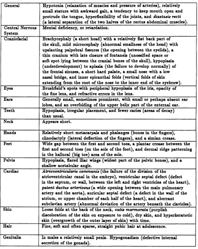
On exam ination of a patient w ith Down syndrome, a dysmorphologist m ay record a variety of anomalies such as congenital m ental retard atio n (which can range from m oderate to severe), a sloping forehead, low set ears, small ear canals, oriental appearance of the eyes, gray or very light spots at periphery of iris (known as Brushfield Spots), short broad hands w ith a single pahnzir crease (or simian crease ^), a flat nose or absent bridge and generally dwarfed physique. These signs would all support the diagnosis. In the literature, syndrome descriptions are documented m ore system atically and m ay include clinical and radiological photographs of recorded patients w ith family histories. Table 1.1
Having three of a particular chromosome instead of the usual two.
General Hypotonia (relaxation of muscles and pressure of arteries), relatively small stature with awkward gait, a tendency to keep mouth open and protrude the tongue, hyperflexibility of the joints, and dia$ta»ia recti
(a lateral separation of the two halves of the rectus abdominal muscles). Central Nervous
System
Mental defldency, or retardation.
Craniofacial Brachycephaly (a short head) with a relatively flat back part of the skull, mild microcephaly (abnormal smallness of the head) with upslanting palpebral Assures (the opening between the eyelids), a thin cranium with late dosure of fontanels (unossifled space or soft spot lying between the cranial bones of the skull), hypoplasia (underdevelopment) to aplasia (the failure to devdop normally) of the frontal sinuses, a short hard palate, a small nose with a low nasal bridge, and inner epicanthal folds (vertical folds of skin extending from the root of the nose to the inner end of the eyebrow). Eyes Brushfleld’s spots with peripheral hypoplasia of the iris, opadty of
the flne lens, and refractive errors in the lens.
Ears Generally small, sometimes prominent, with small or perhaps absent ear lobes, and an overfolding of the upper helix part of the external ear. Teeth Hypoplasia, irregular placement, and fewer caries (areas of decay)
than usual. Neck Appears short.
Hands Relativdy short metacarpals and phalanges (bones in the Angers), clinodactyly (lateral deflection of the Angers), and a simian crease. Feet Wide gap between the Arst and second toes, a plantar crease between the
Arst and second toes (on the sole of the foot), and dermal ridge patterning in the hallucal (big toe) area of the sole.
Pelvis Hypoplasia, flared iliac wings (widest part of the pelvic bones), and a shallow acetabular angle.
Cardiac Atrioventrieularia communia (the failure of the division of the atrioventricular canal in the embryo), ventricular septal defect (defect in the septum, or wall, between the left and right ventricles of the heart),
patent ductua arterioaua (a wide opening between the main pulmonary artery and the aorta), auricular septal defect (a defect in the wall of the atrium , or upper chamber of each half of the heart), and aberrant subclavian artery (abnormal deviation of the artery beneath the clavicles). Skin Loose folds at the back of the neck, cutia marmorata (purplish
discoloration of the skin on exposure to cold), dry skin, and hyperkeratotic skin (overgrowth of the outer layer of sldn) with time.
Hair Fine, soft and often sparse, straight pubic hair a t adolescence.
Genitalia In males a relatively small penis. Hypogonadism (defective internal secretion of the gonads).
lists the m ain abnormalities associated with Down syndrome. ^
. [ /\((
1.3 .1 D ia g n o s tic ity and O ccasion al F ea tu res *
---Table 1.1 provides a comprehensive set of anomalies th a t ai^ i n f a n t ^ i t h Down syn drome might exhibit. A p attern of features from this set m ay together distinguish Down syndrome from other known disorders. A patient m ay not necessarily exhibit aU the abnormalities listed, although a sufHciently conclusive subset of typical features m ust generally be evident in order to establish a diagnosis. The significance of each individual feature w ith respect to the diagnosis will vary. Some features are m ore im portant than others in order to establish, or distinguish, a syndrome, and therefore carry greater di agnostic weight. Indeed, an abnorm ality m ay be so diagnostic to a p articu lar syndrome th a t it may be considered essential for th a t particular diagnosis. The existence of very diagnostic features m ay enable a specialist to focus very quickly on a particular syn drome. However, it is im portant to note th a t generally a set of features will together support the diagnosis (hence the term ‘p a tte rn ’ is used). An isolated anom aly m ight not be particularly diagnostic, but when seen along w ith certain other abnorm alities its significance may increase. Thus, the diagnostic weight, or diagnosticity^ of an individual feature can vary according to its known relationship w ith other anomalies.
th a t disorder. However, a correlation between frequency and diagnosticity cannot always be made, and should not be assumed. For example, m ental retard atio n can occur in over six hundred different syndromes and so provide a poor handle by which to search for relevant syndromes. Conversely, it is such an im p ortan t feature th a t it m ay be essential to the diagnosis of certain conditions.
This tentative linV between diagnostic signifrcmce and incidence of occurrence is further dem onstrated by w hat are term ed occastona/features. A p a tte rn of méilformations may occasionally cause other anomalies, sometimes thought of as a kind of developmental noise. The list of m ain abnorm alities in Table 1.1 consists of defects th a t occur in at least 25 per cent and usually m ore th a n 50 per cent of patients. Sm ith denotes occasional anomalies as having a frequency of 1 to 25 per cent (most commonly 5 to 10 per cent), edthough anomalies w ith greater incidence m ay also be classed as occasional (a set of occasionals is often listed along w ith the m ain diagnostic p attern ). The existence of an occasional feature for a patient will be of no surprise to an experienced physician who has seen m any infants w ith Down syndrome. Occasional features are not random for a particular syndrome, and they do carry some diagnostic value, or weight, bu t this tends to be relatively low and can only be positive. T h at is to say, occasional features can only add support to a possible diagnosis. They do not carry enough weight in order for their absence to count against a diagnosis. Below is a list of occasional features th a t are specific to Down syndrome w ith an indication of their incidence.
• Seizures (less th an 5 per cent).
• Strabism us, or squinting (33 per cent).
• C ataract (1.3 per cent).
• Nystagmus, an involuntary movement of the eyeball (15 per cent).
• Incomplete fusion of the vertebral arches of the lower spine (37 per cent).
• Cryptorchidism, a condition in which there is a failure of the testicles to descend into the scrotum (27 per cent from b irth to nine years and 14 per cent after 15 years).
As stated earlier, to fonnnlate a diagnosis the dysmorphologist m ust use the p a tte rn of patient findings to reference and m atch the patient w ith known syndromes (this may involve techniques such as diagnostic imaging along w ith clinical exam ination). An ex pert m ay be particularly fa m iliar w ith certain syndromes or syndrome families, which m ay enable a differential diagnosis to be compiled a lm o st immediately. Otherwise, the physician m ay have to search the h teratu re for a m atch. E ither way, to estabhsh a m atch, the subject (the p atient) and the referent (the syndrome) m ust m atch across a conclu sive p attern of anomahes. A diagnostic set is a conclusive p a tte rn of anom ahes exhibited by the in fan t th a t can provide the necessary proof, or confidence, for a diagnosis. The anomahes th a t comprise the diagnostic set tend to be principal features ^ of relatively high diagnosticity, although occasional features m ay be included. S m ith’s com pendium notes the fohowing set of principal features for Down syndrome based on high incidence in recorded cases. The hst is intended as a guide for neonatal exam ination of the in fant. H ypotonia (evident in 80% of cases), a poor Moro reflex ® (85%), hyperflexibihty of joints (80%), excess skin on the back of the neck (80%), a flat facial profile (90%), slanted palpebral fissures (80%), smomalous external ears (60%), dysplasia ® of the pelvis (70%), dysplasia of the m idphalanx of the fifth finger (60%), and a sim ian crease (45%). For a conclusive diagnosis of Down syndrome a physician would expect to estabhsh a diagnostic set comprising of anomahes from these principal features.
1 .3 .2 A d d itio n a l D ia g n o stic In fo r m a tio n
There are other contextual factors which m ay also provide diagnostic pointers. For instance, the sex of an infant can be very discrim inating (some syndromes are known to have sex linkage w ith respect to inheritance, for example, Coffin-Lowry syndrome [51]). Age can also be im portant. The status of a child at presentation (or exam ination) refers to w hether the infant is ahve or not. The circumstances of a fata h ty (e.g., neonatal death or abortion) may have relevance to a diagnosis. If a child is ahve, or has hved for a significant period beyond birth, then age can also be an im po rtan t diagnostic pointer (as some syndromes are distinguished as surviving groups).
^Winter and Baraitser think in terms of hard features as abnormalities with high diagnostic signiA-cance, whilst soft features are equivalent to secondary and occasional abnormalities.
*A defensive reflex consisting of the infant’s drawing of its arms across its chest in an embracing manner in response to stimuli produced by striking the surface on which the infant rests.
A further factor concerns the use of normal features during diagnosis. W hen consid ering a diagnosis, a physician will not generally refer to norm al patient findings unless there is a direct conflict w ith syndrome expectations (for instance, if an abnorm ality is common in a syndrome, bu t the organ is norm al for the p atient). Thus, the existence of a norm al finding in the patient can create a kind of negative diagnostic value, especially if a known anomaly is regarded as essential, thus ruling out the particular syndrome under consideration (this would only be true for principal features, occasional features m ay only C2u ry positive diagnosticity). W hilst norm ality can, therefore, be (negatively) diagnostic,
a problem m ay arise if a feature is minimally norm al or abnorm al. Exclusion of the cor rect diagnosis m ay occur because of an incorrect interpretation of a m inor abnormality. It should be noted th a t there is some debate amongst dysmorphology specialists regarding this m a tter [61].
1 .3 .3 E tio lo g y , P r o g n o sis and G e n e tic C o u n se lin g
Establishing an etiology for a condition will, to a great extent, depend on the respective diagnosis. It follows also th a t a diagnosis will increase the chance of a prognosis and enable the physician to provide some degree of genetic counseling to the parents of the infant. A diagnosis of a condition such as Down syndrome will im m ediately suggest an etiology and enable a substantial level of counseling since it is a relatively common disorder w ith much docum entation. For less common conditions, the cause m ay be more difficult to establish, and b o th the prognosis and counseling given to parents will have less certainty. At the extrem e end of the scale, for cases th a t have no diagnosis or belong to a very rzue group w ith few or no references available, it m ay not be possible to establish a prognosis, and so the inform ation th a t can be given to parents will be restricted, for instance, the physician may not be able to predict the risk of recurrence.
greater th an norm al). The parents may be informed of th e risk of recurrence which is generally ziround 1 per cent, although the age of the m other m ay be im portan t. For example, the incidence for a m aternal age between 15 and 29 years is 1:1500, whereas the incidence for a m aternal age from 35 to 39 is 1:270.
1.4
P erform an ce Tasks in D y sm o rp h o lo g y
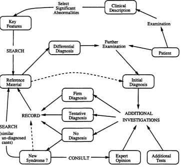
This section provides an overview of the processes th a t are intrinsic to the two key tasks performed by dysmorphology specialists: diagnosis of an affected infant to a recog nised syndrome, and investigation of new syndromes and existing syndrome categories. Figure 1.1 gives a diagram m atic representation of the m ain processes at work when per forming these tasks.
1 .4 .1 D ia g n o sis
Diagnosis is in essence a classification task. The goal of diagnosis is to accurately classify a patient w ith reference to known syndrome categories. Classification implies a process of m atching, or sim ilarity assessment. Much of this skiU em anates &om general medical knowledge in com bination w ith experience specific to the domain. To an experienced specialist some syndromes will be instantly recognisable and will not need a detailed analysis of the dysmorphic features (although an investigation will be required for con firm ation). This ability wiU reflect the dysm orphologist’s specialist medical background. For example, a radiology specialist will be fam iliar w ith syndromes th a t principally affect the skeleton, whereas a geneticist will tend to know about syndromes associated w ith a chromosomal disorder.
of establishing early on what are definite abnorm alities w ith accurate descriptions. For example, hypertelorism can easily be confused w ith telecanthus (in which case the eyes appear widely spaced due to the laterally displaced tn ncr canthal margins ^ ) , ears m ay be described as low set, bu t could actually only appear low set because they are posteriorly ro tated [85]. The interpretation of anomalies has a far reaching effect in dysmorphology through docum entation of individual cases. A physician w ith a rare case at hand may
Select Significant Abnormalities
Climcal Description
Exammation
Further Examination Differential
Diagnosis
Patient
Reference L - - Material
Imtial Diagnosis Firm
Diagnosis
Tentative Diagnosis
ADDmONAL INVESTIGATIONS
No Diagnosis (similar
un-diagnosed cases)
/ --- \
New Expert
^ Opinion
r 'v
Additional ^ Syndrome ? ^ CONSULT ^
I
TestsJ
Figure 1.1: Dysmorphology task analysis diagram.
have to search the literature to find another similar case. Accurate docum entation of anomalies is crucial to establish the sim ilarity between two such cases (docum entation will generally include photographs, which can ease this problem som ew hat). A lthough some of the available texts and com puter systems do attem p t to standetrdise a nom enclature for abnorm al features, the onus remains w ith each dysmorphologist to interpret abnorm alities
Abnormally large width between the eyes.
correctly when recording a case.
The determ ination of patient anomalies m ay also involve detecting feature dependen cies. It has been emphasised edready th a t b oth patient and syndrome descriptions are defined in term s of pattern s of anomalies. The im portance of an individual feature may depend on its relationship w ith other anomalies in the overall feature set. Feature depen dencies may be due to direct clinical or radiological relationships which would be known to medical experts. For instance, an infant m ay be described as having b o th short ribs and elongated clavicles. A radiology speciaHst would know th a t this correlation is usual. Of these features perhaps only one will be recorded which would probably the prim ary feature, short ribs. Then in any future references to this case, the secondary feature would need to be inferred by the excimining physician. So as well as a non-uniformity in feature descriptions, there exists also a non-uniformity in case descriptions. T hat is to say, the detail in docum ented patient descriptions varies greatly. Some patients are recorded merely in term s of one or two principal abnorm alities, whilst some case descrip tions contain a more comprehensive list of dysmorphic features th a t were noted during exam ination. This type of non-uniformity can further complicate the task of m atching
rare cases.
Dependencies between anomalies m ay also p ertain to observations of co-occurring features th a t have been recorded for certain conditions, or a num ber individual cases. These correlations m ay or m ay not have relevance to the diagnosis under consideration, and the examining physician m ay be required to examine the family history of the pa tient in order to establish any significance. C ertain features m ay be evident as harmless tra its in some families, for example, big thum bs and toes, which is a com bination seen in Rubenstein-Taybi syndrome [85]. If a patient has big thum bs and toes it m ust be established w hether or not these features were evident in norm al, healthy ancestors be fore they are included in the overall p a tte rn of anomalies (and therefore given diagnostic relevance). If the family history dictates th a t these features can exist normally, then the physician m ust carefully establish the other known features for Rubenstein-Taybi syndrome before proposing th a t diagnosis.
m ay result from specific knowledge, or experience, of the examining physician. If one pgirticular syndrome stands out as the prim ary candidate, perhaps due to the existence of one or two particularly significant abnorm alities, then further investigation m ay be focussed on establishing th a t diagnosis. For instance, further exam ination m ay be biased in the sense th a t there is a diagnosis ‘in m ind', and the physician m ay look for specific anomalies due to the diagnosis under consideration. This can lead to a quick diagnosis if the physician’s suspicions are confirmed (by the existence of the relevant diagnostic set). However, if th e respective anomalies cannot be found, the physician may be forced to reassess the proposed diagnosis. This can result in a reappraisal of the significance of anomalies recorded on initial exam ination.
If the recorded p attern of abnorm alities matches a num ber of syndromes, then it m ay be necessary for the physician to refine this hst. Further exam ination m ay take place, perhaps w ith greater significance placed on secondary and occasional features. This iterative process of exam ination and re-exam ination is ultim ately geared towards proposing the m ost accurate form of diagnosis given the extent of the inform ation at hand. For some affected infants, no diagnosis m ay be possible. In some cases, perhaps only a differential diagnosis is proposed. If the physician has a degree of certainty, then a firm, or tentative, diagnosis may be p u t forward.
At this point, given some form of initial diagnosis, the examining physician m ay seek further evidence through other means. A dditional investigations can take on a num ber of guises. For example, initial clinical exam ination m ay have suggested a syndrome w ith a known skeletal anomaly, and so the physician m ay want to examine relevant x-rays, or consult w ith a radiologist. The cooperation between dysmorphologists specialising in different medical disciplines is an im po rtan t necessity in diagnosis. C ertain features may require the opinion of a specialist in a relevant field of medicine. For example, the
A d d en d u m
1 .4 .2 R esea rch
The role of research in dysmorphology is associated w ith the ever changing nature of the field and an associated desire to leam and understand more about it. Syndromes th a t once were thought to be very different, and were consequently classified separately, sometimes emerge as different m anifestations of the szime condition, or as the same condition at different points in its n atu ra l history. Alternatively, conditions th a t appear the same may, at a later date, be found to have very different underlying dynamics and prognoses. By m onitoring b o th diagnosed and undiagnosed cases over tim e, the dysmorphologist may be able to spot previously unregistered correlations and establish previously unknown etiologies. Documenting the development of affected infants may enable more accurate prognosis and counseling in future.
Along w ith the compendia cited earlier, which are designed w ith an emphasis on diagnosis, the dysmorphologist also has at hand numerous research publications th a t can assist investigation [92, 93, 94, 95, 96]. These journals provide a m edium by which to communicate w ith other specialists. There are also a num ber of annual conferences and workshops for medical specialists to attend. Comm unication is crucial in order to allow practitioners to announce new findings, discuss p otential links between rare cases, and ultim ately propose new syndromes (or establish revisions of known categories).
One of the results of docum entation, analysis and communication w ith respect to cases of m ultiple malformations has been a nom enclature for syndromes and syndrome families. T h at is to say, there has been some thought towards higher order syndrome taxonomies. However, in keeping w ith the uncertain and ever changing n ature of th e field, nomenclatures are not interpreted rigorously. Compendia such as S m ith’s are structured in a way th a t reflects syndrome nom enclature. In Sm ith’s book, syndromes are grouped across twenty or more general categories such as chromosomal abnormédity syndromes, syndrome characterised by a very small statu re, syndromes w ith facial defects as a m ajor feature, craniosynostosis syndromes and osteochondrodysplasias Specialists within radiology have denoted a m ore specific classification of osteochondrodysplasias [69]. This working group has defined a taxonomy consisting of three general groups: defects of the tubular (and flat) bones an d /o r axial skeleton, disorganised development of cartilagenous
^^Premature closure of the skull sutures
and fibrous components of the skeleton, and idiopathic osteolyses W ithin the first group, affecting tubular bones, twenty four sub-categories have been defined. Similarly osteolyses are divided along four sub-groups. An im portant point about these syndrome groupings (which has relevance to issues discussed in C hapter 2) is th a t they effectively provide a category hierarchy. However, although a hierarchical classification of syndromes is conceivable, there is some reluctance to adhere to hierarchical schemas. The existence of syndrome clusters is generally accepted, but individual syndromes are not necessarily mutucdly exclusive. T h at it is to say, some syndromes m ay belong to more th an one higher order category. It would seem also th a t hierarchies p ertain only to relatively well understood syndrome families.
The specific procedures of learning involve the revision of a syndrome description or a higher order syndrome category. Such revision can occur in a num ber of ways. One process is the straightforward addition of a new disorder to the syndrome nom enclature, either w ith or w ithout reference to existing categories. A new syndrome definition may be based on a link between two or more undiagnosed infants th a t stand isolated w ith reference to the existing syndrome nom enclature. It is im p o rtan t to note th a t some known syndromes are based on the descriptions of only a few cases (an example of this instance is Pfeiffer syndrome). These are precisely the syndromes where diagnosis is the m ost difficult. A new syndrome m ay also be defined w ith reference to a known disorder or family of disorders. If an individual has sim ilarity to a known syndrome family, but not specifically to one of the family members, then it m ay be tentatively linked w ith the syndrome group until there is a greater understanding of th a t group. As further similar cases are registered and placed in the same vicinity of the nom enclature, a new, distinct syndrome may be formed within the syndrome family.
Category inform ation may also be revised as m ore infants become diagnosed. The level of detail of syndrome descriptions can vary according to the num ber of diagnosed children. This is evident w ith the more common conditions for which statistical inform a tion is sufficient to provide accuracy (and confidence) in the syndrome description used for diagnosis. Descriptions of syndrome families also change over tim e. The definition of a syndrome is formed as a generalisation of the features by which diagnosed cases are adjudged to be similar. As m ore cases become registered, it m ay become apparent to the investigator th a t the group m ay be divided along certain tra its, whilst still dem onstrating
some higher order relationship. Thus, knowledge may be derived in two directions: by generalising across a set of similar cases in order to establish a diagnostic set and by increasing the specificity amongst a set of related cases.
1.5
C on clu sion s and G oals o f th e R esearch
Dysmorphology is a complex and diverse medical domain. The state of the domain is incomplete, or weak, in the sense th a t there are m any unknown cases and the level of
understanding of dysmorphology is not total. It is very much a dynamic field of medicine with knowledge about syndromes increasing gradually over tim e. Thus, dysmorphology is not merely a domain for medical diagnosis, b ut also one of medical research. P racti tioners, whether performing diagnoses or acting as researcher, are medical experts who rely on b o th general and specialised medical knowledge, and experience.
There has been a n atu ral progression from texts to com puter systems in order to provide support and assistance to the dysmorphologist. The following chapter reviews computer applications dedicated to dysmorphology. These vary in design and conse quently in their scope. Some are merely syndrome databases. Some, on the other hand, employ artificial intelligence techniques in order to aid diagnosis, and m ay therefore be regarded as expert systems. One aspect common to all these com puter systems is th a t they address the prim ary task of diagnosis, either through an autom ated diagnostic pro cedure or simply through a look-up facility. Some address the potential of using com puter systems to leam new syndromes. However, none have specifically tackled this problem. Such investigation is left as an implicit task for the user.
The general them e of this research is to increase the scope of assistance available to a specialist in dysmorphology from a com puter system. However, it is the learning as pect of the domain, i.e., the recognition of new syndromes and the revision of syndrome categories, th a t provides the specific focus of this work. In particular, the thesis involves the development of a com puter system th a t utilises 'case-based reasoning’. Case-based reasoning (GBR) is a field of artificial intelligence (Al) linked to machine learning. An im portant feature of CBR systems is th a t they have the ability to leam automatically. T h at is to say, they can update their knowledge, which is stored in the form of spe cific and generalised cases, dynamically. A specific aim of the work is to autom ate the
learning process of dysmorphology by designing and implem enting a CBR system. Thus, unlike previous applications, this work specifically addresses the secondeiry learning task performed by dysmorphology specialists. The ability of a CBR system to leam auto m atically depends to a great extent on the stm ctu re of the case database, or case-base, éind the m ethods for assessing the sim ilarity between individual cases. To this end, CBR has much overlap w ith another field of machine learning called ‘increm ental concept for m atio n ’. Following the review of dedicated com puter systems in C hapter 2, the m ain features of these two AI disciplines are discussed w ith reference to the dysmorphology domain.
It has been stated th a t the m ain focus of research is an autom ated, case-based rea soning approach to the learning task of dysmorphology. W ith this in m ind, a background goal of the work is to design a practical solution which enables the prim ary task of diagno sis to be performed as well as specifically autom ating the learning process involved w ith the proposal of new syndromes. An im po rtant tenet of this research is th a t b o th diagno sis and learning can be represented using case-based reasoning. Furtherm ore, the same underlying case-based system architecture can be utilised by b o th ‘calling procedures’: object retrieval (i.e., retrieving similar syndrome or case entities from m em ory), and a learning CBR program (called case-based learning, or CBL). C hapter 3 describes this architecture, which lays the foundation for the subsequent chapters specific to case-based learning, and the general CBR m odel which is discussed in C hapter 7.
on machine learning. They assess, and enhance, a recognised learning algorithm with respect to a real-world, and incomplete medical domain. Added to this. C hapter 6 con siders (eind answers) some of the open issues of sim ilarity assessment w ith regard to CBR in the light of the work th a t has been done.
Finally, C hapter 7 describes a case-based reasoning system th a t facilitates diagnostic assistance as well as the research objective of learning new syndromes. This design provides a general CBR solution, derived &om the work involved w ith learning algorithm s (C hapters 4, 5 and 6), and the case-based architecture described in C hapter 3.
1.6
Sum m ary
The following points summarise the m ain characteristics of the dysmorphology domain and goals of the thesis.
• The domain is incomplete. The acquisition of knowledge concerning dysmorphic syndromes is ongoing and is perform ed by experienced specialists of varying medical backgrounds.
• Two performance tasks exist: diagnosis and research. B oth involve a process of as sessing the sim ilarity between two entities. Diagnosis involves m atching a patient to known syndrome m alform ation p a tte rn s . Research involves comparing undiagnosed infants w ith a view to learning new syndromes.
• W hilst syndrome category structure is very loosely defined, generally a m a tte r of interpretation, and not rigidly adhered to by dom ain experts, syndromes do appear to form groups, or families. The dom ain is not yet at a stage at which a single hierarchical categorisation can be defined.
• Previous com puter systems dedicated to dysmorphology have addressed only the diagnostic task. The research task has not specifically been autom ated to any de gree. This work addresses learning in dysmorphology. More specifically it involves the design and im plem entation of a com puter system th a t autom ates this mode of operation.
maps well w ith facets of the dedicated domain and facilitates autom ated learning of syndromes and revision of syndrome categories.
• The m ain thru st of the work is to design and implem ent a model of case-based reasoning, specifically focussed at addressing the learning task. A standard CBL
model is taken as an initial starting point, im plem ented, tested w ith d ata from the dysmorphology domain, and extended in order to improve performance.
• The complexities of the dysmorphology domain expose problems w ith regard to the case-based learning model and the theoretical assumptions on which it is founded. These issues are addressed, and solved by a m ore general case-based reasoning system th a t incorporates the CBL program , bu t also offers functions aimed at providing asisstance w ith the primeiry task of diagnosis. This approach is driven by a desire to provide a case-based system th a t offers a practical solution w ith regard to bo th diagnosis and research in dysmorphology.
A d d en d u m
C h ap ter 2
D ysm orp h ology, C o m p u ter
S y ste m s and A I
2.1
In tro d u ctio n
The utilisation of reference m aterials (books, journals and com puter systems) is an im p o rtan t, if not crucisd, step in bo th diagnosis and research. The general structure of syndrome compendia has already been described in the previous chapter. This chap ter reviews com puter apphcations w ithin dysmorphology. Some of these apphcations are essentially computerised equivalents of compendia, i.e., database systems. A typical database system would offer increased efficiency in a search for possible syndromes or similarly affected infants (if stored in the database) by using a relevant keyword indexing mechanism. A num ber of reseeu’chers, however, have endeavoured to extend the basic performance goal of a com puter system beyond th a t of a straightforw ard look-up facility. In such cases energy has been directed towards modelling some of the processes at work during diagnosis.
diagnostic system, applicable across all known syndromes, then one can probably expect to establish a weak diagnostic model which at best provides a m anageable differential diagnosis (which is small enough to refine through further exzimination). This is the real istic scope of a database application. On the other hand, as has gdready been indicated, syndromes do appear to form groups which m ay be described or specified across certain chromosomal, clinical or radiological criteria. If a system is intended to follow a diagnosis procedure for a specific, distinctive group, then it m ay be feasible to acquire sufficient knowledge firom a specialist who is particularly familiar w ith th a t group in order to cre ate a strong diagnostic model. This would allow the development of a more intelligent system th a t produces greater accuracy in the differential diagnosis.
The following sections describe two systems in detail: the aforeneimed London Dys morphology D atabase which, in term s of operation, typifies a database application, and the Skeletal Dysplasia D iagnostician [15, 36], a m odel-based expert system which demon strates a knowledge engineering approach to development of a system dedicated to the diagnosis of a specific group of syndromes. These two systems correspond to the two above m entioned extremes in term s of system design and operation. O ther com puter applications are subsequently discussed w ith reference to these two systems. The topic of discussion will then switch to theoreticzd ideas &om w ithin artificial intelligence (AI) th a t are relevant to this research, and in particular to the system design issues th a t have been highlighted.
2.2
T h e London D y sm o rp h o lo g y D a ta b a se
Diagnostic aids such as reference texts and com puter systems have arisen due to b o th the sparsity of expertise and the necessity to cope w ith the increasing num ber of dysmorphic syndromes. For database systems, two m otivating factors have influenced development: portability and upgradabihty. The general increase in use of com puters, particularly personal com puters, has m eant th a t a system designed using well established software is inherently portable and C2ui be readily distributed to medical centres around the world.