Visualization of Irreparable Ischemic Damage in Brain by Selective
Labeling of Double-Strand Blunt–Ended DNA Breaks
Vladimir V. Didenko, Hop Ngo, Candace L. Minchew, Denise J. Boudreaux, Marsha A. Widmayer, and David S. Baskin
Department of Neurosurgery, Baylor College of Medicine and VAMC, Houston, TX, USA
Accepted September 15, 2002
Abstract
Background:Double-strand DNA breaks with blunt ends represent the most serious type of DNA damage, and can-not be efficiently repaired by cells. They are generated in apoptosis or necrosis and are absent in normal or tran-siently damaged cells. Consequently, they can be used as a molecular marker of irreparable cellular damage. We evaluated the effects of focal brain ischemia using selec-tive labeling of blunt-ended DNA breaks as a marker of ir-reversible tissue damage. A new approach permitting such analysis in situ is introduced.
Materials and Methods:Rat brain sections taken 6, 24, 48 and 72 hr after the onset of focal brain ischemia were used. Double-strand DNA breaks were detected directly in the tissue sections via ligation of blunt-ended hairpin-shaped oligonucleotide probes. The probes were attached to the ends of the breaks by T4 DNA ligase. Conventional
Address correspondence and reprint requests to: David S. Baskin, 6560 Fannin, Suite 944, Houston, TX 77030. Phone: 713-798-4696; fax: 713-798-3227;
e-mail: [email protected]
cresyl violet co-staining and terminal transferase based la-beling (TUNEL) were employed to analyze the distribu-tion of labeled cells.
Results:Double-strand blunt-ended DNA breaks rapidly accumulate in brain cells after focal brain ischemia. At 24 hr, they concentrate in the peripheral areas of stroke, which are prone to ischemia-reoxygenation. By 48–72 hr, this type of DNA damage spreads inward, covering the in-ternal areas of the ischemic zone.
Conclusions: Selective labeling of blunt-ended DNA breaks delineates the dynamics of stroke-induced irre-versible DNA damage and provides highly specific detec-tion of brain cells with irreparable DNA injury. It can be used for comparing the efficiency of various anti-ischemic drugs, particularly those that target DNA damage, as well as for monitoring stroke-induced damage.
Introduction
Molecular markers of cellular phenomena are im-portant for the assessment of experimental inter-ventions. Markers of cell death are most widely used. In cell cultures, a general assessment of cell death is performed using vital dyes, which pene-trate into cells only if membrane permeability is severely disturbed. In this case, irreversible mem-brane damage is used as a general molecular marker of cell death. However, similar analysis is not pos-sible in sectioned tissues, where cellular membrane integrity is compromised. Because tissue sectioning is a fundamental element of many analyses, it is important to have a general molecular marker of cell death applicable for this situation. Convention-ally used terminal transferase based labeling (TUNEL) detects all possible DNA break configu-rations, is nonselective, and labels not only dead but also transiently damaged cells (1). In this report, we introduce detection of double-strand blunt-ended DNA breaks as a new selective marker of cell death, which is applicable for research using sectioned tissue.
Double-strand blunt-ended DNA breaks consti-tute the most serious type of DNA damage. Neither DNA strand is able to provide physical integrity or information content necessary for successful repair, as can occur with single-strand breaks, where one strand in the duplex remains intact. Among various types of double-strand breaks, which include dou-ble-strand breaks with 3⬘ or 5⬘ end extensions, the blunt-ended variety represents the least efficiently repaired type. Even when ligated, these breaks still result in irreversible alteration of the DNA and cell death (2,3). Generation of significant amounts of blunt-ended DNA breaks is characteristic of both apoptosis and necrosis (4,5), whereas reversible cel-lular damage after ischemia or chemical interven-tions is not associated with their production (1). Other types of DNA damage, such as breaks with 3⬘ overhangs or single-strand breaks, are either re-stricted to apoptotic DNA cleavage (4) or are repro-duced in both transiently damaged and dying cells (1,4). Thus, blunt-ended double-strand breaks rep-resent a more specific death-associated type of DNA damage, and can serve as a biochemical common marker for cell death in both apoptosis and necrosis (4,6).
that they are sometimes viewed as a single, ischemic brain-specific type of cell death (9).
In this study, we introduce a new approach to visualize irreparable stroke damage in tissue sec-tions by detecting unrepairable double-strand blunt-ended DNA breaks. We use a new in situ ligation technique to detect the breaks, which serve as mol-ecular markers of cell death. The approach exclu-sively labels lethally damaged cells regardless of the apoptotic or necrotic nature of their death, and pro-vides high cell-to-cell resolution.
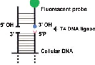
The assay detects blunt-ended 5⬘ phosphory-lated double-strand breaks using hairpin-shaped blunt-ended oligoprobes, which are attached to the ends of the breaks by T4 DNA ligase. This permits selective detection of only a single type of DNA damage (4,10,11).
Using the new approach, we demonstrate that double-strand blunt-ended DNA breaks rapidly ac-cumulate in brain cells after focal brain ischemia, and delineate stroke-induced progression of cell death. This new approach may be useful for assess-ment of the efficiency of anti-ischemic therapies, particularly those that target DNA damage, as well as for monitoring stroke-induced damage.
Methods
The animal care and use portion of the following protocol was reviewed and approved by the Institu-tional Animal Care and Use Committee of the VA Medical Center and the Animal Protocol Review Committee of Baylor College of Medicine. All pro-cedures were carried out with strict adherence to the protocol and the National Institutes of Health animal research guidelines.
Focal Brain Ischemia
All surgical techniques were performed under gen-eral inhalational anesthesia. Occlusion of the right middle cerebral artery (MCA) was performed in male Long Evans rats through an incision made midway between the right eye and ear. Using the operating microscope, the right MCA was exposed transcra-nially via a subtemporal craniotomy without damage to the zygomatic bone. Ten to twelve millimeters of the MCA, beginning ventral to the olfactory tract, was permanently occluded using bipolar coagulation.
The animals were transcranially perfused with paraformaldehyde 6, 24, 48, and 72 hr after occlu-sion, and the brains were removed and post-fixed overnight. A 2-mm thick coronal block was cut from the center of the MCA territory, beginning ⬃0.5 mm anterior to the optic chiasm and ending ⬃0.5mm posterior to the optic chiasm. The tissue was embed-ded in paraffin and cut in 6-m thick sections. Sec-tions used in the current study were taken from the middle area of the tissue block. Each staining condi-tion was repeated in four separate seccondi-tions from each of the eight animals.
Cresyl Violet Staining
The extent of the ischemic injury was determined by cresyl violet staining. The sections were deparaf-finized with xylene, dehydrated in 100% alcohol (3⫻2 min) and stained in 0.02% cresyl violet solu-tion for 60 min. They were briefly washed in 95% and 100% ethanol, cleared in xylene, and covered with Permount. The area of cellular damage was clearly identified using this technique.
The Oligoprobe for In Situ Ligation
The oligoprobe sequence was designed to avoid mis-folding into any structure other than a blunt-ended hairpin, so that it was able to ligate to blunt-ended DNA breaks. Biotin was incorporated into the probes by chemical insertion of biotin triethylene glycol (TEG) phosphoramidite (Glen Research, Sterling, VA, USA) directly into the oligonucleotide backbone (In-tergen Company, Purchase, NY, USA). The sequence of the probe was: 5⬘GCG CTA GAC C5*G GTC TAG CGC 3⬘ (5*- represents biotin-TEG spacer). Probes were detected in a reaction with a streptavidin-FITC conjugate (Vector Laboratories, Burlingame, CA, USA) (see In Situ Ligation).
In Situ Ligation
In situ ligation using hairpin shaped oligoprobes was performed as previously described (9). Briefly, 6-m thick sections were deparaffinized with xy-lene, rehydrated in graded alcohol concentrations, washed in water, and treated with proteinase K (50 g/ml) for 15 min. Sections were rinsed with water and incubated in 80 l of the ligation buffer without the probe (66 mM-Tris HCl, pH 7.5, 5 mM MgCl2, 0.1 mM dithioerythritol, 1 mM ATP, and
15% polyethylene glycol-8000) for 15 min to ensure even saturation. The buffer was aspirated, and the full ligation mix containing the ligation buffer with the hairpin probe (35 g/ml) and T4 DNA ligase (250 U/ml) was applied to the sections. As a mock control reaction, an equal volume of 50% glycerol in water was substituted for T4 DNA ligase.
Sections were incubated in a humidified cham-ber for 16 hr at 23⬚C, and were then briefly washed in water. The signal was visualized using TSA-direct kit (NEN Life Science Products, Boston, MA, USA), with color development stopped on all sections si-multaneously. Sections were then counterstained with 4,6-diamidino-2-phenylindole (1 g/ml).
Terminal Transferase Based Nick-End Labeling
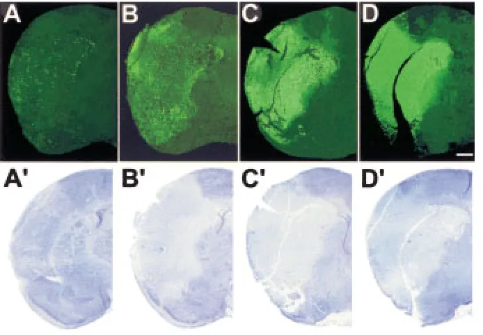
irreversibly damaged cells are located scattered throughout the zone. After 24 hr of focal brain is-chemia, cell death and DNA injury are much more pronounced (Fig. 2B). Interestingly, at 24 hr, the dy-ing cells with extensive double-strand blunt-ended DNA breaks are concentrated at the boundaries of the stroke. The signal is particularly strong in many areas neighboring undamaged tissue. This suggests that ischemia-reoxygenation episodes may con-tribute to the distribution of DNA damage and cell death. These reoxygenation episodes frequently oc-cur at the boundary between normal and ischemic tissue.
After 48 hr of continuing ischemia, the progression of cell death results in the appearance of large numbers of dying cells in the central parts of the ischemic zone. Seventy-two hours after ischemia onset, cells with high numbers of double-strand blunt-ended DNA breaks occupy all parts of the ischemic zone.
The comparison of in situ ligation-based assess-ment of brain damage with cresyl violet staining il-lustrates the much higher sensitivity of the new ap-proach, especially at the earliest time points. Because the new approach is able to resolve the signal at a single cell level, it permits both a general overview of the damaged area, as well as an analysis of cellu-lar populations affected by ischemia (Fig. 2).
The general territory occupied by stroke at 72 hr is already outlined at 6 hr by in situ ligation, but not with cresyl violet staining. The cresyl violet staining does not show the dynamics of stroke progression in comparable detail to the new approach, which clearly visualizes several stages.
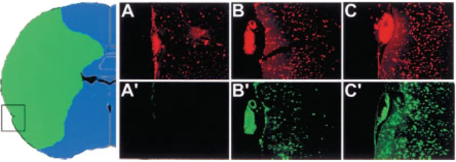
Figure 3 demonstrates the early generation of double-strand breaks at the boundaries of stroke, and the subsequent progression of their generation and cell death inward into the ischemic zone. This early cell death at the boundary of stroke can be ex-plained by the repeated episodes of ischemia-reoxy-genation, which have been shown to occur in the pe-ripheral areas of brain ischemia on the boundary with unaffected tissue and can worsen cellular dam-age in these areas via production of free radicals (13). Interestingly, in areas not bordering healthy tis-sue, the in situ ligation signal can appear at a later time compared to the internal brain areas (Fig. 4). Colabeling using TUNEL demonstrates that ischemia-damaged cells in this area can initially become TUNEL positive, and then later show the in situ ligation signal (Fig. 4).
Discussion
The detection of blunt-ended double-strand DNA breaks represents a new modality useful for deter-mining the extent and distribution of irreversible cel-lular damage and death in ischemic brain. This type of break is associated with lethal injury, thus transiently damaged cells are not labeled. In previous reports, we describe the use of in situ ligation for detection of
Results
Figure 1 represents the principle of the assay for se-lective in situ labeling of blunt-ended DNA breaks. It demonstrates a double-strand oligonucleotide probe with blunt ends ligating to a genomic blunt-ended DNA break. The ligation occurs if a break possesses a 5⬘PO4group, needed for the creation of
a bond by T4 DNA ligase. The 5⬘phosphates at the ends of the breaks are either produced in the cleav-age reaction by apoptotic or nonspecific cellular nu-cleases, or are introduced by kinases and trans-ferases acting on 5⬘OH bearing DNA ends generated by the lysosomal DNase II.
To make a rough estimate of the sensitivity of the assay in our conditions, in preliminary experi-ments we established that our detection system composed of an Olympus IX-70 microscope with an attached MicroMax digital videocamera, can visual-ize 1.25 fmole of FITC spotted as a 1.3 mm dot (not shown). This corresponds to ⬃45,000 FITC mole-cules per 80 m2, the area of a 10-m diameter nu-cleus. Using a TSA-direct kit for fluorescence devel-opment, the signal can be increased by two orders of magnitude. As the assay places a single biotin at the end of each break, detection of several thousand double-strand blunt-ended breaks per nucleus is observable. This number of breaks far exceeds the repair capabilities of any cell, and is comparable to the total number of breaks observed in the initial stages of apoptosis (12).
Figure 2 illustrates the dynamics of the irrepara-ble brain tissue damage at different time-points after the onset of focal brain ischemia in rat, as visualized by labeled cells with high concentrations of blunt-ended DNA breaks (upper panel; green fluores-cence). A traditional cresyl violet staining is pro-vided for comparison (lower panel).
After 6 hr of ischemia, the boundary of the is-chemic zone is already clearly delineated by the cells with extensive DNA damage labeled by in situ ligation (Fig. 2A) and relatively small numbers of
specific DNA breaks created by the apoptotic nucle-ases, with particular emphasis on detection of short 3⬘ overhangs (4,10,11). In this paper, however, attention is shifted to detection of blunt-ended breaks, which expands the utility of the approach.
Blunt-ended DNA breaks are not found in nor-mal brain cells. Very snor-mall numbers of blunt-ended breaks (one or several/cell) can occur during early B-and T-cell development in a process of V(D)J recom-bination (3). However the detection of these single breaks is below the limit of sensitivity of the in situ ligation technique (4) and, therefore, will not pro-duce false positives.
Several other labeling techniques are currently employed for the determination of cell injury in brain ischemia. Cresyl violet staining is traditionally used for visualization of stroke in tissue sections. The tech-nique labels damaged areas via negative staining, be-cause the injured cells do not retain the dye. Being simple, it is frequently used. However, the absence of detection of an identifiable molecular marker of dam-age makes the technique less appealing. In addition, its ability to visualize stroke damage, especially
early, is much less specific compared to the new approach (Fig. 2).
Terminal transferase labeling (TUNEL staining) is also often employed for visualization of cell dam-age in stroke. Yet the method indiscriminately de-tects any exposed 3⬘OH groups, some of which can be present in transiently damaged (1) and even nor-mal dividing cells (14). In addition it is prone to artifactual labeling, as single-strand breaks of various kinds can be created in tissue fixation (15) and processing such as microwave treatment in co-staining with antibodies (4) and even in the process of cutting sections by microtome (16). Assessment of the effectiveness of anti-apoptotic interventions us-ing TUNEL is difficult due to the fact that survivus-ing cells often still contain high numbers of reparable single-strand DNA breaks (1,17).
Although the in situ ligation signal appears sev-eral hours later compared to the TUNEL signal, it is far less ambiguous and has more value in comparing the effects of different interventions, because in such assessments, it is critical to discriminate between lethally and nonlethally damaged cells.
Fig. 3. Detection of lethally damaged cells at the boundary of stroke by in situ ligation at 6 and 72 hr after the onset of ischemia. The first dying cells appear at the boundary at the earliest time point of 6 hr. Large numbers of dying cells are demonstrated within the stroke area at a later time of 72 hr post-ischemia. The arrow in the inset indicates the direction of progression of DNA damage and cell death as time goes by. (Bar, 150 m).
tutes of Health (D.S.B.), by grant 004949-054 from the Texas Higher Education Coordinating Board (D.S.B. and V.V.D.), and by grants from DeBakey Medical Foundation (V.V.D.), Baylor College of Medicine (V.V.D.), The Taub Foundation, The Henry J.N. Taub Fund for Neurosurgical Research, The George A. Robinson, IV Foundation, The Blanche Greene Estate Fund of The Pauline Sterne Wolff Memorial Foundation, and The Seigo Arai and Kop-pelman Funds of The Neurological Research Foun-dation (D.S.B.).
References
1. Didenko VV, Wang X, Yang L, Hornsby PJ. (1999) DNA dam-age and p21WAF1/CIP1/SDI1 in experimental injury of the rat adrenal cortex and trauma-associated damage of the human adrenal cortex. J. Pathol.189:119–126.
2. Lips J, Kaina B. (2001) DNA double-strand breaks trigger apoptosis in p53-deficient fibroblasts. Carcinogenesis22:579– 585.
3. Lieber MR. (1998) Pathological and physiological double-strand breaks: roles in cancer, aging, and the immune system. Am. J. Pathol.153:1323–1332.
4. Didenko VV, Hornsby PJ. (1996) Presence of double-strand breaks with single-base 3⬘ overhangs in cells undergoing apoptosis but not necrosis. J. Cell Biol.135:1369–1376. 5. Al-Lamki RS, Skepper JN, Loke YW, et al. (1998) Apoptosis
in the early human placental bed and its discrimination from necrosis using the in-situ DNA ligation technique. Hum. Reprod.13:3511–3519.
6. Al-Lamki RA, Skepper JN, Loke YW, et al. (1998) Apoptosis in the early human placental bed and its discrimination from necrosis using the in situ DNA ligation technique. Hum. Reprod.13:3511–3519.
7. Morita-Fujimura Y, Fujimura M, Yoshimoto T, Chan PH. (2001) Superoxide during reperfusion contributes to caspase-8 expression and apoptosis after transient focal stroke. Stroke 32:2356–2361.
8. Moroni F, Meli E, Peruginelli F, et al. (2001) Poly(ADP-ribose) polymerase inhibitors attenuate necrotic but not apoptotic neuronal death in experimental models of cerebral ischemia.Cell Death Differ.8:921–932.
9. Fukuda T, Wang H, Nakanishi H, et al. (1999) Novel non-apoptotic morphological changes in neurons of the mouse The new approach detects irreversible tissue
damage only when it is accompanied by extensive DNA degradation, which occurs in ischemia, reoxy-genation, apoptosis, radiation exposure, and anti-cancer treatment with DNA-targeting drugs. In some other situations, such as instant cell death caused by chemical agents disrupting cellular membranes or by rapid freezing, cell death is not accompanied by extensive DNA damage. We believe that the major utility of this new approach is in assessing the effi-ciency of various anti-ischemic, antineoplastic, and antiradiation interventions, where it could provide valuable quantitative comparisons of the extent of irreversible DNA damage.
Detection of a marker for lethal cell injury is also important in the analysis of ischemic brain tissue. The absence, or considerable variability, of the classical markers of apoptotic cell death was demonstrated in ischemic brain. For example, different sets of activated caspases were detected in various apoptotic brain cells (18). In addition, the absence of classical apoptotic morphology has recently been demonstrated in a number of apoptotic brain cells (19). It is also impor-tant that the necrotic component of cell injury cannot be selectively labeled; its detection requires morpho-logic evaluation on a cell-by-cell basis (19).
Because in situ ligation detects an important and early point of no return for lethal cell injury of all types, we believe that it will be of value for delin-eation of lethal DNA damage in brain tissue. It is not intended to replace traditional staining techniques used to delineate infarct size, such as cresyl violet and TTC. Unlike other available techniques, it fo-cuses on a parameter of paramount importance, namely, a molecular marker of lethal cell injury.
Acknowledgments
This research was supported by grant R01 CA78912 from the National Cancer Institute, National
hippocampus following transient hypoxic-ischemia. Neurosci. Res.33:49–55.
10. Didenko VV, Tunstead JR, Hornsby PJ. (1998) Biotin-labeled hairpin oligonucleotides: probes to detect double-strand breaks in DNA in apoptotic cells. Am. J. Pathol.152:897–902. 11. Didenko VV, Boudreaux DJ, Baskin DS. (1999) Substantial background reduction in ligase-based apoptosis detection us-ing newly designed hairpin oligonucleotide probes. Biotech-niques27:1130–1132.
12. Walker PR, Leblanc J, Carson C, et al. (1999) Neither caspase-3 nor DNA fragmentation factor is required for high molecu-lar weight DNA degradation in apoptosis. Ann. N.Y. Acad. Sci. 887:48–59.
13. Kontos HA. (2001) Oxygen radicals in cerebral ischemia: the 2001 willis lecture. Stroke32:2712–2716.
14. Li X, Darzynkiewicz Z. (1995) Labelling DNA strand breaks with BrdUTP. Detection of apoptosis and cell proliferation. Cell Prolif.28:571–579.
15. Grasl-Kraupp B, Ruttkay-Nedecky B, Koudelka H, et al. (1995) In situ detection of fragmented DNA (TUNEL assay) fails to discriminate among apoptosis, necrosis, and autolytic cell death: a cautionary note. Hepatology21:1465–1468. 16. Sloop GD, Roa JC, Delgado AG, et al. (1999) Histologic
sec-tioning produces TUNEL reactivity. A potential cause of false-positive staining. Arch. Pathol. Lab. Med.123:529–532. 17. Dikomey E, Lorenzen J. (1993) Saturated and unsaturated
re-pair of DNA strand breaks in CHO cells after X-irradiation with doses ranging from 3 to 90 Gy. Int. J. Radiat. Biol.64:659–667. 18. Velier JJ, Ellison JA, Kikly KK, et al. (1999) Caspase-8 and
caspase-3 are expressed by different populations of cortical neurons undergoing delayed cell death after focal stroke in the rat. J. Neurosci.19:5932–5941.