VOLUME 13 | ISSUE 2 | 2018 | 267
Single resistance training session leads to muscle
damage without isometric strength decrease
GUSTAVO BARQUILHA1,2, JEAN CARLOS SILVESTRE3, YURI LOPES MOTOYAMA1 , PAULO
HENRIQUE SILVA MARQUES DE AZEVEDO1
1Grupo de Estudos e Pesquisas em Fisiologia do Exercício (GEPEFEX), Universidade Federal de São Paulo,
Brazil
2Universidade Cruzeiro do Sul, Brazil
3Universidade Metropolitana de Santos (UNIMES), Brazil
ABSTRACT
Here we demonstrated that a single resistance exercise session causes muscle damage, delayed onset muscle soreness (DOMS), higher creatine kinase (CK) and lactate dehydrogenase (LDH) activity, and increased IL-6 concentration without changes in muscle strength. Sixteen healthy untrained subjects performed five exercises consisting of three sets of 10 maximum repetitions for each exercise and 1 minute rest period between sets and exercises. Blood samples were taken after 30 minutes, 24, 48 and 72 hours and before exercise. Muscular performance was assessed by maximum isometric strength (MIS) before, 24h, 48h and 72h exercise session. We have concluded that the single resistance exercise session, performed on this study, led to muscle damage and this variable cannot be evaluated through maximal isometric strength. Among those markers, CK was more sensitive to muscle damage. This information might be
important for adequate recovery between training sessions. Key words: CK, IL-6, LDH, DOMS, MAXIMAL
ISOMETRIC STRENGTH.
1Corresponding author. Grupo de Estudos e Pesquisas em Fisiologia do Exercício (GEPEFEX), Universidade Federal de São
Paulo,Brazil.http://orcid.org/0000-0002-7207-387X
E-mail: [email protected] Submitted for publication August 2017 Accepted for publication January 2018 Published March 2018
JOURNAL OF HUMAN SPORT & EXERCISE ISSN 1988-5202 © Faculty of Education. University of Alicante
doi:10.14198/jhse.2018.132.02
Cite this article as:
268 | 2018 | ISSUE 2 | VOLUME 13 © 2018 University of Alicante
INTRODUCTION
For scientists and coaches, the assessment of muscular damage is important to control recovery and training periodization in resistance training (Halson, 2014; Raeder et al., 2016). There are markers of muscle damage like Creatine Kinase (CK), Lactate Dehydrogenase (LDH) and Interleukin 6 (IL-6) that represent muscle membrane disruption and inflammation, however this represents a more expensive or invasive method for assessment (Brancaccio, Maffulli, & Limongelli, 2007; Lima & Denadai, 2015; Tartibian, Azadpoor, & Abbasi, 2009; Uchida et al., 2009). Because of low costs and non-invasive methods, the evaluation of damage (consequently recovery) in resistance exercise, coaches use dynamic strength tests like one-repetition maximum performance (1RMtest) (Halson, 2014) to make decision about next training session. However
dynamic strength tests could induce additional fatigue (Halson, 2014). Another option used by coaches is to assess recovery through perceived pain, like delayed onset muscle soreness (DOMS), which is widely used to monitor fatigue and predict cellular damage (Schoenfeld & Contreras, 2013).
There is a positive correlation between muscle damage and strength decrease when resistance exercise is performed in isokinetic device (Raastad, Risoy, Benestad, Fjeld, & Hallen, 2003). However, this type of device differs from those used in training centers more often and the volume training sessions used in question are different from reality. There is a lack in studies about exercise inducing damage and recovery with a single resistance exercise session involving multiple sets (upper and lower limbs) performed in conventional machines leading to muscle damage. Resistance exercise can cause muscle damage (Newham, McPhail, Mills, & Edwards, 1983) and induce DOMS (Uchida et al., 2009). The DOMS is characterized by discomfort and soreness, reaching a peak of pain between 24 - 72 hours after the exercise practice (Tricoli, 2001). The muscle damage can trigger an inflammatory response, with changes in plasma cytokines, such as IL-6 (Petersen & Pedersen, 2006). Increased IL-6 by exercise is due to decreased muscle glycogen stores, reactive oxygen species (ROS) production and skeletal muscle cell contraction (Petersen & Pedersen, 2006). If these enzymes remain elevated, the integrity of the muscle cell is not restored. So, new training session is not recommended, especially for athletes that use strength and power in their sports because CK elevation has been associated with a failure of rhabdomyolysis treatment (Huynh et al., 2016).
We hypothesize that the assessment of maximal isometric strength (MIS) may be an optimal method to evaluate fatigue and muscular damage because it takes less time to apply, do not use expensive equipment, does not induce additional fatigue and it seems to have a protective effect (Chen, Chen, Pearce, & Nosaka, 2012; Halson, 2014). Considering MIS a non-invasive measure to assess serum markers of damage, we propose a design for questioning some inconsistences in scientific literature: i) exercise-induce damage used in most methods is different from a real training session; ii) DOMS seems to be inaccurate in predicting cell muscular damage; iii) MIS could be an alternative strategy to control training periodization and assess muscular damage.
MATERIALS AND METHODS
Participants
VOLUME 13 | ISSUE 2 | 2018 | 269
Experimental procedures
To resemble the volume training performed in training situations, the single resistance training session consisted of performing three sets of 10 maximum repetitions in five resistance exercises (bench press, shoulder press, parallel squat, leg curl and lat pulldown). Participants performed three sets with one minute of interval between sets and exercises.
Before resistance training session participants had blood samples collected from median cubital vein. Were taken at four moments: 30 minutes after exercise and 24, 48 and 72 hours too, from CK, LDH and IL-6 analysis.
Before the training session, the subjects performed a warm-up, which consisted of three sets of 12 submaximal repetitions with approximately 50% of the estimated load chosen by subjects. After the warm-up and immediately before the training session, participants were tested for the ability to generate maximal isometric strength (MIS) of the right leg knee extensors (dominant). The MIS test was performed on a leg extension machine instrumented with a load cell (Lider Scales, Araçatuba-SP). During testing, participants were seated with the knee flexed to 90 degrees and hip to 75° (0° degree was equal to the hip fully extended). The participants were requested to place the distal part of the leg in a horizontal padded rod and, after a signal, to push it as hard and as fast as possible and relax after 4 seconds. Other undesirable movements were restricted by belts positioned around the thigh and trunk and fixed on the machine. The knee rotation axis was aligned with the leg extension machine rotation axis. The rod was positioned just above the ankle. The MIS was considered the high strength obtained among three trials (Brown & Weir, 2001).
Determination of the concentration of CK, LDH and IL-6
The CK and LDH activity were measured according to methods established by Oliver (Oliver, 1955)and
Zammit (Zammit & Newsholme, 1976). The plasma activity of CK and LDH was analyzed using a commercial kit (Bioclin®, São Paulo, Brazil). The IL-6 plasma level was determined using the ELISA method with the R&D
Systems commercial kit (Quantikine®, R & D Systems, Minneapolis, MM, USA), according to the kit guide
(Cavaglieri et al., 2003).
Analysis and data processing of maximal strength of knee extensors
Besidesthe assessment of maximal isometric strength of knee extensors in the pre-test before the training session, participants were assessed 24, 48 and 72 hours after the exercise session. Isometric maximum strength was performed after the blood sample collection. The strength data from the load cell were acquired at 1000 values per second, transformed from analog to digital by an A/D board (National Instruments®),
converted into Newtons and filtered by a Butterworth digital filter with frequency and low pass threshold of 20 Hz. The strength temporal series record and analysis were made via a computational routine written in graphic language (LabView, National Instruments®). The attempt which the subject reached the MIS on each
test session (pre, 24, 48 and 72 hours) was considered for further analysis (Taylor, Christou, & Enoka, 2003).
DOMS evaluation
270 | 2018 | ISSUE 2 | VOLUME 13 © 2018 University of Alicante
Statistical analysis
Shapiro-Wilk test was used to examine the normality distribution of data. Variance analysis for repeated measures (ANOVA) was performed to test the effect of the test time on concentration of CK, LDH and IL-6, maximal strength and DOMS. Possible degrees of violation in sphericity of data were corrected by Mauchly W test. Analysis of variance with repeated measures (ANOVA) followed by Bonferroni post-hoc was applied for comparison between sets. Alpha level of P≤0.05 was accepted. To calculate Effect Size (ES) the Hedge’s
g (Berben, Sereika, & Engberg, 2012) was used. To classify the ES we used a qualitative scale developed by Cohen adapted by Rosenthal (Rosenthal, 1996) and to estimate the probability of the superior outcome of one treatment over another we used the common language effect size statistic (Grissom & Kim).
RESULTS
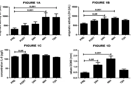
To show a sensibility of different markers (biochemical, psychophysiological and performance) of muscle damage we analyzed the time effects acutely to 72-h after single session of resistance training in sedentary subjects. It was observed (figure 1) for CK concentrations [F(4.56)=2.9; P<0.005], LDH [F(4.56)=3.9; P<0.05], VAS [F(3,48)=0.8; P<0.05] and IL-6 cytokine [F(4.56)=4.1, P<0.05] that we had a time effect showing that these markers are sensible to indicate muscle damage after a single resistance training session, but there was no time effect for MIS [F(3,48)=0.05, P >0.05] (Table 1) demonstrating that it is a poor marker of muscle damage and cannot be used to assess muscle damage and monitor training periodization.
VOLUME 13 | ISSUE 2 | 2018 | 271
The ES for all variables is described in table 2 with values of Confidence Intervals (IC). All post values of MIS were small when compared with pretest. The IL6 shows a large ES when compared pretest with 72h posttest. All other variables showed a very large ES.
DISCUSSION
272 | 2018 | ISSUE 2 | VOLUME 13 © 2018 University of Alicante
2001). This conflict may be due to the different exercise protocols used in those studies. Our data shows that while inflammatory, muscle damage and pain markers are elevated, the isometric muscle strength does not change. Therefore, isometric muscle strength does not appear to be suitable for assessing the recovery period between training sessions. One hypothesis that could support this finding is that the strength training protocol used does not damage the same group of fibers used in MIS evaluation. There is a difference between activation pattern during an isometric and anisometric (shortening and lengthening) twitch. During an isometric twitch there is a lower neuromuscular activation (based on EMG), and based on a size principle, it may explain different motoneuron and consequently different fibers utilization during the MIS (Altenburg, de Ruiter, Verdijk, van Mechelen, & de Haan, 2009). Due to a lack of studies that found the same results, there is a need for future studies to understand this unknown mechanism.
Here we show that just after (30 minutes) of a single session of high-intensity resistance training, the muscle damage and inflammatory markers were increased and remained elevated up to 72 hours. DOMS increased reaching the higher value in 48 hours (ES= 8.2) and remained high about 72 hours (ES= 2.9) compared to pre-exercise. If we follow the MIS, this data can lead us to a wrong interpretation that muscle is able to be exercised again 24h later, but when carefully examined we can see that DOMS, CK, LDH and IL-6 remain elevated (table 2), showing that the next exercise session should occur after the 72 hours. Future researches are needed to detect the optimal moment for the next exercise session.
The CK and LDH enzymes concentrations were elevated in plasma when the muscle fiber was damaged (Brancaccio et al., 2007). The peak concentration occurs between 9 hours and 4 days after strength exercise (Friden & Lieber, 2001; Raastad et al., 2003; Uchida et al., 2009). Here, the peak concentrations were found from 48 hours (LDH) to 72 hours (CK). This difference can be explained by the different exercise protocols, since one study (Uchida et al., 2009)did not find increases of CK for several days due to using lower volumes of training compared to our protocol and other studies (Friden & Lieber, 2001; Raastad et al., 2003). One important data is that 30 minutes after training session the CK and LDH concentrations were high (ES of 2.4 and 3.0 respectively), it shows us that muscle damage can be detected acutely. This data suggests that training based on monitoring these enzymes values for load adjustments is necessary to adjust the time of recovery.
Releasing IL-6 after a stressful situation - such as the training protocol used - represents the proinflammatory role of this cytokine in response to muscle damage (Pedersen et al., 2004; Petersen & Pedersen, 2006). In this study, an increase in IL-6 was found immediately after the training session (equivalent to our training volume). This increase (ES of 3.8) is associated with a satellite cell proliferation about 24h after the training protocol and differentiation of satellite cell already proliferated during resting period (between 24h and 48h) (Guerci et al., 2012; Peake, Della Gatta, Suzuki, & Nieman, 2015). If the coach does not know how to monitor the training loads, executing a new exercise session in the wrong period may decrease the tissue recovery and start a overreaching process. An indirect marker of muscle damage used on this study was DOMS. The present study demonstrated the pain peak 48 hours and very large ES after the activity, similar to others studies (Uchida et al., 2009). In practice, DOMS can be used to control when a person could exercise again without muscle discomfort and injury risks. However, caution is needed with this application of DOMS, because here, we demonstrated that enzymes activity remains elevated even with the decrease of pain sensation (DOMS) at 72h after the training session. Even with a high ES compared with pre-values, DOMS was less sensitive for muscle damage than CK, LDH or cytokines activity because its values were lower about 72 hours while enzymes and cytokines remained elevated 9. CK and LDH activity remain elevated 72
VOLUME 13 | ISSUE 2 | 2018 | 273
needed before training again. Furthermore, DOMS can be influenced by other variables like training experience and trained muscular group.
CONCLUSIONS
The volume-training often used in gyms and training venues - that we used in this research - may lead to a muscular damage. Noninvasive methods like maximal isometric strength test and DOMS are not a good measure to assess muscular damage, consequently, the recovery. Muscle damage can be detected after a single session of high-intensity resistance exercise and, based in our training protocol, the recovery needs to be over 72 hours to complete muscle regeneration, therefore, the monitoring of training loads is necessary for an optimal periodization.
ACKNOWLEDGMENTS
The authors would like to thank the participants in this investigation who made this work possible and the English revision made by Carmen Andrea Perez.
AUTHOR CONTRIBUTIONS
Joel conceived, designed the experiments and collected the data; Silvestre, Motoyama and Azevedo wrote and made the statistical approach, reviewed and made the intellectual contribution.
CONFLICTS OF INTEREST
The authors declare no conflict of interest.
REFERENCES
Altenburg, T. M., de Ruiter, C. J., Verdijk, P. W., van Mechelen, W., & de Haan, A. (2009). Vastus lateralis surface and single motor unit electromyography during shortening, lengthening and isometric contractions corrected for mode-dependent differences in force-generating capacity. Acta Physiol (Oxf), 196(3), 315-328. https://doi.org/10.1111/j.1748-1716.2008.01941.x
Berben, L., Sereika, S. M., & Engberg, S. (2012). Effect size estimation: methods and examples. Int J Nurs Stud, 49(8), 1039-1047. https://doi.org/10.1016/j.ijnurstu.2012.01.015
Brancaccio, P., Maffulli, N., & Limongelli, F. M. (2007). Creatine kinase monitoring in sport medicine. Br Med Bull, 81-82, 209-230. https://doi.org/10.1093/bmb/ldm014
Brown, L. E., & Weir, J. P. (2001). ASEP procedures recommendation I: accurate assessment of muscular strength and power. Professionalization of Exercise Physiology, 4(11).
Byrne, C., & Eston, R. (2002). The effect of exercise-induced muscle damage on isometric and dynamic knee extensor strength and vertical jump performance. J Sports Sci, 20(5), 417-425. https://doi.org/10.1080/026404102317366672
Byrne, C., Eston, R. G., & Edwards, R. H. (2001). Characteristics of isometric and dynamic strength loss following eccentric exercise-induced muscle damage. Scand J Med Sci Sports, 11(3), 134-140. https://doi.org/10.1046/j.1524-4725.2001.110302.x
anti-274 | 2018 | ISSUE 2 | VOLUME 13 © 2018 University of Alicante
inflammatory cytokines by cultured lymphocytes. Life sciences, 73(13), 1683-1690. https://doi.org/10.1016/S0024-3205(03)00490-9
Chen, T. C., Chen, H. L., Pearce, A. J., & Nosaka, K. (2012). Attenuation of eccentric exercise-induced muscle damage by preconditioning exercises. Med Sci Sports Exerc, 44(11), 2090-2098. https://doi.org/10.1249/MSS.0b013e31825f69f3
Friden, J., & Lieber, R. L. (2001). Serum creatine kinase level is a poor predictor of muscle function after
injury. Scand J Med Sci Sports, 11(2), 126-127.
https://doi.org/10.1034/j.1600-0838.2001.011002126.x
Grissom, R., & Kim, J. Effect sizes for research: A broad practical approach. 2005. Mahwah, NJ: Earlbaum.
Guerci, A., Lahoute, C., Hebrard, S., Collard, L., Graindorge, D., Favier, M., . . . Sotiropoulos, A. (2012). Srf-dependent paracrine signals produced by myofibers control satellite cell-mediated skeletal muscle hypertrophy. Cell Metab, 15(1), 25-37. https://doi.org/10.1016/j.cmet.2011.12.001
Halson, S. L. (2014). Monitoring training load to understand fatigue in athletes. Sports Med, 44 Suppl 2, S139-147. https://doi.org/10.1007/s40279-014-0253-z
Huynh, A., Leong, K., Jones, N., Crump, N., Russell, D., Anderson, M., . . . Johnson, D. F. (2016). Outcomes of exertional rhabdomyolysis following high-intensity resistance training. Intern Med J, 46(5), 602-608. https://doi.org/10.1111/imj.13055
Lima, L. C., & Denadai, B. S. (2015). Attenuation of eccentric exercise-induced muscle damage conferred by maximal isometric contractions: a mini review. Frontiers in physiology, 6. https://doi.org/10.3389/fphys.2015.00300
Newham, D. J., McPhail, G., Mills, K. R., & Edwards, R. H. (1983). Ultrastructural changes after concentric and eccentric contractions of human muscle. J Neurol Sci, 61(1), 109-122. https://doi.org/10.1016/0022-510X(83)90058-8
Oliver, I. T. (1955). A spectrophotometric method for the determination of creatine phosphokinase and myokinase. Biochem J, 61(1), 116-122. https://doi.org/10.1042/bj0610116
Peake, J. M., Della Gatta, P., Suzuki, K., & Nieman, D. C. (2015). Cytokine expression and secretion by skeletal muscle cells: regulatory mechanisms and exercise effects. Exerc Immunol Rev, 21, 8-25. Pedersen, B. K., Steensberg, A., Fischer, C., Keller, C., Keller, P., Plomgaard, P., . . . Febbraio, M.
(2004). The metabolic role of IL-6 produced during exercise: is IL-6 an exercise factor? Proc Nutr Soc, 63(2), 263-267. https://doi.org/10.1079/PNS2004338
Petersen, A. M., & Pedersen, B. K. (2006). The role of IL-6 in mediating the anti-inflammatory effects of exercise. J Physiol Pharmacol, 57 Suppl 10, 43-51.
Raastad, T., Risoy, B. A., Benestad, H. B., Fjeld, J. G., & Hallen, J. (2003). Temporal relation between leukocyte accumulation in muscles and halted recovery 10-20 h after strength exercise. J Appl Physiol (1985), 95(6), 2503-2509. https://doi.org/10.1152/japplphysiol.01064.2002
Raeder, C., Wiewelhove, T., De Paula Simola, R. A., Kellmann, M., Meyer, T., Pfeiffer, M., & Ferrauti, A. (2016). Assessment of fatigue and recovery in male and female athletes following six days of intensified strength training. J Strength Cond Res. https://doi.org/10.1519/JSC.0000000000001427 Rosenthal, J. A. (1996). Qualitative descriptors of strength of association and effect size. J Soc Serv Res,
21(4), 37-59. https://doi.org/10.1300/J079v21n04_02
Schoenfeld, B. J., & Contreras, B. (2013). Is Postexercise Muscle Soreness a Valid Indicator of Muscular Adaptations? Strength & Conditioning Journal, 35(5), 16-21.
VOLUME 13 | ISSUE 2 | 2018 | 275
Taylor, A. M., Christou, E. A., & Enoka, R. M. (2003). Multiple features of motor-unit activity influence force fluctuations during isometric contractions. Journal of neurophysiology, 90(2), 1350-1361. https://doi.org/10.1152/jn.00056.2003
Tricoli, W. (2001). Mechanisms involved in delayed onset muscle soreness etiology. Rev. Bras. Ciên. e Mov, 9(2), 39-44.
Uchida, M. C., Nosaka, K., Ugrinowitsch, C., Yamashita, A., Martins, E., Jr., Moriscot, A. S., & Aoki, M. S. (2009). Effect of bench press exercise intensity on muscle soreness and inflammatory mediators. J Sports Sci, 27(5), 499-507. https://doi.org/10.1080/02640410802632144
World Medical Association Declaration of Helsinki: ethical principles for medical research involving human subjects. (2013). JAMA, 310(20), 2191-2194. https://doi.org/10.1001/jama.2013.281053 Zammit, V. A., & Newsholme, E. A. (1976). The maximum activities of hexokinase, phosphorylase,
phosphofructokinase, glycerol phosphate dehydrogenases, lactate dehydrogenase, octopine dehydrogenase, phosphoenolpyruvate carboxykinase, nucleoside diphosphatekinase, glutamate-oxaloacetate transaminase and arginine kinase in relation to carbohydrate utilization in muscles from marine invertebrates. Biochem J, 160(3), 447-462. https://doi.org/10.1042/bj1600447