Polarized Morphogenesis and Virulence of
Aspergillus fumigatus
Jarrod R. Fortwendel,fPraveen R. Juvvadi,aLuise E. Rogg,aYohannes G. Asfaw,cKimberlie A. Burns,dScott H. Randell,d,eand William J. Steinbacha,b
Department of Pediatrics, Division of Pediatric Infectious Diseases, Duke University Medical Center, Durham, North Carolina, USAa
; Department of Molecular Genetics and Microbiology, Duke University Medical Center, Durham, North Carolina, USAb
; Division of Laboratory Animal Resources, Duke University Medical Center, Durham, North Carolina, USAc
; Cystic Fibrosis/Pulmonary Research and Treatment Center, University of North Carolina, Chapel Hill, North Carolina, USAd
; Department of Cell and Molecular Physiology, University of North Carolina, Chapel Hill, North Carolina, USAe
; and Department of Microbiology and Immunology, University of South Alabama, Mobile, Alabama, USAf
Ras is a highly conserved GTPase protein that is essential for proper polarized morphogenesis of filamentous fungi. Localization of Ras proteins to the plasma membrane and endomembranes through posttranslational addition of farnesyl and palmitoyl resi-dues is an important mechanism through which cells provide specificity to Ras signal output. Although theAspergillus fumiga-tusRasA protein is known to be a major regulator of growth and development, the membrane distribution of RasA during polar-ized morphogenesis and the role of properly localpolar-ized Ras signaling in virulence of a pathogenic mold remain unknown. Here we demonstrate thatAspergillus fumigatusRasA localizes primarily to the plasma membrane of actively growing hyphae. We show that treatment with the palmitoylation inhibitor 2-bromopalmitate disrupts normal RasA plasma membrane association and decreases hyphal growth. Targeted mutations of the highly conserved RasA palmitoylation motif also mislocalized RasA from the plasma membrane and led to severe hyphal abnormalities, cell wall structural changes, and reduced virulence in murine in-vasive aspergillosis. Finally, we provide evidence that proper RasA localization is independent of the Ras palmitoyltransferase homolog, encoded byerfB, but requires the palmitoyltransferase complex subunit, encoded byerfD. Our results demonstrate that plasma membrane-associated RasA is critical for polarized morphogenesis, cell wall stability, and virulence inA. fumigatus.
T
he Ras GTPases are membrane-associated binary switches thatcontrol the activity of a multitude of signal transduction path-ways. Specificity of Ras signal output occurs in part via subcellular localization of the Ras protein to various membranes, as well as through compartmentalization to specific regions with the
mem-brane (23,37). Differential localization of Ras proteins is
accom-plished through posttranslational modification of the hypervari-able domain, the region in which Ras homologs are most divergent. The hypervariable domains of nascent Ras proteins contain an extreme C-terminal “CAAX” motif that serves as a
recognition sequence for cytoplasmic farnesyltransferase (5). The
farnesyltransferase enzyme modifies the CAAX motif by addition of a farnesyl lipid moiety to the cysteine residue. Farnesyl modifi-cation has been shown to be necessary and sufficient for direction of the Ras protein to the endoplasmic reticulum (ER) and Golgi
membranes (5). Once localized to the endomembrane system, Ras
proteins are further processed by the activity of a Ras-converting enzyme (Rce), which catalyzes the proteolytic cleavage of the
CAAX residues from the farnesylated CAAX motif (43). The
farnesylated cysteine is then carboxymethylated by action of an
isoprenylcysteine carboxymethyltransferase (19,40).
For Ras to complete transit via the exocytosis machinery of the ER and Golgi complex and become associated with the plasma membrane, another ER resident protein complex, the palmitoyl-transferase, must modify the Ras protein through addition of a
palmitoyl lipid moiety (1). This modification occurs at conserved
cysteine residues located just upstream of the CAAX motif, within
the hypervariable domain (42). Palmitoylation leads to
transloca-tion of the Ras protein to the plasma membrane, as this modifica-tion is proposed to impart greater affinity for cholesterol-rich
membrane domains (17). Ras palmitoyltransferase complexes
have been identified and characterized in humans as well as in the
yeastSaccharomyces cerevisiae(27,46). Ras protein palmitoylation
is reversible, and plasma membrane-localized Ras has been shown to cycle back to the ER and Golgi membranes through the activity
of thioesterase enzymes at the plasma membrane (2, 15). One
thioesterase, APT1, which is responsible for depalmitoylation of
Ras at the plasma membrane, has been identified (8).
The importance of subcellular localization of Ras proteins for function in specific morphological processes has been explored in several fungi. Early evidence for this is found in the model yeast
Schizosaccharomyces pombe. When the Ras1 palmitoylation motif ofS. pombeis ablated, Ras1 localization is restricted to endomem-branes, where it interacts with the guanine nucleotide exchange factor (GEF) Efc25p and signals through a Cdc42-mediated
path-way to control cell morphology (34). In contrast,S. pombeRas1p
restricted to the plasma membrane is activated by a different GEF, Ste6p, and signals through a mitogen-activated protein (MAP)
kinase pathway to mediate the mating response (34). Ras
mem-brane distribution in the fungal pathogenCryptococcus
neofor-mansis also important for differential activation of morphology
and mating pathways. InC. neoformans, plasma
membrane-local-ized Ras1 is required for morphology, high-temperature growth,
Received20 March 2012Accepted24 April 2012
Published ahead of print4 May 2012
Address correspondence to Jarrod R. Fortwendel, [email protected].
Copyright © 2012, American Society for Microbiology. All Rights Reserved.
doi:10.1128/EC.00091-12
on September 8, 2020 by guest
http://ec.asm.org/
and virulence (32). Mutation of the highly conserved
“dual-palmitoylation” motif ofC. neoformansRas1 results in a
mislocal-ized Ras protein that cannot support morphology and high-tem-perature growth yet is still able to support the mating response
(32). Most recently, the importance of Ras membrane localization
has also been explored in the yeast pathogenCandida albicans.
Cells expressing a palmitoylation-deficientC. albicansRAS1 are
able to produce hyphae and grow at wild type-levels in rich media,
although the mutant hyphae are more variable in length (36).
However, polarized morphogenesis of the palmitoylation-defi-cient mutant is drastically reduced when cells are embedded in a matrix, a condition requiring Ras signaling for hyphal induction
(36). In addition, expression of a constitutively activated RAS1
overcomes the embedded filamentation defect, arguing that
mis-localization ofC. albicansRas1 may negatively affect activity (36).
Together, these studies support the hypothesis that fungal Ras signal transduction from different cellular locations can lead to unique biological outputs, a general paradigm of Ras protein reg-ulation that is apparent in many organisms.
The conservation of this paradigm among the yeast organisms described above leads to questions about the role of this form of Ras regulation in pathogenic molds, in which polarized morpho-genesis is required for normal growth and virulence. In the human
pathogenAspergillus fumigatus,rasAis not essential, but it plays a
critical role in hyphal morphogenesis and cell wall integrity (10,
12). From what membrane(s) does Ras mediate hyphal
morpho-genesis, and therefore likely virulence, in filamentous fungal or-ganisms? Despite the emerging data describing its role in growth and development, the subcellular localization of Ras proteins in a mold has not been explored. Here, we show for the first time that
RasA localizes to theA. fumigatusplasma membrane. Mutation of
the highly conserved palmitoylation motif resulted in mislocaliza-tion of RasA from the plasma membrane to endomembrane com-partments, leading to defects in hyphal morphogenesis, cell wall formation, and virulence. In addition, the putative RasA
palmi-toyltransferase subunit genes were identified and deleted:erfB, the
homolog ofS. cerevisiae ERF2, anderfD, the homolog ofS.
cerevi-siae ERF4. Our data establish the importance oferfD for RasA
plasma membrane localization inA. fumigatus, whereaserfBis not
required. Our findings provide novel insights into the regulation
of Ras signal transduction inA. fumigatusand may potentially be
extrapolated for studies with other filamentous fungal pathogens.
MATERIALS AND METHODS
Strains, culture conditions, and growth rate analyses.Aspergillus fu-migatuswild-type strain H237, the⌬rasAstrain (10), and each of the green fluorescent protein (GFP)-RasA fusion strains were maintained on glu-cose minimal medium (GMM) (10). For quantification of growth rates, a 10-l drop containing 10,000 conidia was inoculated onto GMM agar. The colony diameter of each strain was measured daily over the first 72 h of growth, and images were taken at the end of day 3.
Live-cell microscopy was employed for analysis of morphogenesis during germination, as previously described (11). Briefly, conidia from each strain were preincubated in GMM for 4 h at 37°C in a glass-bottom tissue culture plate. Plates were subsequently used for live-cell analysis on a Zeiss Axio Observer inverted microscope equipped with a 37°C environ-mental chamber. For analysis of hyphal morphogenesis during branch formation, 100 conidia from each strain were inoculated onto a thin layer of GMM agar solidified on a glass-bottom petri dish. Cultures were incu-bated at 37°C for the indicated amount of time, and images were captured
on an inverted Nikon Diaphot microscope mounted with a Nikon D50 digital camera.
To analyze the effects of 2-bromopalmitate (2-BP) (Sigma) treatment, 106conidia of the GFP-RasA strain were inoculated into GMM broth supplemented with the indicated concentrations of 2-BP and incubated for 24 h at 37°C. Mycelia from each treatment were collected by centrifu-gation, and excess water was expressed onto filter paper using a sterile spatula. Hyphal mats were then allowed to dry for 6 h at room tempera-ture before weights were recorded. To assess the effect of 2-BP on RasA localization, 100-l aliquots were removed from each culture prior to collection of mycelia and mounted for fluorescence microscopy.
Construction of GFP-RasA fusion strains.For construction of a vec-tor expressing a green fluorescent protein (GFP)-RasA fusion, therasA
coding sequence was PCR amplified from pooledA. fumigatus cDNA using primers incorporating NotI restriction sites to both 5=and 3=ends. The amplifiedrasAcDNA was restriction digested with NotI and cloned into expression vector pAGRP. Vector pAGRP was generated by cloning enhanced GFP (eGFP), amplified as an N-terminal fusion from pUCGH (22), into vector pARP (11) as a BamHI and NotI restriction fragment. The GFP-RasA expression vector was then introduced into the⌬rasA
mutant via protoplast transformation, as previously described (10). Eight transformants positive for fluorescence were found to have identical lo-calization of GFP signal to the hyphal periphery. To introduce mutations in the dual-palmitoylation motif, therasAcDNA was PCR amplified using reverse primers that incorporate the C206S, C207S, or C206S C207S mu-tations. TherasApalmitoylation mutated cDNAs were then cloned into pAGRP and verified by sequence analysis. Each GFP-RasA palmitoyl-ation mutant vector was transformed into the⌬rasAbackground, and multiple transformants were analyzed for RasA localization patterns. To generate the GFP-DARasAC206/207S strain, the previously con-structed DArasA cDNA (12) was amplified using the same primers employed for the C206S C207S mutation described above. The DArasAand DArasAC206/207ScDNAs were then cloned, sequenced, and transformed into the ⌬rasA mutant. All strains were analyzed by Southern blot analysis to verify that a single, ectopic integration event occurred.
Western blot analysis.Western blot analysis for the GFP-RasA and GFP-RasAC206/207Smutant was performed as previously described (22). In short, each strain was cultured in liquid GMM for 24 h at 37°C with shaking at 250 rpm. Total protein extracts were prepared by homogeniz-ing mycelia in liquid nitrogen, followed by addition of extraction buffer (50 mM Tris-HCl [pH 7.5], 150 mM NaCl, 50 mM KCl, 0.01% Triton X-100, 1 mM phenylmethylsulfonyl fluoride [PMSF], and 1:100 protease cocktail inhibitor). Lysates were centrifuged at 4,000 rpm for 8 min to remove debris. Supernatants were removed, and protein content was quantitated by Bradford assay. Fifty micrograms of total protein from each strain was resolved on a 12% SDS-polyacrylamide gel using a Mini-protean tetracell (Bio-Rad). Proteins were then electroblotted to a poly-vinylidene difluoride membrane (Bio-Rad) and probed with a polyclonal rabbit anti-GFP antibody. A horseradish peroxidase-conjugated anti-rab-bit IgG was used as a secondary antibody, and detection was performed using SuperSignal West Pico chemiluminescent substrate (Thermo Scien-tific).
Identification and deletion ofA. fumigatus erfBanderfD.TheA. fumigatus ERF2homolog,erfB(Afu3G06470), was previously identified (14). TheA. fumigatus ERF4homolog,erfD(Afu5G7920), was identified using BLAST search analysis of theA. fumigatusgenome database with the
S. cerevisiaeErf4p sequence as a query. To deleteerfBanderfD, 1.5 kb of upstream and downstream flanking genomic sequence was PCR ampli-fied for each gene. Flanking sequences were cloned into vector pHygR, carrying a hygromycin resistance cassette, generating a deletion construct for each gene. TheerfBanderfDdeletion constructs were PCR amplified, and 2g of each purified product was used for transformation intoA. fumigatusprotoplasts, as previously described (10). Hygromycin-resis-tant transformants were screened for integration by PCR and homologous
on September 8, 2020 by guest
http://ec.asm.org/
recombination confirmed by Southern blot analysis. To generate strains expressing GFP-RasA in either the⌬erfBor⌬erfDbackground, the con-struct generated for GFP-RasA expression (vector pAGRP) was trans-formed into the⌬erfBand⌬erfDmutants as described above. Multiple phleomycin-resistant transformants were isolated for both strains, and RasA localization patterns were analyzed in each. Six⌬erfB⫹GFP-RasA isolates were identified and found to have unaltered RasA plasma mem-brane localization patterns. Five⌬erfB⫹GFP-RasA strains were isolated, and each displayed similar mislocalization of RasA to endomembrane structures.
Fluorescence microscopy for localization analysis and cell wall staining.Ten thousand conidia of each strain were inoculated in 3 ml of GMM broth and incubated in a petri dish on sterile coverslips. After 16 h of growth at 37°C, the strains were observed for fluorescence using an Axioskop 2 Plus microscope (Zeiss) equipped with a digital camera. Im-ages were captured using AxioVision 4.6 imaging software.
Calcofluor white and aniline blue staining was performed as previ-ously described (10,25). Calcofluor white staining of the wild-type and RasAC206/207S strains was performed by culturing conidia on coverslips immersed in 5 ml GMM broth and grown for 16 h at 37°C. The coverslips were briefly rinsed in sterile water and inverted onto a 500-l drop of 0.4-g/ml fluorescent brightener 28 (Sigma) for 5 min at room tempera-ture. Mycelia were then rinsed twice for 5 min each in sterile water and observed by fluorescence microscopy.-Glucan staining with aniline blue was performed by rinsing coverslips in sterile water and inverting them onto a 500-l drop of aniline blue (0.1 mg/ml) for 5 min at room tem-perature. Stained mycelia were directly observed by fluorescence micros-copy.
Electron microscopy.For transmission electron microscopy (TEM) of the wild type and the RasC206/207Smutant, 10,000 conidia of each strain were inoculated into 10 ml GMM broth and grown for 48 h at 37°C. Mycelia were collected by centrifugation and washed three times in dis-tilled water. Washed mycelia were then fixed in 5% glutaraldehyde in 0.1 M cacodylate buffer. The mycelia were embedded in low-viscosity Spurr’s resin, ultrathin sectioned, and then stained with uranyl acetate and lead citrate. Images were obtained on Zeiss EM900 electron microscope, and negatives were scanned for image analysis.
For scanning electron microscopy (SEM), coverslip cultures of 10,000 conidia/strain were grown in 10 ml of GMM in a petri dish containing sterile 22- by 22-mm (no. 2) coverslips. The wild type was grown for 24 h at 37°C, and the RasAC206/207Smutant was grown for 48 h at 37°C due to reduced hyphal growth. The mycelia on the coverslips were briefly rinsed in 50 mM PIPES [piperazine-N,N=-bis(2-ethanesulfonic acid)] buffer (pH 7) and then fixed with 50 mM PIPES buffer (pH 7) containing 3.5% formaldehyde for 60 min. Following fixation, coverslips were washed three times for 10 min each in 50 mM PIPES buffer (pH 7). The fixed mycelia were then dehydrated with 50, 70, and 90% ethanol for 10 min each, followed by 99.5% ethanol three times for 10 min each. The cover-slips were then allowed to dry overnight. Samples were then sputter coated with gold and observed on a field emission SEM (FE-SEM) (FEI model XL-30) using an accelerating voltage of 7 kV.
Virulence studies.A transient-immunosuppression model of inva-sive aspergillosis was used as previously described (38). Six-week-old CD-1 male mice (Charles River) (20 per group) were immunosuppressed with a single dose of triamcinolone acetonide (40 mg/kg of body weight injected subcutaneously) on day⫺1 of infection. The mice were anesthe-tized with 3.5% isoflurane and inoculated intranasally with 2 ⫻106 conidia in 20l of sterile saline on day 0. Mortality was monitored twice daily for the next 15 days, and mice that appeared moribund were sacri-ficed by CO2euthanasia. The lungs of all mice were plated onto inhibitory mold agar (Becton Dickinson), and the phenotypes of recovered isolates were analyzed to confirm infection with the appropriate strain. Survival was plotted on a Kaplan-Meier curve for each group, and a log rank test was used for pairwise comparison of survival, as previously described (45).
RESULTS
RasA localizes to the plasma membranes of germlings and ac-tively growing hyphae.To investigate the distribution of RasA
duringA. fumigatusgrowth and development, a GFP-RasA fusion
was generated and expressed in the⌬rasAbackground. To limit
artifactual cellular distribution, the endogenousrasApromoter
was used to drive GFP-RasA expression (11). Expression of
GFP-RasA in the⌬rasAmutant fully complemented the compact
col-ony morphology and conidiation defects associated with loss of
rasA(10), suggesting that the GFP-RasA fusion protein is
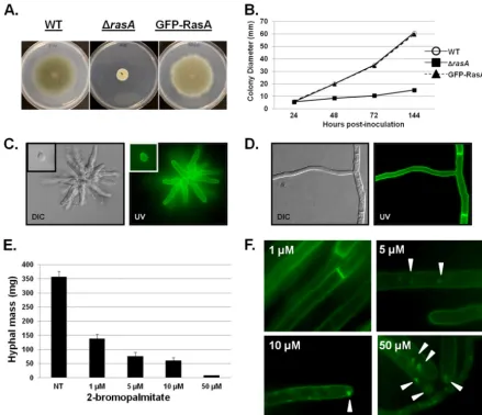
func-tional (Fig. 1AandB). During early growth, GFP-RasA was visible
at the cell periphery of swollen conidia (Fig. 1C, inset at upper left)
and distributed around the periphery of nascent germlings (Fig.
1C). During fully polarized growth, GFP-RasA was visible at the
outer edge of hyphae, with strong localization to septa, and no
GFP signal was associated with internal compartments (Fig. 1D).
These findings are consistent with RasA localization to theA.
fu-migatusplasma membrane, with the strong septal localization likely stemming from plasma membrane apposition along each side of the septum.
Palmitoylation of the Ras protein on conserved residues is re-quired for plasma membrane localization and full function of Ras
signaling in various organisms (4,7,32,36,49). However, nothing
is known about the importance of Ras membrane distribution during invasive growth and virulence of a filamentous pathogen,
such asA. fumigatus. To investigate, we cultured the GFP-RasA
strain in the presence of increasing concentrations of
2-bromo-palmitate (2-BP) (1 to 50M), a nonreversible inhibitor of
pal-mitoylation (20), and assayed hyphal mass. 2-BP inhibited hyphal
growth in a dose-dependent manner (Fig. 1E), whereas dimethyl
sulfoxide (DMSO) vehicle controls produced no effect (data not shown). To examine the effects of increasing concentrations of 2-BP on RasA localization, aliquots were removed from each cul-ture and hyphae were observed using fluorescence microscopy.
Although 1M 2-BP treatment reduced growth, no detectable
disruption of RasA localization was noted (Fig. 1F). However,
higher concentrations of 2-BP (5, 10, and 50M) caused
mislo-calization of GFP-RasA to internal patches (Fig. 1F). These results
suggested that, similar to the case inC. albicansandC. neoformans,
palmitoylation may be important for proper RasA localization during polarized growth and that mislocalization of RasA may contribute to decreased hyphal growth.
Mutation of the conserved RasA palmitoylation motif inhib-its hyphal growth and mislocalizes RasA.Nichols et al. recently
characterized the importance of Ras palmitoylation toC.
neofor-mansgrowth and virulence, describing a “dual-palmitoylation
motif” that is conserved among the filamentous fungal Ras
ho-mologs (32). InC. neoformans, mutation of both cysteine residues
to serine completely blocked palmitoylation of the Ras1 protein
and caused mislocalization to internal membranes (32). Further,
mutation of the single conserved palmitoylation residue ofC.
al-bicansRas1 causes mislocalization from the plasma membrane to
endomembrane structures (36). To investigate the role of the
pal-mitoylation motif in RasA localization and function duringA.
fumigatuspolarized morphogenesis, mutations were introduced to replace each conserved cysteine residue of the
dual-palmitoyla-tion motif with serine, either singly (RasAC206Sand RasAC207S) or
in tandem (RasAC206/207S). Using the same vector constructed for
GFP-RasA, each of the new RasA palmitoylation motif mutants
on September 8, 2020 by guest
http://ec.asm.org/
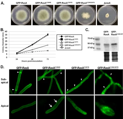
was expressed in the ⌬rasA background. Expression of either
GFP-RasAC206Sor GFP-RasAC207Swas able to fully complement
the hyphal growth defects associated with⌬rasA, indicating that
only one of the conserved cysteines is sufficient for RasA function
in hyphal growth (Fig. 2AandB). However, expression of
GFP-RasAC206/207S, representing complete blockade of RasA
palmitoyl-ation, only partially restored hyphal growth of the⌬rasAmutant
(Fig. 2AandB). Because mutation of the palmitoylation domain inC. albicansRas1 caused reduced protein abundance (36), we compared steady-state levels of the RasA protein in the GFP-RasA
and GFP-RasAC206/207Sstrains. Western blot analysis of whole-cell
protein lysates revealed the expected band size for a GFP-RasA
fusion at⬃51 kDa in both strains (Fig. 2C). In contrast to theC.
albicansdata, RasA protein levels in the GFP-RasAC206/207Smutant
were more abundant than those in the GFP-RasA strain (Fig. 2C),
suggesting that the RasAC206/207Smutation does not negatively
affect protein abundance inA. fumigatus. These data suggest that
the RasAC206/207Smutant phenotypes are not due to decreased
protein abundance and are more directly attributable to mislocal-ization of RasA.
Under fluorescence microscopy, the GFP-RasAC206S mutant
displayed no alteration in plasma membrane association in the subapical sections of hyphae, whereas the apical hyphal sections
showed internal patches of GFP fluorescence (Fig. 2D, arrows). In
contrast, the GFP-RasAC207Smutant displayed mislocalized GFP
signal in both the subapical and apical regions (Fig. 2D). Although
GFP-RasAC206Swas associated with internal patches only in apical
compartments whereas GFP-RasAC207S was found on internal
patches in both the apical and subapical sections, both were still associated with the plasma membrane, as evidenced by clear
lo-FIG 1RasA localizes to the plasma membrane and is disrupted by 2-bromopalmitate. (A) Colony morphology comparison of the wild-type (WT),⌬rasA, and GFP-RasA strains. Ten thousand conidia were inoculated onto glucose minimal media (GMM) and incubated for 98 h at 37°C. (B) Quantification of the average colony diameter for each strain at 24, 48, 72, and 98 h postinoculation. (C and D) Two hundred conidia from the GFP-RasA strain were inoculated into GMM broth and cultured on coverslips at 37°C. Coverslips were removed from culture at defined time points, and GFP fluorescence in swollen conidia (C, inset), germlings (C), and hyphae (D) was analyzed. (E) Conidia (106) of the GFP-RasA strain were inoculated into 50 ml of GMM broth with the indicated
concentration of 2-bromopalmitate and incubated for 24 h at 37°C and 250 rpm. Hyphae were harvested by centrifugation and dried on Miracloth for 6 h before weighing. Each culture was performed in triplicate, and the graph represents the mean hyphal mass⫾standard deviation (SD). (F) Aliquots (100l) were removed from each culture from panel E, and hyphae were analyzed by fluorescence microscopy. Arrowheads indicate the presence of GFP-RasA patches in the tips and subapical compartments of hyphae treated with 5, 10, or 50M 2-bromopalmitate.
on September 8, 2020 by guest
http://ec.asm.org/
calization of GFP signal to septa (Fig. 2D, arrowheads). In contrast to either single mutation, simultaneous mutation of both residues
within the dual-palmitoylation motif (GFP-RasAC206/207S)
re-sulted in a dramatic reduction of GFP signal at the plasma
mem-brane. The GFP-RasAC206/207Smutant was mislocalized from the
hyphal periphery and septa to the cytosol and internal patches,
presumably to compartments of the endomembrane system (Fig.
2D). These data suggest that whereas a single palmitoylation site is
sufficient for RasA function in supporting polarized morphogen-esis, complete blockade of RasA palmitoylation through ablation of the entire dual-palmitoylation motif disrupts plasma mem-brane association and causes decreased hyphal growth. However,
the radial growth difference between the RasAC206/207Sand⌬rasA
mutants suggests that the palmitoylation-deficient mutant is par-tially functional.
It is possible that the aberrant growth phenotypes noted in the
GFP-RasAC206/207Sstrain were due to accumulation of RasA on the
endomembranes and not to loss of RasA at the plasma membrane. To
test this, we generated a strain expressing therasAC206/207Sallele in the
wild-typeA. fumigatusbackground. As with the GFP-RasAC206/207S
mutant, this new strain (GFP-RasAC206/207S⫹WT) exhibited an
en-hanced GFP-RasA signal in the cytosol and on the endomembranes (data not shown). However, no alteration in growth or morphology was observed (data not shown). Taken together, these data argue that
aberrant growth in the GFP-RasAC206/207Smutant was due to loss of
RasA localization at the plasma membrane.
FIG 2TheA. fumigatusRasA dual-palmitoylation motif is required for hyphal growth and RasA localization. (A) Colony morphology of the GFP-RasA palmitoylation mutants. Ten thousand conidia from each strain were inoculated onto GMM agar and incubated for 72 h at 37°C. (B) Quantification of radial growth of the GFP-RasA palmitoylation mutants. Data points represent the mean colony diameter of each strain after 24, 48, and 72 h of growth at 37°C. (C) Western blot analysis of total protein extracts from the GFP-RasA and GFP-RasAC206/207Sstrains using an anti-GFP antibody. A Coomassie blue-stained protein
gel is shown (lower panel) for a loading control. (D) Fluorescence microscopy images of the GFP-RasA palmitoylation mutant strains. Two hundred conidia from each strain were cultured on glass coverslips immersed in GMM broth for 24 h at 37°C. Arrowheads denote positions of septa. Note that the GFP-RasAC206/207S
fluorescence pattern does not appear along the hyphal periphery or septa. Arrows denote internal patches of GFP fluorescence in apical regions of the GFP-RasAC206Smutant.
on September 8, 2020 by guest
http://ec.asm.org/
Plasma membrane-localized RasA signaling controls early polarized growth and branch emergence. Normal vegetative
growth of Aspergillus species progresses from resting conidia
through germination to fully polarized hyphae. Deletion ofrasA
inA. fumigatuscauses delayed germination and subsequent for-mation of wide, blunted hyphae that continually switch polarized
growth axes (10). To examine the contribution of plasma
mem-brane-localized Ras activity to these important morphogenetic processes, we compared polarization phenotypes during
germina-tion and early hyphal growth of the GFP-RasAC206/207Smutant to
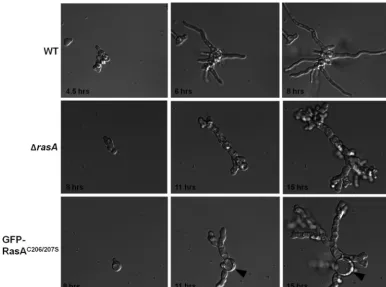
those of the wild-type and⌬rasA strains. Live-cell microscopy
analysis revealed nearly identical growth defects in the⌬rasAand
GFP-RasAC206/207Smutants during germination. Both mutants
displayed low germination rates and formed stunted, highly
branched germlings over the initial 15 h of culture (Fig. 3),
sug-gesting a nonfunctional Ras protein. The only notable difference between the mutant strains throughout the initial growth stages was the exaggerated swelling of the conidium during germ tube
extension in the GFP-RasAC206/207Sstrain (Fig. 3, arrowheads). In
contrast, the germination rates and morphologies of the
GFP-RasAC206Sand GFP-RasAC207Smutants were similar to those of
the GFP-RasA control strain (data not shown).
However, after 24 h of growth, the highly branched, stunted
hyphae of the GFP-RasAC206/207Smutant developed into
morpho-logically normal hyphae (Fig. 4B, arrows). In contrast, the⌬rasA
mutant grew only as stunted, meandering hyphae at all stages
(data not shown). Although the GFP-RasAC206/207Smutant
devel-oped grossly normal hyphae after the initial 24 h of culture, sub-sequent branching resulted in reversion to the early growth
phe-notype (Fig. 4C). The altered morphology of germination and
branch formation in the GFP-RasAC206/207Smutant mimicked the
phenotype of the⌬rasAstrain (10). Taken together, these results
further suggest partial Ras functionality of the GFP-RasAC206/207S
mutant during invasive growth. Although exogenous 2-BP
inhib-ited hyphal growth (Fig. 1EandF), 2-BP treatment did not
phe-nocopy the RasAC206/207Sphenotypes noted inFig. 4. These
find-ings are likely due to the inability of 2-BP to completely inhibit RasA palmitoylation and therefore to perfectly mimic the
rasAC206/207Smutation.
Plasma membrane localization is required for full Ras func-tion in cell wall integrity,-glucan deposition, and virulence.
Deletion of RasA causes decreased cell wall integrity as evidenced
by increased susceptibility to cell wall-destabilizing agents (10).
To examine the contribution of plasma membrane-localized RasA activity to cell wall integrity, we first compared the susceptibilities of
the wild type and the GFP-RasAC206/207Smutant to cell
wall-damag-ing agents. The GFP-RasAC206/207Smutant displayed greater
sensitiv-ity to the chitin-perturbing compounds nikkomycin Z and calcofluor
white than the wild-type strain. Nikkomycin Z (0.25g/ml)
treat-ment yielded 10% versus 52% inhibition (P⬍0.01) of radial growth
rates for the wild-type and GFP-RasAC206/207Sstrains, respectively.
Furthermore, growth in the presence of calcofluor white (3g/ml)
yielded 3% inhibition of the wild-type strain versus 32% inhibition of
the GFP-RasAC206/207Sstrain (P⬍0.01). The GFP-RasAC206Sand
FIG 3Plasma membrane-localized RasA controls the timing and morphogenesis of germination. Conidia from the wild-type (WT),⌬rasA, and GFP-RasAC206/207S
strains were inoculated into GMM broth in a 6-well glass-bottom culture dish and preincubated for 4 h at 37°C. Following preincubation, plates were used for live-cell analysis on a Zeiss Axio Observer inverted microscope equipped with a 37°C environmental chamber and a digital camera. Images from time points that represent similar germination stages for each strain were used for comparison. The arrowheads denote the swollen initial conidium in the GFP-RasAC206/207Sstrain. Images of each strain
were acquired at identical magnifications.
on September 8, 2020 by guest
http://ec.asm.org/
GFP-RasAC207Smutants displayed no increase in sensitivity to either
cell wall-damaging agent compared to the wild type (data not shown). Because increased sensitivity to cell wall-disruptive agents
was apparent in the GFP-RasAC206/207Smutant, cell wall
organiza-tional differences were examined using scanning electron microscopy (SEM) and transmission electron microscopy (TEM). SEM analysis revealed the presence of an extracellular matrix associated with
cul-tures of the GFP-RasC206/207Smutant that was not evident in the wild
FIG 4Plasma membrane-localized RasA regulates morphology of branch formation. Conidia of the GFP-RasAC206/207Sstrain were inoculated onto a thin layer
of GMM agar solidified on a glass-bottom petri dish and incubated at 37°C. Bright-field microscopy images were taken during germination (A) and at 24 h (B) and 48 h (C) of hyphal growth. Arrows indicate the appearance of morphologically normal hyphae after 24 h of growth. Arrowheads denote formation of branches after 48 h of growth that revert to the early growth phenotype.
FIG 5RasA contributes to cell wall structure and integrity from the plasma membrane. (A) Twenty-four-hour coverslip cultures of the wild-type (WT) and GFP-RasAC206/207Sstrains were used for scanning electron microscopy analysis. The lower panels are magnified images of the areas indicated in the upper panels
by dashed boxes. Arrowheads indicate extracellular matrix produced by the GFP-RasAC206/207Smutant. (B) Twenty-four-hour coverslip cultures of each strain
were utilized for transmission electron microscopy analysis. Note the disorganized outer layer and translucent middle layer of the GFP-RasAC206/207Scell wall. (C)
Conidia of the wild-type (WT),⌬rasA, and GFP-RasAC206/207Sstrains were grown on glass coverslips for 16 h at 37°C. Following incubation, coverslips were
rinsed briefly and hyphae stained in 0.1% aniline blue. Arrowheads indicate low staining of hyphal tips in the⌬rasAand GFP-RasAC206/207Smutants.
on September 8, 2020 by guest
http://ec.asm.org/
type (Fig. 5A, arrowheads). Subsequent TEM analysis revealed a
GFP-RasAC206/207Scell wall that was disorganized compared to that of
the wild type, with fibrillar material protruding from the outer layers
of hyphae (Fig. 5B).
We hypothesized that the extracellular fibrillar matrix was likely to be cell wall material that was inadequately incorporated at the growing hyphal tip. To visualize the distribution of chitin and
-glucan, major cell wall components of A. fumigatus, hyphae
from each strain were stained with calcofluor white or aniline
blue. Although calcofluor white staining of the⌬rasAand
GFP-RasAC206/207Shyphae revealed slightly brighter fluorescence than for the wild type, all strains displayed similar patterns of chitin distribution (data not shown). This finding coincided with the TEM analysis, which revealed that the innermost portion of the cell wall, consisting mostly of chitin, was relatively unaltered in
the mutant strain (Fig. 5B). In contrast, hyphal staining with
ani-line blue revealed an altered distribution of-1,3-glucan in the
⌬rasA and GFP-RasAC206/207Scell walls. After 16 h of culture,
the wild-type strain stained intensely at the hyphal tip, whereas the
subapical hyphal walls stained at relatively low levels (Fig. 5C). In
contrast, both the⌬rasAand RasAC206/207Smutants displayed
re-duced aniline blue staining at the hyphal tips accompanied by
intense staining along the subapical hyphal walls (Fig. 5C). Thus,
normal-glucan deposition appears to be dependent on plasma
membrane-associated RasA.
InC. neoformans, the deletion of Ras1 and mutation of the Ras1 palmitoylation domain both result in avirulence, due to lack
of growth at 37°C (32). To determine the contribution of plasma
membrane localization of RasA to A. fumigatus virulence, the
⌬rasA and GFP-RasAC206/207S mutants were compared to the
wild-type strain in a murine model of invasive aspergillosis. This model utilizes a single dose of triamcinolone acetonide to induce
transient immunosuppression (38). Virulence of both the⌬rasA
and GFP-RasAC206/207Smutants was significantly attenuated (P⬍
0.0001), with infected mice displaying 25% and 20% mortality at
15 days postinoculation, respectively (Fig. 6). This was in contrast
to the wild-type and complemented (⌬rasA⫹rasA) strains, both of
which caused higher mortality (Fig. 6), consistent with previously
published results using wild-typeA. fumigatusin the same mouse
model (38). Proper RasA localization to the plasma membrane is
therefore essential for full virulence inA. fumigatusinfection.
Plasma membrane localization is required for full induction of the activated RasA phenotypes of the DArasA1mutant.We recently reported novel phenotypes caused by the expression of
constitutively activatedA. fumigatusRasA, including unchecked
vacuolar expansion, hyphal swelling, and lysis of hyphal
compart-ments (11). Recent studies withC. albicansrevealed that
constitu-tive activation of CaRas1p can overcome the effects of mislocal-ization, suggesting that CaRas1p localization at the plasma membrane may be more critical for full Ras protein activity than
for engagement of the proper effector proteins (36). To test this
hypothesis inA. fumigatus, we mutated the dual-palmitoylation
motif of the previously generated DArasA1cDNA to generate a
palmitoylation-deficient DArasA1 mutant (DArasA1C206/207S).
The DArasA1and DArasA1C206/207Salleles were then expressed as
GFP fusion proteins in the⌬rasAbackground using the pAGRP
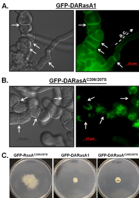
vector. Expression of GFP-DArasA1produced the same
pheno-types as previously reported for the untagged DARasA1 strain
(11), including development of large, vacuolated subapical hyphal
segments intermingled with normal apical compartments (Fig.
7A). Similarly to the wild-type RasA protein, GFP-DARasA1
lo-calized primarily to the plasma membrane, along the hyphal
periphery, and at septa (Fig. 7A). In contrast, mutation of the
dual-palmitoylation motif caused the accumulation of
GFP-DARasA1C206/207Sin the cytosol and on punctate endomembrane
structures but not at the plasma membrane (Fig. 7B). Phenotypic
analysis revealed that the GFP-DARasA1C206/207Smutant
devel-oped hyphae that were stunted and highly branched (Fig. 7B).
These hyphal phenotypes were more similar to those of the GFP-RasAC206/207Smutant (Fig. 3andFig. 4A) than to those of the
GFP-DARasA1 mutant (Fig. 7A) (11), suggesting that
constitu-tively activated RasA does not overcome the defects caused by RasA mislocalization. Although the hyphal morphology was more
similar to that of the GFP-RasAC206/207Sstrain, the radial growth
and colony morphology of the GFP-DARasA1C206/207Smutant
re-sembled that of the DARasA1 strain. Both strains formed more compact, slowly growing colonies, compared to the more
substan-tial radial outgrowth generated by the GFP-RasAC206/207Smutant
(Fig. 7C). Together, these data show that constitutively active RasA mislocalized to the endomembranes only partially induces activated Ras phenotypes. Therefore, although plasma membrane localization of RasA may be required for full induction of RasA activity, our studies do not rule out the lack of proper effector engagement by RasA restricted to the endomembrane system as a
plausible scenario inA. fumigatus.
TheA. fumigatus Erf4p homolog, ErfD, is important for proper RasA localization, whereas the Erf2p homolog, ErfB, is not required.InS. cerevisiae, the Ras palmitoyltransferase
com-plex is known to consist of two proteins, Erf2p and Erf4p (27,50).
Erf2p is a DHHC palmitoyltransferase enzyme responsible for
transferring the palmitoyl moiety onto the Ras protein (1,30,31).
The second subunit, Erf4p, was originally copurified with Erf2p
and later found to be a peripheral ER membrane protein (1,21).
Although Erf4p does not contain the DHHC domain required for palmitoyl transfer, deletion of either subunit causes
mislocaliza-tion of Ras2p from the plasma membrane (50). TheA. fumigatus
ERF2homolog,erfB, was previously identified in a study of fungal
DHHC palmitoyltransferases (14). To explore the importance of
the palmitoyltransferase complex to RasA localization, we first
identified the putative Erf4p homolog inA. fumigatus. The S.
cerevisiaeErf4p sequence was used in a BLAST search of theA. FIG 6RasA mislocalization affects virulence in a murine model of invasive
aspergillosis. Groups of 20 6-week-old CD1 male mice (20 to 26 g) were im-munosuppressed with a single dose of triamcinolone acetonide (40 mg kg⫺1)
on day⫺1 and inoculated intranasally with 2⫻106conidia from each strain.
Survival was assessed over a 15-day period. The⌬rasAand GFP-RasAC206/207S
mutants displayed statistically significantly reduced virulence compared to the wild type.
on September 8, 2020 by guest
http://ec.asm.org/
fumigatusgenome database (http://www.aspgd.org). This search
identified a single A. fumigatus open reading frame (ORF)
(Afu5G7920) which contains a Golgin subfamily A member 7/Erf4 superfamily domain. This protein shares 24% identity with
S. cerevisiaeErf4p and was named ErfD. A Clustal V alignment of the Erf4 superfamily domain, with the highest identity between theS. cerevisiaeandA. fumigatusproteins, is shown inFig. 8A. To investigate whether the putative Ras-palmitoyltransferase com-plex is required for filamentous growth and/or RasA localization,
strains with eithererfBorerfDdeleted were generated. Loss oferfB
(⌬erfB), the DHHC palmitoyltransferase enzyme homolog,
caused no alteration in colony morphology or growth rate
com-pared to those of the wild-type strain (Fig. 8BandC). In contrast,
deletion oferfD(⌬erfD) led to a minor, but statistically significant
(P⬍0.01), reduction in radial growth rate (Fig. 8BandC). No
alteration in hyphal morphology was noted in the mutant strains (data not shown). Furthermore, expression of GFP-RasA in the mutant backgrounds revealed punctate RasA localization in the
⌬erfDstrain, whereaserfBdeletion caused no disruption in RasA
localization (Fig. 8D). Mislocalization of the GFP-RasA signal to
internal patches, resembling that in the GFP-RasAC206/207Sand
2-BP-treated strains, was evident in the⌬erfDmutant background
(Fig. 8D). These data suggest a role forerfDin RasA localization during hyphal growth.
DISCUSSION
In the present study, mutations of the conserved Ras
palmitoyl-ation motif were introduced intoA. fumigatusto delineate the
importance of this domain to RasA localization and to understand the role of properly localized RasA in polarized morphogenesis. We found that RasA localized primarily to the plasma membrane, including at septa. Mislocalization of RasA from the plasma mem-brane caused defects in germination and hyphal morphogenesis, aberrant cell wall formation, and decreased virulence. The
major-ity of these phenotypes were shared by the⌬rasAmutant (10),
suggesting a mostly nonfunctional RasAC206/207Sprotein.
How-ever, hyphal growth of the RasAC206/207Smutant was significantly
greater than that seen in the complete absence of RasA, suggesting that mislocalized RasA remains partially competent to mediate some aspects of fungal growth and/or morphogenesis. This partial functionality was not attributable to reduced protein abundance,
as was seen with a similar mutation inC. albicansRas1 (36),
be-cause steady-state RasA protein levels were not negatively affected by the palmitoylation mutation in our model. Although temporal regulation of RasA protein activity is also important for
RasA-mediated growth (12,35,44), our findings support the hypothesis
that spatial regulation of RasA through proper localization to the plasma membrane is required for engagement of RasA signaling pathways controlling polarized morphogenesis. At present, three possible models for spatial regulation of Ras signal transduction in fungi are as follows: (i) fungal Ras proteins employ the plasma membrane and endomembranes as separate signaling platforms, engaging distinct effectors at each membrane; (ii) fungal Ras pro-teins can potentially interact with all effectors equally from either membrane, but plasma membrane localization is required for ac-tivation and, therefore, function; or (iii) Ras activity is not affected by membrane distribution, and the partial functionality of the RasAC206/207Smutant could be explained by the presence of a pool
of Ras that is still active at or near the plasma membrane. Each of these models is at least partially supported by the current litera-ture, and some combination of the three is likely to provide a more complete picture of spatiotemporal control of Ras signaling in fungi.
Differential localization of human Ras proteins to the plasma membrane and endomembrane propagates distinct signal trans-duction events through the utilization of distinct effector proteins
in human and yeast (3,4). Our data, along with recent reports for
the yeasts, provide some support for a model of spatially segre-gated Ras-effector interactions in fungal organisms as well. For example, the utilization of specific membrane platforms for Ras signaling to morphogenetic programs appears to be conserved for
C. neoformans, C. albicans, S. pombe, and, as our data show,A. fumigatus. At the plasma membrane,C. neoformansRas1 controls
FIG 7Plasma membrane association is required for full development of the DARasA1 phenotype. (A and B) The DArasAor DArasAC206/207ScDNA was
cloned into vector pAGRP and expressed in the⌬rasAbackground. Conidia from each strain were inoculated onto coverslips immersed in GMM broth and incubated at 37°C for 16 h. Left panels, differential interference contrast (DIC) micrographs. Right panels, UV micrographs. Arrows indicate positions of septa. Note the grossly normal apical compartment (a.c.) mixed with swollen subapical sections in the GFP-DARasA1 mutant, which is not seen in the DARasA1C206/207Sstrain. Plasma membrane localization was evident
through-out the GFP-DARasA strain, whereas mutation of both C206 and C207 mis-localized DARasA to endomembranes. (C) Colony morphologies of the GFP-RasAC206/207S, GFP-DARasA1, and GFP-DARasA1C206/207S strains. Ten
thousand conidia of each strain were inoculated onto GMM agar and incu-bated for 3 days at 37°C. Note the compact colony morphology induced by expression of activated RasA, seen in both palmitoylated and palmitoylation-deficient mutants.
on September 8, 2020 by guest
http://ec.asm.org/
cell shape and high-temperature growth, whileC. albicansRas1
regulates the yeast-to-hypha transition (32). Our data similarly
suggest thatA. fumigatusRasA may utilize the plasma membrane
as a platform to interact with distinct effectors mediating
polar-ized growth and hyphal morphogenesis. Further,C. neoformans
Ras1 restricted to endomembranes is functional for the support of
the mating response (32). In contrast,S. pombeutilizes plasma
membrane-localized Ras1 for the mating response, whereas
endo-membrane-localized Ras1 mediates proper morphology (4,32).
Although the specific membrane platforms utilized for Ras signal
output differ, the current data argue that bothC. neoformansand
S. pombemay be able to spatially segregate the activation of Ras-mediated morphology and mating pathways through activation of
distinct effectors (4,32). As a sexual cycle forA. fumigatushas
recently been described, it will be of interest to analyze the ability
of a RasAC206/207Smutant to mate (33). Such studies would reveal
whether activation of the mating pathway in a mold can be accom-plished when Ras localization or activity is not concentrated at the
plasma membrane. In addition to morphogenesis,-glucan
dis-play properties and cell wall integrity were altered in both the
⌬rasA and RasAC206/207Smutants, again suggesting that plasma
membrane localization is important for these roles. Host
recogni-tion of exposed-glucan of theA. fumigatuscell wall is an
impor-tant mediator of host-pathogen interactions (25,28). Future work
in this area will focus on quantitative measurement of-glucan
exposure and the subsequent immune response generated by RasA mutant strains.
Although differential activation of distinct effectors from each membrane is an attractive model, we cannot yet rule out the pos-sibility that mislocalization of RasA results in greatly decreased activation due to the inability of Ras to interact with its cognate
GEF. Data from studies withC. albicansseem to support this
al-ternative, as expression of a constitutively activated palmitoyla-tion-deficient Ras1 can overcome the embedded filamentation
defect of the Ras1 palmitoylation mutant (36). However, in S.
cerevisiae, where expression of activated Ras2p results in hyper-sensitivity to heat shock and nutrient stress, loss of Ras2p
palmi-toylation reverses activated Ras phenotypes (6). This supports a
model in which Ras must be localized to the plasma membrane to
engage proper effectors. Our data suggest that regulation ofA.
fumigatusRasA may involve a combination of these two scenarios.
Expression of DARasA1C026/207Scaused loss of some hyphal
phe-notypes associated with DARasA1 expression, suggesting that the constitutively activated RasA was unable to interact with needed effectors. However, mislocalization of DARasA1 did not cause
complete reversion to the RasAC206/207S phenotype, as colony
morphology was compact, similar to that of the DARasA1 mutant. A specific, biochemical analysis of Ras activity will be required to decipher this question. Similarly, we cannot rule out the potential for a pool of active RasA near the plasma membrane that may be able to propagate the signals needed to produce the limited hyphal
growth observed in the RasAC206/207Smutant. This caveat was also
noted in recent studies with the Ras1 protein ofC. neoformans
(32). Current models of human Ras protein trafficking suggest a
FIG 8TheA. fumigatus ERF4homolog,erfD, is required for proper RasA localization. (A) Partial alignment of the Erf4 superfamily domain ofS. cerevisiae(Sc) Erf4p and the putative protein sequence ofA. fumigatus(Af) ErfD. Conserved residues are highlighted in gray. Amino acid position numbers represent theA. fumigatusErfD sequence. (B) Colony morphologies of the⌬erfBand⌬erfDmutants. Ten thousand conidia from each strain were inoculated into GMM agar and incubated for 4 days at 37°C. (C) Quantitative comparison of radial growth for the wild-type (WT) and mutant strains. Data points are the mean colony diameter in millimeters from triplicate growth experiments for each time point. (D) Localization of GFP-RasA in the⌬erfBand⌬erfDmutant backgrounds. Coverslip cultures of each strain were utilized for fluorescence microscopy. Note the increased presence of endomembrane GFP-RasA signal in the⌬erfDmutant background.
on September 8, 2020 by guest
http://ec.asm.org/
complicated system involving Golgi-mediated exocytosis through recycling endosome (RE) and non-RE pathways accompanied by palmitoylation-mediated, reversible association with the plasma
membrane (29). It is possible that RasA could signal, albeit less
efficiently, from recycling endosomes near the plasma membrane to support limited hyphal growth. More-in-depth studies of Ras trafficking in fungal organisms will elucidate the complex rela-tionships between Ras localization and effector engagement.
Another finding of the current study is the ability of the palmi-toylation inhibitor 2-BP to disrupt RasA localization and to
in-hibitA. fumigatushyphal growth in a dose-dependent manner.
Although 2-BP has been used to inhibit palmitoylation of yeast
proteinsin vitro(20) and to inhibitTrypanosoma bruceigrowth in
culture (9), this is the first study providing data on the sensitivity
of fungal growth to 2-BP. Although treatment of the GFP-RasA strain with 2-BP caused mislocalization of RasA and inhibited hyphal growth, increasing concentrations of 2-BP did not
pheno-copy the RasAC206/207S mutant. This discrepancy could be
ex-plained by an inability of 2-BP to completely inhibit RasA palmi-toylation, therefore not mimicking phenotypes rendered by complete mutation of the dual palmitoylation motif. In this situ-ation, it may be more likely that decreased growth caused by 2-BP treatment is due to inhibition of palmitoylation of the myriad other proteins involved in hyphal growth. Regardless, these find-ings highlight the importance of palmitoylation as a posttransla-tional modification controlling cellular processes required for in-vasive growth.
The importance of palmitoyltransferases to various cellular activities of microbes has now been studied in depth for both
yeast (41) andT. brucei(9). TheA. fumigatusgenome contains
six predicted DHHC palmitoyltransferases, including one with
significant homology to S. cerevisiae Erf2p, the yeast
Ras-palmitoyltransferase (14). Although Erf2p is considered the
main palmitoyltransferase for Ras2p, palmitoylation of S.
cerevisiae Ras2p occurs in the absence of Erf2p (41). These findings suggest that palmitoyltransferase activity may be pro-miscuous, such that nonspecific palmitoylation by other palmitoyltransferase enzymes may compensate for Erf2p loss. In fact, recent data suggest that palmitoylation of many periph-eral membrane proteins (e.g., Ras) may actually be a promis-cuous process, whereas integral membrane protein
palmitoyl-ation occurs via specific palmitoyltransferase enzymes (13,39).
Because deletion oferfBdid not affect RasA localization, our
data suggest that RasA may be a peripheral membrane protein whose palmitoylation can be accomplished through the activity of multiple palmitoyltransferases. In contrast, deletion of the
ERF4homolog,erfD, caused partial disruption of RasA
local-ization. However, mislocalization of RasA due toerfDdeletion
did not cause morphological abnormalities mimicking those of
the RasAC206/207Smutant, likely due to incomplete inhibition of
RasA plasma membrane association. It is tempting to speculate that the ErfD protein may be an important subunit of
palmi-toyltransferase complex formation inA. fumigatus, regardless
of the DHHC enzyme that is present. Further studies on the roles of the various palmitoyltransferases and the protein makeup of the complexes in which they function will help to elucidate their importance to Ras localization and fungal growth in general.
In summary, the present study reports the importance of RasA plasma membrane localization for full function in the hyphal
morphogenesis, cell wall integrity, and virulence ofA. fumigatus.
Regardless of the mechanistic reason for decreased protein
func-tion of the RasAC206/207Smutant (i.e., decreased RasA activity
ver-sus inability to interact with distinct effectors), plasma mem-brane-directed RasA signaling is required for the majority of roles
played by RasA inA. fumigatusgrowth and virulence. What are
those signaling pathways that are engaged by RasA at the plasma membrane to control the phenotypes mentioned above? Effectors likely to be immediately downstream of RasA signaling are the
conserved GTPase proteins ModA (AspergillusCdc42 homolog)
and RacA, for activation of mitogen-activated protein kinase or
p21-activated kinase pathways (18,24,47). Other interesting
can-didates that may physically or genetically interact with RasA at the plasma membrane or endomembranes are the septins and Rho proteins, both of which play roles in germination and branch
for-mation ofA. nidulans(16,26,48). Future work in this area will
focus on the identification of RasA effectors to construct im-proved molecular models of fungal growth and morphogenesis.
ACKNOWLEDGMENTS
These studies were supported by Duke University Molecular Mycology and Pathogenesis Training Program grant 5T32-AI052080 and NIH/ NIAID Research Scholar Development Award 1 K22 AI89786-01A1 to J.R.F. W.J.S. was supported by NIH/NIAID grant 1R56AI077648-01A2. L.E.R. was supported by The Hartwell Foundation Biomedical Research Fellowship.
We thank J. Andrew Alspaugh for helpful suggestions and review of the manuscript.
REFERENCES
1.Bartels DJ, Mitchell DA, Dong X, Deschenes RJ.1999. Erf2, a novel gene product that affects the localization and palmitoylation of Ras2 in Saccha-romyces cerevisiae. Mol. Cell. Biol.19:6775– 6787.
2.Camp LA, Hofmann SL.1993. Purification and properties of a palmitoyl-protein thioesterase that cleaves palmitate from H-Ras. J. Biol. Chem. 268:22566 –22574.
3.Casar B, et al.2009. Ras subcellular localization defines extracellular signal-regulated kinase 1 and 2 substrate specificity through distinct utili-zation of scaffold proteins. Mol. Cell. Biol.29:1338 –1353.
4.Chang E, Philips M. 2006. Spatial segregation of Ras signaling: new evidence from fission yeast. Cell Cycle5:1936 –1939.
5.Choy E, et al.1999. Endomembrane trafficking of Ras: The CAAX motif targets proteins to the ER and Golgi. Cell98:69 – 80.
6.Dong X, et al.2003. Palmitoylation and plasma membrane localization of Ras2p by a nonclassical trafficking pathway inSaccharomyces cerevisiae. Mol. Cell. Biol.23:6574 – 6584.
7.Dudler T, Gelb MH.1996. Palmitoylation of Ha-Ras facilitates mem-brane binding, activation of downstream effectors, and meiotic matura-tion in Xenopus oocytes. J. Biol. Chem.271:11541–11547.
8.Duncan JA, Gilman AG.1998. A cytoplasmic acyl-protein thioesterase that removes palmitate from G protein␣subunits and p21(RAS). J. Biol. Chem.273:15830 –15837.
9.Emmer BT, et al. 2011. Global analysis of protein palmitoylation in African trypanosomes. Eukaryot. Cell10:455– 463.
10. Fortwendel JR, et al.2008.Aspergillus fumigatusRasA regulates asexual development and cell wall integrity. Eukaryot. Cell7:1530 –1539. 11. Fortwendel JR, Juvvadi PR, Rogg LE, Steinbach WJ.2011. Regulatable
Ras activity is critical for proper establishment and maintenance of polar-ity inAspergillus fumigatus. Eukaryot. Cell10:611– 615.
12. Fortwendel JR, Panepinto JC, Seitz AE, Askew DS, Rhodes JC.2004.
Aspergillus fumigatus rasAandrasBregulate the timing and morphology of asexual development. Fungal Genet. Biol.41:129 –139.
13. González Montoro A, Chumpen Ramirez S, Quiroga R, Valdez Taubas J.2011. Specificity of transmembrane protein palmitoylation in yeast. PLoS One6:e16969. doi:10.1371/journal.pone.0016969.
14. González Montoro A, Quiroga R, Maccioni HJF, Valdez Taubas J.2009. A novel motif at the C-terminus of palmitoyltransferases is essential for Swf1 and Pfa3 function in vivo. Biochem. J.419:301–308.
on September 8, 2020 by guest
http://ec.asm.org/
15. Goodwin JS, et al.2005. Depalmitoylated Ras traffics to and from the Golgi complex via a nonvesicular pathway. J. Cell Biol.170:261–272. 16. Guest GM, Lin X, Momany M.2004.Aspergillus nidulansRhoA is
in-volved in polar growth, branching, and cell wall synthesis. Fungal Genet. Biol.41:13–22.
17. Hancock JF, Cadwallader K, Paterson H, Marshall CJ.1991. A CAAX or a CAAL motif and a second signal are sufficient for plasma membrane targeting of a ras proteins. EMBO J.10:4033– 4039.
18. Harris SD.2011. Cdc42/Rho GTPases in fungi: variations on a common theme. Mol. Microbiol.79:1123–1127.
19. Hrycyna CA, Sappersein SK, Clarke S, Michaelis S.1991. The Saccha-romyces cerevisiaeSTE14 gene encodes a mthyltransferase that mediates C-terminal methylation of a-factor and RAS proteins. EMBO J.10:1699 – 1709.
20. Jennings BC, et al.2009. 2-Bromopalmitate and 2-(2-hydroxy-5-nitro-benzylidene)-benzo[b]thiophen-3-one inhibit DHHC-mediated palmi-toylation in vitro. J. Lipid Res.50:233–242.
21. Jung V, Chen L, Hofmann SL, Wigler M, Powers S.1995. Mutations in the SHR5 gene ofSaccharomyces cerevisiaesuppress Ras function and block membrane attachment and palmitoylation of Ras proteins. Mol. Cell. Biol.15:1333–1342.
22. Juvvadi PR, et al.2011. Localization and activity of the calcineurin cata-lytic and regulatory subunit complex at the septum is essential for hyphal elongation and proper septation inAspergillus fumigatus. Mol. Microbiol. 82:1235–1259.
23. Konstantinopoulos PA, Karamouzis MV, Papavassiliou AG.2007. Post-translational modifications and regulation of the RAS superfamily of GTPases as anticancer targets. Nat. Rev. Drug Discov.6:541–555. 24. Kwon MJ, et al.2011. Functional characterization of Rho GTPases in
Aspergillus nigeruncovers conserved and diverged roles of Rho proteins within filamentous fungi. Mol. Microbiol.79:1151–1167.
25. Lamaris GA, et al.2008. Caspofungin-mediated-glucan unmasking and enhancement of human polymorphonuclear neutrophil activity against Aspergillus and non-Aspergillus hyphae. J. Infect. Dis.198:186 –192. 26. Lindsey R, Cowden S, Hernandez-Rodriguez Y, Momany M. 2010.
Septins AspA and AspC are important for normal development and limit the emergence of new growth foci in the multicellular fungusAspergillus nidulans. Eukaryot. Cell9:155–163.
27. Lobo S, Greentree WK, Linder ME, Deschenes RJ.2002. Identification of a Ras palmitoyltransferase in Saccharomyces cerevisiae. J. Biol. Chem. 277:41268 – 41273.
28. Mattila PE, Metz AE, Rapaka RR, Bauer LD, Steele C.2008. Dectin-1 Fc targeting ofAspergillus fumigatusbeta-glucans augments innate defense against invasive pulmonary aspergillosis. Antimicrob. Agents Chemother. 52:1171–1172.
29. Misaki R, et al.2010. Palmitoylated Ras proteins traffic through recycling endosomes to the plasma membrane during exocytosis. J. Cell Biol.191: 23–29.
30. Mitchell DA, Mitchell G, Ling Y, Budde C, Deschenes RJ.2010. Muta-tional analysis ofSaccharomyces cerevisiaeErf2 reveals a two-step reaction mechanism for protein palmitoylation by DHHC enzymes. J. Biol. Chem. 285:38104 –38114.
31. Mitchell DA, Vasudevan A, Linder ME, Deschenes RJ.2006. Protein palmitoylation by a family of DHHC protein S-acyltransferases. J. Lipid Res.47:1118 –1127.
32. Nichols CB, Ferreyra J, Ballou ER, Alspaugh JA. 2009. Subcellular localization directs signaling specificity of theCryptococcus neoformans
Ras1 protein. Eukaryot. Cell8:181–189.
33. O’Gorman CM, Fuller HT, Dyer PS.2009. Discovery of a sexual cycle in the opportunistic fungal pathogenAspergillus fumigatus. Nature457:471– 474.
34. Onken B, Wiener H, Philips MR, Chang EC.2006. Compartmentalized signaling of Ras in fission yeast. Proc. Natl. Acad. Sci. U. S. A.103:9045– 9050.
35. Osherov N, May G.2000. Conidial germination inAspergillus nidulans
requires RAS signaling and protein synthesis. Genetics155:647– 656. 36. Piispanen AE, et al.2011. Roles of Ras1 membrane localization during
Candida albicanshyphal growth and farnesol response. Eukaryot. Cell 10:1473–1484.
37. Plowman SJ, Hancock JF.2005. Ras signaling from plasma membrane and endomembrane microdomains. Mol. Cell Res.1746:274 –283. 38. Richie DL, et al.2009. A role for the unfolded protein response (UPR) in
virulence and antifungal susceptibility inAspergillus fumigatus. PLoS Pat-hog.5:e1000258. doi:10.1371/journal.ppat.1000258.
39. Rocks O, et al.2010. The palmitoylation machinery is a spatially organiz-ing system for peripheral membrane proteins. Cell141:458 – 471. 40. Romano JD, Schmidt WK, Michaelis S.1998. TheSaccharomyces
cerevi-siaeprenylcysteine carboxyl methyltransferase Ste14p Is in the endoplas-mic reticulum membrane. Mol. Biol. Cell9:2231–2247.
41. Roth AF, et al.2006. Global analysis of protein palmitoylation in yeast. Cell125:1003–1013.
42. Roy S, et al.2005. Individual palmitoyl residues serve distinct roles in H-Ras trafficking, microlocalization, and signaling. Mol. Cell. Biol.25: 6722– 6733.
43. Schmidt WK, Tam A, Fujimura-Kamada K, Michaelis S.1998. En-doplasmic reticulum membrane localization of Rce1p and Ste24p, yeast proteases involved in carboxyl-terminal CAAX protein process-ing and amino-terminal a-factor cleavage. Proc. Natl. Acad. Sci. U. S. A.95:11175–11180.
44. Som T, Kolaparthi VS. 1994. Developmental decisions in Aspergillus nidulansare modulated by Ras activity. Mol. Cell. Biol.14:5333–5348. 45. Steinbach WJ, et al.2006. Calcineurin controls growth, morphology, and
pathogenicity inAspergillus fumigatus. Eukaryot. Cell5:1091–1103. 46. Swarthout JT, et al.2005. DHHC9 and GCP16 constitute a human
pro-tein fatty acyltransferase with specificity for H- and N-Ras. J. Biol. Chem. 280:31141–31148.
47. Virag A, Lee MP, Si H, Harris SD.2007. Regulation of hyphal morpho-genesis by cdc42 and rac1 homologues inAspergillus nidulans. Mol. Mi-crobiol.66:1579 –1596.
48. Westfall PJ, Momany M.2002.Aspergillus nidulansseptin AspB plays pre- and postmitotic roles in septum, branch, and conidiophore develop-ment. Mol. Biol. Cell13:110 –118.
49. Willumsen B, Cox A, Solski P, Der C, Buss J.1996. Novel determinants of H-Ras plasma membrane localizaiton and transformation. Oncogene 13:1901–1909.
50. Zhao L, Lobo S, Dong X, Ault AD, Deschenes RJ.2002. Erf4p and Erf2p form an endoplasmic reticulum-associated complex involved in the plasma membrane localization of yeast Ras proteins. J. Biol. Chem.277: 49352– 49359.