www.impactjournals.com/oncotarget/
Oncotarget, 2018, Vol. 9, (No. 15), pp: 12487-12502
The role of exosomal non-coding RNAs in cancer metastasis
Jin-Peng Wang
1,2,3, Yan-Yan Tang
1,2, Chun-Mei Fan
2, Can Guo
2, Yan-Hong Zhou
2,
Zheng Li
2, Xiao-Ling Li
2, Yong Li
2,4, Gui-Yuan Li
1,2,3, Wei Xiong
1,2,3, Zhao-Yang
Zeng
1,2,3and Fang Xiong
11The Key Laboratory of Carcinogenesis of the Chinese Ministry of Health, Xiangya Hospital, Central South University, Changsha, Hunan, China
2The Key Laboratory of Carcinogenesis and Cancer Invasion of the Chinese Ministry of Education, Cancer Research Institute, Central South University, Changsha, Hunan, China
3Hunan Key Laboratory of Nonresolving Inflammation and Cancer, Disease Genome Research Center, The Third Xiangya Hospital, Central South University, Changsha, Hunan, China
4Department of Cancer Biology, Lerner Research Institute, Cleveland Clinic, Cleveland, OH, USA Correspondence to: Fang Xiong, email: [email protected]
Zhao-Yang Zeng, email: [email protected]
Keywords: exosome; non-coding RNA; tumor microenvironment; tumor metastasis
Received: July 10, 2017 Accepted: October 28, 2017 Published: December 21, 2017
Copyright: Wang et al. This is an open-access article distributed under the terms of the Creative Commons Attribution License 3.0 (CC BY 3.0), which permits unrestricted use, distribution, and reproduction in any medium, provided the original author and source are credited.
ABSTRACT
An increasing number of studies has confirmed that many cells can secrete vesicles or exosomes in eukaryotes, which contain important nucleic acids, proteins and lipids and play important roles in cell communication and tumor metastasis. This paper summarizes the comprehensive function of exosomal non-coding RNAs. Although some studies have shown that exosomes mediate tumor signal transduction, the functional mechanism of the tumor metastasis remains to be elucidated. In this paper, we reviewed the role of exosomal non-coding RNAs in mediating cancer metastasis in the tumor microenvironment to provide new ideas for the study of tumor pathophysiology.
INTRODUCTION
Tumor metastasis is the main cause of cancer death
[1–6]. Metastasis is a multiple-level process that includes
tumor cell transit through the basement membrane
into the vasculature, survival in the circulation system,
gradual outward invasion, and finally colonization
to distant organs, dissemination of cancer cells and
adaptation to the microenvironment to promote cancer
development [7–12]. In the process of tumor metastasis
in the tumor microenvironment, studies have confirmed
that cancer cells communicate with each other and that
the surrounding stromal cells could lead to the occurrence
and development of tumor metastasis [13–20]. Exosomes
play important roles in the tumor microenvironment and
the mechanism of malignant tumor metastasis.
Exosomes consist of a phospholipid bilayer, which
is composed mainly of proteins, lipids, carbohydrates
and nucleic acids [21–23]. Studies on exosomal
non-coding RNAs (ncRNAs) have raised in recent years,
such as microRNAs (miRNAs), long non-coding RNAs
(lncRNAs), and circular RNAs (circRNAs). Exosomes
can carry these functional biomolecules to receptor cells
to promote non adjacent intercellular communication
[24, 25]. The main sources of exosomes in lysosome
particles include invagination and the formation of
multi-vesicular bodies, multi-multi-vesicular bodies and serosa release
exosomes: receptor cells can ingest exosomes via direct
fusion, endocytosis or fusion with exosome surface
markers [26]. Exosomes can transmit information to the
extracellular matrix to regulate the receptor cells [27–29].
The function of exosomes depends on the cell type [29],
which may be involved in the immune response, antigen
presentation, cell migration, cell differentiation, tumor
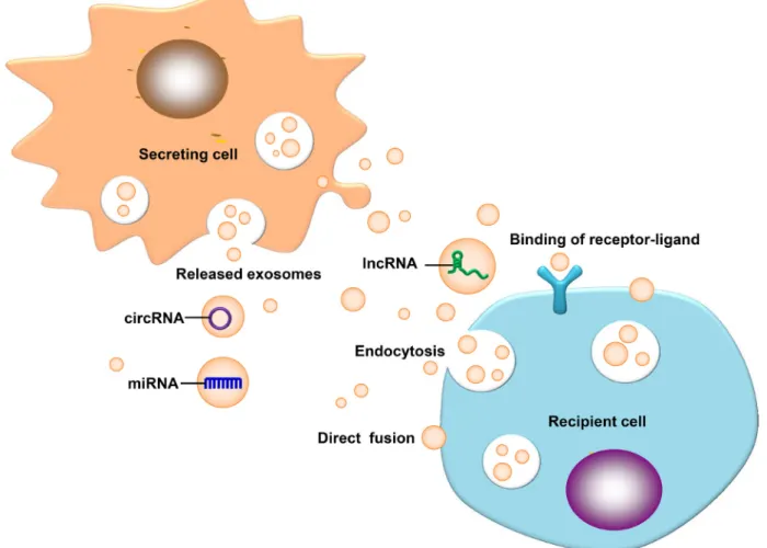
invasion and other aspects [30]. The above concepts are
illustrated in Figure 1.
Exosomes promote the growth of tumor cells in
recipient cells by promoting angiogenesis, invasion,
proliferation and diffusion [31, 32]. Exosomes target
to stromal cells, immune cells and vascular cells in the
tumor microenvironment to promote the occurrence and
development of cancer [33]. Studies have shown that
exosomes can directly regulate the invasion and migration
of tumor cells and can promote the directional movement
of cells via the activity of biological components of the
extracellular matrix (ECM) [34, 35]. Tumor exosomes can
promote the growth and spread of tumor cells by affecting
the permeability of blood vessels or anchoring distant
metastasis sites to change the physiological functions of
adjacent or distant cells [36–40]. In this review, we focus
on the study of the role of exosomal non-coding RNAs in
the development of cancer, which are important molecules
in cancer metastasis.
THE ROLE OF EXOSOMAL NON-CODING
RNAS IN TUMOR METASTASIS
Exosomal miRNAs in tumor metastasis
MicroRNAs (miRNAs) are approximately
22-nucleotide-long small molecules of RNA. miRNAs
bind to the target mRNA 3 'UTR to prevent the translation
of the target mRNA or promote the degradation of target
mRNA at the post transcriptional level, to negatively
regulate the target genes, and thus to inhibit or promote
tumor metastasis [41–43]. Recently, exosomal miRNAs
have been found to play key roles in physiological
and pathological processes in eukaryotes [44–47].
The tumor-derived miRNAs and stromal cells of the
microenvironment are interrelated with each other, which
regulate the tumor progression, angiogenesis, invasion
and metastasis and can escape from immune surveillance
[48]. The role of exosomal miRNAs in tumor metastasis
is summarized in Table 1.
Epithelial-mesenchymal transition (EMT) refers
to the loss of the characteristics of epithelial cells and
gaining the process of interstitial cell phenotype. This
process includes changes in the cell’s morphology and
phenotype, with invasion and distant metastasis of tumor
cells as the initial step [49–51]. Scholars confirmed that
stromal cell-derived exosomes transport miRNAs to tumor
cells, leading to EMT changes in the morphology and
biochemistry of tumor cells. Intercellular communication
also affects the occurrence and development of the EMT
process. Research has shown that exosomes contain
special proteins and miRNAs that could induce phenotypic
changes [52]. Recently, researchers have confirmed that
exosomes contain miR-23a, which can influence the
occurrence and development of EMT by regulating
TGF-β. Exosomes induce the activation of TCF/LEF and
the Wnt pathway in lung cancer cells, which affect the
occurrence and development of tumors [53–55]. Exosomal
miRNAs can be transferred to the target cells, where they
directly modify their target mRNAs [56–60].
The development of tumor metastasis requires the
tumor cells to adapt to the new metastasis site. However,
for when and how the tumor cells can develop metastasis,
the underlying mechanisms remain unclear. Recently,
researchers have shown that when a primary tumor reaches
to the brain, tumor cells that normally express PTEN lose
its expression. When PTEN-deficient metastatic brain
cancer cells leave the brain, the expression of PTEN
will return to normal levels, and the reversal of PTEN
deletion occurs via astrocyte-derived exosomal miR-19a
in the brain and spinal cord. The miR-19a targets PTEN to
complete the biological behavior [8]. The loss of PTEN in
brain tumor cells could increase the secretion of cytokine
CCL2, which can restore microglia to metastatic tumors to
promote the growth of tumor cells and can protect cancer
cells from death. This study provides a new hope for the
treatment of cancer metastasis [61–64].
Glycolysis and carbohydrate uptake are aberrantly
active during carcinogenesis. Recent studies have shown
that exosomal miR-122 can be found in breast cancer cells,
which can inhibit the proliferation of primary tumor and
increase the effectiveness of nutrients of the precancerous
metastasis niche, thereby inducing cancer metastasis.
miR-122 inhibits carbohydrate uptake by niche cells
by inhibiting glycolytic enzymes and pyruvate kinase.
The inhibition of miR-122 can reduce the incidence of
brain and lung metastases in vivo [65]. By changing the
efficiency of glucose utilization by the susceptible anterior
metastatic niche cells, exosomal miR-122 can be adapted
to the system’s energy metabolism program to promote
cancer progression.
DNA methylation, histone modification and
miRNAs play important roles in the process of tumor
progression by regulating the microenvironment and
immune surveillance. Exosomal miRNAs can not only be
used as potential markers of malignant tumors, but they
are also involved in the internal communication between
cancer cells and the microenvironment. The expression
of miR-24-3P was significantly higher in the plasma of
nasopharyngeal carcinoma patients. The miR-24-3P
inhibited the proliferation of T cells and the differentiation
of Th1 and Th17 and induced the regulation of T cells.
MiR-24-3P regulated the proliferation and differentiation
of T cells, decreased the expression of P-ERK, P-STAT1
and P-STAT3, and increased the expression of P-ATAT5
in cells [66]. FGF11 is a direct target of miR-24-3P in the
regulation of proliferation and differentiation of T cells.
Many studies have demonstrated that exosomal miRNA
is involved in regulating the tumor microenvironment
[67–71].
Exosomes can be involved in tumor resistance
through intercellular communication by carrying
biological information and drug factors. Studies have
shown that the occurrence and development of breast
cancer are closely related to miR-134 and that the
expression level of miR-134 can reduce STAT5B, Hsp90,
and Bcl-2 to reduce cell proliferation and promote
apoptosis by cisplatin [71]. This study suggests that
miR-134 can be used as a diagnostic biomarker of breast cancer
have shown that exosomal miR-939 is associated with
poor breast cancer prognosis, inhibits VE-cadherin and
destroys the barrier function of endothelial cells [72].
Gemcitabine (GEM) is a key drug used in
pancreatic ductal adenocarcinoma (PDAC), but PDAC
cells can produce chemical resistance after long-term
administration. Because resistance is rapidly expanding
into PDAC tissue, it is important to look for the
mechanisms that produce resistance to chemotherapy.
Researchers found that the long-term use of GEM
increased the expression of miR-155 in PDAC cells
[73]. The increase in miR-155 induced the secretion of
exosomes via chemotherapy resistance, which promoted
anti-apoptotic activity. Exosomes deliver miR-155 to
other PDAC cells. Targeting miR-155 may thus effectively
reduce drug resistance. This study found a new therapeutic
target for GEM in PDAC as well as a new approach to
address metastasis.
Studies have revealed breast cancer-derived
exosomal miR-200, which can be taken by recipient cells
to acquire metastatic ability [70]. Exosomal miR-1246
can induce cell invasion and migration by regulating the
DENN/MADD Domain containing 2D (DENND2D) in
breast cancer cells [68]. Exosomal miR-105 could destroy
vascular endothelial cells to promote the occurrence and
metastasis of ovarian cancer by targeting the tight junction
protein ZO-1 [67]. Exosomal miR-200 can directly target
E-cadherin’s mRNA transcriptional repressors ZEB1 and
ZEB2 to increase the expression of E-cadherin and inhibit
the movement ability of tumors [74, 75]. Other studies
have shown that exosomal miR-23a increased significantly
in hypoxic lung cancer cells. miR-23a can promote
angiogenesis by directly targeting prolyl hydroxylase1/2
(PHD1/PHD2), leading to the accumulation of
hypoxia-inducible factor-1α (HIF-1α) in endothelial cells. At
the same time, exosomal miR-23a inhibits the tight
junction protein (ZO-1), thereby increasing the vascular
permeability and migration ability. Hypoxic lung cancer
cell-derived exosomal miR-23a can promote cancer
progression by modulating the tumor vasculature [76].
Glioblastoma is a highly invasive malignant brain
tumor that is highly vascularized and associated with high
mortality. Kininogen-1 (KNG1) is associated with tumor
suppression and antiangiogenic activity in glioblastoma.
CTU1, KIAA1274 and RAX regulate KNG1 via miR-138.
Following the knockdown of the expression of CTU1,
KIAA1274 and RAX in U87 cells and immortalized
human endothelial cells (iHEC), KNG1 expression is
down-Figure 1: Exosomal ncRNAs ingested by a receptor cell.
Exosomal ncRNAs ingested by a receptor cell via direct fusion, endocytosis and binding of receptor ligands.regulated, which results in the up-regulation of oncogenic
EGFR signaling and induces angiogenesis in a mouse
xenograft model. Knockdown of the expression of KNG1,
CTU1, KIAA1274 or RAX can increase the expression
of U87 cell-derived exosomal miR-138 and enhance the
angiogenesis of iHEC to promote cancer development [77].
Exosomes are new mediators of intercellular
communication. In addition to the fact that the exosomes
can affect the regulation of recipient cells, whether the
release of exosomes affects donor cells remains uncertain.
Recently, scholars have innovatively proposed that major
vault protein (MVP)-mediated tumor suppressor miRNAs
can be selectively separated to the body to promote the
development of the tumor process [28, 78]. Many studies
have confirmed that miR-193a can interact with the MVP
and that the MVP promotes the release of miR-193a from
Table 1: The biological functions of exosomal miRNAs in tumor metastasis
Exosomal miRNA Role in tumor metastasis Tumor type Reference miR-23a Induce EMT by targeting TGF- β
Directly target PHD1/PHD2 and ZO1 to promote lung cancer cell permeability, angiogenesis and migration across endothelial cells
Lung canner
Lung cancer [54][76]
miR-19a Promote brain metastasis by inducing PTEN loss Breast cancer [8]
miR-122 Regulate niche cells, glycolytic enzymes, pyruvate kinase,
inhibit glycolysis, and promote tumor metastasis Breast cancer [65]
miR-24-3p Targeting FGF11 to inhibit the function of T cells Nasopharyngeal carcinoma [66]
miR-134 Inhibit STAT5B, Hsp90 and Bcl2, thereby inhibiting tumor proliferation, invasion and metastasis while promoting cisplatin-induced apoptosis
Breast cancer [71]
miR-939 Inhibit VE-cadherin and destroy endothelial barrier
function Breast cancer [72]
miR-1246 Regulate cell movement and invasion by regulating
DENND2D of oral squamous cell carcinoma Oral squamous cell carcinoma [68]
miR-105 Targeting ZO-1 damages vascular endothelial cells and
promotes metastasis Breast cancer [67]
miR-200 Directly target the mRNA transcriptional repressor ZEB1, E-cadherin, ZEB2, up-regulate the expression of E-cadherin, and inhibit motility in cancer
Ovarian cancer [74]
It is absorbed by recipient cells so they can develop
metastasis Breast cancer [70]
miR-193a Interaction with the MVP, which can block the release of miR-193a after exocytosis, can cause the accumulation of miR-193a in donor cells and inhibit tumor development
Colon cancer [28]
miR-142-3P It is selectively sorted out to promote malignant
phenotypes both inside and outside the cell Oral carcinoma [79]
miR-320a
miR-21
MiR-320a-PBX3 inhibits the activation MAPK pathway and the occurrence of EMT, and it down-regulates the expression of CDK2 and MMP2, thereby inhibiting tumor development
Exosomes transport the stromal cell derived miR-21 to ovarian cancer cell. MiR-21 targeting APAF1 induces resistance to paclitaxel in ovarian cancer cells Down-regulation of PTEN to activate the PIK3/AKT signaling pathway and promote the cisplatin resistance of gastric cancer cells
Liver cancer Ovarian cancer Gastric cancer [80] [63] [120] miR-155 miR-214 miR-221 miR-138
Exosomes transport miR-155 to other PDAC cells, effectively attenuating drug resistance
Down-regulation of the expression of PTEN protein results in the secretion of large amounts of IL-10 by Tregs, which results in the immune escape of the host and tumor growth
Exosomes transfer miR-221 to tumor cells and can bind to hormone therapy targets, resulting in tumor HTR The expression of miR-138 can increase angiogenesis. CTU1, KIAA1274 and RAX regulate KNG1 by miR-138
Pancreatic ductal adenocarcinoma Lung cancer Breast cancer Glioma [73] [117] [122] [77]
the donor cells via the circulation of blood, consequently
promoting tumor development. The amount of exosomal
miR-193a released would decrease after knocking down
the MVP, which would result in the accumulation of
miR-193a in cells and decrease the development of tumor.
miR-193a raises from the Ccnd2 and cMyc by targeting
Caprin1, which retards the cell cycle G1 phase and inhibits
cell proliferation. At the same time, they proposed that
advanced patients had higher levels of exosomal
miR-193a.
Through further investigation, we found that oral
cancer cells can release miR-142-3P by selecting the
extracellular vesicles, thus promoting the malignant
phenotype both inside and outside the cells [79]. Inhibiting
the exosomal protein Rab27A output, which can stop the
release of exosomes, is thought to increase the expression
levels of miR-142-3p, miR-150-5p, and miR-223-3p.
Increasing miR-142-3p-targeted TGFBR1, which can
downgrade the expression of TGFBR1 in donor cells, can
reduce the formation of the malignant phenotype. The
reverse validation by increasing the release of
miR-142-3p in the donor cells, which can be ingested by receptors
on endothelial cells, reduced the activity of TGFBR1,
eventually enabling the development of tumor.
The loss of cancer-associated fibroblast-derived
exosomal miR-320a can promote the proliferation and
metastasis of hepatocellular carcinoma cells. MiR-320a
can be combined with the downstream target genes PBX3
as a tumor suppressor miRNA to inhibit the liver cancer
cell proliferation, migration and metastasis.
miR-320a-PBX3 inhibits the active pathway of the MAPK and EMT
and reduces the expression of dependent cytokinase 2
(CDK2) and MMP2 to inhibit tumor development [80].
Exosomal lncRNAs in tumor metastases
LncRNA are nucleotides longer than 200 nucleotides
in length that do not encode proteins. The approaches to
regulate genes of lncRNAs are varied. One of the most
important ways is to directly target the transcription of
target genes through base complementation and binding
to target genes or by indirectly regulating target genes
upstream or downstream of gene transcription [62, 81–88].
Although this process plays an important role in tumor
development, the mechanisms underlying the relationship
between lncRNAs and tumor metastasis need further
study to identify more treatment approaches [17, 89–93].
The role of exosomal lncRNAs in tumor metastases is
summarized in Table 2.
Recent studies have confirmed that lncRNA HOX
transcript antisense RNA (HOTAIR) is associated with
poor prognosis for many types of cancers. There are a
variety of lncRNAs in the urine of bladder cancer patients,
including HOTAIR, HOX-AS-2, the MALAT1, SOX2,
OCT4 HYMA1, LINC00477, LOC100506688, and
OTX2-AS1. Knockdown of HOTAIR in bladder cancer cell lines
will reduce invasion and migration. More significantly, the
loss of the HOTAIR will affect the changes of related EMT
genes, e.g., SNAI1, TWIST1, ZEB1, ZO1, MMP1, LAMB3,
and LAMC2 [62]. The exosomal lncRNAs originating
from urine can be used as new molecular markers and
targets for molecular therapy, thus providing cancer
treatments with more effective solutions.
CD90+ cancer cells are cancer stem cells that can
promote cancer invasion and metastasis. The exosomes
released by CD90+ cells (not parent liver cancer cells) are
rich in lncRNAH19, which can regulate endothelial cells
and promote angiogenesis and adhesion between cells.
This study showed a new kind of regulating mechanism.
The cancer stem cell sample CD90+ cells can promote
angiogenesis and the tumor microenvironment [94].
This research proposed that lncRNAH19 can also serve
as a potential therapeutic target in liver cancer. Other
studies showed that exosomal lncRNA TUC339, which
derives from liver cancer cells, can regulate the tumor
microenvironment and can be used to adjust the growth
and adhesion ability of tumor cells via the horizontal
transformation of exosomes in cells [95].
In recent years, researchers have found that an
lncRNA called lncARSR (lncRNA Activated in RCC
with Sunitinib Resistance) is closely related to sunitinib
resistance in clinical treatment [60]. lncARSR can be
used as a competitive endogenous RNA (ceRNA),
when combined with miR-34/miR-449, to promote the
expression of AXL, c-MET in renal carcinoma cells,
and then realize the promotion of sunitinib resistance.
However, this bioactive lncARSR is delivered to the
recipient cells by exosomes, thereby contributing to the
spread of sunitinib drug resistance. Targeted blockade of
lncARSR or the use of AXL/c-MET inhibitors may aid in
treating renal cell carcinoma that is resistant to sunitinib.
lncARSR may serve as an effective molecular marker and
intervention target to improve the therapeutic efficacy
of sunitinib in renal cell carcinoma and provide a new
approach and perspective for the individualized target
therapy of renal cell carcinoma.
Many studies have shown that the expression of
long noncoding RNA ZFAS1 has increased in cancers,
and some scholars have linked ZFAS1 to the exosomes.
The expression of long noncoding RNA ZFAS1 is
up-regulated in the serum of patients with gastric cancer,
in whom exosomes can promote the proliferation and
migration of gastric cancer cells by transmitting the ZFAS1
[96]. Exosomes can up-regulate cyclin D1 and accelerate
the cell cycle. Bcl-2 simultaneously increases time the
down-regulation of Bax, which can increase the level of
ERK phosphorylation. Exosomal ZFAS1 can increase the
proliferation and invasion of gastric cancer cells. Many
studies confirmed that the expression of ZFAS1 is
up-regulated in cancers and proved that it can compete with
miR-150 as ceRNA; further, the expression of miR-150
ZFAS1 is associated with EMT in gastric carcinoma [98].
Whether these biological mechanisms are associated with
biological function of exosomes requires further research.
The study confirmed that ZFAS1 may play a role in the
tumorigenesis of gastric cancer and can serve as a novel
target for treating gastric cancer.
Research found that the lncRNAs in plasma
can be protected by exosomes and expressed stably,
and researchers have confirmed that the expression of
LINC00152 is significantly increased in the plasma of
gastric cancer patients. Its expression level is almost equal
in plasma and exosomes [99]. LINC00152 can be used as
a potential and stable molecular marker of gastric cancer.
Some studies confirmed that exosomal lncRNAs play
important roles in tumor development and progression.
HOTAIR, MALAT1 and MEG3 can be used as targets of
cervical cancer in early diagnosis [100]. The lncRNA
H19 can increase the expression of angiogenic factor
VEGF and its receptor VEFG-R1, both of which can be
used as potential drug targets in hepatocellular carcinoma
[101]. In the development of cancer, many studies have
confirmed that a variety of lncRNAs is closely related
to the development of cancer. The lncRNAs HULC,
lncRNAH19, HEIH, and MVIH can promote the growth
of cancer cells. The lncRNAs HOTAIR and PVT1 can
promote the proliferation of cancer cells. The lncRNAs
MALAT1, LET and ATB can promote the metastasis of
cancer cells. Whether these exosomal lncRNAs are closely
related to cancers deserves further discussion [62, 100,
101].
Exosomal circRNAs in tumor metastasis
circRNA is formed by a downstream 5 splice site
that is associated with an upstream 3 splice site and is more
stable than linear RNA. Its formation can be divided into
the phases of intron cyclization, Lasso driving cyclization,
intron pairing, driving and cyclization. In the beginning,
circRNAs were considered a by-product of splicing errors
and did not receive extensive attention. In recent years,
with the development of sequencing technology, more
circRNAs have been identified [102–105].
Exosomal circRNA is novel and stable. It may serve
as a new tumor marker and provide new ideas for the
diagnosis and prognosis of tumors. circRNAs may play an
important role in cell communication. Some recent studies
have confirmed that circRNAs can be used as molecular
sponges for miRNAs [106–108], which can competitively
inhibit the biological activity of miRNA. CiRS-7 can
be used as a natural molecular sponge for miRNA-7 to
regulate gene expression, regulate transcription and
splicing, and regulate parental gene transcription [81,
109]. Researchers speculated that functional exosomal
circRNAs participate in intercellular communication.
ciRS-7/CDR1 can serve as a miRNA molecular sponge,
which can combine with special miRNA, and can transfer
biological information to a specific position to achieve
the efficient transmission of biological information and
thereby promote the spread of cancer [102].
Studies have shown that the development of colon
cancer is closely related to circRNA. The content of
exosomal circRNAs is far greater than that found in cells
[110]. CircRNAs may regulate the development of colon
cancer and serve as molecular markers for cancer.
The expression levels of exosomal circRNAs
are stable and abundant. However, the mechanism is
unknown and may be attributed to the special protection
of exosomes, the features of a specific sequence, or the
protection of certain proteins [103]. Exosomal circRNAs
are likely to become new markers for cancer therapy and
molecular therapeutic targets.
THE MECHANISM OF EXOSOMAL
NON-CODING RNAS INVOLVED IN TUMOR
METASTASIS
Invasion and metastasis are the basic characteristics of
malignant tumors. They are also the pathological bases for
tumor recurrence, disease deterioration and, ultimately, death.
There are many mechanisms that affect tumor metastasis
and are related to a variety of pathways. Exosomal
non-coding RNA can also contribute to tumor metastasis through
a variety of mechanisms [111]. Figure 2 shows that the
pathways of exosomal ncRNAs regulate tumor metastasis.
Transmission of oncogenes
Exosomal non-coding RNA can affect the
proliferation, invasion and migration of tumor cells
through specific biological action and can further affect
the occurrence and development of tumor metastasis
[39]. miR-1246, for instance, regulates the invasion and
metastasis by regulating DENND2D. miR-105 destroys
vascular endothelial cells by targeting the tight junction
protein ZO-1 and promotes metastasis. The lncRNA
ZFAS1 promotes the proliferation and migration of
gastric cancer cells via the transmission of the exocrine.
miR-134 can inhibit STAT5B, Hsp90 and Bcl2 and can
further inhibit the proliferation, invasion and metastasis
of tumor cells. miR-142-3P can be selectively isolated
from exosomes, thus promoting the development of the
malignant phenotype of tumor cells.
Interfere with the immune system to promote
tumor metastasis
Tumor cells can evade the immune response through
various mechanisms, such as a lack of histocompatibility
complex (MHC)I and the expression of MHC II, mutation
and degradation of tumor associated antigen, and blocking
immune molecules to increase the tolerance of the
immune system. Tumor cells can also inhibit NK cells
and raise CTL cells by regulating the output of VEGF,
IL-10, TGF-β, and adenosine ROS immunosuppressive
molecules, thus damaging the immune response [112],
and are not recognized by immune cells to escape
immune surveillance [113, 114]. The improvement of
tumor cell proliferation and the increased instability can
cause the development of different types of antigens.
Exosomal CD39 and CD73 exist in cancer cells. These
two molecules can generate adenosines, which can
restrain immunity; then, tumor cells can regulate immune
cells [115]. Exosomes can influence the development of
tumor metastasis by affecting the immune function, such
as miR-24-3p targeting FGF11 to inhibit T cell function.
Macrophages can secrete exosomes that contain abundant
miR-155, which promotes the expression of TNF-a,
IL-6, and IL-23 while increasing the expression of CD40,
CD63, CD81, MCH-I, MyD88, and NF-kB. The research
has shown that miR-155 is involved in the inflammatory
response [116]. Studies have reported that tumor-derived
exosomal miR-214 inhibited the PTEN protein expression
level to induce the secretion of IL-10 by Tregs, leading
to the host organism immune escape to promote tumor
growth [117].
Participate in drug resistance to promote tumor
metastasis
Multiple drug resistance in tumor cells and tumor
metastasis are the two main factors contributing to
the failure of cancer treatment. Multidrug resistance
(MDR) in tumor cells can cause resistance to drugs
that are administer to inhibit cell growth and can cause
toxicity. Numerous studies have shown that exosomes
blockade can block the response to cell growth therapy
[118]. Studies have reported that breast cancer cells can
transmit miR-100, miR-222, miR-30a, and miR-17 via
exosomes to decrease the drug resistance to doxorubicin
and paclitaxel [119]. Exosome can participate in the
mechanisms underlying cancer drug resistance to promote
tumor metastasis. miR-21 moves from the stromal cells to
the ovarian cancer cells via the transport of exosomes and
targets APAF1 to cause Taxol resistance in ovarian cancer
cells [63]. There have also been reports that macrophages
can deliver miR-21 to the stomach cancer cells via
exosomes, thereby inhibiting the apoptosis of gastric
cancer cells by down-regulating PTEN to activate the
PIK3/AKT signaling pathway, and can promote cisplatin
resistance in gastric cancer cells [120]. The competitive
combination of lncARSR with miR-34/miR-449 increases
the expression levels of AXL and c-met though exosomes
to promote the spread of drug resistance. An increase in
miR-155 in the pancreatic duct adenocarcinoma (PDAC)
can induce exosome secretion. By promoting the
anti-apoptotic activity produced by chemotherapy drug
resistance, exosomal miR-155 will be delivered to other
PDAC cells and can effectively weaken gemcitabine
(GEM) resistance [76, 121]. The fibroblasts secrete
miR-221 into tumor cells, which can bind to hormone therapy
targets, resulting in tumor hormone therapy resistance
(HTR) [122].
Involvement in tumor metabolism to promote
tumor metastasis
Tumor cell metabolism refers to normal cells that
are under the action of a carcinogenic factor and develop
a continuous proliferation of tumor cells. Similar to
increases in glucose intake, lactic acid accumulation
or nucleic acid synthesis can reinforce metabolic
Table 2: The biological functions of exosomal lncRNAs in tumor metastasis
Exosomal lncRNA
Role in tumor metastasis
Tumor type
Reference
HOTAIR,
MALAT1,
MEG3
As a target for the early diagnosis of cervical cancer
Cervical cancer
[100]
lncARSR
The competitive combination of miR-34/miR-449
increase the expression levels of AXL and c-MET
and promote the dissemination of sunitinib resistance
through exosomes
Renal carcinoma
[60]
H19
Increasing the expression of VEGF and its receptor
VEGF-R1, which may serve as a potential target for
HCC
Liver cancer
[101]
ZFAS1
LINC00152
TUC339
Promote the proliferation and migration of gastric
cancer cells through the transmission of the exosome
The expression in exosome increased significantly in
patients
Regulating the microenvironment of hepatoma cells
by intracellular transfer of the exosome, thereby
regulating the growth and adhesion of tumor cells
Gastric cancer
Gastric cancer
Liver cancer
[96]
[99]
[95]
changes. The three most remarkable features of tumor
cells, including immortality, mobility and the loss of
contact inhibition, have the most direct relationship with
tumor cell metabolism. Molecular biological studies of
metastatic tumors have found that the main carcinogenic
signaling pathways converge at the end of the tumor
cell metabolism. Metabolic changes are essential in the
development of tumor cells. Therefore, some research
teams have explored the mechanisms of tumor metastasis
from the perspective of tumor sugar metabolism; e.g.,
miR-122 inhibits glycolysis by downregulating the pyruvate
kinase. Recent studies have reported that the pyruvate
kinase M2 (PKM2) plays an important role in the release
of exosomes [65]. This process is a key step in exosome
secretion, and the release of tumor cells is closely related
to the general and metabolic transformation of tumor cells.
Participate in EMT to promote tumor metastasis
EMT is an important process in the early stage
of tumor invasion and metastasis. An important marker
of EMT is the down-regulation of E-cadherin. Many
transcription factors can inhibit the expression of
E-cadherin. For example, Snail/Slug family proteins,
Twist, ZEB1, SIP1 and the E12/E47, Wnt, TGF-β, and
nuclear factor-κB (NF-κB) pathways play an important
role in the EMT process. They can promote the occurrence
of EMT by affecting the transcription inhibitory factor of
EMT [51]. Many studies have shown that a variety of
exosomal miRNAs may influence the signaling molecules
that are related to the EMT pathway. Exosomal miRNAs
can promote or inhibit tumor metastasis by influencing the
occurrence of EMT. For example, exosomal miR-23a can
regulate the development of EMT by influencing TGF-β.
Exosomes can induce the transcriptional activation of
TCF/LEF and activate the Wnt pathway in lung cancer
cells to affect the occurrence and development of tumor
metastasis. miR-939 inhibits VE-cadherin and destroys
endothelial barrier function. miR-200 can directly target
E-cadherin’s transcriptional repressors ZEB1 and ZEB2
and can up-regulate the expression of E-cadherin and
inhibit the motility of mRNA. miR-320a-PBX3 can inhibit
the activation pathway of MAPK, thereby inhibiting the
occurrence of EMT and down-regulating the expression of
cyclin dependent kinase 2 (CDK2) and MMP2. Therefore,
miR-320a-PBX3 can inhibit tumor development. The loss
of p85α expression in the stroma of breast cancer can
cause stromal fibroblasts to acquire the characteristics
of cancer associated fibroblasts (CAF). Exosomes carry
Wnt10b and induce EMT via the classical Wnt pathway,
Figure 2: The mechanisms of the exosomal ncRNAs involved in cancer metastasis.
(1). Horizontal transmission of oncogenes, (2). Regulate the immune system as immunomodulators, (3). Participate in tumor chemotherapy resistance, (4). Regulate tumor metabolism, (5). Influence the progression of EMT and cancer premetastatic niches, (6). Promote angiogenesis.which can promote cancer progression by modifying the
stromal cells and epithelial tissue remodeling of the tumor
micro-environment [123].
Modulate vascular system to promote tumor
metastasis
miR-138, an exosomal ncRNA that is derived from
glioblastoma, enhances tumor angiogenesis and promotes
cancer development. The exosomes originating from
glioma stem cells promote the angiogenesis of endothelial
cells via the miR-21/VEGF signaling pathway [124]. The
increased expression of angiogenic factor (VEGF) and
its receptor VEGF-R1 caused by lncRNA H19 affects the
development of metastatic tumors.
Many researchers have also explored the effects and
functions of exosomal ncRNAs through other approaches.
For example, some non-coding RNAs in exosomes can
play vital biological roles by acting as ceRNA. Further,
lncARSR combined with miR-34/miR-449 can increase
the expression levels of AXL and c-MET. This promotes
the development of metastatic disease by promoting the
dispersion of drug resistance. miR-19a promotes brain
metastasis by inducing the loss of PTEN [8]. We need to
conduct more in-depth studies to explore new pathways by
which exosomal ncRNAs can promote cancer metastasis.
Summary
In recent years, studies on the mechanism of
exosomal non-coding RNAs have increased. However,
deeper mechanisms are still to be found in the exosomes.
The main challenges in studying exosomes include how
they participate in the physiological and pathological
processes and dissecting the transfer of exosomes from
cell to cell [125, 126]. In this review, we have summarized
various exosomal non-coding RNAs that play key roles
in tumor metastasis. This review provides a detailed
description of the function of exosomal ncRNAs, which
will offer new insights and provide novel therapeutic
approaches in malignant tumor metastasis.
Exosomes are membranous vesicles that are
secreted into the extracellular space by a variety of cells
and play important biological functions in intercellular
communication. Many sources of exosomal ncRNAs have
been shown to be closely related to human malignancies.
As the potential functions of exosomes are gradually
discovered, it is critical to explore the structures of these
vesicles and their social domains. This will give aid in
studying the deeper functional mechanisms of exosomes
[13, 127, 128]. Currently, the main research approach
is to explore the relative miRNAs and protein levels
of exosomes [129, 130]. We believe that the lncRNA,
circRNA, and other new components will be used in
a broader field of exosome investigations [131]. This
method of transferring small packages will provide new
perspectives for cancer treatment and prognosis and will
give more novel ideas to examine tumor metastasis and
unique clinical paths.
Exosomal ncRNAs may serve as very important
biomarkers for disease diagnosis, prognosis, and
treatment. Many studies have reported that there are
important cancer-associated RNAs and protein markers
in exosomes, but because of their small size and low
density, it is not easy to isolate exosomes. Because of
the complexity of extraction methods, it is difficult to
study the exosomes. The current mainstream exosome
isolation techniques include ultracentrifugation-based
isolation techniques, ultrafiltration, immunoaffinity
capture-based techniques, and exosome precipitation
[132, 133]. Recently, a new method for the rapid isolation
and identification of exosomes has been found. The core
of this technology is an alternating current electrokinetic
(ACE) microarray device. Such a device may have more
than 1000 electrodes, and its surface is coated with a thin
layer of porous water gel. In the dielectrophoretic high
field area, the exosome mass is enriched. Exosomes are
enriched at the chip electrodes and can be analyzed and
identified directly via scanning electron microscopy
and immunofluorescence [134]. In addition, the novel
microfluidics-based isolation techniques may make
it more convenient to explore the exosomal ncRNAs
[135, 136]. There are also many emerging technologies
for extracting exosomes. The advent of more advanced
exosome extraction techniques will greatly accelerate the
development of exosomal ncRNA research.
Exosomal ncRNAs play important roles in malignant
tumor metastasis. They are hoped to have an indispensable
role in targeted drug delivery and clinical therapy. At the
same time, the development of more efficient exosome
separation techniques will promote exosome research. The
anomalous expression levels of exosomal ncRNA may
indicate cancer or cancer progression. Exosomal ncRNAs
are thus promising biomarkers. Further study of exosomal
ncRNAs may provide an effective minimally invasive
strategy for the diagnosis, prognosis and treatment of
cancer. The discoveries of exosomal ncRNAs have opened
a potential market for more exciting clinical applications
in the near future.
Abbreviations
ncRNA: coding RNA; LncRNA: Long
non-coding RNA; miRNA: micro RNA; ECM: Extracellular
matrix; EMT: Epithelial-mesenchymal transition; PDAC:
Pancreatic ductal adenocarcinoma; GEM: Gemcitabine;
HIF-1 α: Hypoxia-inducible factor-1 α; ZO-1:Tight
junction protein; iHEC: immortalized human endothelial
cells; MVP: Major vault protein; HOTAIR: HOX transcript
antisense RNA; ceRNA: Competitive endogenousRNA;
lncARSR: lncRNA Activated in RCC with Sunitinib
Resistance; CAF: Cancer associated fibroblasts; MDR:
Multidrug resistance; HTR: Hormone therapy resistance;
PTEN: Phosphatase and tensin homolog.
Author contributions
WX and ZYZ designed and revised the manuscript.
JPW wrote the manuscript and drew figures. YYT and
CMF collected the related paper and created the tables.
FX, CG, YHZ, ZL, XLL, YL and GYL participated in the
design of the review. All the authors read and approved the
final version of the review.
ACKNOWLEDGMENTS
Not applicable.
CONFLICTS OF INTEREST
None.
FUNDING
This work was supported by the National Natural
Science Foundation of China (81672683, 81472531 and
81402009), the Natural Science Foundation of Hunan
Province (2015JJ1022, 2016JC2035 and 2017SK2105),
and the 111 project (111-2-12).
REFERENCES
1. Zhang W, Fan S, Zou G, Shi L, Zeng Z, Ma J, Zhou Y, Li X, Zhang X, Li X, Tan M, Xiong W, Li G. Lactotransferrin could be a novel independent molecular prognosticator of nasopharyngeal carcinoma. Tumour Biol. 2015; 36:675–83. https://doi.org/10.1007/s13277-014-2650-1.
2. Yan Q, Zeng Z, Gong Z, Zhang W, Li X, He B, Song Y, Li Q, Zeng Y, Liao Q, Chen P, Shi L, Fan S, et al. EBV-miR-BART10-3p facilitates epithelial-mesenchymal transition and promotes metastasis of nasopharyngeal carcinoma by targeting BTRC. Oncotarget. 2015; 6:41766–82. https://doi. org/10.18632/oncotarget.6155.
3. Bo H, Gong Z, Zhang W, Li X, Zeng Y, Liao Q, Chen P, Shi L, Lian Y, Jing Y, Tang K, Li Z, Zhou Y, et al. Upregulated long non-coding RNA AFAP1-AS1 expression is associated with progression and poor prognosis of nasopharyngeal carcinoma. Oncotarget. 2015; 6:20404–18. https://doi. org/10.18632/oncotarget.4057.
4. Zeng Z, Huang H, Zhang W, Xiang B, Zhou M, Zhou Y, Ma J, Yi M, Li X, Li X, Xiong W, Li G. Nasopharyngeal carcinoma: advances in genomics and molecular genetics. Sci China Life Sci. 2011; 54:966–75. https://doi. org/10.1007/s11427-011-4223-5.
5. Yang L, Tang Y, He Y, Wang Y, Lian Y, Xiong F, Shi L, Zhang S, Gong Z, Zhou Y, Liao Q, Zhou M, Li X, et al. High Expression of LINC01420 indicates an unfavorable
prognosis and modulates cell migration and invasion in nasopharyngeal carcinoma. J Cancer. 2017; 8:97–103. https://doi.org/10.7150/jca.16819.
6. Tang Y, He Y, Shi L, Yang L, Wang J, Lian Y, Fan C, Zhang P, Guo C, Zhang S, Gong Z, Li X, Xiong F, et al. Co-expression of AFAP1-AS1 and PD-1 predicts poor prognosis in nasopharyngeal carcinoma. Oncotarget. 2017; 8:39001–11. https://doi.org/10.18632/oncotarget.16545. 7. Aleckovic M, Kang Y. Welcoming Treat:
Astrocyte-Derived Exosomes Induce PTEN Suppression to Foster Brain Metastasis. Cancer Cell. 2015; 28:554–6. https://doi. org/10.1016/j.ccell.2015.10.010.
8. Zhang L, Zhang S, Yao J, Lowery FJ, Zhang Q, Huang WC, Li P, Li M, Wang X, Zhang C, Wang H, Ellis K, Cheerathodi M, et al. Microenvironment-induced PTEN loss by exosomal microRNA primes brain metastasis outgrowth. Nature. 2015; 527:100–4. https://doi.org/10.1038/nature15376.
9. Liao Q, Guo X, Li X, Xiong W, Li X, Yang J, Chen P, Zhang W, Yu H, Tang H, Deng M, Liang F, Wu M, et al. Prohibitin is an important biomarker for nasopharyngeal carcinoma progression and prognosis. Eur J Cancer Prev. 2013; 22:68– 76. https://doi.org/10.1097/CEJ.0b013e328354d351. 10. Yang Y, Liao Q, Wei F, Li X, Zhang W, Fan S, Shi L, Li X,
Gong Z, Ma J, Zhou M, Xiang J, Peng S, et al. LPLUNC1 inhibits nasopharyngeal carcinoma cell growth via down-regulation of the MAP kinase and cyclin D1/E2F pathways. PLoS One. 2013; 8:e62869. https://doi.org/10.1371/journal. pone.0062869.
11. Song Y, Li X, Zeng Z, Li Q, Gong Z, Liao Q, Li X, Chen P, Xiang B, Zhang W, Xiong F, Zhou Y, Zhou M, et al. Epstein-Barr virus encoded miR-BART11 promotes inflammation-induced carcinogenesis by targeting FOXP1. Oncotarget. 2016; 7:36783–99. https://doi.org/10.18632/ oncotarget.9170.
12. Wang M, Zhao J, Zhang L, Wei F, Lian Y, Wu Y, Gong Z, Zhang S, Zhou J, Cao K, Li X, Xiong W, Li G, et al. Role of tumor microenvironment in tumorigenesis. J Cancer. 2017; 8:761–73. https://doi.org/10.7150/jca.17648.
13. Costa-Silva B, Aiello NM, Ocean AJ, Singh S, Zhang H, Thakur BK, Becker A, Hoshino A, Mark MT, Molina H, Xiang J, Zhang T, Theilen TM, et al. Pancreatic cancer exosomes initiate pre-metastatic niche formation in the liver. Nat Cell Biol. 2015; 17:816–26. https://doi. org/10.1038/ncb3169.
14. Melo SA, Sugimoto H, O’Connell JT, Kato N, Villanueva A, Vidal A, Qiu L, Vitkin E, Perelman LT, Melo CA, Lucci A, Ivan C, Calin GA, et al. Cancer exosomes perform cell-independent microRNA biogenesis and promote tumorigenesis. Cancer Cell. 2014; 26:707–21. https://doi. org/10.1016/j.ccell.2014.09.005.
15. Zhou Y, Liao Q, Li X, Wang H, Wei F, Chen J, Yang J, Zeng Z, Guo X, Chen P, Zhang W, Tang K, Li X, et al. HYOU1, Regulated by LPLUNC1, Is Up-Regulated in Nasopharyngeal Carcinoma and Associated with Poor Prognosis. J Cancer. 2016; 7:367–76. https://doi. org/10.7150/jca.13695.
16. Zeng Z, Fan S, Zhang X, Li S, Zhou M, Xiong W, Tan M, Zhang W, Li G. Epstein-Barr virus-encoded small RNA 1 (EBER-1) could predict good prognosis in nasopharyngeal carcinoma. Clin Transl Oncol. 2016; 18:206–11. https://doi. org/10.1007/s12094-015-1354-3.
17. Zeng Z, Bo H, Gong Z, Lian Y, Li X, Li X, Zhang W, Deng H, Zhou M, Peng S, Li G, Xiong W. AFAP1-AS1, a long noncoding RNA upregulated in lung cancer and promotes invasion and metastasis. Tumour Biol. 2016; 37:729–37. https://doi.org/10.1007/s13277-015-3860-x.
18. Xu K, Xiong W, Zhou M, Wang H, Yang J, Li X, Chen P, Liao Q, Deng H, Li X, Li G, Zeng Z. Integrating ChIP-sequencing and digital gene expression profiling to identify BRD7 downstream genes and construct their regulating network. Mol Cell Biochem. 2016; 411:57–71. https://doi. org/10.1007/s11010-015-2568-y.
19. Xiao K, Yu Z, Li X, Li X, Tang K, Tu C, Qi P, Liao Q, Chen P, Zeng Z, Li G, Xiong W. Genome-wide Analysis of Epstein-Barr Virus (EBV) Integration and Strain in C666-1 and Raji Cells. J Cancer. 20C666-16; 7:2C666-14–24. https://doi. org/10.7150/jca.13150.
20. Tu C, Zeng Z, Qi P, Li X, Yu Z, Guo C, Xiong F, Xiang B, Zhou M, Gong Z, Liao Q, Yu J, He Y, et al. Genome-Wide Analysis of 18 Epstein-Barr Viruses Isolated from Primary Nasopharyngeal Carcinoma Biopsy Specimens. J Virol. 2017; 91:e00301–17. https://doi.org/10.1128/JVI.00301-17. 21. de Oliveira JC, Scrideli CA, Brassesco MS, Morales AG,
Pezuk JA, Queiroz RP, Yunes JA, Brandalise SR, Tone LG. Differential miRNA expression in childhood acute lymphoblastic leukemia and association with clinical and biological features. Leuk Res. 2012; 36:293–8. https://doi. org/10.1016/j.leukres.2011.10.005.
22. Shi W, Alajez NM, Bastianutto C, Hui AB, Mocanu JD, Ito E, Busson P, Lo KW, Ng R, Waldron J, O’Sullivan B, Liu FF. Significance of Plk1 regulation by miR-100 in human nasopharyngeal cancer. Int J Cancer. 2010; 126:2036–48. https://doi.org/10.1002/ijc.24880.
23. Subra C, Grand D, Laulagnier K, Stella A, Lambeau G, Paillasse M, De Medina P, Monsarrat B, Perret B, Silvente-Poirot S, Silvente-Poirot M, Record M. Exosomes account for vesicle-mediated transcellular transport of activatable phospholipases and prostaglandins. J Lipid Res. 2010; 51:2105–20. https://doi.org/10.1194/jlr.M003657.
24. Bang C, Thum T. Exosomes: new players in cell-cell communication. Int J Biochem Cell Biol. 2012; 44:2060–4. https://doi.org/10.1016/j.biocel.2012.08.007.
25. Tang MK, Wong AS. Exosomes: Emerging biomarkers and targets for ovarian cancer. Cancer Lett. 2015; 367:26–33. https://doi.org/10.1016/j.canlet.2015.07.014.
26. Batista BS, Eng WS, Pilobello KT, Hendricks-Munoz KD, Mahal LK. Identification of a conserved glycan signature for microvesicles. J Proteome Res. 2011; 10:4624–33. https://doi.org/10.1021/pr200434y.
27. Isola AL, Chen S. Exosomes: The Link between GPCR Activation and Metastatic Potential? Front Genet. 2016; 7:56. https://doi.org/10.3389/fgene.2016.00056.
28. Teng Y, Ren Y, Hu X, Mu J, Samykutty A, Zhuang X, Deng Z, Kumar A, Zhang L, Merchant ML, Yan J, Miller DM, Zhang HG. MVP-mediated exosomal sorting of miR-193a promotes colon cancer progression. Nat Commun. 2017; 8:14448. https://doi.org/10.1038/ncomms14448.
29. Subra C, Laulagnier K, Perret B, Record M. Exosome lipidomics unravels lipid sorting at the level of multivesicular bodies. Biochimie. 2007; 89:205–12. https:// doi.org/10.1016/j.biochi.2006.10.014.
30. Tkach M, Thery C. Communication by Extracellular Vesicles: Where We Are and Where We Need to Go. Cell. 2016; 164:1226–32. https://doi.org/10.1016/j. cell.2016.01.043.
31. Raposo G, Stoorvogel W. Extracellular vesicles: exosomes, microvesicles, and friends. J Cell Biol. 2013; 200:373–83. https://doi.org/10.1083/jcb.201211138.
32. Li X, Wang S, Zhu R, Li H, Han Q, Zhao RC. Lung tumor exosomes induce a pro-inflammatory phenotype in mesenchymal stem cells via NFkappaB-TLR signaling pathway. J Hematol Oncol. 2016; 9:42. https://doi. org/10.1186/s13045-016-0269-y.
33. Caballero JN, Frenette G, Belleannee C, Sullivan R. CD9-positive microvesicles mediate the transfer of molecules to Bovine Spermatozoa during epididymal maturation. PLoS One. 2013; 8:e65364. https://doi.org/10.1371/journal. pone.0065364.
34. Zoller M. Tetraspanins: push and pull in suppressing and promoting metastasis. Nat Rev Cancer. 2009; 9:40–55. https://doi.org/10.1038/nrc2543.
35. Zhang H, Deng T, Liu R, Bai M, Zhou L, Wang X, Li S, Wang X, Yang H, Li J, Ning T, Huang D, Li H, et al. Exosome-delivered EGFR regulates liver microenvironment to promote gastric cancer liver metastasis. Nat Commun. 2017; 8:15016. https://doi.org/10.1038/ncomms15016. 36. Yu DD, Wu Y, Shen HY, Lv MM, Chen WX, Zhang XH,
Zhong SL, Tang JH, Zhao JH. Exosomes in development, metastasis and drug resistance of breast cancer. Cancer Sci. 2015; 106:959–64. https://doi.org/10.1111/cas.12715. 37. Vella LJ. The emerging role of exosomes in
epithelial-mesenchymal-transition in cancer. Front Oncol. 2014; 4:361. https://doi.org/10.3389/fonc.2014.00361.
38. Suchorska WM, Lach MS. The role of exosomes in tumor progression and metastasis (Review). Oncol Rep. 2016; 35:1237–44. https://doi.org/10.3892/or.2015.4507.
39. Rivoltini L, Chiodoni C, Squarcina P, Tortoreto M, Villa A, Vergani B, Burdek M, Botti L, Arioli I, Cova A, Mauri G, Vergani E, Bianchi B, et al. TNF-Related Apoptosis-Inducing Ligand (TRAIL)-Armed Exosomes Deliver Proapoptotic Signals to Tumor Site. Clin Cancer Res. 2016; 22:3499–512. https://doi.org/10.1158/1078-0432.CCR-15-2170.
40. Luo Z, Wang Q, Lau WB, Lau B, Xu L, Zhao L, Yang H, Feng M, Xuan Y, Yang Y, Lei L, Wang C, Yi T, et al. Tumor microenvironment: The culprit for ovarian cancer
metastasis? Cancer Lett. 2016; 377:174–82. https://doi. org/10.1016/j.canlet.2016.04.038.
41. Gong Z, Yang Q, Zeng Z, Zhang W, Li X, Zu X, Deng H, Chen P, Liao Q, Xiang B, Zhou M, Li X, Li Y, et al. An integrative transcriptomic analysis reveals p53 regulated miRNA, mRNA, and lncRNA networks in nasopharyngeal carcinoma. Tumour Biol. 2016; 37:3683–95. https://doi. org/10.1007/s13277-015-4156-x.
42. Li Q, Chen P, Zeng Z, Liang F, Song Y, Xiong F, Li X, Gong Z, Zhou M, Xiang B, Peng C, Li X, Chen X, et al. Yeast two-hybrid screening identified WDR77 as a novel interacting partner of TSC22D2. Tumour Biol. 2016; 37:12503–12. https://doi.org/10.1007/s13277-016-5113-z. 43. Liang F, Li Q, Li X, Li Z, Gong Z, Deng H, Xiang B,
Zhou M, Li X, Li G, Zeng Z, Xiong W. TSC22D2 interacts with PKM2 and inhibits cell growth in colorectal cancer. Int J Oncol. 2016; 49:1046–56. https://doi.org/10.3892/ ijo.2016.3599.
44. Hornick NI, Doron B, Abdelhamed S, Huan J, Harrington CA, Shen R, Cambronne XA, Chakkaramakkil Verghese S, Kurre P. AML suppresses hematopoiesis by releasing exosomes that contain microRNAs targeting c-MYB. Sci Signal. 2016; 9:ra88. https://doi.org/10.1126/scisignal. aaf2797.
45. Roccaro AM, Sacco A, Maiso P, Azab AK, Tai YT, Reagan M, Azab F, Flores LM, Campigotto F, Weller E, Anderson KC, Scadden DT, Ghobrial IM. BM mesenchymal stromal cell-derived exosomes facilitate multiple myeloma progression. J Clin Invest. 2013; 123:1542–55. https://doi. org/10.1172/JCI66517.
46. Zeng Z, Huang H, Huang L, Sun M, Yan Q, Song Y, Wei F, Bo H, Gong Z, Zeng Y, Li Q, Zhang W, Li X, et al. Regulation network and expression profiles of Epstein-Barr virus-encoded microRNAs and their potential target host genes in nasopharyngeal carcinomas. Sci China Life Sci. 2014; 57:315–26. https://doi.org/10.1007/s11427-013-4577-y.
47. Liao Q, Zeng Z, Guo X, Li X, Wei F, Zhang W, Li X, Chen P, Liang F, Xiang B, Ma J, Wu M, Tang H, et al. LPLUNC1 suppresses IL-6-induced nasopharyngeal carcinoma cell proliferation via inhibiting the Stat3 activation. Oncogene. 2014; 33:2098–109. https://doi.org/10.1038/onc.2013.161. 48. Li D, Liu J, Guo B, Liang C, Dang L, Lu C, He X, Cheung
HY, Xu L, Lu C, He B, Liu B, Shaikh AB, et al. Osteoclast-derived exosomal miR-214-3p inhibits osteoblastic bone formation. Nat Commun. 2016; 7:10872. https://doi. org/10.1038/ncomms10872.
49. Xiao D, Barry S, Kmetz D, Egger M, Pan J, Rai SN, Qu J, McMasters KM, Hao H. Melanoma cell-derived exosomes promote epithelial-mesenchymal transition in primary melanocytes through paracrine/autocrine signaling in the tumor microenvironment. Cancer Lett. 2016; 376:318–27. https://doi.org/10.1016/j.canlet.2016.03.050.
50. Chao YL, Shepard CR, Wells A. Breast carcinoma cells re-express E-cadherin during mesenchymal to epithelial
reverting transition. Mol Cancer. 2010; 9:179. https://doi. org/10.1186/1476-4598-9-179.
51. Banyard J, Bielenberg DR. The role of EMT and MET in cancer dissemination. Connect Tissue Res. 2015; 56:403– 13. https://doi.org/10.3109/03008207.2015.1060970. 52. Syn N, Wang L, Sethi G, Thiery JP, Goh BC.
Exosome-Mediated Metastasis: From Epithelial-Mesenchymal Transition to Escape from Immunosurveillance. Trends Pharmacol Sci. 2016; 37:606–17. https://doi.org/10.1016/j. tips.2016.04.006.
53. Qin W, Tsukasaki Y, Dasgupta S, Mukhopadhyay N, Ikebe M, Sauter ER. Exosomes in Human Breast Milk Promote EMT. Clin Cancer Res. 2016; 22:4517–24. https://doi. org/10.1158/1078-0432.CCR-16-0135.
54. Kim J, Kim TY, Lee MS, Mun JY, Ihm C, Kim SA. Exosome cargo reflects TGF-beta1-mediated epithelial-to-mesenchymal transition (EMT) status in A549 human lung adenocarcinoma cells. Biochem Biophys Res Commun. 2016; 478:643–8. https://doi.org/10.1016/j. bbrc.2016.07.124.
55. Gao D, Joshi N, Choi H, Ryu S, Hahn M, Catena R, Sadik H, Argani P, Wagner P, Vahdat LT, Port JL, Stiles B, Sukumar S, et al. Myeloid progenitor cells in the premetastatic lung promote metastases by inducing mesenchymal to epithelial transition. Cancer Res. 2012; 72:1384–94. https://doi. org/10.1158/0008-5472.CAN-11-2905.
56. Wen D, Peng Y, Liu D, Weizmann Y, Mahato RI. Mesenchymal stem cell and derived exosome as small RNA carrier and Immunomodulator to improve islet transplantation. J Control Release. 2016; 238:166–75. https://doi.org/10.1016/j.jconrel.2016.07.044.
57. Wang S, Li X, Zhu R, Han Q, Zhao RC. Lung cancer exosomes initiate global long non-coding RNA changes in mesenchymal stem cells. Int J Oncol. 2016; 48:681–9. https://doi.org/10.3892/ijo.2015.3272.
58. Wang F, Li T, Zhang B, Li H, Wu Q, Yang L, Nie Y, Wu K, Shi Y, Fan D. MicroRNA-19a/b regulates multidrug resistance in human gastric cancer cells by targeting PTEN. Biochem Biophys Res Commun. 2013; 434:688–94. https:// doi.org/10.1016/j.bbrc.2013.04.010.
59. Tanaka Y, Kamohara H, Kinoshita K, Kurashige J, Ishimoto T, Iwatsuki M, Watanabe M, Baba H. Clinical impact of serum exosomal microRNA-21 as a clinical biomarker in human esophageal squamous cell carcinoma. Cancer. 2013; 119:1159–67. https://doi.org/10.1002/cncr.27895.
60. Qu L, Ding J, Chen C, Wu ZJ, Liu B, Gao Y, Chen W, Liu F, Sun W, Li XF, Wang X, Wang Y, Xu ZY, et al. Exosome-Transmitted lncARSR Promotes Sunitinib Resistance in Renal Cancer by Acting as a Competing Endogenous RNA. Cancer Cell. 2016; 29:653–68. https://doi.org/10.1016/j. ccell.2016.03.004.
61. Elshelmani H, Rani S. Exosomal MicroRNA Discovery in Age-Related Macular Degeneration. Methods Mol Biol. 2017; 1509:93–113. https://doi.org/10.1007/978-1-4939-6524-3_10.
62. Berrondo C, Flax J, Kucherov V, Siebert A, Osinski T, Rosenberg A, Fucile C, Richheimer S, Beckham CJ. Expression of the Long Non-Coding RNA HOTAIR Correlates with Disease Progression in Bladder Cancer and Is Contained in Bladder Cancer Patient Urinary Exosomes. PLoS One. 2016; 11:e0147236. https://doi.org/10.1371/ journal.pone.0147236.
63. Au Yeung CL, Co NN, Tsuruga T, Yeung TL, Kwan SY, Leung CS, Li Y, Lu ES, Kwan K, Wong KK, Schmandt R, Lu KH, Mok SC. Exosomal transfer of stroma-derived miR21 confers paclitaxel resistance in ovarian cancer cells through targeting APAF1. Nat Commun. 2016; 7:11150. https://doi.org/10.1038/ncomms11150.
64. Alexander M, Hu R, Runtsch MC, Kagele DA, Mosbruger TL, Tolmachova T, Seabra MC, Round JL, Ward DM, O’Connell RM. Exosome-delivered microRNAs modulate the inflammatory response to endotoxin. Nat Commun. 2015; 6:7321. https://doi.org/10.1038/ncomms8321. 65. Fong MY, Zhou W, Liu L, Alontaga AY, Chandra M,
Ashby J, Chow A, O’Connor ST, Li S, Chin AR, Somlo G, Palomares M, Li Z, et al. Breast-cancer-secreted miR-122 reprograms glucose metabolism in premetastatic niche to promote metastasis. Nat Cell Biol. 2015; 17:183–94. https:// doi.org/10.1038/ncb3094.
66. Ye SB, Zhang H, Cai TT, Liu YN, Ni JJ, He J, Peng JY, Chen QY, Mo HY, Jun C, Zhang XS, Zeng YX, Li J. Exosomal miR-24-3p impedes T-cell function by targeting FGF11 and serves as a potential prognostic biomarker for nasopharyngeal carcinoma. J Pathol. 2016; 240:329–40. https://doi.org/10.1002/path.4781.
67. Zhou W, Fong MY, Min Y, Somlo G, Liu L, Palomares MR, Yu Y, Chow A, O’Connor ST, Chin AR, Yen Y, Wang Y, Marcusson EG, et al. Cancer-secreted miR-105 destroys vascular endothelial barriers to promote metastasis. Cancer Cell. 2014; 25:501–15. https://doi.org/10.1016/j. ccr.2014.03.007.
68. Sakha S, Muramatsu T, Ueda K, Inazawa J. Exosomal microRNA miR-1246 induces cell motility and invasion through the regulation of DENND2D in oral squamous cell carcinoma. Sci Rep. 2016; 6:38750. https://doi.org/10.1038/ srep38750.
69. Tomasetti M, Lee W, Santarelli L, Neuzil J. Exosome-derived microRNAs in cancer metabolism: possible implications in cancer diagnostics and therapy. Exp Mol Med. 2017; 49:e285. https://doi.org/10.1038/emm.2016.153.
70. Le MT, Hamar P, Guo C, Basar E, Perdigao-Henriques R, Balaj L, Lieberman J. miR-200-containing extracellular vesicles promote breast cancer cell metastasis. J Clin Invest. 2014; 124:5109–28. https://doi.org/10.1172/JCI75695. 71. O’Brien K, Lowry MC, Corcoran C, Martinez VG, Daly
M, Rani S, Gallagher WM, Radomski MW, MacLeod RA, O’Driscoll L. miR-134 in extracellular vesicles reduces triple-negative breast cancer aggression and increases drug sensitivity. Oncotarget. 2015; 6:32774–89. https://doi. org/10.18632/oncotarget.5192.
72. Di Modica M, Regondi V, Sandri M, Iorio MV, Zanetti A, Tagliabue E, Casalini P, Triulzi T. Breast cancer-secreted miR-939 downregulates VE-cadherin and destroys the barrier function of endothelial monolayers. Cancer Lett. 2017; 384:94–100. https://doi.org/10.1016/j. canlet.2016.09.013.
73. Mikamori M, Yamada D, Eguchi H, Hasegawa S, Kishimoto T, Tomimaru Y, Asaoka T, Noda T, Wada H, Kawamoto K, Gotoh K, Takeda Y, Tanemura M, et al. MicroRNA-155 Controls Exosome Synthesis and Promotes Gemcitabine Resistance in Pancreatic Ductal Adenocarcinoma. Sci Rep. 2017; 7:42339. https://doi.org/10.1038/srep42339. 74. Park SM, Gaur AB, Lengyel E, Peter ME. The miR-200
family determines the epithelial phenotype of cancer cells by targeting the E-cadherin repressors ZEB1 and ZEB2. Genes Dev. 2008; 22:894–907. https://doi.org/10.1101/ gad.1640608.
75. Hu Y, Li D, Wu A, Qiu X, Di W, Huang L, Qiu L. TWEAK-stimulated macrophages inhibit metastasis of epithelial ovarian cancer via exosomal shuttling of microRNA. Cancer Lett. 2017; 393:60–7. https://doi.org/10.1016/j. canlet.2017.02.009.
76. Hsu YL, Hung JY, Chang WA, Lin YS, Pan YC, Tsai PH, Wu CY, Kuo PL. Hypoxic lung cancer-secreted exosomal miR-23a increased angiogenesis and vascular permeability by targeting prolyl hydroxylase and tight junction protein ZO-1. Oncogene. 2017; 36:4929–42. https://doi.org/10.1038/onc.2017.105. 77. Ren Y, Ji N, Kang X, Wang R, Ma W, Hu Z, Liu X, Wang Y.
Aberrant ceRNA-mediated regulation of KNG1 contributes to glioblastoma-induced angiogenesis. Oncotarget. 2016 Oct 14. https://doi.org/10.18632/oncotarget.12659. [Epub ahead of print].
78. Feng Q, Zhang C, Lum D, Druso JE, Blank B, Wilson KF, Welm A, Antonyak MA, Cerione RA. A class of extracellular vesicles from breast cancer cells activates VEGF receptors and tumour angiogenesis. Nat Commun. 2017; 8:14450. https://doi.org/10.1038/ncomms14450. 79. Dickman CT, Lawson J, Jabalee J, MacLellan SA, LePard
NE, Bennewith KL, Garnis C. Selective extracellular vesicle exclusion of miR-142-3p by oral cancer cells promotes both internal and extracellular malignant phenotypes. Oncotarget. 2017; 8:15252–66. https://doi.org/10.18632/ oncotarget.14862.
80. Zhang Z, Li X, Sun W, Yue S, Yang J, Li J, Ma B, Wang J, Yang X, Pu M, Ruan B, Zhao G, Huang Q, et al. Loss of exosomal miR-320a from cancer-associated fibroblasts contributes to HCC proliferation and metastasis. Cancer Lett. 2017; 397:33–42. https://doi.org/10.1016/j.canlet.2017.03.004. 81. Ahadi A, Brennan S, Kennedy PJ, Hutvagner G, Tran N.
Long non-coding RNAs harboring miRNA seed regions are enriched in prostate cancer exosomes. Sci Rep. 2016; 6:24922. https://doi.org/10.1038/srep24922.
82. Zhang W, Huang C, Gong Z, Zhao Y, Tang K, Li X, Fan S, Shi L, Li X, Zhang P, Zhou Y, Huang D, Liang F, et al. Expression of LINC00312, a long intergenic non-coding