AND SIZE IN THE TEMPORAL CORTEX OF THE
MACAQUE
Elisabeth Wachsmuth
A Thesis Submitted for the Degree of PhD
at the
University of St Andrews
1996
Full metadata for this item is available in
St Andrews Research Repository
at:
http://research-repository.st-andrews.ac.uk/
Please use this identifier to cite or link to this item:
http://hdl.handle.net/10023/14647
CODING OF OBJECT PARTS, VIEW, ORIENTATION AND SIZE
IN THE TEMPORAL CORTEX OF THE MACAQUE.
by:
Elisabeth Wachsmuth
June, 1995All rights reserved
INFORMATION TO ALL USERS
The quality of this reproduction is dependent upon the quality of the copy submitted.
In the unlikely event that the author did not send a com plete manuscript and there are missing pages, these will be noted. Also, if material had to be removed,
a note will indicate the deletion.
uest
ProQuest 10166360
Published by ProQuest LLO (2017). Copyright of the Dissertation is held by the Author.
All rights reserved.
This work is protected against unauthorized copying under Title 17, United States C ode Microform Edition © ProQuest LLO.
ProQuest LLO.
789 East Eisenhower Parkway P.Q. Box 1346
52’000 words in length, has been written by me, that is the record of work carried out by me and that it has not been submitted in any previous application for a higher degree.
Signed: Date:
I was admitted as a research student in October 1992 and as a candidate for the degree of Doctor of Philosophy in October 1992; the higher study for which this is a record was carried out in the University if St. Andrews between 1992 and 1995.
Signed: Date:
I hereby certify that the candidate has fulfilled the conditions of the Resolution and Regulation appropriate to the degree of Doctor of Philosophy in the university of St. Andrews and that the candidate is qualified to submit this thesis in application for that degree.
THESIS COPYRIGHT DECLARATION Unrestricted^
In submitting this thesis to the University of St. Andrews I understand that I am giving permission for it to be made available for use in accordance with the regulations of the University Library for the time being in force, subject to any copyright vested in the work not being affected thereby. I also understand that the title and the abstract will be published, and that a copy of the work my be made and supplied to any bona fide library or research worker.
given me during these three years, and for his helpful comments and contributions in producing this thesis. His unlimited enthusiasm (especially in the lab), encouragement and enormous knowledge of the different issues of visual perception have guided me throughout this three year period and prepared me to move on in my courier and life. Also, I would like to thank Mike Oram for all his help and patience with teaching and introducing me to the methods of electrophysiological recordings and statistical analysis of the data obtained. Thanks to Lesley Harrison and Nathan Emery for sharing constant battles with the equipment and helping to collect the data. I am also grateful to Hilda Dickie, Wendy Taylor, Jo Hutchison, Mary Latimer, all the workshop technical staff and Sean Earnshaw for helping with the monkeys, the equipment and the photography respectively.
Abstract
A b stra ct...
C hapter I ... 11
General Intro ductio n... 11
Problem of object recognition...11
What method is appropriate for the aim of this investigation?...12
Thesis outline... 13
C hapter I I . ... ...14
An ato m ical pathw ays fo r objectr e c o g n it io n...14
The segregation of visual information in early visual cortex...15
The dorsal and ventral visual processing pathways...17
Ventral pathway... 18
Receptive field sizes... 19
Anterior STS... 20
Summary of visual processing stages for object recognition... 21
C hapter I I I ...«...23
Ov erview of Fundam ental Object Recognition Mo d e l s...23
Psychological object recognition models... 23
Template matching...24
Feature Analysis...25
Structural description... 28
The role of components in object recognition ... 28
Hierarchical organisation: Bottom-up vs Top-down processing...35
Physiological evidence for the role of component parts in object recognition 37 View discrimination: object-centred vs viewer-centred...42
a) Object-centred representations...42
b) Viewer-centred representation...44
Single view representation... 45
Multiple view representations...45
Physiological evidence: object-centred vs viewer-centred... 45
C hapter IV ... 49
General experim ental m eth o d s: single unit r eco rding in the m a c a q u e... 49
Pre-surgical training and fixation task...49
Implant construction...51
Surgery... 52
Recording techniques... 53
Data analysis... 55
Recording sites...56
Perfusion and Histology... 58
Chapter V ...60
The role of com ponentparts in object r e c o g n it io n... 60
Intro du c tio n... 60
Cellular sensitivity to objects and their parts...61
Me t h o d s... 63
Static visual stim uli... 63
Testing methods...64
Testing visual stimuli in motion... 64
Single cell data analysis...65
Population data analysis...65
Re s u l t s... 66
Coding of parts...67
a) Cells only responsive to the head... 67
b) Cells only responsive to the body (torso and limbs)...68
Coding the entire body...68
Cells only responsive to whole body... 68
Cells responsive to multiple body parts...68
Population estimates of time course responses... 69
Cell population coding one component part (Head alone)... 69
Cell population coding multiple parts... 70
Cell population coding Whole Body alone... 70
Overall population estimates of time course responses... 71
Coding of parts in motion...71
Cells only responsive to one component part in motion... 72
Coding the entire body in motion...72
Cells responsive to multiple body parts in motion...72
Cells only responsive to whole body in motion... 72
Histological Localisation... 73
Disc u s s io n... 73
Cell sensitivity to the body... 73
Implications for models...74
a) Cells selective for one body part... 74
b) Cells selective for multiple body parts...75
c) Cells selective for the whole body... 76
Coding body parts in motion... 76
Coding of isolated object parts... ....77
Learning the association between parts... 77
Population estimates of time course responses... 79
Chapter V I... 81
View Specificityand Com ponent Parts...81
Me t h o d s... 84
Visual static stimuli...84
Visual stimuli in motion...84
Data analysis...85
View discrimination index... 85
Results ... 85
Cell responses to static heads/bodies...86
Cells with viewer-centred properties... 86
Cells coding only one static body part...86
Coding the entire body... 87
Cells responsive to multiple static body parts...87
Cells only responsive to the static whole body... 87
Cells with object-centred properties...87
Cell responses to heads/bodies in motion... 88
Motion sensitive cells with viewer-centred properties... 88
Coding of single body parts in motion...88
Coding the entire body in motion... 88
Cells responsive to multiple body parts in motion...88
Cells responsive to only the whole body in motion... 89
Motion sensitive cells with object-centred properties...89
View discrimination indices ... 89
a) View discrimination: Static whole body vs static head alone... 89
b) View discrimination: Static whole body vs body alone...90
c) View discrimination: Whole body in motion vs effective body part alone in m otion... 90
Population estimates of time course responses... 90
Histological Localisation... 91
Disc u s s io n... 91
Object-centred coding... 91
Viewer-Centred coding... 92
View discrimination occurs for both the whole body and its effective parts 92 Associations and learning configurations... 93
Population estimates of time course responses...93
C hapter V II...94
Effect of rotation on object r e c o g n it io n... 94
In tro du c tio n... 94
Behavioural studies in Humans...94
Short term memory...94
Simultaneous matching of shapes...94
Successive matching of shapes... 95
Long term memory...95
Identification and naming tasks... 95
Models explaining rotation effects... 97
Behavioural studies in other species: monkeys, apes and pigeons... 99
Generalising across orientations... 99
Inversion effects...100
Neuropsychological studies in humans...104
Ventral lesions: Visual agnosia...106
Dorsal lesions: Optic ataxia...106
Neuropsychological studies in monkeys...106
Ventral lesions... 106
STS lesions... I ll Single cell studies...112
Orientation specificity... 112
Orientation generalisation...113
Me t h o d... 114
Visual stimuli... 115
Testing methods... 115
Data analysis... 116
Re s u l t s...117
Generalising across orientation... 118
Generalising across all orientations with some tuning...118
Selectively responsive to a particular orientation...118
Distribution of orientation tuning... 118
Orientation Discrimination Index... 119
Population estimates of time course of responses...120
Histological reconstruction... 120
Disc u ssio n ... 121
The more anterior the more generalisation...122
Build to object-centred representations from viewer-centred representations 124 Familiarity and experience... 124
Response latencies at the population and single cell level... 126
Advantage of orientation specific coding...128
Orientation coding in dorsal and ventral stream... 128
C hapter V III... 130
Effect ofsize change on object r e c o g n it io n... 130
Intro ductio n... 130
Behavioural studies in humans...130
Models explaining size transformations...131
Exceptions to the rule... 132
Neuropsychological studies in humans...134
Neurological studies in monkeys... 135
Single cell studies...137
Size specificity and generality... 137
Effects of size change on memory for stimulus shape... 140
Evoked potential studies in humans... 141
Me t h o d s ... 142
Data analysis... 143
Re s u l t s...144
Size Discrimination Index... 145
Population estimates of time course of responses... 145
Histological reconstruction... 146
Dis c u s s io n...146
What is size generalisation and specificity important for?... 146
What exactly is generalisation and specificity?... 147
Previous and empirical findings...147
Familiarity and Experience... 150
Coding different image sizes... 150
Population response pattern to different stimulus sizes... 151
C hapter IX ...152
Gen eral d isc u ssio n... 152
Cells which code information by inhibition... 152
What kind of information do single cells code?... 153
Little object-centred coding at more anterior parts of the processing pathway 155 An outline of a object processing scheme...155
R eferences...158
General Introduction 11
Chapter I
General Intro duction
"We who investigate the workings of the human eye occupy a quite special place in the body o f science, fo r our concern is both with the objective and subjective. The eye is the window in that wall which separates the body from the mind and we may look both out and in. Never in history has the view been more exciting than it is today when new techniques on every side invite us to explore gardens that fo r centuries have been locked to all but speculations. "
(W. A. H. Rushton, Prentice Lecture, 1963)
Problem of object recosnition
Visual object recognition is a fundamental part of our everyday activity. An object can be recognised despite variations in the object's orientation, distance and size, part occlusions, perspective view and lighting conditions. These different viewing conditions induce profound changes in the shape of the image on the retina. So how can the brain cope with this constantly changing retinal information, achieving perceptual invariance and constancy^ ? Furthermore, the word ‘recognition’ implies that the viewed object has been encountered and registered before. This suggests that there must be some sort of representation for the encoding of that object held, by some means, by the neuronal mass of the brain. The neuronal activity responsible for the representation of an object may reflect long-term memory. So that when an object is viewed, the incoming (retinal image) information selectively triggers a particular neuron, or set of neurons (see later), within high levels of the visual processing pathway. The firing pattern and the output of this neuronal representation is relatively unique to the viewed object, allowing recognition to occur. How are these representations formed and what is their configuration? Once a representation of an object is formed, what (if any) transformational processes of the incoming information have to occur to match these representations?
object (or objects of the same class). The following questions will be addressed: What are the underlying mechanisms for the brain to achieve recognition that is independent of object part occlusion, retinal view, orientation and size? What is the role of object parts in representing the entire object? And how are specific retinal object views, orientations, and sizes coded?
What method is apyropriate for the aim of this investieation?
Damage or disconnection to the temporal cortex of man or monkey results in defects in object processing. It has, therefore, been suggested that there are similarities between how the two species process and code visual information about objects. Hence, great emphasis has been placed on the study of the monkey brain in attempting to understand how complex perceptual processes such as object recognition occur in both human and non-human primates.
General Introduction 13
these methods is high, spatial resolution is relatively low. The methods mentioned above would fail to indicate neuronal populations selectively coding different information, if the populations were anatomically intermixed. Furthermore, if a brain area is shown to be significantly more active than other areas during a PET scan, it is difficult to determine whether such neurons are activated because of the visual stimuli or the experimental task involved. Single cell studies, on the other hand, provide us with information at the neuronal level rather than measuring the neuronal response of a population. Hence, if two or more populations of neurons perform different functions in the same anatomical area, then selectivity can be observed at the single cell level. Thus, the individuality or similarity between the selectivity of different neurons can be established. The study reported here examined the mechanisms underlying object recognition. This was achieved by examining neuronal responses of single cells when the subject is viewing an object in a particular circumstance (transformation).
Thesis outline
Chapter II
Anato m icalpath w ays fo r object reco g nitio n
This chapter will provide an brief overview of the anatomical structures and pathways involved in visual object recognition. It is to provide the reader with sufficient anatomical background to aid understanding and discussions on possible integration of information from different visual cortical areas discussed in subsequent chapters.
Figure 2.1. Connections of cortical areas: The topological organisation of the entire macaque cortical processing system. A total of 758 connections between the 73 areas are represented, of which 136 (18%) are one-way (reproduced from Young, 1993).
[image:18.618.84.505.171.469.2]anatomical, neurophysiological and neuropsychological grounds (Newcombe and Russell, 1969; Ungerleider and Mishkin, 1982; Mishkin et al., 1983; Maunsell and Newsome, 1987; Young, 1992).
The sesresation of visual information in early visual cortex
When an image is projected onto the retina at the back of the eye, the visual information is carried from the retina to the lateral geniculus nucleus (LGN); a complex structure consisting of six layers. Visual information of motion, form and colour are believed to be segregated by the early visual system into parallel anatomical pathways (extensively reviewed elsewhere; Livingstone and Hubei, 1984; 1987; Zeki, 1990a, b). In the LGN, cells of the upper four layers forming parts of the parvocellular (P) system, tend to be wavelength selective. Cells in the lower two layers, on the other hand, are part of the magnocellular (M) system and are sensitive to contrast, dynamic form (form in motion) and motion of the visual stimulus.
The striate cortex (also called VI or the primary visual cortex, corresponding to the human cortical area 17) receives inputs from the retina via the LGN and optic radiations in a point to point manner. The M layers project mainly to layer 4C a of VI. Cells in this cortical layer give rise to two new kinds of signal, direction and orientation selectivity of line stimuli (Hubei and Wiesel, 1962; 1968). The P layers project to layers 4Cp of VI, where the cells also detect two new kinds of signals. Cells in these layers can be wavelength or orientation selective. Furthermore, the cortical colour system is obtained from the P input to the striate cortex and the cortical motion system from the M input. Information about stimulus orientation, however, results from both the M and P input.
The cortical form system is divided into two parallel pathways (Zeki, 1990a, b). One system (VI interblob - V2 interstripe - V4) is concerned with static form, intimately linked to colour and hence derived from the P-system. The other system (VI layer 4B - V2 thick stripe - V3) is concerned with dynamic form (independent of colour) derived from the M-system (Livingstone and Hubei, 1984; Shipp and Zeki,
Anatomical Pathways 16
The prestriate visual areas, which are specialised to process different kinds of information such as form, colour and motion, are divided into area V2 surrounding VI (also called secondary visual cortex, corresponding to the human area 18), the V3 complex (corresponding to human area 19), the V4 complex, and MT (also called V5) and its satellite areas (Albright, 1984, 1992; Albright, Desimone, et al., 1984; Zeki, 1976; 1990a, b). However, these areas are not connected to each other in a single hierarchical chain. Sub-regions of the striate cortex (VI) send separate outputs to each of these visual areas.
As mentioned above, neuronal signals carrying specific information about the visual scene are already distinct before they reach the specialised areas of the prestriate visual cortex. Layers 2 and 3 of VI contain regions commonly referred to as 'blobs' revealed by cytocrome oxidase staining. Approximately half of the cells found in the blobs are wavelength selective but not orientation selective, whereas cells found outside the blobs are orientation selective but not wavelength sensitive. This suggests that the pathways for form and colour are segregated within and may be distributed separately by VI. Such segregation of information occurs across the depth of the cortex in individual layers (Zeki, 1990a, b; 1991).
VI projects information either directly to the different prestriate cortical divisions (V2, V3, V4, MT, PO and PIP), or via an indirect pathway passing through V2 (see e.g. Kuypers et al., 1965; Cragg, 1969; Van Essen and Zeki, 1978; Zeki, 1978a, b; Rockland and Pandya, 1981; Livingstone and Hubei, 1984; Shipp and Zeki, 1985; Shipp and Zeki, 1989; Zeki, 1989; 1990). Studies have shown that V2 is arranged in sets of alternative thick and thin stripes defined by staining characteristics, separated by very thin interstripes (Tootell et al., 1983; Livingstone and Hubei, 1987; Zeki, 1989). Cells found in V2 thin stripes receive input from cells located in the blobs of VI and project to V4 (De Yoe and Van Essen, 1985; Shipp and Zeki, 1985; Zeki, 1989). These V4 cells are wavelength or colour selective. If the V4 cells are truley colour selective, they show colour constancy, where the cell response is selective to the colour of the image independent of the ambient light's wavelength composition (Zeki,
responsive to the form and orientation of the stimulus independent of colour. Other cells are selectively responsive to a particular form in a particular colour (Zeki, 1976; Van Essen and Zeki, 1978; Zeki, 1978a, b; Desimone, Schein et al., 1985; Desimone and Schein, 1987; Kobatake and Tanaka, 1993; 1994). Nonetheless, some V4 cells, whether sensitive to stimulus orientation or not, show sensitivity to wavelength or colour.
Projections from V4 cells are directed mainly to the temporal lobe and the cortex of the ventral superior temporal sulcus (STS). V4 cells also project to the parietal cortex (see e.g. Felleman and Van Essen, 1991; 1992; Young and Yamane,
1992).
The dorsal and ventral visual vrocessins mthwavs
Lesion studies of various areas in the brain have enlightened us a great deal about the anatomical arrangements of the visual processing pathways. Striate cortex damage results in a blind region in the visual field depending on which part of the striate cortex is damaged. Damage to anatomical areas which occur later in the visual pathway, for example the temporal or parietal lobe, can produce quite different results (Newcombe and Russell, 1969). Temporal cortex damage results in agnosia (Milner, 1958; Kimura, 1963; Landsdell, 1968; Benson et al,, 1974; Meadows, 1974), posterior damage results in motion processing impairments (Zihl et al., 1983) and damage to the parietal cortex can cause visual spatial impairments (McFie et al., 1950; Ratcliff and Newcombe, 1973).
Anatomical Pathways 18
since it seems to be involved in visumotor control (Goodale, 1993; Milner and Goodale, 1993). The dorsal pathway, however, does not only project to the parietal areas, but also projects from area MST/FST via the STS to the temporal cortex [posterior and anterior superior polysensory area (STPp and STPa)] (see Fig. 2.2). (b) The ventral pathway projects from V4 via the inferior longitudinal fasciculus to the temporal lobe [posterior, central and anterior IT (PIT, GIT, AIT) to STPa]. This second pathway, is suggested to be responsible for the recognition of objects and hence has been named the "what" pathway (see below).
The temporal lobe is divided by the superior temporal sulcus (STS) which runs along its length into the superior temporal gyrus (primary auditory cortex including area KA) (dorsally) and the inferior temporal gyrus (ventrally).
This thesis is mainly concerned with the ventral pathway of object recognition. However, it should be noted that the two pathways are not completely independent and anatomically distinct, rather there are many interconnections between these two main streams of visual information processing. Direct correspondence between the two subcortical areas and their pathways are suggested to occur (though see e.g. Livingstone and Hubei, 1987; Maunsell and Newsome, 1987).
Ventral pathway
Effects of bilateral removal of the inferior temporal cortex (IT) in the monkey have been reviewed by Wilson (1957); Cowey and Weiskrantz (1967); Gross (1973); Dean and Weiskrantz (1974); Dean (1976). These IT lesions resulted in severe impairment in visual discrimination, especially discriminating 2D- and 3D-shapes and colours.
LIP VIP
MT f
V2
V4 VI
PIT
STPa CIT
[image:23.618.91.528.122.465.2]Anatomical Pathways 19
the anterior part, area AIT or also called TE, would appear to be involved in object representation and visual memory (Iwai and Mishkin, 1968; Cowey and Gross, 1970; Iwai and Mishkin, 1990).
Von Bonin et al. (1942) suggested that the ventral cortical pathway is responsible for object recognition. Each prestriate area of this pathway sends projections across the splenium of the corpus callosum to the prestriate area of the other hemisphere. In addition, the tail of caudate, the claustrum, the amygdala and the hippocampal complex all receive input from the areas in the ventral pathway (Yeterian and van Hoesen, 1978). These areas, however, are believed to play a greater role in higher cognitive information processing rather than in pure object recognition processes (Brothers and Ring, 1993). Ungerleider and Mishkin (1982) argue that even though the ventral pathway (V1-V4-PIT-AIT) is essential for the analysis and coding of the physical dimensions of visual stimuli and the recognition and identification of visual objects, it is unlikely that this pathway is involved in even higher visual processing such as motivation and emotions. It was suggested that for object recognition the IT cortex might be the final stage and that the amygdala codes the emotional and social significance of the recognised object (Ungerleider and Mishkin, 1982).
Receptive field sizes
does not influence cell responses. The inferior temporal cortex, therefore, could provide the neuronal mechanism for stimulus equivalence across the whole retina (Gross, Rocha-Miranda et al., 1972; Gross et al., 1977).
Anterior STS
Visual information is then passed from the anterior IT to the anterior superior temporal polymodal area (STPa) in the superior temporal sulcus (STS). The STS runs from the lunate sulcus (LS) and area PG by the occipitoparietal border to area TG in the temporal pole (Seltzer and Pandya, 1978; 1989). Seltzer and Pandya (1989) suggested that the superior temporal gyrus can be subdivided into several areas: Tsl, Ts2, Ts3, paAlt, Tpt and KA (primary auditory area). In addition, the STS is divided into the upper bank, lower bank and the fundus. The upper bank of the STS is divided into three regions: area TAa, TPO (1-4) and PGa, whereas the lower bank is subdivided into areas IPa, TEa, TEm (present in anterior STS) and QAa (present in posterior STS) (Fig. 2.3). Cells found in the upper bank and fundus of the STS (area PGa and IPa) have been found to be polymodal (i.e. responsive to visual, auditory and somatosensory stimuli) and hence the area has been named the superior temporal polymodal (STP) area (Bruce et al., 1981). The STP is equivalent to T3 of Jones and Burton (1976) and overlaps with area TPO and PGa of Selzer and Pandya (1978). Cells selectively responsive to only visual stimuli (unimodal responses) have been found in areas TAa, TEa and QAa (see e.g. Gross, Rocha-Miranda et al., 1972).
Figure 2.3. The lateral surface of the cerebral hemisphere of the macaque monkey showing the architectonic parcellation of the superior temporal sulcus (STS) (enclosed in heavy dark lines), superior temporal gyrus, and inferotemporal region. Below, coronal sections of the hemisphere, taken at the levels indicated on the lateral surface, to show the architectonic areas of the STS (reproduced from Selzer and Pandya, 1989).
c s
AS P S
OA
LS
TPO TAa
paAll
Ts3 OA
Ts2
OS^ Tsl
T^o
TEa IPa
CS C S
IPS IPS
CINQ 8,
ILF LF
'ts3
[image:26.618.129.501.164.568.2]parahippocampal areas IPL and STG, to orbital areas 11 and 14 and to lateral areas 10, 12 and 46 of the frontal lobe. More anterior regions of the STP, such as area TEa and TEm, receive visual input from area TEl, TE2 and TE3 of the inferior temporal cortex and projects back to these areas, and in addition projects to areas in the ventral temporal lobe such as perirhinal cortex and areas TF and TL of the parahippocampal gyrus (Seltzer and Pandya, 1991; Barnes and Pandya, 1992). Furthermore, areas TEa and TEm also project to orbitofrontal areas 11 and 12 and lateral frontal areas 8 and 46 (Seltzer and Pandya, 1989).
In this thesis I will investigate the neuronal response of visual selective cells found in the anterior STS, predominantly from the upper and lower bank of the STPa.
Summary of visual vrocessine stasres for object recoenition
It has been suggested (Perrett and Oram, 1993; 1994) that there are several (at least six) visual processing stages involved in object recognition: VI is involved in extracting edges and contours from the visual scene. This information is passed on to V2 where cells are responsible for defining more general contours, which leads to the more precise definition of simple edge configuration in V4. By combining the selectivity to these edge configurations in PIT, simple shape selectivity is achieved. By merging this information on specific simple shape, more complex shape selectivity occurs in CIT. Selectivity increasing in shape complexity is accomplished by combining information obtained from earlier areas, resulting in high and complex shape selectivity in PIT of abstract forms and object-features. These forms and features may even be highly view-, orientation- and size-dependent. Finally, the STPa may be involved in constructing an object-description which is able to generalise across different visual attributes such as the orientation, size and view of the object. Such object-centred descriptions, however, could also be found in cortical areas subsequent to STPa, for example, the temporal pole.
Anatomical Pathways 22
Chapter III
Overview of Fundamental Object Recognition Models
This chapter will provide an overview of some models and theories of object recognition relevant to the empirical studies of this thesis. Historically, earlier models of object recognition put forward by Marr (1978) and Biederman (1987) have a profound impact on the object recognition literature and are therefore described and discussed here. More detailed information about experiments and models which are specifically relevant to chapters concerned with coding orientation and size information are described and discussed in the empirical chapters. Some overlap and repetition can therefore be expected which will be kept to a minimum.
Psvcholoeical object recognition models
Literature Overview 24
Many models of object recognition (e.g. Ullman, 1989) have been derived from early two-dimensional (2D) pattern recognition models. These early models can be divided into three groups: a) Template analysis, where a pattern representation consists of a series of 'templates' each of which fit some corresponding image/picture-like representation; b) Feature analysis, where the pattern representation consists of sets of unchanging features (each coded by feature detectors, e.g. 'demons'); and c) more recently suggested structural descriptions, which provide a language with which one is able to construct a theory of pattern and object recognition.
Template matching
Early work on pattern recognition examined the recognition of alpha-numeric patterns. Stimulus constancy for alpha-numeric patterns, however, is quite different from stimuli constancy for object recognition, since letters and numbers are two dimensional (2D), whereas objects are three dimensional (3D). There might be a change in the form, size and orientation of 2D letters or numbers as observed in 3D objects, but other problems unique to 3D object recognition, such as different view points and shadows, cannot be taken into account with alpha-numeric stimuli. Nonetheless, it is still worthwhile to discuss the problem of recognising alpha-numeric patterns, since it gives us an opportunity to question certain theories for recognising 2D and 3D objects.
rotation of input images could occur in 3D, so that images of 3D objects could be aligned with a template of a 'prototypical' view of that object. The 'template' consists of an image-like representation of the possible form of the relevant alpha-numeric character.
These standardisation procedures used on the incoming image (arising from the original descriptive template theories) are, however, questionable, since they may not be sufficient to reliably carry out recognition of the pattern/object. The resulting pattern of a hand-written character after size and orientation normalisation could still be substantially different from the corresponding template of the character, so that it could still be classified or matched against the wrong template. This could easily occur with letters which appear very similar, such as 'U' and 'V. To achieve pattern recognition with a system of templates, it seems that there would have to be a whole series of templates for each pattern, requiring a very large storage capacity of the brain. In addition, if the incoming pattern is matched to all the templates present in the brain in a serial fashion, then it would make it difficult to understand the process of recognition, since the time required for success would be far too long. If, on the other hand, there was an alternative system of template matching utilising parallel processing and high tolerance of each template, object recognition models using templates might be plausible (see below).
Feature Analysis
Literature Overview 26
Neisser (1967), and Lindsay & Norman (1972) to a system suitable for alpha-numeric pattern recognition. This scheme for processing visual patterns has a number of subcomponents called 'demons' - which could be analogous to neuronal feature detectors. The incoming pattern is first analysed by 'feature demons', which selectively respond to particular local configurations, such as right angles, vertical lines, curves etc. (see Fig. 3.1). The information from the feature demons is then combined and presented to the 'cognitive demons' which represent a particular letter (or object). For example, a 'T-demon' requires input from a vertical- and a horizontal-bar-feature detector and a third feature detector reporting the perpendicular interaction of the vertical bar dissecting the horizontal bar at its mid-point and at the top of the vertical bar. Other cognitive demons would also be activated, though to a lesser extent, by some of the same features, e.g. a demon for the letter 'J'. Finally, the 'decision demon' selects the right letter based on which cognitive demon "shouts" loudest. This "shouting" (the activity of the demon) could be compared with proportional neuronal firing from feature detectors such as those found by Hubei and Wiesel (1962; 1968). In this model, individual incoming pattern are represented as sets of critical features. The processing of the pattern is in a hierarchical fashion which becomes more and more abstract as information is processed.
FEATURE
DEMONS COGNITIVEDEMONS
IMAGE
R
VERTICAL
HORIZONTAL LINES
w i '
OBLIQUE LINES\ / RIGHT
ACUTE ANGLES
DISCONTINUOUS CURVES
3 4
CONTINUOUS CURVES
DECISION DEMON
[image:33.618.131.491.135.497.2]Literature Overview 27
recognition. Single unit recordings in the macaque have shown that there are cells responsive to the identity of one face (but not to others) and capable of generalising across viewing conditions (Perrett et al., 1984, Young, 1993). On the other hand, cells have been found which responded to the human face/body regardless of its identity. One could therefore suggest, that there are different systems in the brain responsible for high specificity, such as grandmother cells, and other systems with greater and varying tolerance for recognition.
The finding of highly visual selective cells to complex objects such as faces, hands, bodies and fractal patterns (see e.g. Gross, Rocha-Miranda et al., 1972; Perrett et al., 1982; 1984; Miyashita, 1988; Miyashita and Chang, 1988; 1991; Wachsmuth et al., 1993; 1994) found in IT and anterior STS would support sparse population coding, where single cells show great specificity for behaviourally relevant stimuli (Konorski, 1967; Barlow, 1972; 1985; Young and Yamane, 1993). If, however, the neuronal coding was very tight, as tight as a 1:1 cell to concept mapping, then one would expect to find that the system has a problem in deciding which cell represents the best ‘fit’ of the incoming image. Furthermore, if there was only one cell which was to respond when viewing e.g. your grandmother, then this neuronal response might not be detectable above spontaneous activity (noise) of other surrounding non-grandmother cells.
In summary, feature analysis models (or related models described above) are able to 'learn' to give more or less emphasis to a feature depending on the importance of this feature in discriminating between the different incoming patterns. However, more out-dated feature analysis models are insufficient for describing pattern and object recognition, since incoming patterns are described as sets of features which act like many different templates (Neisser, 1967). This would bring one back to the same problems as described for original restricted template matching models; i.e. too many templates would have to exist to cover every possible view, orientation, size etc. of an object ever seen. Several newer models of object recognition have attempted to solve this problem and are described above (see also e.g. Rolls, 1994). Nonetheless, the models described above are very weak in describing the spatial relationship between the different features/parts in relation to a reference frame of an object (see below).
Structural description
Structural description provides a symbolic 'language' with which one can construct a model of how pattern or object recognition is achieved. This language, however, is not linguistic, but rather consists of a set of abstract or prepositional descriptions about a particular object configuration. Thus, an object or a pattern is described in terms of its components, which allows precise description of the structural arrangements of the object's components (Selfridge, 1959).
There are two different approaches to structural descriptions for object recognition: 1) Parts of objects are specifically related to a perceptual reference frame, such as the major axis of the object (Marr and Nishihara, 1978; Hinton, 1981; Marr, 1982; Humphreys, 1983; Lowe, 1985); or 2) Objects are recognised on the basis of their volumetric parts and the parts relative position to each other (Biederman, 1987): recognition by components (RBC).
The role of comvonents in object recoenition
Literature Overview 29
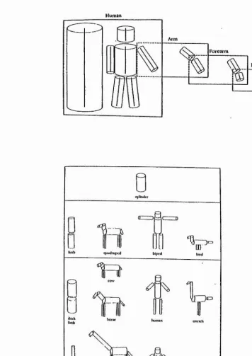
components of the object are described in relation to each other, or to the major axis of the object (see later). In their model, information about an object is processed through a series of four stages: The first stage (primal sketch), represents an object in terms of its edges, bars, and blobs which carry characteristics of orientation, width, length, contrast, and position. In the second stage, the IVi D sketch, cues for depth, surface texture, and contour occlusion are added. A 3D structural description is achieved in the third stage, by describing the object in an object-centred fashion and classifying the object with the help of stored structural descriptions. Finally, in the fourth stage, the structural description is used for semantic interpretation. Marr and Nishihara (1978) suggest that for describing an object, one has first to set up a co-ordinate system which is determined by the overall shape of the object itself. Each 3D model description specifies the following: 1) the model axis: a single axis which defines the overall volumetric shape of the object; 2) the relative spatial arrangement, orientations and the length of the component axes; and 3) the volumetric primitives (generalised cones) which are associated with each axis. 4) Then, repeatedly, each component axis becomes a model axis for its sub-components, until the wanted level of detail is reached (see Fig. 3.2a). Alternatively, for recognising parts of the whole object the model allows direct access to the information at any subordinal level. For example, a limb of a human body can be described as an (model) axis of an component part relative to its major axis (of the whole body). This model axis of the arm becomes a major axis if components such as the upper arm, forearm and the hand are described in relation to their major axis (arm itself) (Mair and Nishihara, 1978).
To describe these volumetric primitives, Marr's model of object recognition uses 'generalised cones'. A generalised cone “refers to the surface created by moving a cross section along a given smooth axis. The cross section may vary smoothly in size, but it shape remains constant.” (Marr, 1982). These generalised cones do not reveal any detail of the object's part such as surface pattern or texture, but have a cylinder like property. This simplifies the object structure substantially, resulting in a 3D 'stick- figure' (see Fig. 3.2b). In addition, this kind of representation does not use information obtained from surface patterns.
specificity (reproduced from Marr and Nishihara, 1978).
Human
a)
Arn»
Forearm
Hand
b)
cylinder
human
"0
[image:37.618.111.467.149.652.2]Literature Overview 30
constructed by using a number of different 3D shapes. However, we are ail capable of recognising a silhouette as the same 3D object. He therefore suggested that there must be some kind of constraints on the possible range of interpretations of the contour, which might be due to assumptions made by the visual system. Such assumptions were suggested to be that 1) each point of a silhouette represents a point on the surface of the 3D object; 2) two points close together in a silhouette will also be close together on the object; and 3) all the points on the contour line of a silhouette will lie in a single plane of the object viewed. These assumptions lead Marr to conclude that any silhouette of an object (and therefore the 3D object itself) is based on one or several generalised cones which are described in relation to the principal axis of the object (Marr, 1982).
The next question addressed was how these axes are derived from the object's occluding contours. Marr and Nishihara (1978) suggested that the complex contour is segmented into a number of distinct parts (generalised cones). They gave an example of a toy donkey silhouette and segmented its contour into distinct parts by finding the different concavities in the contour (Fig. 3.3). This was done by first labelling the different concave sections, which brings out the strong segmentation points. Then the outline was divided into a set of smaller segments using the strong segmentation points just defined. Next, these points are connected to other strong, neighbouring segmentation points of the contour in a straight line. Finally, the component axes are determined which are then linked up and related to each other.
rules for connecting these to other points on the contour, e) The component axis is found for each segment, f) The axes are related to one another (this lines).(Reproduced from Marr and Nishihara, 1978.)
<cl (ill
[image:39.616.129.395.188.617.2]Literature Overview 31
and Waxman, 1991) is implausible. Marr does not provide an answer to this problem in his theory.
It was put forward by Hoffman and Richards (1984) that different parts of an object intersect at a contour of concave discontinuity of their tangent planes. In other words, whenever two surfaces interpenetrate, they will always meet in a concave discontinuity. So that where one part penetrates the other there is a concave cusp formed. Therefore, any concavity of an object will indicate the division between the contours of two intersecting parts of the object. They also pointed out that in the contours of a smooth object, the concavities can be found by looking at the sites of the object contour with the greatest negative curvature, i.e. the most inward pointing part of the silhouette of the object will indicate the presence of a concavity.
In contrast to MaiT and Nishihara's (1978) theory, however, Hoffman and Richards (1984) suggest that the segregation does not depend on the parts being generalised cones, but rather is independent of the nature of the parts of the object.
Examples such as the 'face-vase' image support suggestions that the parts of an object can be predicted from the reversal of their figures. If the vase in this image is perceived, the concavities point inwards, defining the base, the stem and the bowl of the vase. However, when the faces are perceived, the concavities point into the face, defining the parts of the chin, nose, mouth, forehead etc.. Hence, depending on how the contour of the image is divided into parts, by looking at interpreting the concavities prior to recognition, different objects can be perceived (in this example either a vase or two faces each with its own distinct set of parts).
(reproduced from Biederman, 1987).
Geons
1
2
Objects
o
<o ^
0
^
Literature Overview 32
compared to Marr's model, though Marr's model does offer more precise information about the spatial arrangements of the components.
Biederman’s (1987) model of object recognition starts off with extracting edges from the image of a viewed scene of 3D objects. The edges enhance the presence of non-accidental properties, and hence indicate that these properties are reflected by the image from the real world. So that if, for example, a set of parallel lines are present in an image, it can be assumed that these lines are a property of the edges of an object in the real world and are parallel in 3D space. Biederman points out that because these non-accidental properties are constant over different view-points they can be used as additional information to determine the geons used for the object representation stored in memory. The other main information needed to determine the geons, is derived by segmenting the silhouette of the object image at regions of sharp concavity, resulting in a set of object parts (as done in Marr’s model, 1977). These determined parts are then matched against the representations of the different geons. From the nature and anangements of these geons, the structural model of the object can be defined (every known object has its own structural model based on its geons, the geon's relative sizes, orientations, place of attachment, etc.), resulting in the recognition of the object. It is very important to note the spatial arrangement between the geons of an object, since this builds the base of recognition of that object. Two different objects may have the same number and type of geons, but different spatial arrangements between them. Biederman (1987) uses terms like “on top o f’ or “attached at the side” to specify the spatial arrangement between the geons. Some of these descriptions (e.g. on top of), however, are based on a viewer-centred co-ordinate system, since they describe the position of the geon in relation to the viewer. Such view based terms should not be used in Biederman’s object-centred description, since he explicitly points out that there is no top or bottom in a representation of an object.
Edge extraction t:2
Detection of non-accidental
properties Parsing of regions of concavity
Determination of components __________(geons)__________
Matching of components to object representation
Object identification
Biederman (1987) carried out some experimental work to support his theory of object recognition by components. Subjects were to name as fast as possible simplified line drawings of objects presented for 100 ms. The complexity of these objects ranged from complete objects composed of only two geons to complete objects composed of nine geons. Randomly interspersed between the presentation of complete images of objects, were images of different incomplete objects. For example, a 9-component object could have been presented with 1, 2, 3, 4, 5 or 6 components missing. Alternatively a 3-component object could have been presented with only two components present. Biederman found that subjects take longer to name and make more recognition errors if some parts of an object are missing irrespectively of how complex the complete version of the object was. However, for the more complex objects, recognition error was low (< 10%) if more than three geons of the object were presented (Biederman, 1987). Furthermore, the addition of colour, brightness and texture did not significantly improve recognition response times (Biederman and Ju,
198&X
Literature Overview 34
concavity so that coliniarity and smooth curvature of the segments bridges the concavity (Fig, 3.5). The results showed that recognition was slightly faster and more accurate (approximately by an error rate of 30%) if the information for the geons was present. Biederman (1987) concluded that, as in the previously described experiment, recognition of an object is carried out on the basis of the object's geons. However, the suggestion that recognition of an object is better if regions of sharp concavities are left intact in a fragmented image, than the recognition of an image where the object is fragmented in such a way that the concavities are removed, may be questionable. Biederman and Gerhardstein (1992) argued that in the first instance geons are more easily found for recognition purposes than when the concavities are removed from the image. However, this is debatable since priming and recognition of an image is best if enough information is available in the image to support perceptual closure. Perceptual closure is based on the Gestalt principle of filling in gaps in an contour to achieve the most meaningful forms of an image (Snodgrass and Feenan, 1990). Biederman's image of a fragmented object where regions of sharp concavities are intact could aid in Gestalt principle closure rather than an image of a fragmented object where the concavities are removed. Hence, this could influence the results found in these experiments.
Right column: parsing at sites of concavities, resulting in colliniaiity or smooth curvature of the segments bridging the concavities (reproduced from Biederman, 1987).
W f' "Vi/
( '* - 4
[image:45.615.174.405.189.505.2]
Literature Overview 35
priming occurred with a different exemplar of the same object class as the target image, RTs and error rates were higher than when using the other priming conditions. Biederman concluded that visual priming was not based on image vertices and edges and that a visual image representation would have to be more global than that specifying the image features. This would leave two plausible types of the representations named above: either a representation based on the component parts of the object, or a representation which is a specific object model itself (e.g. a grand piano). However, when the component parts (geons) of an object were removed, RTs and error rates were much better if the target image was primed with the identical image, rather than with the complementary image. Hence, priming only occurred when the convex components (geons) were present in priming image (as well as in the primed image). It was concluded that “a// the visual priming in object naming can be attributed to the explicit (actual) presentation of the components in the image” and the spatial arrangement between them (Biederman and Cooper, 1991b).
Hierarchical organisation: Bottom-up vs Top-down processing
Nniurul-ilisconnected
n
o o
Nniutal-icranihled
O '‘'V)
UniiBlural-diicoiinccicd
"7
Unnatural-acrambled
■ = .
Parla-removed
OrV
<
s>
[image:47.614.103.406.118.579.2]
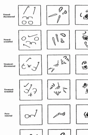
Literature Overview 36
disconnected object parts (natural or unnatural parse) were scrambled, naming RT and error rates were higher than when the parts were simply disconnected from each other. It was concluded that one critical factor for recognition of an object was the spatial relationship between the parts of the object (scrambled or proper organisation). Furthermore, Baker-Cave and Kosslyn (1993) found no interaction between the effect of (i) the way an object was parsed and (ii) the arrangement on the page (scrambled or unscrambled). Baker-Cave and Kosslyn (1993) suggested on the basis of their results that in normal viewing conditions the overall shape of an object is encoded without the image being parsed into parts.
Unfortunately, Baker-Cave and Kosslyn (1993) did not look in greater detail at the effect of part deletion, which might influence and/or diminish the quality of object recognition. They only showed that a whole intact object can be recognised and named faster than an image of the same object with some of its parts removed. According to Marr's theory, part deletion should not greatly influence the recognition process, since recognition can start at different levels of the 'stick-like model', i.e. one can access the model at different levels of detail depending, on the need for specific information on e.g. a particular part of the object. On the other hand, Biederman's theory of object recognition very much depends on the object' components, so that deletion of parts would have a greater impact on the recognition process.
Physiological evidence for the role of component parts in object recognition
It has been proposed that the inferotemporal cortex (IT) plays a central role in visual pattern and object recognition (Gross, 1973; Ungerleider and Mishkin, 1982; Schwartz et al., 1983; Desimone et al., 1984; Tanaka and Fujita, 1991). Many physiological studies have been carried out in this region of the monkey brain, some of which will be discussed below.
One of the attempts to study shape selectivity of individual cells in the IT cortex examined how IT cells can extract information about the overall shape of an object by analysing the local boundaries of the image (Schwartz, Desimone et al., 1983). Schwartz et al. (1983) investigated whether any object can be described in terms of its boundaries curvatures (the contours). They argued that the visual system describe these boundaries in two steps. First, the boundary orientation function of the shape, which is the orientation of the shape’s boundaries measured at regular intervals around the perimeter, is determined. This boundary orientation is then expanded in a Fourier series. These Fourier series, which have a particular frequency (number of lobes), amplitude (lobe indentation) and phase (orientation), were named 'Fourier descriptors' (FD). A FD can also be described as a shape which is systematically varying in boundary curvatures. According to Schwartz’s theory, every shape is described by its own sets of FD,
Literature Overview 38
of cells in the IT cortex, since some cells have been found to be sensitive to a particular shape in a particular colour or texture, or sensitive to 3D-objects, faces or hands (Desimone, Albright et al., 1984; Tanaka and Fujita, 1991; 1993; Kobatake and Tanaka, 1994) - all which normally are much more complex stimuli than FD. Some of these cells, nonetheless, were in addition sensitive to FD's. The conclusion drawn, was that selectivity for boundary curvature is not a characteristic of all IT cells, nor is it the only response property of many of these cells.
Another systematic attempt to determine the mechanism for selectivity for a particular stimulus dimension, such as shape, was conducted by Desimone and colleagues (1984). Previous work concentrated on recordings of cells selectively responsive to stimuli which were either simple, as for example white and coloured bars, or complex such as patterns, hands and faces (see e.g. Gross, Rocha-Miranda et al., 1972; Perrett, Rolls et al., 1982). Desimone et al. (1984) tested cells in the IT cortex systematically with simple stimuli, such as bars with different length and width, 2D patterns, and with a wide range of 3D objects. Once the preferred object/pattern of a cell was identified, they tried to isolate the critical stimulus which was responsible for the response of the cell. They also studied cells in the superior temporal sulcus which were selectively responsive to face stimuli.
This investigation showed that most cells in the inferior temporal cortex responded to many different visual stimuli and therefore it was concluded that these cells cannot be narrowly tuned 'detectors' for a particular object. This is in contrast to theories of 'Grandmother cells' and 'Yellow Volkswagen detectors' discussed earlier.
Half of the IT cells which showed some selectivity seemed to be selective for shape, colour, texture or a combination the three (Desimone et al., 1984). Of these cells, some showed the same kind of sensitivity to length, width, or a coloured bar as the cells found in earlier stages of the visual pathway (striate and prestriate cortex). As pointed out in chapter II, the receptive field of IT cells are much larger (almost always include the centre of gaze and usually extend into both the contralateral and ipsilateral visual field), than the receptive fields of cells found in the striate and prestriate cortex.
simple stimuli such as a bars. It was concluded that the IT cortex must play an important role in higher shape recognition.
IT cells which respond selectively to faces or hands were first identified by Gross et al. (1969). In 1972, Gross and his colleagues showed that there are FT cells which best respond to photographs of faces. Later investigations (Desimone, Albright et al., 1984), however, showed that cells which are selective for a specific object are very rare throughout the IT cortex. Perrett et al. (1982) demonstrated a more frequent appearance of cells selectively responsive to faces in the anterior part of the superior temporal sulcus (STS) which partially overlaps with the TT cortex. Nonetheless, the cells appeared to be distributed non homogeneously - a particular clumped distribution for particular features with cell type patches of 1-4 mm" (Perrett, Smith et al., 1984;
1985; Harries and Perrett, 1991).
Perrett and colleagues (1982; 1983; 1984) tested face sensitive cells for their neuronal responses to the sight of faces with component parts, such as the eyes, the mouth or the hair, being occluded from sight. It was reported that most of these cells responded significantly better to the face with certain parts of it occluded than to various control objects and base line activity (S/A). However, most cells responded better to the whole face than to just isolated parts of the face. Different cells were found to be maximally activated by different individual component parts of the face. Furthermore, some cells only required one face part present whereas other cells required several parts present. For the remaining cells the response magnitude was highly reduced when any part of the face was occluded, i.e. the entire face was necessary to drive the cell.
The overall configuration of the component parts of a face also seem to play a highly important role in the neuronal firing of these face responsive cells (Perrett, Rolls et al., 1982; 1984; Desimone et al., 1984). A face has several different shapes, contours, colours and textures which all have to be arranged in a certain order to compose a face. Perrett et al. (1982; 1983) found that when this arrangement was upset (i.e. jumbled faces) then the cell response of most ceils was greatly reduced.
Literature Overview 40
the r r cortex is the pattern of activity of a population of cells rather than one specific cell (see also Tanaka, 1993). However, face and hand selective cells are an exception to this system (Desimone et al., 1984). This might be due to the fact that, for primates, faces play an extremely important role in social communication and therefore, might have been in favour during selective evolutionary pressure to build a separate (but not necessarily independent) neuronal mechanism coding biologically important objects such as faces and hands. Nonetheless, faces may not be extraordinary exceptions from objects since face stimuli could activate many millions of cells selective for face features, which might be analogous to the representation of other complex objects.
More recent studies of object selectivity in different parts of the FT cortex have been carried out by Tanaka et al. (1991) and Kobatake and Tanaka (1994). They suggested that the IT cortex is divided into two areas: the posterior (PIT) and anterior inferior temporal cortex (AIT). PIT receives its information from V4 and in turn projects to a wide posterior-anterior range of AIT. V4 also projects to the posterior part of area AIT ( see chapter II and e.g. Desimone et al., 1980; Baizer et al., 1991).
Tanaka and his colleagues (1991; Kobatake and Tanaka, 1994) tested single cells in different cortical areas of the ventral pathway (areas V2, V4, PIT and AIT). All the cells recorded from in the these areas were classified into six groups:
1) Primary cells: Maximally activated by slits or dots, where the exact shape of the stimulus is not crucial;
2) Texture cells: Maximally activated by textures such as stripe or dot patterns;
3) Elaborate cells: Maximally activated by pattern features which are more complex than only orientation of contours, colour blocks or simple textures;
4) Others: Cells which responded to a change in colour of a block or some specific movement;
5) Weak: Cells responding only very weakly to any stimuli tested; and 6) Unresponsive: Did not at all respond to any of the visual stimuli tested.
features (e.g. bars or discs with adjusted size, orientation and/or colour) increased up to 49% in PIT. The highest relative proportion of elaborate cells, however, were found in the remaining anterior part of the IT cortex. Between 67% and 75% of AIT cells were highly selective in their response to complex features (such as a particular shape with a particular texture and/or colour, faces or hands) and did not respond to other complex features tested (Tanaka et al., 1991; Kobatake and Tanaka, 1994). There seemed to be an abrupt change in the distribution of the cells between the posterior and anterior part of the r r (Tanaka and Fujita, 1991). Hence, Tanaka concluded that the IT cortex is divided into posterior and anterior IT cortex, and cells in the anterior part require more complex visual stimuli than only orientation of a contour or a certain colour of the object. Therefore, object recognition and its coding advances significantly in the anterior IT, where an integration process of pattern information occurs based on the selectivity of earlier V4 and PIT cells (Kobatake and Tanaka, 1994).
Tanaka et al. (1992) proposes that modules in area AIT are selectively activated when an object is viewed. This activity is specific to a subset of modules, which represents a particular partial feature of the object. However, the feature is not coded in the activity of one specific cell, but rather in the pattern activities of many cells in the same module. The whole object is then coded as the activity patterns of a particular subset of modules, whereas the individual features which make up the object, are coded in activities distributed over a population of cells in corresponding modules.
View discrimination: obiect-centred vs viewer-centred
A further question is addressed: Is the representation which is produced object-centred, Le. view-independent (Man' and Nishihara, 1978; Marr, 1982; Biederman,
1987), or view-centred, i.e. view-dependent (Koenderink and van Doom, 1976; Perrett, Smith et al., 1985; Tan and Pinker, 1989; Ullman, 1989; Kosslyn, Flynn et al., 1990; Poggio and Edelman, 1990; Seibert and Waxman, 1992a, b; Baker-Cave and Kosslyn,
1993)?
a) Obiect-centred representations
An object-centred representation allows the object to be recognised when the object or the parts are seen from any view-point (hence is view-independent). A major advantage of this kind of representation is that only a single description of each object's spatial structure would have to be stored in long-term memory, to allow recognition of the object from any (even a previously inexperienced) view.
Literature Overview 43
early representations of the object which need further processing to establish a representation with a co-ordinate system independent of the view observed.
Marr and Nishihara's (1978) model of object recognition is based on an object- centred co-ordinate system in which the position of every generalised cone is described ultimately with respect to the model axis, i.e. the principal axis of the major component. In this hierarchical system, each component axis becomes a major/model axis for its subcomponents. Therefore, only one representation is needed, since everything can be related back to this axis. This structural description of an object is totally view-independent and is based on the spatial arrangements between the object and its parts.
Having an object-centred representation, the question arises how, from different views of the same object, is the same object-centred description accessed? Marr (1982) gives a vague answer to this question, by suggesting that the major axis of an object (which is essential to the recognition of the object), is derived from the image itself. Hence, the same object-centred description will be produced irrespective of the view of the object.