Positive-feedback systems are rare in biology but are utilized in situations that require a phasic, unambiguous signal. In endocrinology, the classic example of such a system is the control of ovulation in mammals in which the hypothalamus and ovary interact positively to generate the luteinizing hormone surge that coordinates ovulatory events. In the case of insect physiology, the process of ecdysis likewise requires an unambiguous signal. Its success is dependent on the precise coordination of behaviour with ongoing events of the moult and, once initiated, these events cannot be reversed.
The insect goes through a sequence of behavioural phases as the time for ecdysis approaches (Carlson, 1977; Hughes, 1980). Late in a moult, as the synthesis of the new cuticle and the digestion of the old one near completion, an insect typically becomes restless and seeks a suitable site for ecdysis. As this preparatory phase progresses, the animal anchors itself to the substratum and begins movements that loosen the connections between the old and new cuticles. The preparatory phase is variable in duration, but it eventually culminates in the ecdysis attempt. The ecdysis phase starts with abdominal peristaltic waves that begin to move the old cuticle posteriorly. These are coordinated with other behavioural subroutines to extract the appendages from the cuticle as it is shed and with changes in
blood distribution and with air-swallowing that expand the anterior end of the animal and aid in rupturing the old cuticle (Reynolds, 1980). The initiation of ecdysis is typically associated with irreversible changes in the new cuticle so that the failure to accomplish ecdysis in a timely manner results in crippling deformities or an animal permanently trapped within its old skin.
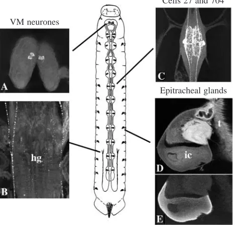
Most of the studies on the hormonal control of ecdysis have been carried out on various stages of the tobacco hornworm Manduca sexta. Two endocrine systems have been implicated in initiating ecdysis (Fig. 1). One is in the brain and includes two pairs of neurones (the VM neurones) whose axons project through the length of the central nervous system (CNS) as well as to peripheral neurohaemal sites (Truman and Copenhaver, 1989). These cells are the source of the neuropeptide eclosion hormone (EH). The second endocrine system is a peripheral one consisting of a set of three-celled glands, the epitracheal glands, one of which is located on the trachea near each of the 18 spiracles (Fig. 1D,E; Zitnan et al. 1996). The Inka cell component of each gland produces a peptide hormone, ecdysis triggering hormone (ETH; Zitnan et al. 1996). Injection of either ETH (Zitnan et al. 1996) or EH (Copenhaver and Truman, 1982; Truman et al. 1983) Printed in Great Britain © The Company of Biologists Limited 1997
JEB0784
A successful ecdysis in insects requires the precise coordination of behaviour with the developmental changes that occur late in a moult. This coordination involves two sets of endocrine cells: the peripherally located Inka cells, which release ecdysis triggering hormone (ETH), and the centrally located neurosecretory neurones, the VM neurones, which release eclosion hormone (EH). These two sets of endocrine cells mutally excite one another: EH acts on the Inka cells to cause the release of ETH. ETH, in turn, acts on the VM neurones to cause the release of EH. This positive-feedback relationship allows the Inka cells and the VM neurones to be the peripheral and central halves, respectively, of a decision-making circuit. Once conditions
for both halves have been satisfied, their reciprocal excitation results in a massive EH/ETH surge in the blood as well as a release of EH within the central nervous system. This phasic signal then causes the tonic activation of a distributed network of peptidergic neurones that contain crustacean cardioactive peptide. The relationship of the latter cells to the subsequent maintenance of the ecdysis motor programme is discussed.
Key words: eclosion hormone, ecdysis triggering hormone, crustacean cardioactive peptide, neuromodulation, feedback,
Manduca sexta.
Summary
Introduction
CONTROL OF INSECT ECDYSIS BY A POSITIVE-FEEDBACK ENDOCRINE
SYSTEM: ROLES OF ECLOSION HORMONE AND ECDYSIS TRIGGERING
HORMONE
JOHN EWER, STEPHEN C. GAMMIE AND JAMES W. TRUMAN*
Department of Zoology, University of Washington, Box 351800, Seattle, WA 98195-1800, USA
Accepted 5 December 1996
induces ecdysis, but ETH triggers the behaviour more rapidly than does EH and behavioural sensitivity to ETH occurs earlier in the moult. Zitnan et al. (1996) proposed that EH caused the release of ETH which, in turn, caused ecdysis behaviour. Although the evidence for this endocrine pathway is compelling, this relationship by itself fails to account for some known aspects of ecdysis control (Hesterlee and Morton, 1996). For example, debrained larvae injected with EH show good pre-ecdysis behaviour but not ecdysis (A. Novicki and J. C. Weeks, unpublished results, cited in Hesterlee and Morton, 1996), and back-firing of the VM neurones that contain EH can lead to the rapid onset of ecdysis behaviour (Hewes and Truman, 1991).
Besides the VM neurones and Inka cells, the neurones of the cell 27/704 group are another set of peptidergic cells that are associated with ecdysis (Fig. 1C). These segmentally reiterated neurones include both interneurones and neurosecretory cells. In most insects, the neurones of this group contain crustacean cardioactive peptide (CCAP) (Davis et al. 1993; Dircksen, 1994). In Manduca sexta (Ewer et al. 1994) and a wide variety of other insects (Ewer and Truman, 1996), these neurones show a striking accumulation of the second messenger cyclic 3′,5′-guanosine monophosphate (cGMP) just prior to the onset of ecdysis. The rise in intracellular cGMP level enhances the
excitability of these cells (Gammie and Truman, 1977), presumably facilitating the release of their neuropeptides. Since some of these neurones innervate the foregut and the alary muscles of the heart (Braünig, 1990; Dircksen et al. 1991; Ewer et al. 1994), they are the likely candidates for causing the air-swallowing and circulatory changes that accompany ecdysis (Reynolds, 1980).
This paper reports that the VM neurones, the Inka cells and the CCAP neurones constitute a neuroendocrine circuit that initiates and maintains ecdysis behaviour. The VM neurones and the Inka cells are two halves of a positive-feedback circuit that ensures that peripheral and central conditions are appropriate for ecdysis. A key outcome of their interaction is the activation of the neurones of the cell 27/704 group whose peptides then shift the nervous system into the ecdysis phase itself.
Materials and methods Experimental animals
Larvae of Manduca sexta (Johansson) were raised on an artificial diet at 26 °C under a 17 h:7 h L:D photoperiod. Some experiments were performed on larvae during the moult from the first to the second instar. Fifth-instar larvae were also used. Larvae were selected at the time of head-capsule slippage (HCS, when the old head capsule moves down over the forming mandibles of the next larval stage) and were maintained at room temperature (18–20 °C) until ecdysis approximately 16 h later. The larvae showed obvious tanning of the new mandibles (brown mandible stage) by 10 h after HCS. The time course of events in moulting fifth-stage larvae is described in Truman (1972) and Curtis et al. (1984).
Immunocytochemistry
For immunocytochemistry of moulting second-instar larvae, staged animals were cut along the dorsal midline, pinned out under 4 % buffered formaldehyde, and fixed for 1–2 h. Tissues were then pre-incubated in 2.5 % normal goat serum (NGS) and 2.5 % normal donkey serum (NDS) for 1 h in phosphate-buffered saline with 0.3 % Triton X-100 (PBS-TX), and then overnight at 5 °C in a cocktail of primary antibodies [a monoclonal antibody against the molluscan small cardioactive peptide (SCP; Masinovsky et al. 1988), which recognizes the C terminus of ETH (Zitnan et al. 1996), was used at a dilution of 1:500, a rabbit anti-cGMP antiserum (De Vente et al. 1987) was used at a dilution of 1:4000, and a rabbit anti-EH antiserum (Truman and Copenhaver, 1989) was used at a dilution of 1:250] in PBS-TX with 1 % NGS and NDS. After repeated washes, the tissues were incubated overnight at 5 °C in 1:1000 dilutions of a FITC-conjugated donkey anti-rabbit IgG and a Texas-Red-conjugated goat anti-mouse IgG (both Jackson Laboratories) in PBS-TX with 1 % NGS and NDS. After repeated washes, the tissues were dehydrated, mounted in DPX (Fluka) and examined using a BioRad 600 confocal microscope. EH and cGMP immunoreactivity were distinguishable from one another because they occur in distinct Cells 27 and 704
[image:2.609.52.288.78.304.2]Epitracheal glands VM neurones
cell populations. In a limited number of preparations, the spatial distinctness of the EH and cGMP signals was confirmed using an anti-cGMP antiserum that had been raised in sheep rather than rabbit.
For analysis of fifth-stage larvae, individual tissues were dissected, fixed overnight, rinsed in PBS-TX, treated in 0.5 mg ml−1collagenase (Sigma Chemical Co.) for 30 min at room temperature to aid in antibody penetration, and processed for immunocytochemistry as described above.
Immunoassays for ETH
The amount of ETH in the bathing medium was determined by an antibody-capture immunoassay (Harlow and Lane, 1988). 5µl samples of the medium were bound to the bottom of wells of polystyrene 96-well plates (Corning, no. 25802). Wells were incubated overnight with 1:1000 anti-SCP monoclonal antibody, followed by 1:1000 peroxidase-conjugated goat anti-mouse IgG. The amount of bound antibody–enzyme complex was determined using a Peroxidase Substrate kit (Bio-Rad), and the intensity of product was measured using a Microplate Autoreader (BioTek Instruments). Each sample was run in duplicate. Absorbance from each well was compared with a standard dilution series of Inka cell extract.
Hormonal materials
The eclosion hormone used in these studies were from two sources. Some studies utilized recombinant EH produced from baculovirus that carried a copy of the Manduca EH gene (Eldridge et al. 1991). We also used EH that had been purified from corpora cardiaca–corpora allata complexes from pharate adult Manduca sexta (Terzi et al. 1988).
For preparation of epitracheal gland extracts, freshly dissected glands from prepupae together with some adhering tracheal tissue were homogenized in acidified methanol (90:9:1 methanol:water:acetic acid). After centrifugation for 1 min in a PicoFuge (Stratagene), the supernatant was blown to dryness under a stream of nitrogen and resuspended in saline at a concentration of one epitracheal gland equivalent per 10µl. Synthetic ETH was prepared by the Howard Hughes Macromolecular Synthesis Facility at the University of Washington.
Pharate second-stage larvae were injected using glass microneedles that were tip-filled with the appropriate solution. The needles were then fitted into an oil-filled injection system that delivered 1µl into the larva. Pharate fifth-instar larvae were injected using 50 or 100µl syringes.
Electrophysiological techniques
Nervous systems were dissected from pharate fifth-instar larvae that were approximately 5 h before ecdysis. The CNS was briefly exposed to 4 % collagenase/dispase (Boehringer Mannheim) to aid in penetration of the sheath. The bathing solution contained (in mmol l−1): 140 NaCl, 5 KCl, 4 CaCl2, 28 D-glucose, and 5 Hepes (pH 7.4). Glass microelectrodes were filled with 2 mol l−1 potassium acetate and 10 mmol l−1 KCl (pH 7.4). VM cell bodies could be identified visually and
are the only neurones in that region of the brain with overshooting action potentials in their somata (Hewes and Truman, 1994).
The signals were amplified using a Getting microelectrode amplifier (Getting Instruments, Iowa City, IA, USA), stored on a VHS recorder (Vetter Co., Rebersburg, PA, USA) and analysed using SuperScope (GW Instruments, Somerville, MA, USA). Any compensation errors made during bridge-balance recordings were corrected during off-line analysis by subtracting the near instantaneous voltage record attributable to the potential drop across the electrode (Hewes and Truman, 1994). Threshold was determined by injecting positive current pulses (600–1000 ms) into the somata and calculating the minimum voltage deflection from the resting potential needed to fire an action potential.
Results
The timing of behavioural and endocrine events at ecdysis
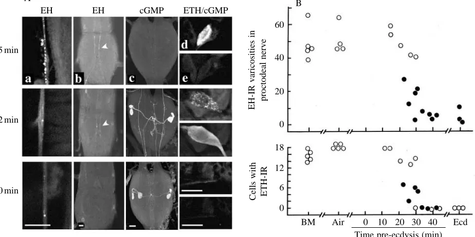
Fig. 2 examines the timing of the endocrine events that occur during ecdysis of second-instar larvae of Manduca sexta. In M. sexta larvae, the preparatory phase consists of the pre-ecdysis behaviour, a set of rhythmic thoracic and abdominal contractions that loosen connections between the old and new cuticles (Copenhaver and Truman, 1982; Miles and Weeks, 1991; Novicki and Weeks, 1993). Prior to the onset of pre-ecdysis behaviour, the proctodeal nerves, the peripheral release site for EH in larvae (Truman and Copenhaver, 1989), were well stocked with EH, showing an average of approximately 50 EH-immunoreactive varicosities per 150µm of nerve (Fig. 2B). EH staining in the proctodeal nerve remained unchanged through the first 25 min of the pre-ecdysis behaviour, but there was then a marked reduction in staining between 25 and 35 min. In larvae that were in this transition period, we occasionally found proctodeal nerves that showed diffuse EH staining in the extracellular stroma surrounding the terminal varicosities (Fig. 2A, 32 min). We interpret this extracellular staining as being due to newly released EH that was fixed before it could diffuse through the stroma and into the circulation. The loss of EH immunoreactivity from peripheral release sites was also accompanied by the disappearance of EH immunoreactivity from the VM cell axons within the CNS (Fig. 2A). Thus, for ecdysis of the second instar, as for pupal ecdysis (Hewes and Truman, 1991), EH is released within the CNS coincident with its liberation into the circulation.
The neurones of the cell 27/704 group showed an abrupt rise in intracellular cGMP levels during the latter phases of the pre-ecdysis behaviour (Fig. 2). These cGMP-immunoreactive neurones were found only in those larvae that had at least begun the process of EH release from the VM cells. These neurones then maintained elevated cGMP levels throughout ecdysis (see also Ewer et al. 1994).
M. sexta larvae have 18 epitracheal glands, one associated
and through the start of pre-ecdysis behaviour (Fig. 2B). ETH immunoreactivity began to disappear abruptly from these cells in larvae that were undergoing EH release at 25–30 min into the pre-ecdysis behaviour. As they were losing their ETH immunoreactivity, these cells also showed elevated intracellular cGMP levels (Fig. 2A, 32 min). Unlike that in the neurones of the cell 27/704 group, however, the increase in cGMP level in the Inka cells was transient and their cGMP levels had declined to below the level of detectability by ecdysis (Fig. 2).
The relationship between EH and ETH release
Experiments involving the incubation of isolated nervous systems with epitracheal glands and EH suggested that EH acts on these glands to cause ETH release (Zitnan et al. 1996). We tested this hypothesis using isolated epitracheal glands attached to a small segment of trachea. Individual epitracheal glands, taken from prepupae at the ‘anterior shrink’ stage (approximately 3–4 h before ecdysis), were incubated with agitation in 50µl drops of Manduca saline (Riddiford et al. 1979) containing 0.02 % BSA and 1 nmol l−1 purified EH (Terzi et al. 1988). After the incubations, the bathing medium was collected and frozen until assayed. As seen in Fig. 3A, the presence of EH resulted in a time-dependent release of ETH
immunoreactivity into the medium. By contrast, control incubations with BSA alone resulted in a low, basal level of ETH secretion.
The time course of the response of the Inka cells to EH exposure in vitro was followed using immunocytochemistry. Inka cells were exposed to EH in vitro and then, at various times thereafter, fixed and double-stained for ETH and for cGMP immunoreactivity. As seen in Fig. 3C, detectable cGMP immunoreactivity became evident in the Inka cells by approximately 5 min after EH addition and was prominent by 10 min. The ETH staining in the resting cell was evident in small granules concentrated in the cortical region of the cell, but by 10–20 min following EH addition these granules began to increase in size and to disappear from the cell cortex (Fig. 3C). We do not know whether the increase in granule size is due to osmotic swelling or to fusion of granules with each other as well as with the cell membrane. ETH staining was severely depleted in these cells by 40–60 min after EH addition. These results clearly support the proposal by Zitnan
et al. (1996) that EH acts on the epitracheal cells to cause the
release of ETH. A direct demonstration of this action of EH on the Inka cells has also been made (T. Kingen, J. Hermesman, D. Zitnan and M. Adams, in preparation).
Zitnan et al. (1996) suggested that injected EH causes its 15 min
32 min
50 min
EH EH cGMP ETH/cGMP B
A
60
40
20
0
18
12
6
0
EH-IR varicosities in
proctodeal nerve
Cells with ETH-IR
[image:4.609.74.536.79.310.2]BM Air 0 10 20 30 40 Ecd Time pre-ecdysis (min)
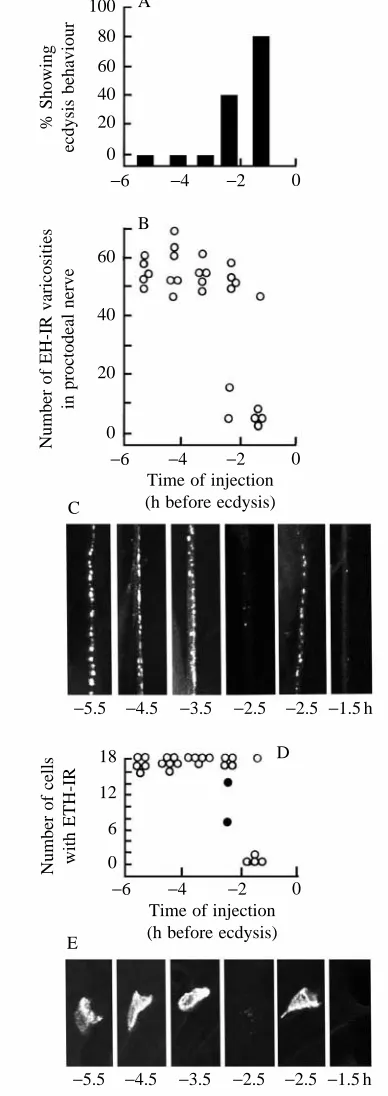
behavioural effects by stimulating ETH release and that the latter, in turn, acts on the CNS to cause ecdysis. Injections of EH are effective in triggering ecdysis behaviour only during the final hours prior to ecdysis (Truman et al. 1983; Copenhaver and Truman, 1982). For example, in pharate second-instar larvae, the onset of EH sensitivity occurred approximately 2.5 h before normal ecdysis (Fig. 4A). As seen in Fig. 4D,E, this time of behavioural sensitivity coincided with the time when EH injection was first able to induce the loss of ETH immunoreactivity from the Inka cells. The ability of animals to respond to injected EH requires a decline in the circulating titres of the ecdysteroids that drive the moulting process (Truman et al. 1983). We had previously interpreted this time as reflecting the time when the CNS became responsive to circulating EH. It now appears that this timing reflects the competence of the Inka cells rather than that of the CNS. Whether these cells become competent through a direct action of ecdysteroids on them or indirectly through the steroid-induced changes in the epidermis on which they reside is not yet known.
Effects of ETH on the activity of VM neurones
Although EH causes ETH release, the situation is reciprocal in that we find that ETH acts on the CNS to excite the VM neurones, resulting in EH release. Outside their normal time of EH release, the VM cells are relatively inexcitable and sustained depolarization results in single action potentials rather than trains of spikes (Hewes and Truman, 1994). For example, intracellular recordings from VM cells in isolated, pharate fifth-instar nervous systems showed neurones with a spike threshold of 27±3 mV (mean ±S.E.M.; N=6) over rest. Addition of epitracheal gland extracts resulted in the onset of tonic firing of these cells within 4–20 min (mean time 10 min;
N=6) (Fig. 5A). Once activated by these extracts, the VM
neurones then maintained tonic firing at 1–2 Hz for at least 20 min.
The epitracheal gland extracts also caused a dramatic broadening of the soma action potential of the VM neurones. Prior to addition of the extracts, the duration of the action potential was 34±3 ms, but it then increased almost threefold to 84±10 ms by 10–15 min after extract addition (N=6; B
C A 0.2
0.1
0
0
2
5
10
20
40
60
C
ETH-IR in medium
Time (min)
[image:5.609.112.505.78.390.2]30 60 30 60 min BSA EH
Fig. 3. Secretory response of isolated epitracheal glands to eclosion hormone (EH) in vitro. (A) Mean + S.E.M. ETH-like immunoreactivity (ETH-IR) released into the medium by single epitracheal glands cultured for 30 and 60 min with 0.02 % bovine serum albumin (BSA) or with BSA with 1 nmol l−1EH. Numbers of cultures are indicated. (B) Dual-channel, optical sections through the Inka cell component of epitracheal
Fig. 5B). Two observations show that this dramatic spike-broadening was not due simply to the repetitive firing of the VM cell. First, driving these neurones at 1–2 Hz in the absence of gland extracts did not result in significant spike-broadening (S. C. Gammie and J. W. Truman, unpublished observations). Second, neurones depolarized after extract addition but prior to the start of the spontaneous spike train already show a markedly broadened soma spike (Fig. 5B, spike b).
The epitracheal glands apparently contain a number of biologically active peptides (Zitnan et al. 1996). Preliminary experiments using synthetic ETH (1–5µmol l−1) show that this peptide is sufficient to induce both spike-broadening and tonic firing of the VM neurones (N=3).
Effects of Inka cell extracts on EH release in vivo
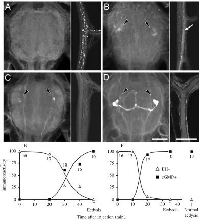
Inka cell peptides promote the release of EH in vivo as well as in vitro. Their effects on EH release were first tested in pharate fifth-instar larvae that were selected 2 h after the head capsule became filled with air (approximately 3 h before ecdysis). Injection of two Inka-cell-equivalents of extract was followed by an abrupt loss of EH immunoreactivity from the proctodeal nerve within 20 min (Fig. 6F) followed by ecdysis at 35±1 min (mean ±S.E.M.; N=12). Injection of similar larvae with about 1.5 ng of recombinant EH also caused the release of endogenous stores of EH but, interestingly, the latency to EH release was longer (30–35 min; Fig. 6E) and ecdysis occurred later (49±2 min; mean ±S.E.M.; N=17) than that seen after Inka cell extract treatment. The longer latency to EH release in the latter case presumably reflects the time required for the injected EH to cause ETH release which, in turn, causes release of endogenous EH stores.
In the case of both EH and Inka cell extract injections, the release of endogenous EH stores coincided with the appearance of cGMP in the cell 27/704 group (Fig. 6A–D). This was then followed by ecdysis 15–20 min later. Thus, irrespective of which peptide was injected, the onset of ecdysis had a constant temporal relationship with the release of endogenous EH and the accompanying increase in cGMP levels in the cell 27/704 group.
The window of development during which M. sexta will show ecdysis behaviour in response to injections of peptide is strikingly different for EH versus ETH. There is only a very narrow window, late in the moult, during which the animal will 100
80 60 40 20
0
% Showing
ecdysis behaviour
−6 −4 −2 0
60
40
20
0
Number of EH-IR varicosities
in proctodeal nerve
−6 −4 −2 0 B
C
D A
Time of injection (h before ecdysis)
Time of injection (h before ecdysis)
−5.5 −4.5 −3.5 −2.5 −2.5 −1.5 h
18
12
6
0
Number of cells with ETH-IR
−6 −4 −2 0
E
[image:6.609.66.260.77.625.2]−5.5 −4.5 −3.5 −2.5 −2.5 −1.5 h
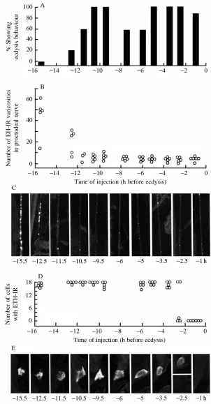
Fig. 4. Development of sensitivity of endocrine centres to injections of EH at various times during the moult to the second larval stage. (A) Percentage of larvae showing ecdysis behaviour within 60 min after injection. (B–E) Immunological assessment of the amount of EH and ETH still stored in the proctodeal nerve and Inka cells, respectively, at 60 min after the injection. (B) Number of EH-positive (EH-IR) varicosities in a 150µm length of proctodeal nerve. (C) Confocal images showing examples of the EH immunoreactivity still present in the proctodeal nerve following EH injection at the indicated times before ecdysis (in h). (D) the number of Inka cells per larva that stained positively for ETH (ETH-IR); filled circles are larvae whose Inka cells also exhibited cGMP immunoreactivity at the time of dissection. (E) Confocal images showing the typical level of ETH-like immunoreactivity present in the Inka cells following EH injection at the indicated times. The two confocal examples given for
respond to injections of EH (e.g. Reynolds et al. 1979; Copenhaver and Truman, 1982), whereas the window of sensitivity to ETH begins many hours earlier (Zitnan et al. 1996). For example, moulting fifth-stage larvae begin to show premature ecdysis behaviour in response to Inka cell extract injections starting shortly after head-capsule slippage (HCS, approximately 28–34 h before ecdysis). By contrast, these larvae respond to EH injections only much later, at approximately 4–5 h before normal ecdysis (Copenhaver and Truman, 1982).
As seen in Fig. 7A, moulting second-instar larvae showed premature ecdysis behaviour in response to injections of Inka cell extracts starting approximately 13 h before normal ecdysis (approximately 3 h after HCS), while EH injections were not effective until approximately 2.5 h before ecdysis (Fig. 4A). These larvae were then all examined 60–90 min after peptide injection to determine their state of EH release. Importantly, all of the animals that showed premature ecdysis in response to either Inka cell extract or EH injection also showed the premature release of their store of EH (Figs 4B,C, 7B,C). These larvae also consistently showed cGMP in the neurones of the cell 27/704 group (data not shown).
Although injections of epitracheal gland extracts were capable of causing EH release starting approximately 13 h before expected ecdysis, we saw no evidence of release of endogenous stores of ETH from the Inka cells until approximately 2.5 h before ecdysis (Fig. 7D,E). This latter time corresponds to when Inka cells first start to show ETH release after injections of EH (Fig. 4D,E). This temporal relationship suggests that the release of ETH after injection of gland extracts is probably a secondary response that follows the release of EH into the circulation. However, it only occurs at late times when the Inka cells finally become responsive to EH.
Relationship of EH and ETH to the activation of the neurones in the cell 27/704 group
Neurones of the cell 27/704 group invariably showed an increase in cGMP levels in association with EH release, whether during a normal, spontaneous ecdysis (Fig. 2) or during an early ecdysis provoked by injection of Inka cell
peptides or EH (Fig. 6E,F). Direct evidence that the VM cells were responsible for this cGMP increase was obtained by repetitively firing the VM neurones with intracellular electrodes (1–2 Hz) or by ‘back-firing’ the VM cell axons through stimulation of the proctodeal nerves as in Hewes and Truman (1991). Both treatments resulted in the appearance of cGMP in the neurones of the cell 27/704 group (in 4 of 10 cells for intracellular stimulation of single VM cells and in 12 of 15 cells for back-firing of the proctodeal nerves). The intracellular stimulation was carried out on isolated nervous systems that lacked tracheae and, hence, epitracheal glands. Consequently, these glands and the ETH that they release are not required for the appearance of cGMP in the cell 27/704 group.
The role of the VM neurones in inducing the cGMP increase in this cell group is also consistent with in vivo experiments in which the ventral nerve cord was transected prior to larvae being challenged with either EH (N=14) or Inka cell peptides (N=9). After peptide challenge, the CNS anterior to the transection subsequently showed EH depletion from the VM axons, the appearance of cGMP in the cell 27/704 homologues and the onset of ecdysis movements. Posterior to the transection, by contrast, there was no EH release, no appearance of a robust cGMP response and no performance of ecdysis behaviour. Thus, rather than acting directly, the signal from peptides circulating in the blood needs to be relayed through the descending signal provided by the activity of the VM neurones.
Discussion
The endocrine trigger for ecdysis behaviour
During the past 25 years it has been thought that a single peptide, eclosion hormone, was responsible for triggering ecdysis behaviour (Truman, 1992). The recent report of a second ecdysis triggering peptide, ETH (Zitnan et al. 1996), showed that the regulation of ecdysis is more complex than originally thought and has sparked a re-evaluation of the role of EH (e.g. Hesterlee and Morton, 1996). In assessing the relationship between EH and ETH, a number of previously held assumptions about EH require re-examination. One assumption has been that EH release in M. sexta occurs prior
B A
20 mV 10 s
9 min
10 mV 20 ms
a c
b a b c
[image:7.609.116.506.74.174.2]EGX
to the start of pre-ecdysis behaviour. Since the injection of EH is always followed by the performance of the pre-ecdysis behaviour (Copenhaver and Truman, 1982; Miles and Weeks, 1991), the normal sequence of events was thought to be: EH release, pre-ecdysis behaviour, then ecdysis behaviour. The data in Fig. 2, as well as results from Novicki and Weeks (1996), are incompatible with this ordering of events, although we cannot exclude the possibility that a small release of EH precedes pre-ecdysis. Nevertheless, the major surge of EH
clearly occurs well into pre-ecdysis behaviour, approximately 15–20 min before the start of ecdysis. This timing is consistent with old data from pupal ecdysis in M. sexta showing EH activity appearing in the haemolymph approximately 20–25 min prior to the start of ecdysis (Truman et al. 1980). The shortness of this interval was surprising at the time since the normal latency between EH injection and pupal ecdysis was 60–65 min.
The isolated abdominal CNS of Hyalophora cecropia was
E F
% Showing
immunoreactivity
0 10 20 30 40 ↑
Ecdysis
Time after injection (min) 100
75
50
25
0
0 10 20 30 ↑ 40
Ecdysis
|
Normal ecdysis 100
75
50
25
0
EH+
cGMP+ 16
17 18
15
16 16 13
[image:8.609.109.499.71.503.2]15 10 13
used to demonstrate that EH acts on the CNS to induce the ecdysis motor programme (Truman, 1978). The success of these experiments, however, required that the tracheal supply to the CNS be included, presumably to ensure an adequate delivery of oxygen to the tissue (Truman, 1978). The results from Zitnan et al. (1996), however, suggest a new
interpretation for the need for the tracheae since these tracheal trunks are the sites on which the epitracheal glands reside. Since ETH can induce an ecdysis motor programme from a M.
sexta CNS that lacks tracheae, these authors suggested that the
EH added in vitro acts on the epitracheal glands to cause ETH release which, in turn, acts on the CNS to cause ecdysis. The 100
80 60
40 20
0
% Showing
ecdysis behaviour
−16 −14 −12 −10 −8 −6 −4 −2 0
60
40
20
0
Number of EH-IR varicosities
in proctodeal nerve
B
C
D A
Time of injection (h before ecdysis)
−15.5 −12.5 −11.5 −10.5 −9.5 −6 −5 −3.5 −2.5 −1 h
18
12
6
0
Number of cells with ETH-IR
E
−16 −14 −12 −10 −8 −6 −4 −2 0
Time of injection (h before ecdysis)
−16 −14 −12 −10 −8 −6 −4 −2 0
[image:9.609.265.567.70.649.2]−15.5 −12.5 −11.5 −10.5 −9.5 −6 −5 −3.5 −2.5 −1 h Fig. 7. Development of sensitivity of endocrine centres
time course of responses of larvae in vivo to injections of EH and epitracheal gland extract support a similar conclusion (Fig. 6E,F): injected EH acts to cause release of ETH, which in turn acts on the CNS to cause ecdysis. A peptide the size of EH (62 amino acids) probably has difficulty passing the blood–brain barrier and, hence, the physiological levels that are normally found in the blood might not have access to target sites within the CNS. The surprising feature of this system, however, is that the appearance of EH inside the blood–brain barrier is then accomplished through ETH-induced release of EH. Both in
vivo (Fig. 7) and in vitro (Fig. 5), ETH serves as a potent
exciter of the VM neurones, resulting in release of endogenous EH stores within the CNS as well as into the circulation. Under all circumstances that we have examined, the release of endogenous EH stores always precedes the onset of ecdysis movements (Figs 2, 6). The importance of centrally released EH in triggering ecdysis movements is also consistent with the results of stimulating the VM neurones by back-firing their axons from the proctodeal nerves (Hewes and Truman, 1991). This treatment leads to the onset of ecdysis behaviour, in some cases within as little as 15 min. The latter latency is much shorter than seen after injection of either EH or ETH.
The presence of the blood–brain barrier confounds a simple
determination of when the CNS becomes responsive to EH. As described above, the onset of behavioural sensitivity to injected EH (e.g. Copenhaver and Truman, 1982; Truman et al. 1980) reflects the time when the Inka cells become responsive to EH. It is now apparent that target elements within the CNS may become responsive to EH at much earlier times. This conclusion is based on the observation that injections of Inka cell extracts shortly after head-capsule slippage promote EH release (Fig. 7), which is always accompanied by a cGMP response in the cell 27/704 group. As detailed below, cGMP induction in target cells is a characteristic feature of EH action.
Control over the initiation of pre-ecdysis and ecdysis behaviours
[image:10.609.111.506.365.618.2]For ecdysis to be successful, it must occur at the proper time relative to the ecdysteroid-regulated digestion of the old cuticle that occurs at the end of the moult. In addition, for many insects, such as grasshoppers Schistocerca gregaria (Hughes, 1980) and crickets Teleogryllus oceanicus (Carlson, 1977), an animal that is ‘ready’ to ecdyse may nevertheless delay the start of the behaviour until it finds a proper perch from which it can hang during ecdysis. Consequently, there are both peripheral and behavioural requirements that must be
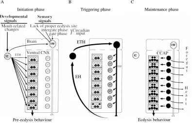
Fig. 8. A proposed general scheme for the sequence interactions that underlie the transition between pre-ecdysis and ecdysis behaviours. These involve the Inka cells (ICs), the VM neurones (VMs), the distributed neurones of the cell 27/704 group (27) and the set of interneurones and motoneurones that generates the motor responses (circles within boxed regions). White, stippled and black refer to cells that are inactive, partially active and fully active, respectively. (A) The initiation phase requires the integration of diverse developmental and sensory stimuli. Ecdysteroid-driven developmental changes in the periphery probably initiate secretion of ecdysis triggering hormone (ETH) from the ICs. ETH probably initiates the pre-ecdysis behaviour and, at higher concentrations, begins to excite the VM neurones. Besides the essential input from ETH, these neurones probably also integrate behaviourally relevant stimuli. Feedback from eclosion hormone (EH) release leads to (B) the triggering phase, during which the mutual excitation of the VM neurones and the Inka cells leads both to a massive surge of EH and ETH levels in the blood and to the local release of EH within the central nervous system (CNS). EH acting within the CNS activates the neurones in the cell 27/704 group which leads into (C) the maintenance phase. The CCAP released from these neurones locally within the CNS serves to evoke the rhythmic ecdysis behaviour; peptide released in the periphery causes ecdysis-related changes in the heart and foregut.
Ecdysis behaviour Pre-ecdysis behaviour
Initiation phase Triggering phase Maintenance phase
ETH
EH
B C
fulfilled before ecdysis is attempted. Once an attempt has begun, however, it is a ‘one-chance’ affair that cannot be interrupted or reinitiated. As summarized in Fig. 8, the endocrine components of this ecdysis-triggering system mirror these features of its behavioural control. The Inka cells and the VM neurones appear to be the peripheral and central halves, respectively, of a decision-making circuit. Once conditions for both halves have been satisfied, their mutually excitatory relationship ensures that ecdysis is initiated by the EH/ETH surge, but the surge can only occur once for a given moult.
Larval ecdysis in M. sexta is an example of a
developmentally controlled ecdysis since pre-ecdysis and
ecdysis occur at a fixed time during the developmental progression of the moult. Their onset can be delayed in a graded fashion by injecting increasing dosages of ecdysteroid that delay the terminal events of the moult (Truman et al. 1983). We had postulated that the declining ecdysteroid titre determined the time of ecdysis through an action on the VM neurones and EH release (Hewes and Truman, 1994). Now, it is more likely that at least some of the effects of ecdysteroids are indirect and mediated through the release of ETH. Presently, ETH secretion is the best candidate for the initiator of pre-ecdysis behaviour (Fig. 8A). Novicki and Weeks (1996) showed that abdomens isolated late in a larval moult, nevertheless, subsequently initiated pre-ecdysis behaviour and did so at the correct time. Hence, the brain and thoracic ganglia are not necessary for the proper initiation of pre-ecdysis behaviour, although they are essential for the subsequent shift to ecdysis (data above and Novicki and Weeks, 1996). Since low levels of ETH rapidly induce pre-ecdysis behaviour (Zitnan et al. 1996), the beginning of a low-level spontaneous release of ETH, perhaps triggered directly or indirectly by declining ecdysteroid titres, probably provides the starting signal. The levels of ETH needed to cause ecdysis are higher than those required for pre-ecdysis (Zitnan et al. 1996). Consequently, a rising titre of ETH, as pre-ecdysis progresses, may then begin to excite the VM neurones. In the case of larval
M. sexta, the excitation provided by ETH is sufficient to
initiate EH release (Fig. 5), but in insects such as grasshoppers, that need an appropriate ecdysis site (Hughes, 1980), the excitatory effects of ETH on the VM cells may be balanced by inhibitory inputs if the animal lacks a proper ecdysis site. With the attainment of an appropriate site, these postulated inhibitory inputs would disappear, allowing the initiation of EH release. The positive-feedback cycle between EH and ETH would then rapidly lead to the massive release of both peptides (Fig. 8B). The rapidity and completeness of peptide release from the VM cells is unprecedented for neurosecretory systems. This rapid depletion probably results from the dramatic spike-broadening seen after ETH exposure (Fig. 5B). If similar changes occur at the terminals of the VM neurones, the prolonged depolarization during the spike may provide the enhanced Ca2+entry needed for the massive mobilization and release of EH. Once begun, the ETH/EH surge is apparently completed within a few minutes (Fig. 2) and is terminated
when the Inka cells and the VM neurones become exhausted of their respective peptides.
Besides developmentally controlled ecdyses, other ecdyses, such as adult ecdysis of many insects, are under a
circadian-gated control. Under these conditions, the time of ecdysis is
partially divorced from the steroid-regulated events of the moult (Truman et al. 1983), and brain transplantation experiments show that the timing of these ecdyses is controlled by the brain (Truman and Riddiford, 1970). Circadian-gated ecdyses are especially intriguing since the circadian clock can either advance or delay the behaviour depending on the insect’s developmental state relative to the circadian cycle (Skopik and Pittendrigh, 1967). Assuming, in these cases, that ETH release is similarly linked to the developmental progression of the moult, as postulated for a larval moult, an ecdysis delay would arise from the circadian oscillator inhibiting the VM neurones if the Inka cells become ready for ecdysis during an ‘intergate’ period. By contrast, circadian excitation of the VM cells due to the arrival of an ecdysis gate could lead to an early peptide surge if the Inka cells had acquired their sensitivity to EH and, hence, could reinforce the excitatory effects of the circadian input. The premature arrival of the gate, however, when the Inka cells were not yet responsive, would not provide this reinforcing feedback, and the insect would have to wait for the opening of the gate on the next day before it could generate a full-scale surge.
Relationship between the triggering events and ecdysis behaviour
Both EH and ETH are completely released 10–15 min before ecdysis begins and, once begun, the ecdysis behaviour may then carry on for an additional hour or more. The key to this sustained maintenance of the ecdysis motor programme appears to be the activation of the peptidergic neurones of the cell 27/704 group (Fig. 8C). The CCAP released from these neurones in the periphery has been thought to be involved in behaviours such as air-swallowing that accompany ecdysis (Ewer et al. 1994; Braünig, 1990). Importantly, recent studies on desheathed abdominal ganglia of M. sexta show that CCAP also acts centrally to induce rapidly and reversibly the ecdysis motor programme itself (S. C. Gammie and J. W. Truman, in preparation). Thus, the sustained performance of the ecdysis motor programme appears to depend on the sustained presence of CCAP within the ganglionic neuropile.
negligible spike-broadening seen in cell 27 (Gammie and Truman, 1997) and CCAP stores are not released to exhaustion. Consequently, cell 27 appears adapted to release peptide in a low-level, tonic fashion rather than in a massive phasic pulse as seen for the release of EH.
Previous experiments showed that the increase in cGMP levels in the neurones of the cell 27/704 group depended on a descending pathway from the brain (Ewer et al. 1994). There are a number of pieces of evidence that suggest that the descending VM neurones constitute this pathway. First, cGMP is the second messenger involved in mediating the action of EH (Morton and Truman, 1985; Morton and Giunta, 1992; Morton, 1996). Second, the appearance of cGMP in the neurones of the cell 27/704 group is invariably linked to the time of EH release (Figs 2, 6; J. Ewer and J. W. Truman, unpublished results). Third, as described in the Results, intracellular stimulation of the VM neurones results in the appearance of cGMP in members of this cell group. While cells 27 and 704 appear to respond to centrally released EH, it is unlikely that they also respond directly to circulating ETH (at least in terms of stimulating cGMP production). This conclusion is supported by the results of experiments in which the nerve cord was transected and animals were then challenged with epitracheal gland extracts. No EH release, cGMP increases and ecdysis behaviour were evident posterior to the transection, although the region showed pre-ecdysis behaviour. Hence, the pathway by which physiological levels of ETH may affect these cells is an indirect one through the ETH stimulation of the VM cells. Moreover, in order to cause EH release, ETH must act at an anterior site, most probably the brain. Whether ETH acts directly on the VM cells has not yet been determined.
Although the cell 27/704 neurones and the Inka cells show a simultaneous appearance of cGMP (Fig. 2B) in response to centrally and peripherally released EH, respectively, the subsequent time courses of this response differed markedly in the two cells. The short-lived cGMP response in the Inka cells (Fig. 2) may reflect a rapid clearing of EH from the circulation (Reynolds et al. 1979). The persistence of the cGMP increase in the neurones, however, is more problematic. We do not know whether this is due to the persistence of EH within the neuropile or to other factors downstream of peptide binding. Interestingly, in some insects, the duration of the cGMP response in the cell 27/704 neurones can be extended by behaviourally relevant stimuli. For example, in ecdysing first-instar grasshoppers Locusta
migratoria, the cGMP response in these neurones remains
elevated for 2–3 times its normal duration in response to stimuli that prolong the performance of ecdysis/digging behaviour (Truman et al. 1996). With the new realization that CCAP is a releaser of ecdysis behaviour, this extended cGMP response in these cells is probably needed for sustained CCAP release to support the continuation of the behaviour. Whether the elevated cGMP level results from the maintenance of guanylate cyclase activity or from the suppression of phosphodiesterase activity is unknown.
Applicability to other insects
EH is found throughout the insects (Truman et al. 1981; Horodyski et al. 1993; Horodyski, 1996) and is widely associated with the VM neurones. CCAP was originally identified from crustaceans (Stangier et al. 1987) and is now known to be present in a highly conserved set of neurones in both crustaceans and insects (Dircksen, 1994). Increases in intracellular cGMP levels are routinely seen in the CCAP neurones at ecdysis in a wide variety of insects with the notable exception of the higher Diptera, which express cGMP in a novel set of neurones (Ewer and Truman, 1996). The phylogenetic distribution of ETH is not yet known, but treatment of Drosophila melanogaster with synthetic Manduca ETH causes both EH release and ecdysis (J. D. Baker, unpublished results). Thus, the components of the ecdysis control system seen in M. sexta are probably ancient ones that are used widely throughout the insects.
We thank Dr J. De Vente for the rabbit and sheep anti-cGMP antisera, Professor N. J. Tublitz for the synthetic CCAP and Professor L. M. Riddiford for helpful discussions and use of equipment. This work was upported by grants from NSF (IBM 9511077) and NIH training grant (GM-07270, S.C.G.).
References
BRAÜNIG, P. (1990). The morphology of suboesophageal ganglion
cells innervating the nervus corporis cardiaci III of the locust. Cell
Tissue Res. 260, 95–108.
CARLSON, J. R. (1977). The imaginal ecdysis of the cricket (Teleogryllus oceanicus), I. Temporal structure and organization into motor programmes. J. comp. Physiol. 115, 299–317. COPENHAVER, P. F. ANDTRUMAN, J. W. (1982). The role of eclosion
hormone in the larval ecdysis of Manduca sexta. J. Insect Physiol.
28, 695–701.
CURTIS, A. T., HORI, M., GREEN, J. M., WOLFGANG, W. J., HIRUMA, K. AND RIDDIFORD, L. M. (1984). Ecdysteroid regulation of the
onset of cuticular melanization in allatectomized and black mutant
Manduca sexta larvae. J. Insect Physiol. 30, 597–606.
DAVIS, N. T., HOMBERG, U., DIRCKSEN, H., LEVINE, R. B. AND
HILDEBRAND, J. G. (1993). Crustacean cardioactive
peptide-immunoreactive neurons in the hawkmoth Manduca sexta and changes in their immunoreactivity during postembryonic development. J. comp. Neurol. 338, 612–627.
DEVENTE, J., STEINBUSCH, H. W. M. ANDSCHIPPER, J. (1987). A new
approach to immunocytochemistry of 3′,5′-cyclic guanosine monophosphate: preparation, specificity and initial application of a new antiserum against formaldehyde-fixed 3′,5′-cyclic guanosine monophosphate. Neuroscience 22, 361–373.
DIRCKSEN, H. (1994). Distribution and physiology of crustacean cardioactive peptide in arthropods. Perspectives in Comparative
Endocrinology (ed. K. G. Davey, R. E. Peter and S. S. Tobe), pp.
139–148. Ottawa: National Research Council of Canada.
DIRCKSEN, H., MULLER, A. AND KELLER, R. (1991). Crustacean cardioactive peptide in the nervous system of the locust, Locusta
migratoria: an immunocytochemical study on the ventral nerve
TRUMAN, J. W., RIDDIFORD, L. M. AND MILLER, L. K. (1991). Expression of an eclosion hormone gene in insect cells using baculovirus vectors. Insect Biochem. 21, 341–351.
EWER, J., DE VENTE, J. ANDTRUMAN, J. W. (1994). Neuropeptide
induction of cyclic GMP increases in the insect CNS: resolution at the level of single identifiable neurons. J. Neurosci. 14, 7704–7712. EWER, J. ANDTRUMAN, J. W. (1996). Increases in cyclic GMP occur at ecdysis in an evolutionarily conserved insect neuronal network.
J. comp. Neurol. 370, 330–341.
GAMMIE, S. C. ANDTRUMAN, J. W. (1997). An endogenous elevation
of cGMP increases the excitability of identified insect neurosecretory cells. J. comp. Physiol. A (in press).
HARLOW, E. AND LANE, D. B. (1988). Antibodies. A Laboratory
Manual. Cold Spring Harbor: Cold Spring Harbor Laboratory
Press.
HESTERLEE, S. ANDMORTON, D. B. (1996). Insect physiology: the
emerging story of ecdysis. Current Biol. 6, 648–650.
HEWES, R. S. ANDTRUMAN, J. W. (1991). The roles of central and
peripheral eclosion hormone release in the control of ecdysis behavior in Manduca sexta. J. comp. Physiol. A 168, 697–707. HEWES, R. S. AND TRUMAN, J. W. (1994). Steroid regulation of
excitability in identified insect neurosecretory cells. J. Neurosci. 14, 1812–1819.
HORODYSKI, F. M. (1996). Neuroendocrine control of ecdysis by
eclosion hormone. J. Insect Physiol. 42, 917–924.
HORODYSKI, F. M., EWER, J., RIDDIFORD, L. M. ANDTRUMAN, J. W.
(1993). Isolation, characterization and expression of the eclosion hormone gene of Drosophila melanogaster. Eur. J. Biochem. 215, 221–228.
HUGHES, T. D. (1980). The imaginal ecdysis of the desert locust, Schistocerca gregaria. I. A description of the behaviour. Physiol. Ent. 5, 47–54.
MASINOVSKY, B., KEMPF, S. C., CALLAWAY, J. C. ANDWILLOWS, A. O. D. (1988). Monoclonal antibodies to the molluscan small cardioactive peptide SCPB: immunolabeling of neurons in diverse
invertebrates. J. comp. Neurol. 273, 500–512.
MILES, C. I. ANDWEEKS, J. C. (1991). Developmental attenuation of the pre-ecdysis motor pattern in the tobacco hornworm, Manduca
sexta. J. comp. Physiol.A 168, 179–190.
MORTON, D. B. (1996). Neuropeptide-stimulated cyclic guanosine
monophosphate immunoreactivity in the neurosecretory terminals of a neurohemal organ. J. Neurobiol. 29, 341–353.
MORTON, D. B. AND GIUNTA, M. A. (1992). Eclosion hormone stimulates cyclic GMP levels in Manduca sexta nervous tissue via arachidonic acid metabolism with little or no contribution from the production of nitric oxide. J. Neurochem. 59, 1522–1530. MORTON, D. B. ANDTRUMAN, J. W. (1985). Steroid regulation of the
peptide-mediated increase in cyclic GMP in the nervous system of the hawkmoth, Manduca sexta. J. comp. Physiol. A 157, 423–432.
NOVICKI, A. ANDWEEKS, J. C. (1993). Organization of the larval pre-ecdysis motor pattern in the tobacco hornworm, Manduca sexta. J.
comp. Physiol. A 173, 151–162.
NOVICKI, A. ANDWEEKS, J. C. (1996). The initiation of pre-ecdysis
and ecdysis behaviors in larval Manduca sexta: the roles of the brain, terminal ganglion and eclosion hormone. J. exp. Biol. 199, 1757–1769.
REYNOLDS, S. E. (1980). Integration of behaviour and physiology in
ecdysis. Adv. Insect Physiol. 15, 475–595.
REYNOLDS, S. E., TAGHERT, P. H., AND TRUMAN, J. W. (1979).
Eclosion hormone and bursicon titres and the onset of hormonal responsiveness during the last day of adult development in
Manduca sexta (L.). J. exp. Biol. 78, 77–86.
RIDDIFORD, L. M., CURTIS, A. T. ANDKIGUCHI, K. (1979). Culture of
the epidermis of the tobacco hornworm Manduca sexta. Tissue
Culture Assn Man. 5, 975–985.
SKOPIK, S. D., ANDPITTENDRIGH, C. S. (1967). Circadian systems. II. The circadian oscillation in the individual Drosophila pupa; its independence of developmental stage. Proc. natn. Acad. Sci. U.S.A.
58, 1862–1869.
STANGIER, J., HILBICH, C., BEYREUTHER, K. ANDKELLER, R. (1987). Unusual cardioactive peptide (CCAP) from pericardial organs of the shore crab, Carcinus maenas. Proc. natn. Acad. Sci. U.S.A. 84, 575–579.
TERZI, G., TRUMAN, J. W. ANDREYNOLDS, S. E. (1988). Purification and characterization of eclosion hormone from the moth, Manduca
sexta. Insect Biochem. 18, 701–707.
TRUMAN, J. W. (1972). Physiology of insect rhythms. I. Circadian
organization of the endocrine events underlying the moulting cycle of larval tobacco hornworms. J. exp. Biol. 57, 805–820.
TRUMAN, J. W. (1978). Hormonal release of stereotyped motor programmes from the isolated nervous system of the Cecropia silkmoth. J. exp. Biol. 74, 151–174.
TRUMAN, J. W. (1992). The eclosion hormone system of insects. Progr. Brain Res. 92, 361–374.
TRUMAN, J. W. ANDCOPENHAVER, P. F. (1989). The larval eclosion
hormone neurones in Manduca sexta: identification of the brain–proctodeal neurosecretory system. J. exp. Biol. 147, 457–470.
TRUMAN, J. W., EWER, J. ANDBALL, E. E. (1996). Dynamics of cyclic
GMP levels in identified neurones during ecdysis behaviour in the locust Locusta migratoria. J. exp. Biol. 199, 749–758.
TRUMAN, J. W. ANDRIDDIFORD, L. M. (1970). Neuroendocrine control of ecdysis in silkmoths. Science 167, 1624–1626.
TRUMAN, J. W., ROUNTREE, D. B., REISS, S. E. ANDSCHWARTZ, L. M. (1983). Ecdysteroids regulate the release and action of eclosion hormone in the tobacco hornworm, Manduca sexta (L). J. Insect
Physiol. 29, 895–900.
TRUMAN, J. W., TAGHERT, P. H., COPENHAVER, P. F., TUBLITZ, N. J.
ANDSCHWARTZ, L. M. (1981). Eclosion hormone may control all
ecdyses in insects. Nature 291, 70–71.
TRUMAN, J. W., TAGHERT, P. H. AND REYNOLDS, S. E. (1980).
Physiology of pupal ecdysis in the tobacco hornworm, Manduca
sexta. I. Evidence for control by eclosion hormone. J. exp. Biol. 88,
327–337.
ZITNAN, D., KINGAN, T. G., HERMESMAN, J. L. ANDADAMS, M. E.