Original citation:
Aaronson, Barak D. B., Lai, Stanley Chi Shing, 1982- and Unwin, Patrick R.. (2014) Spatially resolved electrochemistry in ionic liquids : surface structure effects on triiodide reduction at platinum electrodes. Langmuir, Volume 30 (Number 7). pp. 1915-1919. Permanent WRAP url:
http://wrap.warwick.ac.uk/62023 Copyright and reuse:
The Warwick Research Archive Portal (WRAP) makes this work of researchers of the University of Warwick available open access under the following conditions. Copyright © and all moral rights to the version of the paper presented here belong to the individual author(s) and/or other copyright owners. To the extent reasonable and practicable the material made available in WRAP has been checked for eligibility before being made available.
Copies of full items can be used for personal research or study, educational, or not-for-profit purposes without prior permission or charge. Provided that the authors, title and full bibliographic details are credited, a hyperlink and/or URL is given for the original metadata page and the content is not changed in any way.
Publisher’s statement:
This document is the Accepted Manuscript version of a Published Work that appeared in final form in Langmuir, copyright © American Chemical Society after peer review and technical editing by the publisher. To access the final edited and published work, see
http://pubs.acs.org/page/policy/articlesonrequest/index.html]
The version presented here may differ from the published version or, version of record, if you wish to cite this item you are advised to consult the publisher’s version. Please see the ‘permanent WRAP url’ above for details on accessing the published version and note that access may require a subscription.
Spatially Resolved Electrochemistry in Ionic
Liquids: Surface Structure Effects on Triiodide
Reduction at Platinum Electrodes
Barak D.B. Aaronson,† Stanley C.S. Lai,†,‡ and Patrick R. Unwin†,*
† Department of Chemistry, University of Warwick, Gibbet Hill Rd, CV4 7AL, Coventry, UK
‡ MESA+ Institute for Nanotechnology, University of Twente, PO Box 217, 7500 AE
Enschede, The Netherlands.
ABSRACT:
Understanding the relationship between electrochemical activity and electrode structure is vital for improving the efficiency of dye-sensitized solar cells. Here, the reduction of triiodide to iodide in 1-butyl-3-methylimidazolium tetrafluoroborate ([BMIm][BF4]) room temperature
ionic liquid (RTIL) is investigated on polycrystalline platinum using scanning
electrochemical cell microscopy (SECCM) and correlated to the crystallographic orientation from electron backscatter diffraction (EBSD). Although the rate determining step in all grains was the first electron transfer, significant grain-dependent variations in activity were
polycrystalline electrodes miss vital details for understanding and optimizing electrocatalysts. An additional advantage of the SECCM over single crystal techniques is its ability to probe high index facets.
I. INTRODUCTION
Dye sensitized solar cells (DSSCs) have attracted widespread attention as a low cost alternative to conventional solar cells since the seminal report by O’Regan and Grätzel in the
early 90s.1,2 Typically, a DSSC consists of a dye-sensitized TiO2 photo-electrode and a
platinum counter electrode (CE) that sandwich an organic electrolyte solution containing a redox shuttle. The processes at the sensitized electrode are photo-excitation and electron injection followed by oxidation of the dye. The oxidized form of the dye is then regenerated by the redox shuttle, which is reduced at the CE. Although alternatives to platinum as the CE material3 and the triiodide (I3-) / iodide (I-) redox couple4 as the mediator have been
suggested, these remain the most common components.5 However, compared to other processes occurring in DSSCs, there are relatively few mechanistic studies of the reduction of I3- to I- at platinum in various media.6-9 In this paper we show that the rate of this reaction in
room temperature ionic liquids (RTILs) is strongly influenced by the local structure of the Pt electrodes and identify the optimal surface for electrocatalysis.
DSSCs typically employ acetonitrile2 as the organic electrolyte, but there is now considerable interest in using RTILs10,11 owing to their low vapor pressure, high stability, low toxicity and wide potential window. Although there have been previous macroscale studies on the I3-/I- redox couple at polycrystalline Pt electrodes in RTILs,8,12 such studies prevent an
understanding of structural and dimensional effects that may significantly influence electrocatalytic activity.13-17 This is particularly important for the process of I3- reduction at
been reported in acetonitrile.18-20 As photoelectrodes become more efficient, there will be a need to ensure that cell efficiencies do not become limited by CE processes.
Here we show how scanning electrochemical cell microscopy (SECCM)21,22 can be used to image electrode reaction rates in RTILs that are correlated with local electrode structure (via electron backscatter diffraction (EBSD)). This represents a major new environment for SECCM which has, hitherto, been used only for aqueous electrolyte/electrode interfaces, albeit with some success.13,23 RTILs have generally proven challenging for probe imaging techniques, due to the high viscosity and (for electrochemical probe methods) low (and widely different) diffusion coefficients of redox species.24-26 In contrast, we have found that RTILs can be used readily for SECCM.
2. EXPERIMENTAL
For the present studies the SECCM tip and sample were deployed in a custom-built environmental chamber in which dry nitrogen gas (N2) was flowed to facilitate the removal of
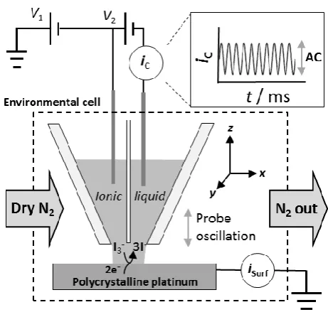
dissolved oxygen as well as to assist in drying of the droplet at the end of the probe (Figure 1).27 Full details are provided in the Supporting Information (SI) section S1. In brief, a dual barrel glass capillary pulled to a fine tip (1.6 µm diameter) was filled with a solution of interest (10 mM triiodide solution in RTIL). The RTIL used, was 1-butyl-3-methylimidazolium tetrafluoroborate [BMIm][BF4]). (≥ 97.0 % purity, (HPLC grade), Sigma
Aldrich) and was used fresh without further treatment. The 10 mM I3- solution was prepared
by dissolving equimolar amounts of I2 (98 % purity, Fischer Chemicals) and
(0.255 mm 3N, MaTeck), rinsed with water and then ethanol, followed by drying under a nitrogen flow was used as quasi-reference/counter electrodes (QRCEs). An Ag QRCE was inserted into each barrel and a bias potential was applied between the two QRCEs to establish an ion conductance current across the meniscus at the end of the pipet, which was monitored. An oscillation (amplitude ~ 200 nm peak to peak) was applied to the probe in the z direction, leading to an alternating current (AC) component of the ionic conductance signal, iC, due to
the periodical compression and expansion of the meniscus at the end of the pipet while in contact with the surface.
[image:5.595.182.413.434.657.2]The substrate of interest (polycrystalline platinum foil, purity > 99.95 %, 0.0125 mm thickness; Advent Research Materials) was flame-annealed prior to experiments and connected as a working electrode (WE) with an area defined by the size of the meniscus contact (roughly the size of the pipet, corresponding to ~ 2-3 × 10⁻⁸ cm2).
Figure 1. Schematic of the SECCM setup, employing a dual barrel glass pipet pulled to a fine tip (1.6 µm diameter). A bias potential (V2) of + 0.2 V was applied between two Ag QRCEs
surface). The working electrode potential (VSurf = -V1 – V2/2)) was varied by changing V1 and
the resulting surface current (iSurf) was recorded.
Electron backscatter diffraction (EBSD) images of platinum substrates were recorded on
a Zeiss SUPRA 55 variable-pressure field emission scanning electron microscope (FE-SEM)
at 20 kV on a 70 tilted sample with an EDAX TSL EBSD system, after the SECCM scans.
EBSD images were constructed from diffraction patterns recorded every 1 µm. The samples
were rinsed with deionized water before insertion into the vacuum chamber of the EBSD.
II. RESULTS AND DISCUSSION
At relatively low concentrations, triiodide (I3-) is formed through the equilibrium
between iodine (I2) and iodide (I-), which lies to the right in organic solvents and ionic
liquids. The redox reactions of interest herein are:7
3 I2 + 2 e⁻ 2 I3⁻ (1)
I3⁻ + 2 e⁻ 3 I⁻ (2)
Reaction 2 has been described to proceed via a complex mechanism in both acetonitrile7 and in RTILs28 but there is some consensus13-15,29 that the reaction may proceed on platinum via:
I3-(sol) I2(sol) + I- (3)
I2(sol) + 2 * 2 I* (4)
2 I* + 2 e- 2 I- (5)
where * denotes the free site on the electrode surface and sol denotes the solution phase.
cathodic direction, the reduction of I3⁻ is observed with an apparent onset potential of + 0.5 V.
Changing sweep direction at - 0.1 V, I- is oxidized back to I3⁻and starting at + 0.65 V , I3- is
further oxidized to form I2. The CV in the SECCM setup is characterized by relatively fast
mass transport rates, such that interfacial kinetic effects are manifested in the CVs (vide infra).
To estimate the formal potential for reaction 2 in [BMIm][BF4], macroscale CVs were
recorded, yielding an apparent formal potential of + 0.51 (± 0.03) V vs. AgQRE (see SI
section S2) at low potential sweep rates, consistent with values in the literature.9 This value was confirmed by open circuit potential measurements of equimolar concentrations of I3- and
[image:7.595.185.411.378.539.2]I- in [BMIm][BF4] (SI, section S2).
Figure 2. SECCM cyclic voltammogram of 10 mM I3- in [BMIm][PF4] on polycrystalline
platinum foil. Scan rate of 100 mV s-1.
SECCM images (Figure 3) were acquired at different electrode potentials for the I-/I3
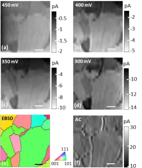
0.45 V to ca. 12 pA at + 0.30 V, in good agreement with the reduction wave in the CV in Figure 2 (taking account of its transient nature). Notably, significant variations in surface current are observed across the SECCM images, with the current changing by more than a factor of two between well-defined regions of low and high activity. A crystallographic orientation map of the same area, obtained with EBSD (Figure 3e), reveals a strong correlation between individual crystallographic grains and the variations in the SECCM surface current maps, highlighting significant electrode structural dependency of the kinetics of the I-/I3- process.
SECCM acquires other maps simultaneously with the surface current image: topography, direct current (DC) conductance and the AC component of the conductance current between the barrels in the probe (Figure 1),21 used as a feedback parameter to maintain a constant tip-substrate separation (meniscus height) throughout a scan.22 The DC and AC components of the conductance current are highly sensitive to changes in wetting and
droplet shape.21,22,30A typical AC amplitude map (set-point value of 20 pA), shown in Figure
3f, verifies the stability of the RTIL meniscus droplet during the SECCM scan. Most importantly, this stability indicates that the electrochemical variations seen in Figures 3(a) - (d) are not due to a change in droplet size and are essentially due to variations in inherent activity. However, in proximity to grain boundaries there appear to be some small perturbation of the droplet, leading to a change in the AC value that can be used to highlight the granular structure of the sample (Figure 3(f) and Figure 4(a)). This effect verifies the spatial correlation found between the SECCM images and EBSD map, as grain boundaries are highlighted by both imaging techniques and can be used as a means of self-referencing between images as well as to provide a precise estimate for the meniscus size (Figure 4(b)).
similar roughness of individual grains on such polycrystalline platinum foils13 excluding the
possibility of significant differences in wetting between individual grains.
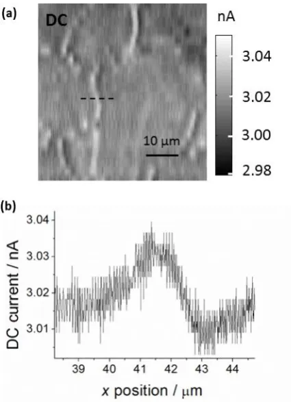
A key feature of SECCM is that we can estimate the magnitude of any ohmic drop via the conductance current between the two QRCEs. This is gained from analysis of the DC conductance current maps, such as the example shown in Figure 4(a). The DC conductance
maps, exhibited a steady DC of ca. 3 nA (corresponding to a resistance at the tip of ca. 66
MΩ for a bias potential of +0.2 V) throughout the scan with minute (but detectable)
variations at grain boundaries. The ohmic drop (iR) associated with the tip resistance during
scanning is negligible and accounts for less than a 1 mV when calculated for the highest
currents observed (~ - 14 pA in Figure 3(d)), highlighting the strength of the SECCM
Figure 3. SECCM and EBSD maps of a Pt electrode. (a) – (d) Spatially resolved surface currents for the reduction of 10 mM I3- in [BMIm][BF4] at 450, 400, 350 and 300 mV vs. Ag
[image:10.595.182.412.73.345.2]Figure 4 – SECCM DC conductance map at an electrode potential of + 0.4 V. (a) Typical DC
conductance map in the SECCM setup exhibiting highly stable values around 3 nA. The bias
voltage between the QRCEs was + 0.2 V. (b) Cross section of the dotted line in (a) where the
meniscus contact area was estimated as < 3 µm in diameter (i.e. of the order of the probe
size) where the DC was perturbed the most (at grain boundaries, shown in Figure 3(e)).
Inspection of the EBSD image (Figure 3(e)) and surface electrochemical images (Figures 3(a) - (d)) indicates that individual grains with a high contribution of the (110) orientation correlate to higher reduction currents at all potentials, whereas grains with a high contribution of the (100) orientation lead to lower reduction currents. The latter observation is in agreement with recent studies of hydrogen oxidation on basal plane Pt electrodes in
RTILs,31 where the surface adsorption of the anion ([BF4-]) was considered to possibly
Pt (110) and Pt (111). Similar trends in activity have been observed in preferentially-shaped nanoparticles in acetonitrile.19
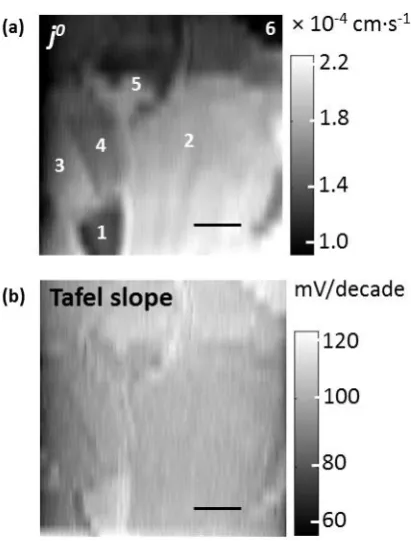
[image:12.595.196.402.311.582.2]To give a qualitative comparison of the variations in activity between grains, effective exchange current densities (j0) were calculated. A Tafel analysis of the four images in Figure 3 was performed (see SI section S3) yielding values of spatially-resolved j0 values and apparent Tafel slopes for the scanned area of the Pt foil. These are shown as images in Figure 5(a) and 5(b), respectively.
Figure 5. Tafel analysis of SECCM images. (a) Spatially resolved exchange current densities (j0)values for the reduction of I3- from the analysis of SECCM images. Several
The variation in effective j0 is between 1.8 (± 0.3) × 10-4 cm·s-1, for the more active
grains (e.g. 2, 3 and 5 in Figure 5(a)) and 0.9 (± 0.1) × 10-4 cm·s-1 for the less active grains
(e.g. 1 and 6 in Figure 5(a)). Figures 3 and 5 highlight key information from SECCM on spatially resolved ET kinetics that is lost in macroscale measurements involving
polycrystalline platinum electrodes. Figure 5(b) shows that the apparent Tafel slopes display little variation across the surface (see also Figure S2 in SI), with all grains between 100 and 120 mV per decade, suggesting that the rate determining step6,8,29 in the mechanism in all grains is the first electron transfer (nominally 120 mV per decade).
III.CONCLUSION
In the context of DSSCs, this study reveals that the rate of the I3- reduction process at
the (polycrystalline) CE is structure-dependent and is influenced by the subtle variations in crystallographic orientation of the electrode. Pt CEs with more (110) character will improve the activity of the process. The unique advantage of SECCM over single crystal
measurements is the ability to investigate high index facets which are difficult to obtain, but are ubiquitous in practical applications.
ASSOCIATED CONTENT
SECCM, Macroscale CVs, SECCM Tafel analysis. This material is available free of charge via the Internet at http://pubs.acs.org.
AUTHOR INFORMATION
Corresponding Author
Notes
The authors declare no competing financial interest.
ACKNOWLEDGMENT
This project was supported by the European Research Council through Advanced Investigator Grant ERC-2009-AdG 247143; “QUANTIF”. SCSL was further supported by a Marie Curie Intra-European Fellowship within the EU’s PF7 Program (project no. 275450; “VISELCAT”). The authors thank Dr. Kim McKelvey for fruitful discussion on data analysis, Dr. Maxim Joseph for contributions to sample preparation, Mr. Anatolii Cuharuc and Dr. Dimitry Momotenko for fruitful discussions, Ms. Changhui Chen for expert advice on EBSD analysis and Mr. Lee Butcher and Mr. Marcus Grant for providing parts for the experimental setup. We appreciate the expert design of custom electronics by Dr. Alex Colburn.
REFERENCES
(1) O'Regan, B.; Grätzel, M. A low-cost, high efficiency solar cell based on dye-sensitized colloidal TiO2 films Nature1991, 353, 737-740.
(3) Wu, M.; Lin, X.; Wang, Y.; Wang, L.; Guo, W.; Qi, D.; Peng, X.; Hagfeldt, A.; Grätzel, M.; Ma, T. Economical Pt-Free Catalysts for Counter Electrodes of Dye-Sensitized Solar Cells J. Am. Chem. Soc.2012, 134, 3419-3428.
(4) Nusbaumer, H.; Moser, J.-E.; Zakeeruddin, S. M.; Nazeeruddin, M. K.; Grätzel, M. CoII(dbbip)22+ Complex Rivals Tri-iodide/Iodide Redox Mediator in Dye-Sensitized
Photovoltaic Cells J. Phys. Chem. B2001, 105, 10461-10464.
(5) Boschloo, G.; Hagfeldt, A. Characteristics of the Iodide/Triiodide Redox Mediator in Dye-Sensitized Solar Cells Acc. Chem. Res.2009, 42, 1819-1826.
(6) Nakata, R.; Okazaki, S.; Fujinaga, T. Polarographic behavior of iodine in non-aqueous solvents J. Electroanal. Chem.1981, 125, 413-420.
(7) Macagno, V. A.; Giordano, M. C.; Arvía, A. J. Kinetics and mechanisms of
electrochemical reactions on platinum with solutions of iodine-sodium iodide in acetonitrile
Electrochim. Acta1969, 14, 335-357.
(8) Rogers, E. I.; Silvester, D. S.; Aldous, L.; Hardacre, C.; Compton, R. G. Electrooxidation of the Iodides [C4mim]I, LiI, NaI, KI, RbI, and CsI in the Room
Temperature Ionic Liquid [C4mim][NTf2] J. Phys. Chem. C2008, 112, 6551-6557.
(9) Zhang, Y.; Zheng, J. B. Investigation on the electro-oxidation of iodide in the room temperature ionic liquid, 1-butyl-3-methylimidazolium tetrafluoroborate at platinum electrode Electrochim. Acta2007, 52, 4082-4086.
(10) Hapiot, P.; Lagrost, C. Electrochemical Reactivity in Room-Temperature Ionic Liquids Chem. Rev.2008, 108, 2238-2264.
(12) Ejigu, A.; Lovelock, K. R. J.; Licence, P.; Walsh, D. A. Iodide/triiodide
electrochemistry in ionic liquids: Effect of viscosity on mass transport, voltammetry and scanning electrochemical microscopy Electrochim. Acta2011, 56, 10313-10320.
(13) Aaronson, B. D. B.; Chen, C.-H.; Li, H.; Koper, M. T. M.; Lai, S. C. S.; Unwin, P. R. Pseudo-Single-Crystal Electrochemistry on Polycrystalline Electrodes: Visualizing Activity at Grains and Grain Boundaries on Platinum for the Fe2+/Fe3+ Redox Reaction J. Am. Chem. Soc.2013, 135, 3873-3880.
(14) Narayanan, R.; El-Sayed, M. A. Shape-Dependent Catalytic Activity of Platinum Nanoparticles in Colloidal Solution Nano Lett.2004, 4, 1343-1348.
(15) Koper, M. T. M. Structure sensitivity and nanoscale effects in electrocatalysis
Nanoscale2011, 3, 2054-2073.
(16) Tian, N.; Zhou, Z.-Y.; Sun, S.-G.; Ding, Y.; Wang, Z. L. Synthesis of Tetrahexahedral Platinum Nanocrystals with High-Index Facets and High Electro-Oxidation Activity Science 2007, 316, 732-735.
(17) Oja, S. M.; Wood, M.; Zhang, B. Nanoscale Electrochemistry Anal. Chem.2012, 85, 473-486.
(18) Bönnemann, H.; Khelashvili, G.; Behrens, S.; Hinsch, A.; Skupien, K.; Dinjus, E. Role of the Platinum Nanoclusters in the Iodide/Triiodide Redox System of Dye Solar Cells
J. Cluster Sci.2007, 18, 141-155.
(19) Zhang, B.; Wang, D.; Hou, Y.; Yang, S.; Yang, X. H.; Zhong, J. H.; Liu, J.; Wang, H. F.; Hu, P.; Zhao, H. J.; Yang, H. G. Facet-Dependent Catalytic Activity of Platinum
in dye-sensitized solar cells: impact of nanoparticle size, crystallinity and surface coverage on catalytic activity Nanotechnology2012, 23, 485405.
(21) Ebejer, N.; Güell, A. G.; Lai, S. C. S.; McKelvey, K.; Snowden, M. E.; Unwin, P. R. Scanning Electrochemical Cell Microscopy: A Versatile Technique for Nanoscale
Electrochemistry and Functional Imaging Annu. Rev. Anal. Chem.2013, 6, 329-351.
(22) Snowden, M. E.; Güell, A. G.; Lai, S. C. S.; McKelvey, K.; Ebejer, N.; O’Connell, M. A.; Colburn, A. W.; Unwin, P. R. Scanning Electrochemical Cell Microscopy: Theory and Experiment for Quantitative High Resolution Spatially-Resolved Voltammetry and
Simultaneous Ion-Conductance Measurements Anal. Chem.2012, 84, 2483-2491.
(23) Lai, S. C. S.; Patel, A. N.; McKelvey, K.; Unwin, P. R. Definitive Evidence for Fast Electron Transfer at Pristine Basal Plane Graphite from High-Resolution Electrochemical Imaging Angew. Chem. Int. Ed.2012, 51, 5405-5408.
(24) Ghilane, J.; Lagrost, C.; Hapiot, P. Scanning Electrochemical Microscopy in Nonusual Solvents: Inequality of Diffusion Coefficients Problem Anal. Chem.2007, 79, 7383-7391.
(25) Walsh, D. A.; Lovelock, K. R. J.; Licence, P. Ultramicroelectrode voltammetry and scanning electrochemical microscopy in room-temperature ionic liquid electrolytes Chem. Soc. Rev.2010, 39, 4185-4194.
(26) Carano, M.; Bond, A. M. Prospects for the Application of Scanning Electrochemical Microscopy in Ionic Liquids Aust. J. Chem.2007, 60, 29-34.
(27) Zhao, C.; Bond, A. M.; Compton, R. G.; O’Mahony, A. M.; Rogers, E. I.
Modification and Implications of Changes in Electrochemical Responses Encountered When Undertaking Deoxygenation in Ionic Liquids Anal. Chem.2010, 82, 3856-3861.
(28) Bentley, C. L.; Bond, A. M.; Hollenkamp, A. F.; Mahon, P. J.; Zhang, J.
platinum, glassy carbon and boron-doped diamond in the room temperature ionic liquid 1-ethyl-3-methylimidazolium bis(trifluoromethanesulfonyl)imide Electrochim. Acta2013, 109, 554-561.
(29) Hauch, A.; Georg, A. Diffusion in the electrolyte and charge-transfer reaction at the platinum electrode in dye-sensitized solar cells Electrochim. Acta2001, 46, 3457-3466. (30) Lai, S. C. S.; Dudin, P. V.; Macpherson, J. V.; Unwin, P. R. Visualizing Zeptomole (Electro)Catalysis at Single Nanoparticles within an Ensemble J. Am. Chem. Soc.2011, 133, 10744-10747.
![Figure 2. SECCM cyclic voltammogram of 10 mM I3- in [BMIm][PF4] on polycrystalline](https://thumb-us.123doks.com/thumbv2/123dok_us/9533012.458282/7.595.185.411.378.539/figure-seccm-cyclic-voltammogram-of-bmim-pf-polycrystalline.webp)