ORIGINAL RESEARCH
Analysis of Central Nervous System Vasculitis
with Diffusion-Weighted Imaging and Apparent
Diffusion Coefficient Mapping of the
Normal-Appearing Brain
M.L. White W.L. Hadley Y. Zhang M.A. Dogar
BACKGROUND AND PURPOSE: Diffusion-weighted imaging (DWI) is a novel method of studying the brain that creates tissue contrast secondary to water diffusion. Central nervous system (CNS) vascu-litis is a rare inflammatory disease that continues to be difficult to diagnose and evaluate with MR imaging. DWI with apparent diffusion coefficient (ADC) analysis may demonstrate abnormalities within the brain that would otherwise be undetected by conventional imaging, thus aiding in the diagnosis and evaluation of patients with CNS vasculitis.
MATERIALS AND METHODS: A retrospective analysis was performed of patients who were diagnosed with CNS vasculitis and had undergone DWI. A total of 15 patients who had DWI with b⫽1000 were analyzed. Regions of interest were drawn in the anterior, central, and posterior regions of white matter (WM) at 3 levels. Regions of interest were also drawn bilaterally in the caudate heads, putamina, thalami, posterior internal capsules, and the cerebellar WM. ADC values were measured and com-pared with 15 healthy controls who were matched for age, sex, and MR imaging scanner.
RESULTS: There was a significant increase in the ADC values (P⬍.00625) for the anterior WM, central WM, thalami, and posterior internal capsules in patients with CNS vasculitis.
CONCLUSION:Diffuse increase in water diffusion was present in the normal-appearing brain in patients with CNS vasculitis, and these abnormalities were not demonstrated by conventional MR imaging sequences. The detection and quantification of ADC abnormalities may provide useful diagnostic information for patients with CNS vasculitis.
C
entral nervous system (CNS) vasculitis is a rare inflamma-tory disease that affects the arteries and the veins of the brain.1,2MR imaging often demonstrates brain lesions inpa-tients with CNS vasculitis.3-7However, there are numerous
examples of conventional MR imaging not demonstrating le-sions in patients with proved vasculitis.5,6The limited sensi-tivity of conventional MR imaging to CNS vasculitis changes is demonstrated by cases in which angiographic abnormalities are present despite normal findings on MR imaging examination.4,5,7,8
Diffusion-weighted imaging (DWI) provides a novel method of investigating the brain, which relies on the molec-ular diffusion of water for tissue contrast.9Apparent diffusion
coefficient (ADC) analysis allows a quantitative measurement of changes within the brain. Brain analysis using ADCs has been found to demonstrate abnormalities that are not detect-able by conventional MR imaging for multiple sclerosis, tuber-ous sclerosis, Behcet’s disease, neurofibromatosis type 1 (NF1), and the aging brain.10-14The detection of
abnormali-ties in the brain by DWI, not otherwise visualized by MR im-aging, may help to determine the extent of abnormalities in patients with CNS vasculitis.
We evaluated the normal-appearing brain as demonstrated by conventional MR imaging with DWI using ADC analysis to
enhance our understanding of the extent and type of changes present in patients with CNS vasculitis. We hypothesized that there would be diffuse abnormalities present within the brain, resulting in increased diffusion and thus increased ADC values.
Materials and Methods
The medical records were searched by computer for patients with the diagnosis of cerebral vasculitis from 1998 to 2005. Since 1998, brain MR imaging has been routinely performed with a DWI sequence. Only cases of inflammatory vasculitis were included. The cases con-sidered to represent vasculitis had to have a clinical diagnosis of vas-culitis and 1 of the following: 1) pathologic evidence, 2) classic vascu-litis pattern on an angiogram, or 3) clinical course and a response to therapy consistent with vasculitis.3,15The diagnosis of vasculitis was
based on clinical history and pathology evidence in 6, clinical history and vasculitis pattern on angiograms in 8, and clinical course with response to therapy in 1. The diagnosis was primary CNS vasculitis in 12 patients and vasculitis secondary to sarcoid in 2 patients.
There were 15 patients who met our entry criteria (4 men, 11 women; mean, 51.9 years of age; range, 39 –74 years of age). Symp-toms in patients had been present from 2 days to 20 days. Eleven patients were imaged within 10 days of the onset of symptoms. The others were imaged after approximately 13, 20, and 45 days of symptoms.
Fourteen subjects underwent scanning with 1.5T; and 1, with 3T MR imaging systems. The MR imaging studies are summarized as follows: 9 studies on Signa (GE Healthcare, Milwaukee, Wis), 1 study on Magnetom Vision (Siemens, Erlangen, Germany), 2 studies on Symphony (Siemens), 2 studies on Avanto (Siemens), and 1 study on Trio (Siemens). Fluid-attenuated inversion recovery (FLAIR) and DWI with b⫽1000 were performed in all patients.
Received April 24, 2006; accepted after revision October 5.
From the Department of Radiology (M.L.W., Y.Z.), University of Nebraska Medical Center, Omaha, Neb; the Department of Radiology (W.L.H.), University Hospital of Cleveland/Case Medical Center, Cleveland, Ohio; and the Department of Radiology (M.A.D.), University of Iowa College of Medicine, Iowa City, Iowa.
Please address correspondence to Matthew L. White, MD, 981045 Nebraska Medical Center, Omaha, NE 68198-1045; e-mail: [email protected]
BRAIN
ORIGINAL
There was a total of 8 cases that were considered to have vasculitis but were not included in the data analysis. One case had corrupt diffusion source data; 1 case was saved on a storage disk that could no longer be accessed; 1 patient had only MR imaging performed outside our institution, the results of which could not be obtained; and 5 of the vasculitis patients had only follow-up imaging at our institution. Additionally, 3 more patients were excluded as result of not having a true inflammatory vasculitis and/or having a complicated history. One patient was scanned after surgery and radiation therapy for an astrocytoma, and there were 2 patients with Susac Syndrome (a clin-ical triad of encephalopathy, hearing loss, and retinal arteriolar occlusions).
Four-vessel angiography was performed using a digital subtrac-tion technique in all patients. The angiograms were reviewed in a blinded fashion by a neuroradiologist, and the reports were also re-viewed. The blinded reviews of the angiograms were all in agreement with the original interpretation.
The control subjects were matched for age, sex, and MR imaging scanner. There were 4 male and 11 female controls with a mean of 50.2 years of age (range, 36 – 67 years of age). Each control MR imaging was interpreted as having normal findings on brain examination. The indications for examinations of the control subjects included the fol-lowing: asymmetric sensorineural hearing loss, headaches, anterior communicating artery aneurysm, bilateral upper extremity numb-ness, paresthesias and facial weaknumb-ness, acoustic neuroma excision, benign positional vertigo, cerebellar signs and left facial droop, optic disk edema and periphlebitis, peripheral visual field loss, and hemi-facial spasm.
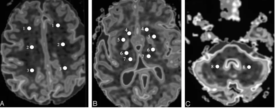
The primary analysis of the brain consisted of evaluation of the cerebral WM. Smaller regions of interest were drawn for sampling than those in previous studies secondary to our concern for avoiding partial volume averaging with adjacent CSF, gray matter, or lesions. Regions of interest were drawn bilaterally in the anterior, central, and posterior regions of WM at each of 3 consecutive levels, with the most caudal level at the superior aspect of the lateral ventricles. These re-gions corresponded to frontal, frontal-parietal, and parietal WM. Re-gions of interest were also drawn bilaterally in the caudate heads, putamina, thalami, posterior internal capsules, and the cerebellar WM (Fig 1). The regions of interest were drawn to avoid areas of
FLAIR and DWI lesions. Similar region-of-interest locations were drawn in patients as well as controls. There was a total of 15 regions of interest for which ADC measurements could not be obtained second-ary to lesion locations that did not allow for placement of the region of interest in the patients with CNS vasculitis. The average size of the region of interest in the patients with CNS vasculitis was 44.4 mm3.
The right-sided and left-sided ADC measurements for the 3 levels at which the WM was measured were averaged together for 1 value to represent each WM region—anterior, central, or posterior WM. Sim-ilarly, the right-sided and left-sided ADC measurements from the caudate heads, putamina, thalami, posterior internal capsules, and the cerebellar WM were averaged together to obtain 1 measurement for each region.
The ADC analysis was performed by using 2-point ADC calcula-tions.16Images with b⫽0 s/mm2and b⫽1000 s/mm2were used. ADC
maps were generated on an ADW 4.0 workstation (GE Healthcare). Thettest was used to compare the average ADCs for each region-of-interest point measured (anterior WM, central WM, posterior WM, caudate heads, putamina, thalami, posterior internal capsules, and the cerebellar WM) in the vasculitis subjects with those of healthy controls. A Bonferroni correction was used, making aPvalue of ⬍.00625 significant.
Results
The ADC values in the normal-appearing brain of the patients are summarized in the Table . There was a significant increase in the ADC values (P⬍.00625) for the anterior WM, central WM, thalami, and posterior internal capsules (Fig 2). In pa-tients 4 and 5, DWI and FLAIR showed no abnormal findings. Both patients overall demonstrated mild-to-moderate eleva-tion of ADCs compared with the average values of the con-trols. There were 9 patients with lesions seen on DWI and 12 patients with lesions seen on FLAIR. Only 2 patients demon-strated more lesions on DWI than on FLAIR. There was a total of 75 DWI lesions and 150 FLAIR lesions.
Discussion
[image:2.585.55.534.44.235.2]imaging findings in CNS vasculitis can be negative when ab-normalities are present at cerebral angiography.4-7
Conven-tional MR imaging findings may even be negative in cases of pathologically proved vasculitis.17Also, angiography and
bi-opsy findings can be negative in patients who have CNS vas-culitis.1,3,4,7,17-19This demonstrates the need for a noninvasive
test that can detect abnormalities that may be missed by con-ventional MR imaging, and even angiography and biopsy. This noninvasive test could potentially be beneficial for both diagnostic and prognostic purposes.
DWI uses a contrast mechanism—water diffusivity—that is unique and quantifiable.9This uniqueness of the contrast
mechanism allows DWI to demonstrate brain changes that routine MR imaging misses. ADCs increase when water mo-tion increases. However, increased diffusion is a nonspecific finding that can be the result of several mechanisms. DWI with ADC analysis has demonstrated the ability to detect subtle changes throughout the brain. Increased ADC values for nor-mal-appearing brain have been found in multiple sclerosis, tuberous sclerosis, Behcet disease, NF1, and the aging brain.10-14Applying ADC analysis to evaluate the brain in
pa-tients with CNS vasculitis may reveal otherwise undetectable changes.
Our results demonstrate that diffuse changes do occur in the normal-appearing brain in patients with CNS vasculitis. The ADCs were increased throughout the WM of the corona radiata and centrum semiovale, the thalami, and the posterior internal capsules. The ADC analysis of the brain helped to elucidate the extent of abnormalities that occur in CNS vascu-litis. This study demonstrated the ability of ADC
measure-ments to detect brain changes that have not been previously noted.
The ADC analysis of the normal-appearing brain may be potentially useful in the diagnostic work-up of patients with suspected CNS vasculitis, given that conventional MR imag-ing findimag-ings may be negative in patients with CNS vasculitis.4-7
For example, normal MR imaging findings but an abnormality of normal-appearing brain in a patient with suspected CNS vasculitis may be strong evidence for pursuing a more invasive diagnostic test such as brain biopsy. This question needs fur-ther evaluation. Even of greater interest would be the possibil-ity that a noninvasive test could help make the diagnosis of CNS vasculitis, and a brain biopsy could be avoided. However, it would be surprising if ADC analysis of the normal-appear-ing brain in patients suspected of havnormal-appear-ing CNS vasculitis could fulfill that role, given the nonspecific meaning of increased ADCs.
We can only hypothesize the meaning of diffuse abnormal-ities detected by ADC analysis. We would conjuncture that they potentially represent diffuse vasogenic edema, brain de-struction (axonal loss), wallerian degeneration, or vascular changes directly due to the vasculitic process. In patients with CNS vasculitis, diffuse pathologic changes in the brain paren-chyma include loss of nerve cells, abnormal nerve cells, perivascular lakes of eosinophilic material, and foci of vacuo-lation.20Changes have been found widespread throughout the
white and gray matter.20At autopsy, diffuse changes of edema,
reactive astrocytosis, ischemic change, and hemorrhage have been found.21The changes in the posterior internal capsules
and the thalami in particular could be due to wallerian degen-ADC in the nonlesional brain of patients
Patient/ Age/ Sex Days from Onset to DWI No. of Lesions on
DWI/FLAIR Angiographic Findings
ADC (⫻10⫺4mm2/s)
Anterior
WM CentralWM PosteriorWM CaudateHeads Putamina Thalami Post IC CerebellarWM 1/40/M 7 21/18 Irregular narrowing of anterior,
posterior circ. bilaterally
6.93–8.24 (7.53) 7.65–8.02 (7.87) 7.54–8.61 (7.92)
8.48 (8.48) 7.94 (7.94) 7.67–8.00 (7.84)
7.62 (7.62) 8.57–8.85 (8.71) 2/46/F 2 6/2 Irregular narrowing of rt. MCA,
lt. ACA, and MCA
7.70–8.41 (8.09) 6.90–7.67 (7.24) 6.98–7.69 (7.38) 7.63–7.94 (7.79) 7.51–7.57 (7.54) 7.39–7.43 (7.41) 6.68–7.02 (6.85) 6.86–6.56 (6.71)
3/45/F 9 0/1 Segmental stenosis of
anterior, posterior circ. bilaterally
7.04–8.50
(7.80) 6.39–7.01(6.85) 7.11–7.64(7.45) 7.51–7.17(7.34) 7.34–7.35(7.35) 7.59–7.86(7.73) 6.75–7.11(6.93) 6.72–7.23(6.98)
4/39/F 5 0/0 Irregular narrowing and serpiginous appearance of rt. PICA 7.29–8.11 (7.67) 5.96–7.80 (6.59) 6.85–7.83 (7.48) 6.71–7.25 (6.98) 7.54–7.84 (7.69) 7.24–7.48 (7.36) 7.54–8.46 (8.00) 6.72–7.31 (7.02)
5/74/M 2 0/0 N/A 7.99–9.09
(8.38) 6.65–7.74 (7.17) 7.25–7.67 (7.46) 7.01–7.02 (7.02) 6.44–6.74 (6.59) 7.41–7.76 (7.59) 6.99–7.16 (7.08) 5.92–6.13 (6.03) 6/58/F 3 2/7 Irregular narrowing of anterior,
posterior circ. bilaterally
7.37–8.50 (7.86) 6.44–7.54 (6.86) 7.01–7.84 (7.47) 6.78–7.03 (6.91) 6.67–7.15 (6.91) 7.60–7.46 (7.53) 6.82–7.18 (7.00) 6.14–6.57 (6.36)
7/56/F 6 5/8 Normal 8.20–8.85
(8.54) 7.03–7.89(7.32) 7.33–8.20(7.75) 8.43–8.69(8.56) 7.42–7.45(7.44) 6.78–7.26(7.02) 7.29–7.37(7.33) 6.13–6.14(6.14) 8/54/F 7 0/1 Narrowing and dilation of lt.
ACA and MCA 6.99–7.97(7.33) 6.49–7.30(6.86) 7.27–7.89(7.58) 7.26–7.65(7.46) 7.47–7.54(7.51) 7.21–7.40(7.31) 7.32–7.36(7.34) 6.66–6.77(6.72) 9/51/F 6 7/11 Occlusion of lt. ACA and MCA;
multiple medium/small vessel stenoses 7.31–9.12 (8.28) 6.72–7.60 (6.96) 6.59–8.42 (7.45) 8.54–8.71 (8.63) 7.53–7.56 (7.55) 7.21–7.60 (7.41) 6.68–7.41 (7.05) 6.05–6.74 (6.40)
10/48/M 13 5/36 Irregular narrowing of anterior,
posterior circ. bilaterally 7.28–9.30(8.07) 6.91–8.45(7.91) 7.35–8.44(7.86) 8.17 (8.17) 7.29–7.36(7.33) 7.50–7.41(7.46) 7.09–8.68(7.89) 7.08–7.20(7.14) 11/51/F 13 13/15 Occlusion in left circ. 6.76–10.10
(7.94) 6.21–8.96 (768) 6.82–7.19 (6.97) 6.89–8.89 (7.89) 6.66–7.39 (7.03) 7.03–7.59 (7.31) 6.88–6.96 (6.92) 5.78–6.11 (5.95) 12/52/F 7 0/23 Diffuse vessel caliber
variations 7.62–8.20 (7.91) 7.06–7.88 (7.51) 7.89–8.61 (8.30) 8.42–8.71 (8.57) 7.81–7.84 (7.83) 7.75–7.76 (7.76) 7.94–8.28 (8.11) 7.63–8.28 (7.96) 13/47/F 20 7/8 Stenosis of MCA and PCA
bilaterally 7.90–8.58(8.18) 8.20–8.81(8.46) 7.53–8.74(8.18) 8.13 (8.13) 7.79 (7.79) 7.64–7.86(7.75) 8.79 (8.79) 7.76–7.83(7.80)
14/61/M 8 7/17 N/A 7.51–8.84
(8.07) 7.57–8.56(8.10) 7.61–8.65(8.00) 7.37–8.33(7.85) 6.83–6.92(6.88) 7.29–7.85(7.57) 7.22–7.46(7.34) 7.82–7.94(7.88) 15/57/F 45 2/3 Irregularity of rt. MCA 7.76–8.41
(8.11) 7.40–8.31 (7.78) 7.12–8.12 (7.54) 7.63–7.64 (7.64) 7.62–8.80 (8.21) 7.49–7.68 (7.59) 7.59–8.42 (8.01) 8.57–8.85 (8.71)
eration secondary to the WM tracts of which they consist (the posterior internal capsules) or the numerous WM connec-tions within them (the thalami).
CNS vasculitis is a relatively rare disorder, and this resulted in the number of cases in our series being small. However, given the rarity of the disease, we believe the number of pa-tients in our series is respectable. Not all of our cases are from the same MR imaging scanner, and this may result in an un-anticipated bias that we may not have corrected even though we matched our healthy controls by the MR imaging scanner used in the patients with vasculitis. We did use the same b value
for all diffusion studies (b⫽1000). Most of our cases were not pathologically proved, but it has been shown that biopsy can be negative in patients who have CNS vasculitis.17,19We were
lim-ited in biopsy material available to confirm the diagnosis of CNS vasculitis secondary to the clinical methodologies used at our in-stitution to make the diagnosis of vasculitis.
Conclusion
[image:4.585.53.538.54.551.2]information can be used to predict patient diagnosis, acute clinical outcome, or long-term disability.
References
1. Calabrese LH, Mallek JA.Primary angiitis of the central nervous system: re-port of 8 new cases, review of the literature, and proposal for diagnostic crite-ria.Medicine (Baltimore)1988;67:20 –39
2. Rhodes RH, Madelaire NC, Petrelli M, et al.Primary angiitis and angiopathy of the central nervous system and their relationship to systemic giant cell arteri-tis.Arch Pathol Lab Med1995;119: 334 – 49
3. Harris KG, Trann DD, Sickels WJ, et al.Diagnosing intracranial vasculitis: the roles of MR and angiography.AJNR Am J Neuroradiol1994;15:317–30 4. Greenan TJ, Grossman RI, Goldberg HI.Cerebral vasculitis: MR imaging and
angiographic correlation.Radiology1992;182:65–72
5. Cloft HJ, Phillips CD, Dix JE, et al.Correlation of angiography and MR imag-ing in cerebral vasculitis.Acta Radiol1999;40:83– 87
6. Wasserman BA, Stone JH, Hellmann DB, et al.Reliability of normal findings on MR imaging for excluding the diagnosis of vasculitis of the central nervous system.AJR Am J Roentgenol2001;177:455–59
7. Pomper MG, Miller TJ, Stone JH, et al.CNS vasculitis in autoimmune disease: MR imaging findings and correlation with angiography.AJNR Am J Neurora-diol1999;20:75– 85
8. Meusser S, Rubbert A, Manger B, et al.99m-Tc-HMPAO-SPECT in diagnosis of early cerebral vasculitis.Rheumatol Int1996;16:37– 42
9. Turner R, Le Bihan D, Maier J, et al.Echo-planar imaging of intravoxel inco-herent motion.Radiology1990;177:407–14
10. Baysal T, Dogan M, Karlidag R, et al.Diffusion-weighted imaging in chronic
Behcet patients with and without neurological findings.Neuroradiology2005; 47:431–37. Epub 2005 May 26
11. Engelter ST, Provenzale JM, Petrella JR, et al.The effect of aging on the appar-ent diffusion coefficiappar-ent of normal-appearing WM.AJR Am J Roentgenol
2000;175:425–30
12. Garaci FG, Floris R, Bozzao A, et al.Increased brain apparent diffusion coeffi-cient in tuberous sclerosis.Radiology2004;232:461– 65. Epub 2004 Jun 23 13. Horsfield MA, Lai M, Webb SL, et al.Apparent diffusion coefficients in benign
and secondary progressive multiple sclerosis by nuclear magnetic resonance. Magn Reson Med1996;36:393– 400
14. Alkan A, Sigirci A, Kutlu R, et al.Neurofibromatosis type 1: diffusion weighted imaging findings of brain.Eur J Radiol2005;56:229 –34. Epub 2005 Jun 15 15. Kissel JT.Neurologic manifestations of vasculitis.Neurol Clin1989;7:655–73 16. Burdette JH, Elster AD, Ricci PE.Calculation of apparent diffusion coefficients
(ADCs) in brain using two-point and six-point methods.J Comput Assist To-mogr1998;22:792–94
17. Chu CT, Gray L, Goldstein LB, et al.Diagnosis of intracranial vasculitis: a multi-disciplinary approach.J Neuropathol Exp Neurol1998;57:30 –38 18. Calabrese LH, Furlan AJ, Cragg LA, et al.Primary angiitis of the central
ner-vous system: diagnostic criteria and clinical approach.Cleve Clin J Med
1992;59:293–306
19. Duna GF, Calabrese LH.Limitations of invasive modalities in the diagnosis of primary angiitis of the central nervous system.J Rheumatol1995;22:662– 67 20. Nurick SW. Blackwood W, Mair WG.Giant cell granulomatous angiitis of the
central nervous system.Brain1972;95:133– 42

![3 {3,3 Dimethylspiro[2,3 dihydro 1H indole 2,3′ (3′H naphtho[2,1 b][1,4]oxazin)] 1 yl}propionic acid](data:image/gif;base64,R0lGODlhAQABAIAAAP///wAAACH5BAEAAAAALAAAAAABAAEAAAICRAEAOw==)