Original Article
MiR-7 involves the activation of Nrf2 pathway
by targeting Keap1 in epileptic seizure
Liang Yu1, Hongbin Sun2, Baomin He2, Supin Li2, Shuai Ma2, Lili Yang2, Yi Guo2, Dong Zhou1
1Department of Neurology, West China Hospital, Sichuan University, Chengdu, Sichuan, People’s Republic of
China; 2Department of Neurology, Sichuan Academy of Medical Sciences & Sichuan Provincial People’s Hospital,
Chengdu, Sichuan, People’s Republic of China
Received November 2, 2015; Accepted January 5, 2016; Epub February 1, 2016; Published February 15, 2016
Abstract: MicroRNAs (miRNAs) are increasingly recognized as diagnostic biomarkers of epileptogenesis as well as targets to prevent or disrupt epilepsy. Keap1, a cytoplasmic inhibitory protein called Kelch-like ECH-associated pro-tein 1, is also reported associated with epilepsy. However, the possible involvement of miRNAs in Keap1-mediated molecular basis for protection from epileptic seizure-induced brain damage is less understood. In the present study, Wistar rats were rapidly kindled in the amygdala. We found a dramatic upregulation of miR-7 and downregulation of Keap1 in the hippocampus of kindled rats, compared with control and sham group. Moreover, luciferase reporter gene assays identified that miR-7 was able to target 3’-untranslated region (3’-UTR) of Keap1 mRNA. To investigate the role of miR-7 in the protection of epilepsy mediated by Keap1/Nrf2 pathway, we used a transfection approach to overexpress miR-7, and then detected a consequential decrease in Keap1 mRNA and protein which subsequently results in an increased Nrf2 expression and cytoprotective enzymes (HO-1 and NQO1) expression. These results indicate that miR-7 may involve the protection of protection through targeting Keap1 and the subsequent activation of Nrf2 pathway.
Keywords: MiR-7, Nrf2, Keap1, epileptic seizure
Introduction
Epilepsy, one of the most common neurological disorders, is a chronic disorder of abnormal electrical activity in the brain, characterized by recurrent unprovoked seizures [1]. The mecha-nisms underlying epileptogenic pathogenesis have been proved to be complex, including oxi-dative stress, glutamate excitotoxicity, calcium overload and so on [2]. Given that pathological processes involved in epileptogenesis are com-plex and interrelated, to find a target, which can interrupt multi-mechanisms underlying seizure, is desirable.
MicroRNA (miRNA), an abundant group of en- dogenous non-coding single strand RNAs wi- th approximately 19~25 nucleotides, regulates the expression of genes at post-transcriptional level by translational repression or degradation of target mRNA. They are critical for normal neuronal development and may be involved in many neurological diseases whose mechanis- ms remain to be explored. For example, Agostini
Animals
Adult male Wistar rats weighing 250-300 g (n = 50) were obtained from the Hebei Medial University. Rats were housed in a room with constant temperature (25 ± 1°C) and humidity (40-60%), and were kept on a 12 h light/dark cycle, with lights on at 8:00 AM and with free access to food and water. Animal experiments were performed according to the regulations of laboratory animal management promulgated by the Ministry of Science and Technology of the People’s Republic of China No. 134, which coincides with internationally recognized NIH guidance. The rats were randomly assigned to three groups. Control group (n = 16) did not receive any treatment. Electrodes were implant-ed in the remaining 34 rats. Two rats could not be included in the experiments because of death during anesthesia or misplacement of the electrode. The remaining 32 rats were ran-domly distributed to the following groups: sham group (n = 16) did not receive any electrical current administration; kindling group (n = 16) were subjected to experimental kindling proce-dure as follow. Twenty-four hours after the last stimulation, all rats were sacrificed. Eight rats in each group were prepared for spectropho-tometry, Western blot and real-time fluores-cence quantitative PCR analysis. Animal care and sacrifice were conducted according to me- thods approved by the Animal Care and Use Committee, Xiangya Medical College, Central South University. All experiments were perfor- med in accordance with the National Institute of Health Guide for the Care and Use of La- boratory Animals.
Electrode implantation
For implantation, animals were anesthetized with chloral hydrate (300 mg/kg, i.p.), the sham and kindling groups were stereotactically im- planted with twisted stainless steel wire bipolar stimulating electrodes in the left basolateral
to prevent infection.
Kindling procedure
The initial after discharge threshold (ADT) was determined using an electric stimulator (SEN-7203; Nihon Kohden) 1 week after electrode implantation [12]. The stimulation parameters were performed by constant current stimula-tions (monophasic square-wave pulses, 60 Hz for 1 s). The stimulus intensity started at 100 μA, and thereafter increased in 40 μA incre-ments at 5 min intervals until an after discharge of at least 3 s duration was elicited (140-340 μA). The next day animals of kindling group were kindled at 10 min intervals stimulation at 120% of their individual ADT. In the course of kindling, stimulation was delivered 40 times for the kindling group, with 20 times every day.
Tissue dissection
Following the behavioral tests, the rats and mice in each group were sacrificed by decapita-tion. The hippocampus was quickly harvested and was frozen in liquid nitrogen and stored at -80°C until further utilization. Bilateral hippo-campus of six rats in each group was pooled from individual rats for spectrophotometry. Unilateral hippocampi of the other six rats in each group were processed into a plastic tube for real-time PCR or Western blot separately.
Primary rat hippocampal neuron cultures
atmosphere and grown in neurobasal medium (NB) supplemented with 2% B27, 0.5 mM l- glutamine, 100 units/ml penicillin and 100 g/ ml streptomycin. Cultures were fed twice we- ekly.
Cell transfection
The miR-7 inhibitors and negative control mol-ecules were purchased from Dharmacon (Au- stin, TX, USA). Cell transfection was performed using DharmFECT1 (Dharmacon) until a final concentration of 20 nM. Medium was changed after 6 h. After transfected and cultured for 48 h, cells were collected for Western blot and qRT-PCR analyses.
Luciferase reporter gene assays
The 3’-UTR of Keap1 containing the putative binding site of miR-7 was amplified and sub-cloned into pGL3 luciferase promoter vector (Promega, Madison, WI, USA). The vector was co-transfected with miR-7 mimics into HEK293 cells for 48 h. The cells were harvested and relative luciferase activity was detected using a dual-luciferase reporter assay kit (Promega) according to the manufacturer’s instructions. All experiments were performed at least three times.
Quantitative real-time polymerase chain reac-tion (RT-PCR)
Total RNA was isolated from tissues and cell lines using the miRNeasy Mini Kit (Qiagen). The miRNA Q-PCR Detection Kit (GeneCopoeia) was used for quantification of miRNA levels accord-ing to the manufacturer’s protocol. For quan- tification of PRMT1 mRNA levels, the RT
reac-tions were conducted with the RevertAid TM H Minus First Strand cDNA Synthesis Kit (Fer- mentas). qRT-PCR was performed using an ABI 7900 System (Bio-Rad). RNU6B and β-actin were used as normalizing controls for miRNA and mRNA quantification, respectively. The 2-ΔΔCt method was employed to calculate the relative expression levels. The primers were as follows: Keap1, forward primer: 5’-CAACTTCG- CTGAGCAGATTGGC-3’ and reverse primer: 5’- TGATGAGGGTCACCAGTTGGCA-3’. Nrf2, forwa- rd primer: 5’-CACATCCAGTCAGAAACCAGTGG-3’ and reverse primer: 5’-GGAATGTCTGCGCCAA- AAGCTG-3’. HO-1 forward primer: 5’-TGTCCC- AGGATTTGTCCGAG-3’ and reverse primer: 5’- ACTGGGTTCTGCTTGTTTCGCT-3’, NQO1 forward primer: 5’-GGGGACATGAACGTCATTCTCT-3’ and reverse primer: 5’-AGTGGTGACTCCTCCCAGAC- AG-3’, GAPDH forward primer: 5’-TGAACGGGAA- GCTCACTG-3’ and reverse primer: 5’-GCTTCA- CCACCTTCTTGATG-3’).
Western blot analysis
[image:3.612.92.521.70.190.2]Cells were rinsed with ice-cold PBS and lysed in PBS containing 2% SDS with protease inhibi-tors cocktail and phosphatase inhibiinhibi-tors (Ro- che). Cell lysates were sonicated for 20 sec, and protein concentration in the lysates was quantified using BCA Protein Assay Reagent (Thermo Scientific). Cell lysates were analyzed by Western blot analysis, using anti-Keap1 (cat-alog #8047, Cell-Signaling), anti-Nrf2 (cat(cat-alog #EP1808Y, Abcam) or anti-β-actin (catalog #A5316, Sigma-Aldrich), followed by incubation with horseradish peroxidase-conjugated anti-rabbit (catalog #HAF008, R&D Systems) or anti-mouse antibody (catalog #HAF007, R&D Systems). ECL detection systems (SuperSignal West Femto, Pierce) were used for detection.
Statistical analysis
Each experiment was repeated at least three times. Data were shown as mean ± s.d and analyzed using SPSS 19.0. Statistical compari-sons between groups were analyzed using Student’s t-test and a two-tailed P < 0.05 was considered to indicate statistical significance. Results
MiR-7 and Keap1 expression in hippocampus
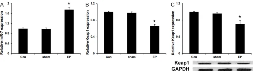
We first detected the miR-7 expression in hip-pocampus was studied by real-time fluores- cence quantitative PCR. As shown in Figure 1A, the expression of miR-7 is much higher in kin-dling groups than that in control and sham groups. Moreover, qRT-PCR assay and western blot assay revealed that the expression of Keap1 mRNA (Figure 1B) and protein (Figure 1C) is much lower in kindling groups compared with control and sham groups.
MiR-7 directly targets Keap1 in hippocampal neurons cells
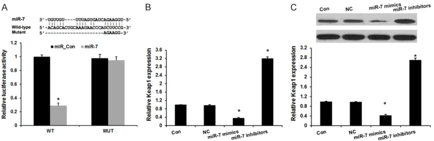
Keap1 is also reported associated with epi- lepsy [13]. However, the relationship between miR-7 and Keap1 in hippocampal neurons cells remains unclear. In this study, the miRNA targ- et prediction websites www.microRNA.org and TargetScan were used and identified a con-served miR-7-binding site in the 3’-UTR of Keap1 mRNA. We then cloned WT or Mut target region sequence of the Keap1 3’-UTR, which was inserted into a luciferase reporter vector
(Figure 2A). Subsequently, these reporter vec-tors were cotransfected with miR-7 mimics and mimics control (mimics_con) into the HEK293 cell line. As shown in Figure 2A, co-transfection of miR-7 mimics suppressed the luciferase activity of the reporter containing wild-type Ke- ap1 3’ UTR sequence, but failed to inhibit that of mutated Keap1 by dual-luciferase reporter assay. These data indicate that Keap1 is one of the direct targets of miR-7.
Next, we transfected miR-7 inhibitors in hippo-campal neurons cells. qRT-PCR and Western blot analysis revealed Keap1 expression we- re significantly increased by transfecting with miR-7 inhibitors, compared with negative con-trol group (Figure 2B and 2C). These results demonstrated that Keap1 is a direct target of miR-7 in hippocampal neurons cells.
MiR-7 regulates Nrf2 signaling pathway
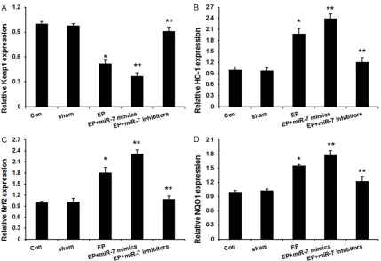
[image:4.612.91.523.71.212.2]Keap1 was reported to involve the regulation of the nuclear factor erythroid 2-related factor 2 (Nrf2) activity [14]. Activated Nrf2 translocates from the cytoplasm to the nucleus, and sequen-tially binds to ARE, which subsquently regulates the expression of a group of cytoprotective enzymes, such as heme oxygenase-1 (HO-1) and NAD(P)H: quinone oxidoreductase-1 (NQ- O1). These Nrf2-dependent gene products go on to protect the cells from oxidative or xenobi-otic damage [11, 12]. Here, the expression of Nrf2, HO-1 and NQO1 mRNA in hippocampus was studied by real-time fluorescence quantita-tive PCR. As shown inFigure 3, Keap1 expres-sion was reduced and Nrf2, HO-1 and NQO1
mRNA expression were increased in kindling groups, which was further induced by miR-7 mimics and reversed by miR-7 inhibitors. Discussion
Epilepsy remains a major medical problem for which there is no effective treatment. Molecular signaling pathways that involves in epilepsy pathogenesis identify new therapeutic targets or biomarkers of intractable epilepsy. In this study, we report a higher level of miR-7 in the hippocampus of kindled rats compared with control and sham group. Moreover, Nrf2 and its two prominent transcriptional targets (HO-1 and GCLM) were significantly upregulated by miR-7 mimics and downregulated by miR-7 in- hibitors, suggesting that miR-7 activates the Nrf2 pathway. In this study, we report the novel function of miR-7 involving protection from epi-leptic seizures via regulating the Nrf2 signaling pathway by targeting the Keap1 mRNA.
MicroRNA (miRNA), an abundant group of en- dogenous non-coding single strand RNAs wi- th approximately 19~25 nucleotides, regulates
[image:5.612.93.522.70.364.2]the expression of genes at post-transcriptional level by translational repression or degradation of target mRNA. Previous genome-wide microR-NA profiling study revealed that various miRmicroR-NAs were deregulated in temporal neocortex of patients with medically intractable temporal lobe epilepsy compared with control subjects [15-17]. MiR-7 has been reported as tumor sup-pressors in various kinds of cancer [18-21]. Recently, Sun et al. revealed miR-7 as potential clinical biomarker for schizophrenia [22]. Ho- wever, the role of miR-7 in epilepsy remains unclear. In our study, we found that the expres-sion of miR-7 was up-regulated after seizure. Moreover, Keap1 expression was down-regulat-ed after seizure and luciferase reporter assay revealed that Keap1 is a direct target of miR-7. Oxidative stress has been reported as an un- derlying mechanism in the epilepsy pathogen-esis, what’s more, excessive oxidative stress contributes to neuronal degeneration in the epileptic focus [23]. Increases in reactive oxy-gen species occur in response to sustained neuronal electrical activity and seizures, indi-cating as the therapeutic strategies for the
stantly polyubiquitinated by CuI3-containing E3 ubiquitin ligase complex and targeted for deg-radation via proteasome
Pathway [26]. In addition, miR-200a has been shown to target Keap1 mRNA in the human breast cancer cell lines, leading to increased Nrf2 activation [14, 27]. In this study, Keap1 expression was reduced and Nrf2, HO-1 and NQO1 mRNA expression were increased in kin-dling groups, which was further induced by miR-7 mimics and reversed by miR-7 inhibitors. These results indicated that miR-7 regulated Nrf2 signal pathway in the epilepsy pathoge- nesis.
In conclusion, our current study demonstrates a novel mechanism by which miR-7 exerts its protective effect against epileptic seizure-in- duced brain damage through targeting Keap1 mRNA and activating Nrf2 pathway.
Disclosure of conflict of interest None.
Address correspondence to: Dr. Dong Zhou, De- partment of Neurology, West China Hospital, Si- chuan University, 37 Guoxue Road, Chengdu 610- 041, Sichuan, People’s Republic of China. E-mail: [email protected]
References
[1] Rowles J and Olsen M. Perspectives on the de-velopment of antioxidant antiepileptogenic agents. Mini Rev Med Chem 2012; 12: 1015-1027.
[2] Farooqui AA, Yi Ong W, Lu XR, Halliwell B and Horrocks LA. Neurochemical consequences of kainate-induced toxicity in brain: involvement of arachidonic acid release and prevention of toxicity by phospholipase A(2) inhibitors. Brain Res Brain Res Rev 2001; 38: 61-78.
[3] Agostini M, Tucci P, Steinert JR, Shalom-Feuer-stein R, Rouleau M, Aberdam D, Forsythe ID, Young KW, Ventura A, Concepcion CP, Han YC,
sociated microRNA, in experimental and hu-man temporal lobe epilepsy. Eur J Neurosci 2010; 31: 1100-1107.
[6] Alsharafi W and Xiao B. Dynamic Expression of MicroRNAs (183, 135a, 125b, 128, 30c and 27a) in the Rat Pilocarpine Model and Tempo-ral Lobe Epilepsy Patients. CNS Neurol Disord Drug Targets 2015; 14: 1096-1102.
[7] Wang XM, Jia RH, Wei D, Cui WY and Jiang W. MiR-134 blockade prevents status epilepticus like-activity and is neuroprotective in cultured hippocampal neurons. Neurosci Lett 2014; 572: 20-25.
[8] Junn E, Lee KW, Jeong BS, Chan TW, Im JY and Mouradian MM. Repression of alpha-synuclein expression and toxicity by microRNA-7. Proc Natl Acad Sci U S A 2009; 106: 13052-13057. [9] Choi DC, Chae YJ, Kabaria S, Chaudhuri AD,
Jain MR, Li H, Mouradian MM and Junn E. Mi-croRNA-7 protects against 1-methyl-4-phenyl-pyridinium-induced cell death by targeting RelA. J Neurosci 2014; 34: 12725-12737. [10] Chaudhuri AD, Kabaria S, Choi DC, Mouradian
MM and Junn E. MicroRNA-7 Promotes Glycoly-sis to Protect against 1-Methyl-4-phenylpyridin-ium-induced Cell Death. J Biol Chem 2015; 290: 12425-12434.
[11] Wang W, Wu Y, Zhang G, Fang H, Wang H, Zang H, Xie T and Wang W. Activation of Nrf2-ARE signal pathway protects the brain from dam-age induced by epileptic seizure. Brain Res 2014; 1544: 54-61.
[12] Wang W, Wang WP, Zhang GL, Wu YF, Xie T, Kan MC, Fang HB and Wang HC. Activation of Nrf2-ARE signal pathway in hippocampus of amyg-dala kindling rats. Neurosci Lett 2013; 543: 58-63.
[13] Liu Z, Yin X, Liu L, Tao H, Zhou H, Ma G, Cui L, Li Y, Zhang S, Xu Z, Yao L, Cai Z, Zhao B and Li K. Association of KEAP1 and NFE2L2 polymor-phisms with temporal lobe epilepsy and drug resistant epilepsy. Gene 2015; 571: 231-236. [14] Eades G, Yang M, Yao Y, Zhang Y and Zhou Q.
miR-200a regulates Nrf2 activation by target-ing Keap1 mRNA in breast cancer cells. J Biol Chem 2011; 286: 40725-40733.
and hypoxia-inducible factor-1 alpha in brain tissue of patients with intractable epilepsy. Int J Neurosci 2015; 1-9.
[16] Henshall DC. MicroRNA and epilepsy: profiling, functions and potential clinical applications. Curr Opin Neurol 2014; 27: 199-205.
[17] Henshall DC. MicroRNAs in the pathophysiolo-gy and treatment of status epilepticus. Front Mol Neurosci 2013; 6: 37.
[18] Liu H, Wu X, Huang J, Peng J and Guo L. miR-7 modulates chemoresistance of small cell lung cancer by repressing MRP1/ABCC1. Int J Exp Pathol 2015; 96: 240-247.
[19] Li YJ, Wang CH, Zhou Y, Liao ZY, Zhu SF, Hu Y, Chen C, Luo JM, Wen ZK and Xu L. TLR9 signal-ing repressed tumor suppressor miR-7 expres-sion through up-regulation of HuR in human lung cancer cells. Cancer Cell Int 2013; 13: 90. [20] Hansen TB, Kjems J and Damgaard CK. Circu-lar RNA and miR-7 in cancer. Cancer Res 2013; 73: 5609-5612.
[21] Okuda H, Xing F, Pandey PR, Sharma S, Wa-tabe M, Pai SK, Mo YY, Iiizumi-Gairani M, Hirota S, Liu Y, Wu K, Pochampally R and Watabe K. miR-7 suppresses brain metastasis of breast cancer stem-like cells by modulating KLF4. Cancer Res 2013; 73: 1434-1444.
[22] Sun XY, Zhang J, Niu W, Guo W, Song HT, Li HY, Fan HM, Zhao L, Zhong AF, Dai YH, Guo ZM, Zhang LY, Lu J and Zhang QL. A preliminary analysis of microRNA as potential clinical bio-marker for schizophrenia. Am J Med Genet B Neuropsychiatr Genet 2015; 168B: 170-178.
[23] Shin EJ, Jeong JH, Chung YH, Kim WK, Ko KH, Bach JH, Hong JS, Yoneda Y and Kim HC. Role of oxidative stress in epileptic seizures. Neuro-chem Int 2011; 59: 122-137.
[24] Vargas MR and Johnson JA. The Nrf2-ARE cyto-protective pathway in astrocytes. Expert Rev Mol Med 2009; 11: e17.
[25] Gopinath K and Sudhandiran G. Naringin mod-ulates oxidative stress and inflammation in 3-nitropropionic acid-induced neurodegenera-tion through the activaneurodegenera-tion of nuclear factor-erythroid 2-related factor-2 signalling pathway. Neuroscience 2012; 227: 134-143.
[26] Liu GH, Qu J and Shen X. NF-kappaB/p65 an-tagonizes Nrf2-ARE pathway by depriving CBP from Nrf2 and facilitating recruitment of HD- AC3 to MafK. Biochim Biophys Acta 2008; 1783: 713-727.