REVERSE SUTURING TECHNIQUE IN PARTIAL PATELLECTOMY
1
Dr. Suhail Bhat,
1
Consultant Orthopedics Health Services Jammu and Kashmir
2
Junior Resident Postgraduate Department of Orthopedics GMC Jammu
3
DNB Resident Dr.Hardas Singh Orthopaedics Hospital and Superspeciality
ARTICLE INFO ABSTRACT
Fractures of the patella constitute almost 1% of all skeletal
indirect trauma. Most patellar fractures are caused by a combination of direct and indirect forces. The most significant effects of fracture of the patella are loss of continuity of the extensor mechanism of the knee and potential incongruity of the patellofemoral articulation.
treatment of patellar fractures. Accepted methods include a variety of wiring techniques, screw fixation, partial patellectomy, and total patellectomy.
result of partial patellectomy by reverse suturing technique, complications associated with the procedure and restoration of range of motion and function of knee and to evaluate the results clinically regarding pai
procedure.
articular, closed and fresh were included. They were treated by partia technique). The age of patients in this study ranged from 20
patients. Most common cause of fracture in this was direct blow to patella due to fall (62.8%). Duration from injury to surgery
days. Time taken to return to previous level of activity ranged from 8 to 12 weeks. Overall excellent results were obtained in 28 patients and good in 7 patients.
Copyright © 2018, Suhail Bhat et al. This is an open access distribution, and reproduction in any medium, provided
INTRODUCTION
Patella fractures account for approximately 1% of all skeletal fractures (Bostrom, 1992) and are seen frequently in the age range of 20 to 50 years more often in males. The majority of patella fractures occur from direct injuries such as a blow to the patella from a fall, a motor vehicle crash, or some combination of these. Indirect injuries occur from a near fall, a fall from a height, or as a combination injury. The diagnosis of a patella fracture is made by performing a complete history and physical examination, and obtaining appropriate X-ray studies.
the patients present with painful swelling and inability to perform a straight-leg raise (Carson, 1984
(AP), lateral, and sunrise views are routinely used in evaluation of patella fractures (Insall, 1984). Other specialized studies such as arthrography, CT, magnetic resonance imaging
and standard tomography are rarely used for the evaluation of a patella fracture (Weber, 1976; Sanders, 1992 and
1985).
*Corresponding author:Dr. Kanav Mahajan
DNB Resident Dr.Hardas Singh Orthopaedics Hospital and Superspeciality Research Centre, Amritsar, Punjab
DOI: https://doi.org/10.24941/ijcr.31078.06.2018
ISSN: 0975-833X
Article History:
Received 13th March, 2018
Received in revised form 20th April, 2018
Accepted 24th May, 2018
Published online 30th June, 2018
Citation: Dr. Suhail Bhat, Dr. Rahul Mahajan and Dr. Kanav Mahajan
of Current Research, 10, (06), xxxxxxxxxx.
Key words:
Reverse; Suturing;
Partial Patellectomy.
RESEARCH ARTICLE
REVERSE SUTURING TECHNIQUE IN PARTIAL PATELLECTOMY
Suhail Bhat,
2Dr. Rahul Mahajan and *
3Dr. Kanav Mahajan
Consultant Orthopedics Health Services Jammu and Kashmir
Resident Postgraduate Department of Orthopedics GMC Jammu
DNB Resident Dr.Hardas Singh Orthopaedics Hospital and Superspeciality Research Centre, Amritsar, Punjab
ABSTRACT
Fractures of the patella constitute almost 1% of all skeletal injuries[1], resulting from either direct or indirect trauma. Most patellar fractures are caused by a combination of direct and indirect forces. The most significant effects of fracture of the patella are loss of continuity of the extensor mechanism of
knee and potential incongruity of the patellofemoral articulation.
treatment of patellar fractures. Accepted methods include a variety of wiring techniques, screw fixation, partial patellectomy, and total patellectomy. The aim of present study was to analyze the result of partial patellectomy by reverse suturing technique, complications associated with the procedure and restoration of range of motion and function of knee and to evaluate the results clinically regarding pain, activities of daily living, range of motion, power, radiologically regarding failure of procedure. A total of 35 cases of inferior pole patella fracture which were comminuted, extra articular, closed and fresh were included. They were treated by partia
technique). The age of patients in this study ranged from 20-50 yrs. Males formed 68.5% of the patients. Most common cause of fracture in this was direct blow to patella due to fall (62.8%). Duration from injury to surgery was an average of 3.56 days and hospital stay was an average of 4.44 days. Time taken to return to previous level of activity ranged from 8 to 12 weeks. Overall excellent results were obtained in 28 patients and good in 7 patients.
access article distributed under the Creative Commons Attribution the original work is properly cited.
fractures account for approximately 1% of all skeletal and are seen frequently in the age range of 20 to 50 years more often in males. The majority of patella fractures occur from direct injuries such as a blow to the tella from a fall, a motor vehicle crash, or some combination of these. Indirect injuries occur from a near fall, a fall from a height, or as a combination injury. The diagnosis of a patella fracture is made by performing a complete history and physical ray studies. Most of the patients present with painful swelling and inability to , 1984). Anteroposterior (AP), lateral, and sunrise views are routinely used in evaluation . Other specialized studies such as arthrography, CT, magnetic resonance imaging (MRI), and standard tomography are rarely used for the evaluation of a , 1992 and Arnoczkyp,
DNB Resident Dr.Hardas Singh Orthopaedics Hospital and Superspeciality
A basictreatment-directed approach classifies the fracture as either nondisplaced or displaced.
such as transverse, stellate or comminuted, longitudinal or marginal, proximal or distal pole, and osteochondral can be used to classify patella fractures. The
fractures is based on the type of fracture and clinic presentation found on physical examination. The overall goals of patella fracture surgery are to
restore continuity of the extensor mechanism, and reduce complications associated with
1977; Ashby, 1975; Black, 1969; 1983; Bostrom, 1972; Chiroff, 1977; Korner, 1905; Lotke, 1981; Seligo, 1971 and Stern, 1980).
Non-operativetreatment; Tension band wiring techniques; Partial patellectomy; Partial patellectomy combined with tension band wiring andTotal excision or patellectomy. Comminuted patellar fractures in which
thepatella is fragmented, leaving a substantial a
normal proximal fragment are treatedby partial patellectomy with repair of the patella tendon using heavy Mersilene or Ethibond (Bostrom, 1972; Depalma
Siwek, 1981; Pritchett, 1997; Nathan International Journal of Current Research
Vol. 10, Issue, 06, pp.70654-70658, June, 2018
Suhail Bhat, Dr. Rahul Mahajan and Dr. Kanav Mahajan. 2018. “Reverse suturing technique in partial patellectomy
Available online at http://www.journalcra.com
REVERSE SUTURING TECHNIQUE IN PARTIAL PATELLECTOMY
Kanav Mahajan
Consultant Orthopedics Health Services Jammu and Kashmir
Resident Postgraduate Department of Orthopedics GMC Jammu
Research Centre, Amritsar, Punjab
injuries[1], resulting from either direct or indirect trauma. Most patellar fractures are caused by a combination of direct and indirect forces. The most significant effects of fracture of the patella are loss of continuity of the extensor mechanism of knee and potential incongruity of the patellofemoral articulation. Opinions differ as to the optimal treatment of patellar fractures. Accepted methods include a variety of wiring techniques, screw e aim of present study was to analyze the result of partial patellectomy by reverse suturing technique, complications associated with the procedure and restoration of range of motion and function of knee and to evaluate the results clinically n, activities of daily living, range of motion, power, radiologically regarding failure of A total of 35 cases of inferior pole patella fracture which were comminuted, extra-articular, closed and fresh were included. They were treated by partial patellectomy (reverse suturing
50 yrs. Males formed 68.5% of the patients. Most common cause of fracture in this was direct blow to patella due to fall (62.8%). was an average of 3.56 days and hospital stay was an average of 4.44 days. Time taken to return to previous level of activity ranged from 8 to 12 weeks. Overall excellent
License, which permits unrestricted use,
directed approach classifies the fracture as either nondisplaced or displaced. Descriptive geometric terms such as transverse, stellate or comminuted, longitudinal or proximal or distal pole, and osteochondral can be used to classify patella fractures. The treatment of patella fractures is based on the type of fracture and clinical physical examination. The overall goals urgery are to preserve patella function, restore continuity of the extensor mechanism, and reduce an articular fracture (Andrews, , 1969; Bostman, 1981; Bostman, , 1977; Dowd, 1982; Insall, 1972; , 1981; Noble, 1984; Nummi, 1971; ). Treatment options include the Tension band wiring techniques; Partial patellectomy; Partial patellectomy combined with tension band wiring andTotal excision or patellectomy. Comminuted patellar fractures in which only the distal pole of thepatella is fragmented, leaving a substantial and relatively normal proximal fragment are treatedby partial patellectomy with repair of the patella tendon using heavy Mersilene or Depalma, 1958; Reider, 1981; Nathan, 2011 and Klassen, 1997).
INTERNATIONAL JOURNAL OF CURRENT RESEARCH
Conventional method of partial patellectomy involves drilling three parallel holes just anterior to thearticular surface of proximal fragment using 2mm K-wire or 2.5mm drill bit from distal to proximaldirection. two heavy nonabsorbable sutures through the patellar tendon, one through themedial and one through the lateral half of the tendon using a suture passer placing one suture end eachthrough the medial and lateral holes
and two through the central hole.With the knee
slightlyhyperextended, tie the sutures securely over the superior pole of the patella (Depalma, 1958; Reider, 1981; Siwek, 1981; Pritchett, 1997; Nathan, 2011; Klassen, 1997; Anderson, 1971; Magnuson, 1933; Payr, 1917; Miskew, 1980; Muller Me, 1979; Mcgreal, 1999; Lotke, 1981; John, 2007; Gosal, 2001; Gardner, 2005 Baran, 2009; Benjamin, 1987; Burvant, 1994; Carpenter, 1997; Chen, 1998; Scilaris, 1998; Weber, 1980; Schauwecker, 1974; Wright, 2009 and Wu, 2001). Reverse suturing technique used in our study involves minor modifications of conventional techniquewith regard to use of suture passer and order of weaving of patellar tendon.
MATERIAL AND METHODS
This prospective study was conducted in the Post Graduate Department of Orthopedics Government Medical College, Jammu during the period from 1st August 2014 to 31st July 2015 with a mean follow up of 1 year.Both male and female patients were included in the study.
Inclusion Criteria: Age group (20 to 50 yrs), Distal fragment
comminution, closed fractures, fresh injuries(less than 7 days), isolated fractures, negative SLR test.
Exclusion Criteria: Proximal fragment comminution, Multiple
injured patients, associated neurovascular injuries, open fractures, cancer or severely ill patients which increases the operation morbidity, patients below 20 and above 50 years, fracture more than 7 days old, pregnancy. All the patients were initially assessed in the emergency section of GMC Jammu. They were given first aid in the form of analgesia, long leg splint, and other resuscitation measures. After selection of the patients for surgery, patients were prepared for elective surgery to be conducted in the elective operation theatre.
Pre-operative evaluation: Pre-operative evaluation included
patients name, age, sex, address, date of injury, associated chronic illness, date of surgery and date of discharge. Every patient was evaluated for swelling, bruising & ecchymosis at the fracture site and visible deformity of the knee joint. Straight leg raising test was done to evaluate the extensor mechanism of the involved knee. A careful neurological and vascular examination of the involved limb was done. All the routine investigations like complete blood count & biochemistry were done. Radiographic evaluation by X-ray of the chest, knee AP & Lat view was done in every patient. Informed and written consent was taken from the patients
Implants: Ethibond suture no.5, 2mm K-wires, 2.5mm drill
bit.
Operative Technique: The patient was positioned in supine
position with a bolster under the knee to keep it in about 30 degrees of flexion. Knee and involved limb were draped under all aseptic conditions. A midline longitudinal skin incision was made centered over the patella. Skin and subcutaneous tissue were reflected medially and laterally to expose the entire
anterior surface of the patella and the quadriceps and patellar tendons. Preserving the intact proximal third of the patella, comminuted distal fragments were removed, joint was cleared of loose segments of bones and c artilages.Small flecks of bone were left within the patellar tendon to make anchorage easier. Articular edge of the proximal fragment was trimmed, and smoothened with a rasp.Beginning on the fracture surface of the proximal fragment just anterior to the articular cartilage, a 2-mm Kirschner wire or 2.5-mm drill bit was used to drill three parallel holes in a proximal direction (one hole in the center and one each in the medial and lateral thirds) (Zhao, 1999; Marder, 1993; Mishra, 1971; Kaufer, 1971; Duthie, 1958; Albanese, 1992). Reverse Suturing Technique used in our study involves the following modifications as compared to conventional technique.
Straightening the Ethibond suture needle with two
needle holders.
Insertion of straightened needle through the lateral hole from distal to proximal with rear end of needle facing upwards (reverse), holding the needle near the tip with a needle holder.
Keeping the needle held within the hole pull out the thread from the proximal end,then remove the needle from the distal hole.
Insertion of same needle through central hole from distal to proximal in conventional manner.
Repeating the above technique through the medial hole
with another ethibond suture needle.
The patellar tendon was then sutured with the patella with the above ethibonds at the front upper surface of patella on a fully extended knee.
The stability of this system was checked at 30º, 60º and
90º of flexion.
Finally, the medial and lateral ratinaculla of patella and capsule were sutured and the joint was closed.
Light immobilization was provided with Robert-Jones
bandage.
Follow-up: All patients were encouraged to engage in
Figure 1. Showing Reverse Suturing Technique
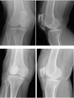
Figure 2. Fracture lower pole of patella and partial patellectomy done Via Reverse Suturing Technique
Muscle atrophy was observed with measurement of the perimeter of the quadriceps muscles using the healthy muscle as a comparison.
RESULTS
Subjective evaluation: Using the Cincinnati evaluation scale
in the 3 months re-evaluation, 15 patients showed excellent results (42.9%), 8 showed good results (22.9%), 8 showed moderate results (22.9%), and 4 showed poor results (11.4%).These results showed a significant improvement at 6 months. At 6 months reevaluation 21 patients (60%) reported knee discomfort, 17 of which after medium exertion, 4 with continuous pain and reduction of activity. 6 patients (17.1%)
complained of knee swelling and a certain amount of movement restriction while working, taking part in sports, or during everyday activities. At the last follow-up examination, 6 patients (17.1%) complained of a “giving way” sensation.
Objective evaluation: Range of Motion (ROM): For 28
patients ROM was above 90% of the norm at a very early stage (3 months examination). This resulted till the end of the evaluation period. For 5 patients, the final ROM was 80-90% and for 2 patient less than 70%, who also had a 10% loss of extension and an obvious limp.
Muscle atrophy: 7 patients had quadriceps atrophy at 1 and 3
months re-evaluation.
Grind Test: From the total of 35 patients that were examined,
16 patients (45.7%) showed a positive patella pressure point.
Complications: In the immediate complications were included
7 superficial infections, which were treated successfully with antibiotics.
DISCUSSION
The reduction of muscle strength in total patellectomy reaches 50% (Watkins, Harris, Wender, Zarins& Rowe, 1983), whereas only 15% in partial patellectomy (Saltzman, Goulet, McClellan, Schneider & Matthews, 1990). For the development of osteoarthritic changes, the results of our series are comparable to those of Saltzman et al. (1990) in which, nevertheless, the early mobilization was not introduced. It would seem that early mobilization does not play a role in producing osteoarthritic changes. Probably, the nature and position of cartilage is a significant factor in the development of secondary changes (Hung et al., 1993). When appropriate selection criteria are utilized, partial patellectomy can yield functional outcomes that are equivalent to open reduction and internal fixation. Multiple authors have reported nearly normal functional outcome when large fragments of the patella and articular congruity are preserved. Retention of small, nonviable fracture fragments or those devoid of caritlage did not improve function, while retention of large fragments provided a lever arm for improved extensor mechanism function. With extensive inferior pole comminution, superior results have been reported with partial patellectomy compared to internal fixation. Bostrom reported 88% good to excellent results with partial patellectomy for transverse patellar fracture with inferior pole comminution, compared to only 74% good to excellent results with internal fixation.
Conclusion
In conclusion, if the above conditions, in relation to the large abutment size, placement and anterior tendon placement on the abutment are met, then partial patellectomy gives satisfactory results. Otherwise an attempt of osteosynthesis should be made, maintaining the patella. In any case, disorders of the articular surface. And this reverse suturing technique in partial patellectomy can be used when curved guide wire is not available without any difficulty.
REFERENCES
Albanese SA, Liivermore JT, Werner FW, et al. 1992. Knee Extensor mechanics after subtotal excision of the patella.
[image:3.595.40.290.263.592.2]Anderson LD. 1971. Campbell’s Operative Orthopaedics. 5th ed. St. Louis, MO: Mosby.
Andrews JR, Hughston JC. 1977. Ttreatment of patellar fractures by partial patellectomy. South Med J., 70:8809-813.
Arnoczkyp, S.P. 1985. Blood supply to the anterior cruciate
ligament and supporting structures. OrthopClin North Am;
16 (1): 15-28.
Ashby M, Shields C, Karmy J. 1975. Diagnosis of osteochondral fractures in acute traumatic patellar dislocations using air arthrography. J Trauma. 15:1032-1033.
Baran O, Manisali M, Cecen B. 2009. Anatomical and biomechanical evaluation of the tension band technique in patellar fractures. IntOrthop., 33:1113-1117.
Benjamin, J, Bried J, Dohm M. 1987. Biomechanical evaluation of various forms of fixation with transverse patella fractures. J Orthop Trauma., 1:219-222.
Black JK, Conners JJ. 1969. Vertical fractures of the patella.
South Med J., 62:1137-1140.
Bostman O, Kiviluoto O, Nirhamo J. 1981.Comminuted displaced fractures of patella. Injury. 13:196-202.
Bostman O, Kiviluoto O, Santavirta S, et al. 1983. Fractures of patella treated by operation. Arch Orthop Trauma Surg.
102:78-81.
Bostrom A 19772. Fracture of the patella. A study of 422 patellar fractures. Actaorthop Scand Suppl; 143:1-80. Bostrom A. 1972. Fracture of patella. Astudy of 422 patellar
fractures. Acta Orthop Scand. 143(suppl):1-80.
Burvant, J., Thomas, K., Alexander, R., et al. 1994. Evaluation of methods of internal fixation of transverse patella
fractures: A biomechanical study. J Orthop Trauma.
8:147-153.
Carpenter J, Kasman R, Patel N, et al. Biomechanical
evaluation of current patella fracture fixation techniques. J
Orthop Trauma. 1997;11:351-356.
Carson, W.G., Jr., James, S.L., Larson, R.L., et al. 1984. Patellofemoral disorders: physical and radiographic
eevaluation. Part II: Radiographic examination.
ClinOrthop; (185): 178-186.
Chen, A., Hou, C., Bao, J, et al. 1998. Comparison of biodegradable and metallic tension-band fixation for patella fractures. 38 patients followed for 2 years.
ActaOrthop Scand., 69:39-42.
Chiroff, R.T. 1977. A new technique for the treatment of comminuted, transverse fractures of patella. SurgGynaecol
Obstet., 145(6):909-912.
Depalma A, 1958. Flynn J. Joint changes following partial and total patellectomy. J Bone Joint Surg Am. 440-A:395-4113.
Dowd GS. 1982. Marginal fractures of patella. Injury. 14:287-291.
Duthie, H, Hutchinson, J. 1958. The results of partial and total excision of the patella. J Bone Joint Surg Br., 40-B:75-81.
Gardner MJ, Griffith MH, Lawrence BD, et al. 2005.
Complete exposure of articular surface for fixation of patellar fractures. J Orthop Trauma., 19:118-123.
Gosal H, singh P, Field RE. 2001. Clinical experience of patellar fracture fixation uusing metal wire or non-absorbable polyester- a study of 37 cases. Injury. 32:129-135.
Insall J, Goldberg V, Salvati E. 1972. Recurrent dislocation and high-riding patella. ClinOrthopRelat Res. 88:67-69. Insall, J.N. 1984. Anatomy of the knee. Surgery of the knee.
New York: Churchill-Livingstone: 1-20.
John J, Wagneer WW, Kuiper JH. 2007. Tension- band wiring oh transverse fractures of the patella. The effect of site of wire twists and orientation of stainless steel wire loop: A biomechanical investigation. IntOrthop. 31:703-707.
Kaufer H. 1971. Mechanical Function of patella. J Bone Joint
Surg Am., 53:1551-1560.
Klassen J, Trousdale R. 1997. Treatment of delayed and nonunion of patella. J Orthop Trauma. 11:188-194. Korner M. 1907. Ein fall von flachenfractur und luxation dur
patella. Deutsche MedizinischeWonchenschrift., 31:996. Lotke PA, Ecker ML. 1981. Transverse fractures of the patella.
ClinOrthopRelat Res., 158:180-184.
Lotke PA, Ecker ML. 1981. Transverse fractures of the patella.
ClinOrthopRelat Res. 158:180-184.
Magnuson P. 1933. Fracture. 2nd ed. Philadelphia, PA: JB Lippincott.
Marder R, Swanson T, Sharkey N, et al. 1993. Effeects of partial patellectomy and reattachment of the patellar tendon on patellofemoral areas and pressures. J Bone Joint
Surg Am., 75-A35-45.
Mcgreal G, Reidy D, Joy A, et al. 1999. The biomechanical evaluation of polyester as atension band for the internal fixation of patellar fractures. J Med Eng Technol.,
23;553-56.
Mishra U. 1972. Late results of patellectomy in fractured patella. ActaOrthop Scand. 433:2256-263.
Miskew, D.B.W., Pearson, R.L., Pankovich, A.M. 1980. Mersilene strip suture in repir of disruptions of the quadriceps and patellar tendons. J Trauma. 20:867-872.
Muller Me, Allgower M, Schneiider R, et al. 1979. Manual of
Internal Fixation. Techniques Recommended by AO Group. Berlin: Springer-Verlag; 248-253.
Nathan ST, Fisher BE, Roberts CS, et al. 2011. The
management of nonunion and delayed union of patella fractures: A systemic review of literature. Int Orthop. 35:791-795.
Noble H, Hakek M. 1984. Boutonniere-type deformity of the knee following patellectomy and manipulations. J Bone
Joint Surg Am., 66-A:137-138.
Nummi J. 1971. Operative treatment of patellar fractures.
ActaOrthop Scand.,42:437-438.
Payr E. 1917. Zur operative behandlung der
kiegelenksteifenachlangdauernderruhigstellung.
ZentralblChir. 44:809.
Pritchett JW. 1997. Nonoperative treatment of widely displaced patella fractures. Am J Kneee Surg. 10:145-147. Reider B, Marshall J, Koslin B, et al. 1981. The anterior aspect
of knee joint. J Bone Joint Surg Am. 19:509-514.
Sanders R 1992. Patella fractures and extensor mechanism injuries. In: Browner BD, Jupiter JB, Levine AM, et al., editors. Skeletal Trauma. Philadelphia: W.B. Saunders Co.: 1685-1710.
Schauwecker R. 1974. The Practice of Osteosynthesis.
Stuttgart, Germany: George Thieme.
Scilaris T, Grantham J, Prayson M, et al. 1998. Biomechanical comparison of fixation methods in transverse patella fracture. J Orthop Trauma., 12:356-359.
Seligo W. 1971. Fractures of the patella.
ReconstrSurgTraumatol. 12:84-102.
Siwek K, Rao JP. 1981. Ruptures of the extensor mechanism of the knee joint. J Bppne Joint Surg Am. 63:932-937. Stern R, Harwin S. 1980. Spontaneous and simultaneous
rupture of both quadriceps tendons. ClinOrthopRelat Res.,
Weber BG, Chech O 1976. Pseudoarthrosis. New York: Grune& Stratton: 224-225.
Weber M, Janecki C, Macleod P, et al. 1980. Efficacy of various forms of fixation of transverse fractures of patella.
J Bone Joint Surg Am. 62-A:215-220.
Wright PB, Kosmopoulos V, Cote RE, ett al. 2009. Fiberwire is superior in strength to stainless steel wire for tension band fixation of transverse patellar fractures. Injury.
40:1200-1203.
Wu CC, Tai CL, Chen WJ. 2001. Patellar tension band wiring: A revised technique. Arch Orthop Trauma Surg. 121:12-16.
Yu JS, Petersilge C, Sartoris DJ, et al. 1994. MR imaging of
injuries of extensor mechanism of the knee.
Radiographics. 14:541-551.
Zhao J, Wu X, Peng X. 1999. Biomechanical experiment and clinical report of modified patellectomy for polar fractures of the patella. Chin J Trauma., 2:122-124.