Original Article
Expression and function of IgSF21 in amacrine cells in a
diabetic retinopathy model
Xiaohui Lin1, Jihong Wang2, Yingying Li1, Xiaocheng Ma1, Zhen Li1, Yihui Zhang1
1Department of Ophthalmology, The Inner Mongolia Autonomous Region People’s Hospital, Hohhot 010017, Inner Mongolia Autonomous Region, China; 2The Second Department of Hand and Foot Microsurgery, The Second Affili-ated Hospital of Inner Mongolia Medical University, Hohhot, Inner Mongolia Autonomous Region, China
Received February 13, 2018; Accepted March 24, 2018; Epub May 15, 2018; Published May 30, 2018
Abstract: Objective: Our aim was to study the role and mechanism of immunoglobulin superfamily member 21 (IgSF21) expression in diabetic retinopathy (DR). Methods: Sprague-Dawley rats were injected with streptozotocin to create the diabetic model. Rats with diabetes were divided into three groups based on their feeding periods: group A (fed for 8 weeks), group B (fed for 12 weeks), and group C (fed for 16 weeks). Levels of IgSF21 mRNA and protein in rat retinal tissues from these groups were measured by quantitative reverse transcription PCR and Western blot. Amacrine cells were isolated from rat eyeballs and miniature inhibitory postsynaptic currents (mIPSCs) were detect-ed using patch clamp technique. Plasmid expressing IgSF21 was constructdetect-ed and transfectdetect-ed into amacrine cells. Co-localization of IgSF21 and neurexin2α and the interaction between these two proteins were analyzed through immunofluorescence (IF) and co-immunoprecipitation (co-IP). The impact of IgSF21 overexpression on mIPSCs in amacrine cells was investigated. Results: As compared with control group, expressions of IgSF21 mRNA in rats with DR in groups A, B and C, as well as expressions of IgSF21 protein in groups B and C were all reduced. Furthermore, the frequencies of mIPSCs in gamma-aminobutyric acid-ergic (GABAergic) amacrine cells in all three groups also decreased, with reduction in group C the greatest (all P<0.05). Through IF assay, it was found that IgSF21 and neurexin2α proteins were co-localized. The co-IP was conducted and direct interaction between these two proteins was detected. Using a patch clamp technique, it was also found that both frequency and current amplitude of mIP-SCs rose when IgSF21 was overexpressed (P<0.05). Conclusion: IgSF21 can act on neurexin2α directly and affect the function of GABAergic amacrine cells while reduction in IgSF21 levels may accelerate progression of DR.
Keywords: Immunoglobulin superfamily member 21, diabetic retinopathy, mechanism research
Introduction
Diabetic retinopathy (DR) is a common compli-cation in patients with diabetes which, in some cases, could even cause blindness. Incidence of DR rises as the course of diabetes prolongs [1]. One of the main manifestations of DR is increase in retinal microvascular permeability which can lead to capillary basal lamina thick-ening and endothelial cell division abnormality, causing loss of transport function in capillaries. The pathogenesis of DR is quite complicated. Some scholars believe that the imbalance between generation and degradation of free radicals within the body can increase the level of reactive oxygen species, disrupt relevant sig-naling pathways, and as a result, affect gene transcription of key proteins in the nucleus [2,
aggrava-neurotransmission [7]. However, it is still un- clear which component regulates neurexin2α. Proteins in the immunoglobulin superfamily (IgSF) have structures similar to that of an anti-body, which can regulate signaling pathways, adjust immune function, and mediate adhesion of lymphocytes to target cells. Immunoglobulin superfamily member 21 (IgSF21) belongs to this group. It can promote the formation of syn-apses and helps with maintenance of neuronal function [8]. At present, mechanisms of IgSF21 and neurexin2α are still unclear. Therefore, our present study investigated expression of IgSF21 in amacrine cells in DR rat model as well as interaction between IgSF21 and neurexin2α, in an effort to obtain some useful information for treating this disease.
Materials and methods
Animal subjects
A total of 80 two-month-old male Sprague-Dawley rats were chosen as subjects (clean grade, 170 ± 25 g, purchased from Anpel Laboratory Technologies (Shanghai) Inc., app- roval No. SYXK (Hu) 2005-0001). Ten rats were fed with a normal diet as control whereas the remaining 70 rats were used to create a dia-betic rat model by receiving a single-dose intra-peritoneal injection of 2% streptozotocin (STZ, diluted in citrate buffer solution, pH=4.5, dos-age: 50 mg/kg) after 3 days of adaptive feed-ing. Rats in the control group were injected with the same volume of normal saline. Blood sugar levels were tested after two days. The model was created successfully if the blood sugar content was above 16.9 mmol/L. After that, rats with DR (symptoms included fundus hem-orrhage and edema) were picked at 8th, 12th and 16th week, respectively, and were assigned into group A, B and C (ten rats in each group). Main reagents and instruments
Main reagents, instruments, and supplier infor-mation were as follows: STZ (Merck, Germany),
Yanjin Biotech Co., Ltd), versatile Arktik PCR device (Thermo, USA), electrophoresis instru-ment (model DYY-12, Beijing Liuyi Biotech Co., Ltd), inverted microscope (model IX51, Olym- pus, Japan), patch-clamp system (Nanion, Germany), confocal fluorescence microscope (3I, USA), and pCMV-Myc plasmid and compe-tent Escherichia coli (E.coli) DH5α (kept in our lab).
Methods
Isolation and culture of amacrine cells: Normal rats and rats with DR were sacrificed by cervical dislocation after anesthesia. The eyeballs were taken out and fasciae around the eyes were re-moved. Under the dissecting microscope, crys-talline lenses were resected and retinal neuro-epithelial layers were stripped. Samples were placed in D-Hank’s solution containing 12 mM HEPES and then put in incubator for trypsin-ization at 37°C for 10 minutes. FBS was then added at 0.5 mL to cease trypsinization. Super-natant was discarded after centrifugation and Dulbecco’s modified eagle medium (DMEM) was added to resuspend retinal cells. Next, some retinal cells were moved to culture plates containing DMEM, with 5-bromo-2’-deoxyuri-dine added in the meantime to inhibit growth of non-neural cells. Samples were incubated at 37°C in an atmosphere of 5% CO2 for 72 hours and were later placed under phase contrast microscope for observing growth of GABAergic amacrine cells. Cell somata were generally over 10 μm in diameter with many slender process-es, which intersected with those in adjacent cells to form a network [9].
clamp was used to set voltage at -70 mV. The whole cell membrane current and fast capaci-tance compensation were recorded. Sampling and filter frequencies in analogue signal were set at 5 kHz and 1 kHz, respectively. All experi-ments were conducted at room temperature.
Detection of expression of IgSF21 mRNA by PCR: Fifty grams of retinal tissues were ground-ed in liquid nitrogen. Total RNA was extractground-ed by TRIzol and checked for quality. After reverse transcription, samples were kept at -20°C. The level of IgSF21 mRNA was detected by SYBR Green quantitative fluorescence PCR. Gene for glyceraldehyde-3-phosphate dehydrogena-se (GAPDH) was udehydrogena-sed as internal control and the primer was designed by Primer 5.0. The IgSF21 forward primer was 5’-CTAAGCTCCAG- CTACTGC-3’, while the reverse primer was 5’-CATGACTGCATAACGCTGAC-3’. GAPDH for-ward primer was 5’-CATACCATTGACTACACTG- AC-3’, and the reverse primer was 5’-CATC- AGACCTACTACGACTC-3’. PCR running param-eters were set as follows: 94°C 6 min, 94°C 20 s, 56°C 25 s and 72°C 40 s for 32 cycles, fol-lowed by 72°C 3 min at the end. Each reaction was done in triplicate. Relative gene expres-
sion of IgSF21 was calculated through 2-ΔΔCt method.
Western blot: Retinal tissue (80 mg) was taken and added with 2 mL tissue lysate for protein ex-traction. Protein concentration was measured by BCA method. Protein samples were loaded into wells (25 μg per well) and separated by 12% SDS-PAGE. Afterward, proteins were trans-ferred from gel to polyvinylidene fluoride mem-brane by tankblot. The memmem-brane was then blocked with tris buffered saline with tween 20 (TBST) containing 5% skim milk powder for 30 minutes. Goat anti-mouse IgSF21 primary antibody was then added and the sample was incubated at 4°C for 8 hours, followed by the addition of secondary antibody conjugat- ed to horseradish peroxidase (HRP) and a 2-hour incubation at room temperature. Sa- mples were washed in TBST solution three times (5 min/per wash) and treated with ECL. After being photographed, the relative level of protein expression was analyzed with Gel Pro software.
[image:3.612.95.517.70.327.2]Construction of eukaryotic expression plasmids pCMV-Myc-IgSF21 and pCMV-HA-neurexin2α:
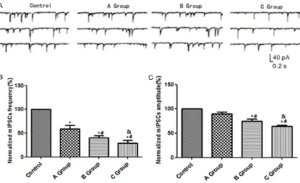
Figure 1. Changes in mIPSCs in GABAergic amacrine cells in rats with DR. A: Levels of mIPSCs measured by patch clamp technique in different groups; B: Histogram of mIPSCs frequencies in different groups; C: Histogram of cur-rent amplitudes of mIPSCS in diffecur-rent groups; *P<0.05 vs. control group, #P<0.05 vs. group A, &P<0.05 vs. group
Amacrine cells were lysed by TRIzol reagent and total RNA was extracted for reverse tran-scription after quality check. Gene primers for IgSF21 and neurexin2α were designed by Prime 5.0. Meanwhile, cutting sites for restric-tion enzymes, BamH1 and EcoR1, were creat-ed. The IgSF21 forward primer was 5’-CGATC- AGTCGCCATGCAT-3’, while the reverse primer was 5’-GTTCAGACGTACTGC-3’; the neurexin2α forward primer was 5’-CAGTCGGATCACTCAGC- 3’, and the reverse primer was 5’-CTGGA-
mids were then extracted, cut by enzyme, and verified by sequencing.
Immunofluorescence (IF) co-localization: GA-BAergic amacrine cells in logarithmic phase were selected. DMEM was renewed one day before transfection and cell density was con-trolled at 5*105/mL. Cells were placed in a 12-well culture plate (0.5 mL per well). Tran- sfection of pCMV-Myc-IgSF21 was performed according to the Lipofectamine-2000 instruc-tion manual. Samples were incubated at 37°C for 72 hours in an atmosphere of 5% CO2, fol-lowed by centrifugation at 1,500 rpm for 10 minutes, and washed in phosphate-buffered saline (PBS) three times. Some cell precipita-tions were taken and spread evenly over cover slips, which were then fixed by 4% formalde-hyde for 20 minutes after getting briefly drying and were washed again in PBS three times. In order to increase membrane permeability, 0.1% Triton was added to cover the cells for 15 minutes. After being blocked for 30 minutes in 10% goat serum, rabbit anti-mouse IgSF21 an-tibody (1:500) and goat anti-mouse neurexin2α antibody were added into the sample for a 6-hour incubation. Afterward, goat anti-mouse secondary antibody (1:500) labeled with fluo-rescein isothiocyanate (FITC) and rabbit anti-goat secondary antibody (1:500) labeled with tetramethylrhodamine (TRITC) were added. Samples were then incubated for another two hours before being stained in 4’, 6-diamidino-2-phenylindole (DAPI) solution for 8 minutes. Next, samples were washed in PBS once and incubated at 37°C for one hour before being washed again in PBS three times. Then, sam-ples were fixed in 70% glycerin and observed under laser scanning confocal microscope.
[image:4.612.91.282.73.481.2]Co-immunoprecipitation (co-IP): Amacrine cells from normal rats were cultured for 24 hours. When cell fusion reached around 60-80%, transfection of eukaryotic expression plasmids pCMV-Myc-IgSF21 and pCMV-HA-neurexin2α was conducted. Transfected amacrine cells were incubated at 37°C for 24 hours, followed
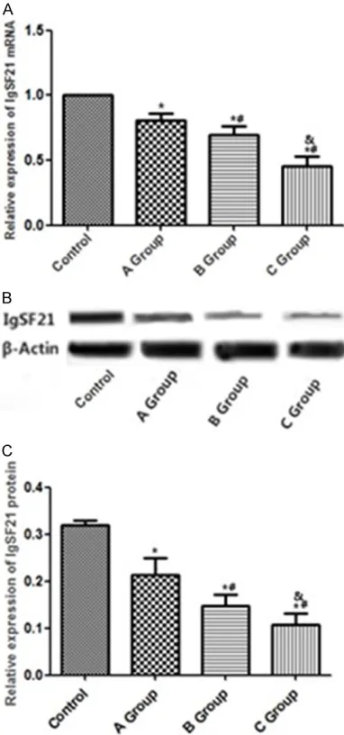
Figure 2. Changes in IgSF21 expression level in rats with DR. A: Levels of IgSF21 mRNA expression in different groups; B: Levels of IgSF21 protein expres-sions in different groups; C: Histogram regarding lev-els of IgSF21 protein expression in different groups;
*P<0.05 vs. control, #P<0.05 vs. group A, &P<0.05 vs.
by centrifugation at 1,500 rpm for 10 minutes. Supernatant was discarded and sample was washed in PBS twice. Next, 1.5 mL pre-cooled radio-immunoprecipitation assay (RIPA) solu-tion was added into the centrifuge tube and tube was placed on ice for 30 minutes. After centrifugation, the supernatant was trans-ferred into Eppendorf tubes labeled No. 1 and No. 2. Anti-IgSF21 and anti-neurexin2α antibodies and immunoglobulin G (IgG) were added, according to the instruction manual. Following a 6-hour agitation, protein A/G was added and samples were being agitated again for 5 hours. After centrifugation, samples were rinsed in RIPA twice and placed in a water bath at 100°C. At the end, Western blot was per-formed to analyze the relative content of pro-tein.
Effects of IgSF21 overexpression on mIPSCs in amacrine cells: GABAergic amacrine cells were extracted from normal rats for culture. Cells in logarithmic phase were selected and cell densi-ty was controlled at 5*105/mL. Cells were plac- ed in a 12-well plate (0.5 mL per well). DMEM was renewed one day before transfection. The transfection of pCMV-Myc-IgSF21 was per-formed according to the instruction manual of Lipofectamine-2000 and samples were incubated at 37°C for 72 hours in an at- mosphere of 5% CO2.Patch clamp technique was employed to measure effects of IgSF- 21 overexpression on mIPSCs in amacrine cells.
Statistical analysis
SPSS 17.0 software was applied for statistical analysis. Measurement data are expressed as mean ± standard deviation. Comparison be- tween two groups was conducted by
indepen-dent-samples t test. Multiple groups were com-pared by one-way analysis of variance and Bonferroni post-hoc test. A value of P<0.05 was considered statistically significant.
Results
Changes in mIPSCs in GABAergic amacrine cells in rats with DR
By using the patch clamp technique, it was found that in comparison with control group, frequencies of mIPSCs in GABAergic amacrine cells in groups A, B and C all declined, with the extent of reduction being proportional to the course of DR (P<0.05). The current amplitude of mIPSCs in groups B and C also decreased (P<0.05). See Figure 1.
Expression of IgSF21 in amacrine cells
Compared with control group, IgSF21 mRNA expression in groups A, B, and C all declined (P<0.05) while the reduction in group C was the greatest (P<0.05). In addition, IgSF21 protein expression in groups B and C also decreased in comparison with control group (P<0.05). See
Figure 2.
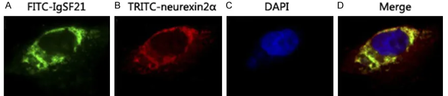
Co-localization of IgSF21 and neurexin2α in amacrine cells
[image:5.612.87.527.73.168.2]Spatial localization of IgSF21 labeled with FITC (green fluorescence) and neurexin2α labeled with TRITC (red fluorescence) were observed through indirect IF. Expressions of the two pro-teins were found in both cell membrane system and cytoplasm. The merged image exhibited relatively strong yellow fluorescence, revealing that these two proteins were close to each other in space and co-localization may exist. See Figure 3.
Interaction between IgSF21 and neurexin2α tested by co-IP
Since co-localization of IgSF21 and neurexin2α in amacrine cells was detected by IF, co-IP was then conducted to check for any direct interac-tion between these two proteins. Co-transfec- tion of pCMV-HA-IgSF21 plasmid labeled with
crine cells from rats with DR, suggesting that the decrease in IgSF21 might be associated with progression of DR. There have been stud-ies that have shown that mIPSCs in poultry can be suppressed when the function of their GABAergic amacrine cells is damaged, thus inducing occurrence of DR [12, 13]. By using the patch clamp technique, it was found that
[image:6.612.92.376.74.119.2]globulin superfamily member 21.
Figure 5. Effects of IgSF21 overexpression on mIPSCS in amacrine cells. A: Levels of mIPSCs in control and transfected groups measured by the patch clamp technique; B: Histogram of mIPSCs frequencies in control and trans-fected groups; C: Histogram of current amplitudes of mIPSCs in control and transfected groups; D: IgSF21 expression levels in the control and trans-fected groups; E: Histogram of IgSF21 expression levels in the control and transfected groups; *P<0.05 vs. control group. mIPCs, miniature inhibitory
postsynaptic currents; IgSF21, immunoglobulin superfamily member 21.
[image:6.612.90.372.198.535.2]ed by Western blot, suggest-ing that neurexin2α could be precipitated by IgSF21. Me- anwhile, Myc tag antibody co- uld co-precipitate neurexin- 2α (labeled with Myc) and HA-IgSF21 was detected by Western blot, indicating that IgSF21 could also be precipi-tated by neurexin2α. See
Figure 4.
Effects of IgSF21 overexpres-sion on mIPSCs in amacrine cells
The patch clamp test showed that in comparison with con-trol, frequencies and current amplitudes of mIPSCs as well as levels of IgSF21 expres-sion in GABAergic amacrine cells in the transfected groups all increased (all P<0.05). See
Figure 5.
Discussion
ama-both frequency and current amplitude of mIP-SCs in GABAergic amacrine cells decreased to some extent, indicating some level of damage in retina amacrine cells. This result was consis-tent with previous findings [14].
Normal neurons can receive both excitatory glutamatergic inputs and inhibitory GABAergic inputs. Issues in either of these transmissions can interfere with neuron function [15, 16]. Neurexin2α is involved in regulating the L-type voltage-gated calcium channel that releases calcium ions out of cells and can activate and mediate PKC signaling pathways, change intra-cellular environment in retinal cells, and stimu-late inhibitory GABAergic transmission. If activ-ity of neurexin2α decreases, the function of GABAergic amacrine cells will be suppress- ed, further inducing DR [17]. In the present study, we performed IF and transfection and observed the spatial localization of IgSF21 and neurexin2α through indirect IF assay. We found expressions of these two proteins in both cell membrane system and cytoplasm. The merged image displayed relatively strong yellow fluores-cence, demonstrating that the two proteins were close to each other in space. Therefore, our results suggest that IgSF21 may act on neurexin2α directly and impact its biological function. Mitchell et al. reported that ezrin fac-tor can bind to neurexin2α, causing changes in mIPSCs of GABAergic amacrine cells in rats with DR [18, 19]. In order to further investigate the correlation between IgSF21 and neurexin2α, co-IP assay was conducted and direct interac-tion between these two proteins was found. Ting et al. reported that IgSF21 protein is relat-ed to DR, which aligns with our result [20]. However, the mechanism behind this has not been verified in their studies. Both the frequen-cy and current amplitude of mIPSCs increased following overexpression of IgSF21 in amacrine cells (P<0.05), suggesting that IgSF21 can greatly affect mIPSCs, and is closely related to progression of DR.
In conclusion, high blood sugar content can induce reduction in IgSF21 levels while IgSF21 can adjust the function of GABAergic amacrine cells by interacting with neurexin2α. Our pres-ent study mainly investigated correlation between these two proteins. Effects of IgSF21 on signaling pathways have yet to be explored. Due to the complexity of DR pathogenesis, the mechanism of impact of high sugar content on
IgSF21 levels was not studied this time. This requires further research in the future.
Disclosure of conflict of interest
None.
Address correspondence to: Xiaohui Lin, Depart- ment of Ophthalmology, The Inner Mongolia Au- tonomous Region People’s Hospital, No. 20 Zhao- wuda Road, Hohhot 010017, Inner Mongolia Au- tonomous Region, China. Tel: +86-15904895373; E-mail: [email protected]
References
[1] Dodo Y, Murakami T, Suzuma K, Yoshitake S, Yoshitake T, Ishihara K, Fujimoto M, Miwa Y and Tsujikawa A. Diabetic neuroglial changes in the superficial and deep nonperfused areas on optical coherence tomography angiography. Invest Ophthalmol Vis Sci 2017; 58: 5870-5879.
[2] Nishikawa S, Kunikata H, Aizawa N and Naka-zawa T. Bullous exudative retinal detachment after retinal pattern scan laser photocoagula-tion in diabetic retinopathy. Case Rep Ophthal-mol 2017; 8: 475-481.
[3] Ratra D, Akhundova L and Das MK. Retinopa-thy of prematurity like retinopaRetinopa-thy in full-term infants. Oman J Ophthalmol 2017; 10: 167-172.
[4] Shah AR, Yonekawa Y, Todorich B, Van Laere L, Hussain R, Woodward MA, Abbey AM and Wolfe JD. Prediction of anti-VEGF response in diabetic macular edema after 1 injection. J Vit-reoretin Dis 2017; 1: 169-174.
[5] Park S, Rhee SY, Jeong SJ, Kim K, Chon S, Yu SY and Woo JT. Features of long-standing ko-rean type 2 diabetes mellitus patients with dia-betic retinopathy: a study based on standard-ized clinical data. Diabetes Metab J 2017; 41: 393-404.
[6] Boss JD, Singh PK, Pandya HK, Tosi J, Kim C, Tewari A, Juzych MS, Abrams GW and Kumar A. Assessment of neurotrophins and inflammato-ry mediators in vitreous of patients with dia-betic retinopathy. Invest Ophthalmol Vis Sci 2017; 58: 5594-5603.
[7] Zhang X, Yang J, Zhong Y, Xu L, Wang O, Huang P, Li C, Qu B, Wang J, Zheng C, Niu M and Yu W. Association of bone metabolic markers with diabetic retinopathy and diabetic macular ede-ma in elderly Chinese individuals with type 2 diabetes mellitus. Am J Med Sci 2017; 354: 355-361.
and molecular genetics study II report no 2. Indian J Ophthalmol 2017; 65: 989-994. [10] Yoshida S, Kobayashi Y, Nakao S, Sassa Y,
Hisatomi T, Ikeda Y, Oshima Y, Kono T, Ishi-bashi T and Sonoda KH. Differential associa-tion of elevated inflammatory cytokines with postoperative fibrous proliferation and neovas-cularization after unsuccessful vitrectomy in eyes with proliferative diabetic retinopathy. Clin Ophthalmol 2017; 11: 1697-1705. [11] Khalaf N, Helmy H, Labib H, Fahmy I, El Hamid
MA and Moemen L. Role of angiopoietins and Tie-2 in diabetic retinopathy. Electron Physi-cian 2017; 9: 5031-5035.
[12] Rho SS, Ando K and Fukuhara S. Dynamic reg-ulation of vascular permeability by vascular endothelial cadherin-mediated endothelial cell-cell junctions. J Nippon Med Sch 2017; 84: 148-159.
[13] Van Katwyk S, Jin YP, Trope GE, Buys Y, Ma-succi L, Wedge R, Flanagan J, Brent MH, El-Defrawy S, Tu HA and Thavorn K. Cost-utility analysis of extending public health insurance coverage to include diabetic retinopathy screening by optometrists. Value Health 2017; 20: 1034-1040.
[14] Shao J, Yin Y, Yin X, Ji L, Xin Y, Zou J and Yao Y. Transthyretin exerts pro-apoptotic effects in human retinal microvascular endothelial cells through a GRP78-dependent pathway in dia-betic retinopathy. Cell Physiol Biochem 2017; 43: 788-800.
[15] Korobelnik JF, Rougier MB and Delyfer MN. Wide field OCT-angiography of a patient with proliferative diabetic retinopathy. J Fr Ophtal-mol 2017; 40: 721-722.
mated detection of photoreceptor disruption in mild diabetic retinopathy on volumetric optical coherence tomography. Biomed Opt Express 2017; 8: 5384-5398.
[18] Mitchell SL, Neininger AC, Bruce CN, Chocron IM, Bregman JA, Estopinal CB, Muhammad A, Umfress AC, Jarrell KL, Warden C, Harlow PA, Wellons M, Samuels DC and Brantley MA Jr. Mitochondrial haplogroups modify the effect of diabetes duration and HbA1c on proliferative diabetic retinopathy risk in patients with type 2 diabetes. Invest Ophthalmol Vis Sci 2017; 58: 6481-6488.
[19] Fenwick EK, Khadka J, Pesudovs K, Rees G, Wong TY and Lamoureux EL. Diabetic retinopa-thy and macular edema quality-of-life item banks: development and initial evaluation us-ing computerized adaptive testus-ing. Invest Oph-thalmol Vis Sci 2017; 58: 6379-6387.