LEABHARLANN CHOLAISTE NA TRIONOIDE, BAILE ATHA CLIATH TRINITY COLLEGE LIBRARY DUBLIN OUscoil Atha Cliath The University of Dublin
Terms and Conditions of Use of Digitised Theses from Trinity College Library Dublin
Copyright statement
All material supplied by Trinity College Library is protected by copyright (under the Copyright and Related Rights Act, 2000 as amended) and other relevant Intellectual Property Rights. By accessing and using a Digitised Thesis from Trinity College Library you acknowledge that all Intellectual Property Rights in any Works supplied are the sole and exclusive property of the copyright and/or other I PR holder. Specific copyright holders may not be explicitly identified. Use of materials from other sources within a thesis should not be construed as a claim over them.
A non-exclusive, non-transferable licence is hereby granted to those using or reproducing, in whole or in part, the material for valid purposes, providing the copyright owners are acknowledged using the normal conventions. Where specific permission to use material is required, this is identified and such permission must be sought from the copyright holder or agency cited.
Liability statement
By using a Digitised Thesis, I accept that Trinity College Dublin bears no legal responsibility for the accuracy, legality or comprehensiveness of materials contained within the thesis, and that Trinity College Dublin accepts no liability for indirect, consequential, or incidental, damages or losses arising from use of the thesis for whatever reason. Information located in a thesis may be subject to specific use constraints, details of which may not be explicitly described. It is the responsibility of potential and actual users to be aware of such constraints and to abide by them. By making use of material from a digitised thesis, you accept these copyright and disclaimer provisions. Where it is brought to the attention of Trinity College Library that there may be a breach of copyright or other restraint, it is the policy to withdraw or take down access to a thesis while the issue is being resolved.
Access Agreement
By using a Digitised Thesis from Trinity College Library you are bound by the following Terms & Conditions. Please read them carefully.
S - ;
' V^rv':;
.
_■'.Yr»ji?'-.'^Vvi*^,^''''
PLATELETS DECREASE
CHEMOTHERAPY-INDUCED CANCER CELL DAMAGE BY
INCREASING CELL SURVIVAL: MECHANISMS
AND SIGNIFICANCE
by
Aneta Radziwon
being a thesis submitted for the degree of Doctor of
Philosophy (Pharmacology)
at
TRINITY COLLEGE DUBLIN
Under the supervision and direction of
Professor Marek W. Radomski
SCHOOL OF PHARMACY AND PHARMACEUTICAL SCIENCES
TRINITY COLLEGE DUBLIN
DECLARATION
This thesis is submitted by the undersigned to the University of Dublin, Trinity
College, for examination for the degree of Doctor of Philosophy. It has not been
submitted as an exercise for a degree at this or any other University. I have
carried out all the practical work except where duly acknowledged. I agree that
the Library may lend or copy this thesis upon request.
TABLE OF CONTENTS
Acknowledgements i
Abstract ii
List of Abbreviations iv
INTRODUCTION AND BACKGROUND...1
1. Cancer and platelets... 1
The platelet: overview...3
The tumour cell... 6
2. Cancer and apoptosis... 10
Apoptosis: overview... 10
3. Cell cycle and cyclins... 12
4. DNA damage repair pathways... 15
5. Mitogen-activated protein (MAP) kinase pathway... 19
6. Telomere and telomerase activity... 22
OBJECTIVES AND AIMS...25
MATERIALS AND METHODS... 27
1. Reagents... 27
2. Blood collection, platelets isolation and platelet releasate... 28
3. Cancer cell culture... 28
4. Cancer chemotherapeutics... 29
7. Real-time PCR...31
8. TILDA (TaqMan® Gene Expression Assays), quality control of RNA....32
9. Proteomics...35
10. Western blotting... 40
11. P C R -E L IS A ... 41
12. Phase contrast microscopy...43
13. Statistics...43
RESULTS...45
1. Effects of platelets on paclitaxel - induced apoptosis in Caco-2 and 59M cells... 45
2. Effects of platelets on 5-fluorouracil - induced apoptosis in Caco-2 and 59M cells... 46
3. Effects of platelet releasate on paclitaxel - induced apoptosis in Caco-2 and 59M cells...48
4. Effects of platelet releasate on 5-fluorouracil - induced apoptosis in Caco- 2 and 59M cells... 48
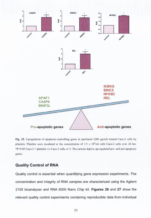
5. Effects of platelets on gene expression in 59M and Caco-2 cells in the presence of paclitaxel... 49
6. Quality control of RNA... 55
7. Effects of platelets on BRCA1 gene expression in 59M cells in the presence of platelets... 60
8. Proteomics of secretome released during interactions of paclitaxel- treated Caco-2 cells with platelets... 61
10. Effects of platelets on cyclins A, B1, D1, E in 59M cells in the presence
of paclitaxel or 5-fluorouracll in G0/G1, S and G2/M phases...73
11. Effects of platelets on DNA damage repair proteins in Caco-2 and 59M cells in the presence of paclitaxel or 5-fluorouracil... 85
12. Effects of platelets on mitogen-activated protein (MAP) kinase pathways in Caco-2 and 59M cells in the presence of paclitaxel or 5-fluorouracil..88
13. Effects of platelets on telomerase activity in Caco-2 cells in the presence of 5-fluorouracil... 92
14. Phase contrast microscopy...93
DISCUSSION... 97
1. Key Novel Findings...97
2. Apoptosis... 97
3. Proteomics... 100
4. Cell cycle and cyclins...102
5. DNA damage repair... 107
6. MAP kinases...110
7. Telomerase...112
8. Pharmacological and clinical significance...113
FUTURE DIRECTIONS... 117
LIST OF FIGURES
Figure 1... 3
Figure 2 ... 4
Figure 3 ... 6
Figure 4 ... 11
Figure 5... 13
Figure 6 ... 14
Figure 7... 15
Figure 8 ... 19
Figure 9 ...22
Figure 10...23
Figure 11...32
Figure 12...35
Figure 13...37
Figure 14...42
Figure 15...45
Figure 16...46
Figure 17...47
Figure 18...47
Figure 19...48
Figure 20...49
Figure 21...50
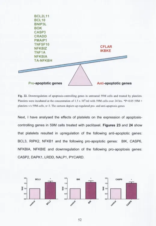
Figure 22...52
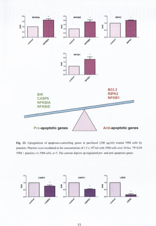
Figure 23...53
Figure 26... 56
Figure 27... 57
Figure 28... 58
Figure 29... 59
Figure 30... 60
Figure 31... 60
Figure 32...70
Figure 33...70
Figure 34...71
Figure 35...71
Figure 36...71
Figure 37...72
Figure 38...72
Figure 39...73
Figure 40...74
Figure 41...75
Figure 42... 75
Figure 43... 76
Figure 44... 77
Figure 45... 78
Figure 46... 78
Figure 47... 79
Figure 48... 79
Figure 49... 80
Figure 50... 80
Figure 52...82
Figure 53...83
Figure 54...83
Figure 55...84
Figure 56...84
Figure 57...85
Figure 58...86
Figure 59...87
Figure 60...87
Figure 61...88
Figure 62...89
Figure 63...90
Figure 64...91
Figure 65...92
Figure 66...92
Figure 67...93
ACKNOWLEDGEMENTS
I would like to emphasise that this research project would not have been possible without the help, assistance and kindness of many people.
SUMMARY
Cancer cells grow without the restraints of feedback control mechanisms that regulate normal tissue or organ growth, such as apoptosis, leading to Increased cancer cell survival. Deregulation of apoptosis has been Implicated In carcinogenesis. Platelets play an Important role in various stages of cancer progression such as anglogenesis. Invasion and metastasis (Jurasz, Alonso- Escolano et al. 2004). Cancer cell survival was affected by platelets. The general objective of my PhD research was to study the role of platelets In chemotherapy-induced cancer cell death and survival. Therefore, the studies focused on the effects of platelets on apoptosis, cell cycle regulation, DNA damage repair, mitogen-activated protein kinase pathways and telomerase activity.
Human colonic adenocarcinoma Caco-2 and human ovarian adenocarcinoma 59M cells were incubated with 5-fluorouracll (1 pg/ml - 3000 pg/ml) or paclitaxel (1 |jg/ml - 200 pg/ml) in the presence or absence of platelets (1.5 x 10®/ml) for 1, 24 or 72 hrs. Following Incubation, cancer cells were harvested and cell survival/death assayed using flow cytometry. Western blotting, ELISA, real-time
PCR, microscopy and TaqMan® Gene Expression Assays. Finally, proteomics
was used to study the release of factors during platelet-cancer cell Interactions. The results of the studies show that platelets and their releasate: (1) have the ability to modulate chemotherapeutic agent-induced cancer cell apoptosis; (2) upregulate the antl-apoptotic genes and downregulate the pro-apoptotic ones; (3) increase the number of cancer cells In the synthesis of DNA (S) and mitosis
phase; (5) upregulate the expression of cyclins A, B1, D1 and E; (6) upregulate
DNA repair proteins such as BRCA1, Chk1, Mre11 and p95/Nbs1; and (7)
upregulate MAPK pathways such as ERK 1, 2; p38 and JNK.
It is concluded that platelets have the ability to decrease cancer cell death and
increase their survival, which may contribute to cancer cell resistance to anti
LIST OF ABBREVIATIONS
A1 Bcl-2-related protein A1
ACTS Beta-actin
ADP Adenosine diphospate
Apaf-1 Apoptotic protease-activating factor-1 AT!\/I Ataxia telangiectasia mutated
ATR Ataxia telangiectasia and Rad3 related BAD Bcl-2-associated death promoter BAK Bcl-2 homologous antagonist/killer BCAP31 B-cell receptor-associated protein 31 BCL3 B-cell lymphoma 3-encoded protein BCL2A1 Bcl-2-related protein A1
BCL2L11 Bcl-2-like protein 11
BCL10 B-cell lymphoma/leukemia 10 BCL-W Bcl-2-like protein 2
BCL-X Bcl-2-associated X protein or Bax BER Base excision repair
Bid pro-apoptotic Bcl-2 protein BIK Bcl-2-interacting killer
BIRC5 Survivin; baculoviral inhibitor of apoptosis repeat-containing 5 BNIP3L Bcl-2/adenovirus E1B 19 kDa protein-interacting protein 3-like BOK Bcl-2-related ovarian killer protein
CASP2 Caspase 2
CASP3 Caspase 3
CASP6 Caspase 6
CASP8AP2 Caspase 8-associated protein 2
CD39 Ectonucleoside triphosphate diphosphohydrolase 1
CD41 Platelet-endothelium attachment receptor, Platelet Glycoprotein
lib
CD61 Platelet-endothelium attachment receptor, integrin beta-3
CD62 Platelet-endothelium attachment receptor, P-selectin
CD95 Tumor necrosis factor receptor superfamily member 6
Cdks Cyclin dependent kinases
CFLAR CASPB and FADD-like apoptosis regulator
Chk1 Checkpoint 1
Chk2 Checkpoint 2
CRADD Death domain-containing protein CRADD
DAPK1 Death-associated protein kinase 1
DKC1 Dyskerin
DNA-PK DNA-dependent protein kinase
DSBs Double strand breaks
E2F Transcription factor
EGF Epidermal growth factor
EGFR Epidermal growth factor receptor
ERK 1, 2 Signal-regulated protein kinase
ERK5 Extracellular regulated kinase 5
FGF Fibroblast growth factor
5FU 5-Fluorouracil
G i phase Gap between mitosis and S phase
G2 phase Gap between S phase and meiosis
Go phase Q uiescent or arrested phase
GAPDH Glyceraldehyde 3-phosphate dehydrogenase
GPlb Glycoprotein lb
G P IIb/llla Glycoprotein llb /llla
GPIa/lla Glycoprotein la/lla
HCC Hepatocellular carcinoma
HR Homologous recombination
Hsp70 Heat shock 70 kDa protein
hTERT Human telom erase reverse transcriptase
hTR Human telom erase RNA
V-H2A.X Histone H2A.X phosphorylated on serine-139
IKBKE Inhibitor o f nuclear factor kappa-B kinase subunit epsilon
IKBKG NEMO, inhibitor of nuclear factor kappa-B kinase subunit
gamma
IL-6 Interleukin 6
JNK c-jun N-terminal kinase
Ku70/Ku80 Complex, DNA double stranded break repair
LPS Lipopolysaccharide
LRDD Leucine-rich repeats and death domain containing
LTB Lymphotoxin-3
MAPK Mitogen-activated protein kinase
NER Nucleotide excision repair
NFkB1 Nuclear factor NF-kappa-B p105 subunit
NFkB2 Nuclear factor NF-kappa-B p100 subunit
NFkBIA Nuclear factor of kappa light polypeptide gene enhancer in
B-cells inhibitor, alpha
NFkBIE Nuclear factor of kappa light polypeptide gene enhancer in
B-cells inhibitor, epsilon
NFkBIZ NF-kappa-B inhibitor zeta
NHEJ Non-homologous end joining
MeCN Acetonitrile
MMP Matrix metalloproteinase
MMP-2 Matrix metalloproteinase-2
MMP-9 Matrix metalloproteinase-9
M phase Phase of mitosis
MMR Mismatch repair
MRN Mammalian complex of Mre11/Rad50/Nbs1
NALP1 Implicated in cell responses to apoptotic and inflammatory
stimuli
NO Nitric oxide
NOS Nitric oxide synthase
p21 protein Cyclin-dependent kinase inhibitor 1
p53 Tumour suppressor gene
p73 Tumour suppressor protein
PARP-1 Poly (ADP-ribose) polymerase 1
PARP Poly (ADP-ribose) polymerase
PDGF Platelet-derived growth factor
PF-4 Platelet factor 4
PGI2 Prostacyclin
PI Propidium Iodide
PMAIP1 Noxa, phorbol-12-myristate-13-acetate-induced protein 1
PMP Platelet-derived microparticles
PLT Platelets
PTX Paclitaxel
PYCARD Apoptosis-associated specl<-lil<e protein containing a CARD
Rad52 gene Protein Rad encoded
RAP1 Ras-proximate-1
REL C-Rel proto-oncogene protein
RIPK2 Receptor-interacting serine/threonine-protein kinase 2
Rb Protein retinoblastoma
RPA Replication protein A
ROS Reactive oxygen species
S phase Phase of DNA synthesis
SDSA Synthesis-dependent strand annealing
SMC Smooth muscle cell
SSBs Single strand breaks
TCIPA Tum our cell-induced platelet aggregation
TEAB Triethylammonium bicarbonate buffer
TGF- (3 Tansforming growth factor-p
TGY Threonine-glycine-tyrosine
TNF Tumour necrosis factor
TNF1A Tum or necrosis factor-a
TNFSF10 TNF-related apoptosis-inducing ligand (TRAIL)
TRAP Telomeric Repeat Amplification Protocol
TRF Terminal Restriction Fragment (method of measuring the length
o f telomere)
TXA2 Throm boxane A2
WRN W erner syndrome protein
VSMCs Vascular smooth muscle cells
VTE Venous thromboembolism
vWF von W illebrand factor
XIAP X-linked Inhibitor of Apoptosis Protein
INTRODUCTION AND BACKGROUND
Cancer and platelets
In 1865, the French physician Armand Trousseau reported a high incidence of venous thrombosis in patients with gastric carcinomas, and the migratory venous thrombosis due to cancer was named Trousseau’s syndrome. Later on, in 1878, Theodor Billroth showed on autopsy that human tumour cells are frequently found in association with thrombi. Recent clinical and experimental data confirm the relationship between cancer cells and blood platelets. For instance, thrombocytosis (increased platelet numbers) is often detected in cancer patients. In addition, thrombocytosis is a poor prognostic factor in stomach, pancreas, liver, ovary, breast, kidney, colon, lung and prostate cancer (Tanaka 1981; Pasquini 1995; Santos, Rodrigues et al. 2001).
Recent experimental evidence has shown that platelets contribute to different stages of cancer progression such as angiogenesis, invasion, intravasation, survival in circulation, extravasation and finally metastasis (Gupta and Massague 2004; Jurasz, Alonso-Escolano et al. 2004).
rich family of receptors (GPIb/IX/V) and selectins (P-selectin) (Larsen, Celi et al.
1989; Radomski 1993; Oleksowicz, Mrowiec et al. 1995; Oleksowicz 1997). In
addition, platelets upon activation release from a-granules and dense bodies a
variety of angiogenesis-regulating factors, such as vascular endothelial growth
factor (VEGF), platelet-derived growth factor (PDGF) and transforming growth
factor-p (TGF- (3) (Amirkhosravi 1999), which can be used by tumour cells for
growth (Honn 1992; Janowska-Wieczorek, Wysoczynski et al. 2005). In
addition, platelets are responsible for facilitating the adhesion of tumour cells to
the endothelium tissue (Mehta 1984). Some studies indicated that platelets coat
cancer cells to avoid immune system response and protect tumour cells from
high shear forces in flowing blood, thus increasing their survival in circulation (Jurasz, Alonso-Escolano et al. 2004).
Platelets also promote cancer cell invasion to disease-free tissues and organs.
In order to invade, tumour cells have the ability to degrade and remodel the
extracellular matrix via release of various proteolytic enzymes. These include
matrix metalloproteinases (MMPs), zinc-dependent endopeptidases, which
break down extracellular matrix proteins (Sternlicht and Werb 2001). MI\/IP-2
and MMP-9 have been implicated in cancer invasion (Jurasz, Chung et al. 2002;
Jurasz, Alonso-Escolano et al. 2004). Our research group has recently reported
that platelets stimulate invasiveness of tumour cells via increased expression of
MMP-9 (Alonso-Escolano, Medina et al. 2006).
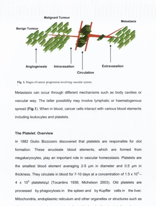
Malignant Tumour
Metastasis
Benign Tumour
Angiogenesis Intravasation Extravasation
Circulation
Fig. 1. Stages o f cancer progression involving vascular system.
Metastasis can occur through different mechanisms such as body cavities or vascular way. The latter possibility may involve lymphatic or haematogenous spread (Fig.1). When in blood, cancer cells interact with various blood elements including leukocytes and platelets.
The Platelet: Overview
[image:27.533.12.520.35.705.2]White 1999; White 2004). Platelets contain three major types of secretory
organelles such as a granules, dense bodies (6 granules) and lysosomes. To
support shape of platelets, actin and myofilaments form the cytoskeleton (White
1983).
The main function of platelets is haemostasis and thrombosis. They are also
natural source of growth factors. Under physiological conditions, platelets are
not- active (resting) and have discoid shape, and this resting state is assured by
such factors as nitric oxide (NO), prostacyclin (PGI2), and adenosine
diphospatase (ADP-ase), which are released from endothelial cells and inhibit
activation of platelets (Michelson 2003) in the circulating blood. After the
vascular injury, the circulating platelets are activated and recruited to repair the
damage. Human platelets can be mainly activated by a number of factors
including thromboxane A2 (TXA2) (Needleman 1976), adenosine diphosphate
(ADP) (Born 1962) and matrix metalloproteinase-2 (MMP-2) (Sawicki 1997).
These agents induce platelet adhesion, activation and aggregation (Nurden
1975). Of note, upon activation platelets change their shape, form pseudopodia
(Fig. 2) and release growth factors.
Fig. 2. TEM images o f hum an platelets. 1. Resting platelets as shown by their discoid shape; 2. Activated platelets. N ote granular centralization and formation o f pseudopodia; 3. A ggregated platelets.
The outer surface of the platelet membrane is very rich in glycoproteins and is
known as glycocalyx. The glycoproteins receptors are necessary to facilitate
adhesion, activation and aggregation of platelets. The glycoprotein GPIb/IXA/
and GPIIb/llla complexes are the principal mobile receptors on platelets. The
outside membrane surface in resting platelets is covered by about 25.000
GPIb/IXA/ and 80.000 GPIIb/llla receptor copies (White 1987).
Firstly, the GPIb/IX/V complex interacts with von Willebrand factor (vWF).
Following damage occurring to the blood vessel, the vWF binds to the
subendothelial collagen. vWF plays a major role in blood coagulation and it is
important in platelet adhesion to wound sites (Sadler 1998). The GPIb/IX/V
complex includes the GPIba subunit, which is essential for vWF biding. GPIb(3
and GPIX are responsible for assembling and anchoring the complex to the
platelet surface. Furthermore, on the platelet surface there are two main
collagen receptors such as GPIa/lla and GPVI. The main function of GPIa/lla is
to facilitate the bonds between platelets and collagen. GPVI acts as a signalling
molecule and fully activates platelets (Clemetson 2001).
Secondly, the binding of vWF to GPIb/IX/X complex leads to the release and
activation of GPIIb/llla (Chen and Lopez 2005; Rivera, Lozano et al. 2009). in
addition, when the platelets are active they translocate P-selectin
(transmembrane adhesion receptor), which is mainly stored in a-granules to the
platelet membrane surface (Stenberg, McEver et al. 1985; Larsen, Celi et al.
1989). The activation of these receptors leads to change in the shape of
platelets. Also upon activation, platelets release growth factors and growth
regulators such as growth factors, interleukin 6 (IL-6), thrombin, fibrinogen
(Amirkhosravi 1999) and angiostatin (Jurasz, Alonso-Escolano et al. 2003;
Finally, the circulating soluble fibrinogen and activated platelets form a
haemostatic plug that is reinforced by the generation of fibrin by the coagulation
cascade, thus forming thrombus (Fullard 2004).
In addition to haemostasis and thrombosis, platelets play also a role in non
haemostatic processes such as innate immune response, wound repair and, as
mentioned before, In carcinogenesis (Jurasz, Alonso-Escolano et al. 2004; Medina, Jurasz et al. 2006). Figure 3 shows platelet receptors involved in the tumour cell-induced platelet aggregation process.
Tumour cell
Endothelial cells Platelets
[b /in a G PIb,
's c le c tin
Aggregate Adhere
Fig. 3. Schematic representation of tumour cell-induced platelet aggregation (TCIPA) and platelet receptors involved in this process.
The Tumour Cell
Malignant neoplasm is a medical term for cancer. There are two different types
of neoplasm {Greek for new growth) - localized and non-localized. Neoplasm that has only the characteristic of localized growth is classified as benign.
Neoplasm with the characteristics of invasiveness and/or the capacity to
metastasize is classified as malignant. The word "tumour", originally defined as
[image:30.533.4.520.24.737.2]"a local swelling", is often used interchangeably with "cancer". The definition of cancer by the British oncologist Willis (Willis 1952):
“A neoplasm is an abnormal mass of tissue, the growth of which exceeds and is uncoordinated with that of the normal tissues and persists in the same excessive manner after cessation of the stimuli which evoked the change.” How normal cells become malignant? This question has occupied scientists for decades. Although the genesis of cancer cells is very complex and multistage in nature, the major factors are likely to involve
> Genetic changes and
> Epigenetic factors
Genetic changes are a result of point mutations, gene amplification, chromosomal translocation or the action of certain viruses and chemical carcinogens. The activation of proto-oncogenes to oncogenes and the inactivation of tumour suppressor genes are the most common issues. The proto-oncogenes are genes, which normally control cell division, apoptosis and differentiation when converted to oncogenes by viral or carcinogen action. They also encode growth factor receptors and signal transduction proteins. The tumour suppressor genes (anti-oncogenes) are genes, which protect cells from cancer. If those genes are mutated, they lead to carcinogenesis, usually in combination with other genetic changes. Epigenetic factors are mechanisms outside the gene such as a cell's exposure to carcinogens or hormones, or genetic variations that modify a gene or its protein by methylation, demethylation, phosphorylation, or dephosphorylation.
> insensitivity to antigrowth signals > evading apoptosis
> limitless replicative potential > sustained angiogenesis
> tissue invasion and metastasis > genomic instability
The first acquired capability of cancer, as discussed by authors is self- sufficiency in growth signals. Normal cells require mitogenic growth factors, which are necessary for cells proliferation. Tumour cells produce own growth factors, thereby reducing their dependence on stimulation from normal tissue microenvironment (Hanahan and Weinberg 2000).
The second acquired capability of cancer is insensitivity to antigrowth signals. Cell proliferation can be blocked by antigrowth factors in two different ways: by being forced into the quiescent Go phase or by being induced to enter into post mitotic state and differentiation.
Another cancer feature Is evading apoptosis. Apoptosis is a programmed cell death which is triggered by two different pathways: extrinsic and intrinsic. The cells undergoing apoptosis are characterized by membrane blebbing, shrinkage and condensation of chromatin. Cancer cells can avoid apoptosis by the mutation of p53 tumour suppressor gene (SIgal and Rotter 2000; Caino 2009). This kind of mutation is very common In over 50 % of tumours (Kaelln 1999). The fourth acquired capability of cancer cells Is a limitless replicative potential. Healthy cells may double up to 60 - 70 times before their death (Hanahan and Weinberg 2000). This limited number of cell proliferation is controlled by the telomeres serving as cellular “internal clock”. Telomeres are sequences of DNA at the end of chromosomes, which become shortened due to each S phase
(Counter 1992). However, 85 - 90 % of cancer types have the ability to maintain their telomere length by upregulating expression of telomerase enzyme. The function of these enzyme is adding the hexanucleotide repeats onto the ends of telomeric DNA (Bryan and Cech 1999).
The fifth capability acquired by tumour cells is a sustained angiogenesis. The essential role of angiogenesis is to form vessels, which supply the oxygen and nutrients for cells in a tissue. Tumour cannot grow more than 1 - 2 milimeters in diameter without creating new blood vessels. There is a balance between promoters of angiogenesis such as VEGF and inhibitors such as angiostatin. Tumour has ability to change the balance between angiogenesis inducers and inhibitors (Hanahan and Folkman 1996). This mechanism is not completely understood.
Cancer and Apoptosis
Apoptosis: Overview
Apoptosis is a programmed cell death, and this process is crucial for such fundamental physiological processes as embryogenesis and homeostasis. Apoptosis requires energy and is characterized by individual cells death, cell shrinkage, condensation of chromatin, membrane blebbing, cell fragmentation, and phagocytosis of the dead cell. Apoptosis is also thought to limit the tumourigenic process (Bold, Termuhlen et al. 1997). When a mutation occurs in a proto-oncogene that converts it into an oncogene, a cell tries to repair the mutation; however, if it is not successful at repairing the damage, the cell will then undergo apoptosis. In contrast, tumour cells have the ability to evade apoptosis for instance by mutation in the p53 tumour suppressor gene, which control the apoptosis (Sigal and Rotter 2000). Moreover, anti- and pro- apoptotic member of the Bcl-2 family may be also mutated as exemplified by non- Hodgkin’s lymphoma, small-cell lung cancer (Reed 1999) and gastrointestinal cancer (Adams and Cory 1998). There are two main pathways of apoptosis: the death receptor pathway and the death mitochondrial pathway. The death receptor pathway is mediated by members of the tumour necrosis factor receptor family (TNF). They activate pro-caspase 8 to an active form, which by cascade of many steps stimulates non-active pro-caspase 3 to caspase 3. The activation of the mitochondrial pathway leads to the release of p53 protein,
which is responsible for activation of subpathway in the mitochondrion. This results in a release of p21 protein and anti-apoptotic members of the Bcl-2 protein family (BCL-X, BCL-W, MCL-1, and A l). This family comprises also agents that stimulate apoptosis known as pro-apoptotic factors (BAX, BAK,
BOK, BAD, BIK and BID). Both anti-apoptotic and pro-apoptotic factors a re p re se n t a t th e surface of mitochondrial m em brane and com pete with e ac h other in regulating apoptosis. Indeed, th e anti-apoptotic factors inhibit re le ase of cytochrom e c while other m em bers prom ote re le a s e of cytochrom e c from mitochondria to cytosol. The re le ase d cytochrom e c, m ak es a com plex with protein apoptotic protease-activating factor-1 (Apaf-1). The com plex activ ates
p ro -c a sp a s e 9 to an active form and finally results in activation of c a s p a s e 3 and 7. C a s p a s e 3 and 7 a ct in cascade-lik e m anner to stim ulate DNA cleavag e by DNAase into fragm ents of b a s e pairs, which c a u s e s cell d eath (Chalah 2008). M echanism s of ap o pto sis a re show n in Fig.4. Given the complexity of ap o p to sis
regulation it is not surprising, that carcin o g en esis may b e a sso c iate d with alterations of expression of pro- or anti-apoptotic factors.
R C E PT O R PATHWAY MITOCHONDRIAL PATHWAY
TNFR Fam ily A ctiv atio n P53 G e iieratio n C a s p a s e -8 A ctivatio n C y to c h ro m e C R e le a s e
C a s p a s e -3 a n d 7 A ctiv atio n
i
A PO PTO SIS Fig. 4. Pathways o f apoptosis
T he cells may die not only by ap o p to sis but also by necrosis. N ecrosis is c au se d by external factors, su ch a s infection or toxins. T he two m ech an ism s of the cells
d e ath a re indep en d en t and opposite. N ecrosis is characterized by large groups of cell rupture, blebbing of outer m em brane and no en erg y is required for this
Cell Cycle and Cyclins
The cell cycle is an event v\/hich prompts the cell to duplicate into two identical daughter cells. Cell division cycle is involved in many physiological and
pathological processes such as growth, repair and tumour development. There
are four different phases of cell cycle G i, S, G2 and M (Fig.5). S phase - phase of DNA synthesis
M phase - phase of mitosis, in which the cell’s chromosomes and cytoplasm are divided between the two daughter cells.
Gi phase - is the gap between mitosis and S phase, during this time the cell is preparing for DNA synthesis.
G2 phase - is the gap between S phase and meiosis, during this time cell is preparing for the mitotic division into two identical cells.
There are two critical events during the cell cycle such as DNA synthesis (S phase) and mitosis (M phase), which entry into both of these phases is strictly
regulated by restriction points or checkpoints. When DNA damage is
occurred the cell cycle is stopped by the checkpoint 1 and/or 2 (C hk1, Chk2).
Therefore, these checkpoints play a crucial role in genetic stability of cell. Cells
may also temporarily or permanently leave the cell cycle and enter a quiescent
or arrested phase known as Go (Rang 2003). The duration of cell cycle phases depend on different kinds of cells. For a typical rapidly proliferating human cell
with a total cycle time of 24 hours, the G i phase might last about 11 hours, S
phase about 8 hours, G2 about 4 hours, and M about 1 hour (Cooper 2009). The
main phases of the cell cycle are shown in Fig. 5.
Chk2
M
Chk1
Fig. 5. Cell cycle phases.
There is a balance between positive and negative regulators of cell division. In mammalian cells, the progression of replicating cells through the cell cycle is controlled by the sequential formation, activation, and subsequent inactivation of a series of proteins such as cyclins and cyclin-dependent kinases (Cdks)
complex with cyclin B and completes mitosis phase (Riabowol, Draetta et al. 1989; Satyanarayana and Kaldis 2009). Both active cyclins, A and B are necessary for passing restriction point 2 (Fig. 6).
The negative regulators of the cell cycle are Rb proteins and two families of Cdks inhibitors, which are hypophosphorylated and hold the cycle in checkpoint 1. Indeed, p21 WAF1, p27KIP1 and p57KIP2 belonging to the CIP family bind to cyclin/Cdk complexes. Another Ink family consist of p15, p16INK1, p18 and p19 inhibitory proteins that bind directly to Cdk4 (Draetta. 1994; Sherr 1994). The growth factors such as FGF, EGF, PDGF, VEGF and TGF-p stimulate the production of positive and negative regulators of cell cycle (Rang 2003).
mitogenic signal
cyclln A/CDC2
cyclln D/CDK4, 6
cyclin A/CDK2
cyclln E/CDK2
c y c lin g cyclin
mitogenic signal
\ cyclin B
Fig. 6. Cyclin/Cdk complexes during cell cycle (Takahashi-Yanaga and Sasaguri 2008).
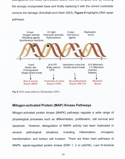
DNA Damage Repair Pathways
The genome of a living cell is constantly at risk of exposure to different
environmental DNA damaging agents. The sources of these factors may be
endogenous and exogenous, e.g. carcinogenic substances, toxins, exposure to
ultraviolet (UV), ionizing radiation (IR), free radicals and chemotherapeutic
agents (Fig. 7).
Fig. 7. DNA damage response pathways. From a Paterson Institute for Canccr Research, The University o f Manchester, www.patcrson.man.ac.uk/dnadamagc/.
Therefore, cells evolved complex signalling networks to carefully monitor the
integrity of the genome and initiate mechanisms to avoid errors such as cell
cycle arrest, activation of repair pathways or apoptosis (Harper and Elledge
2007). DNA damage induces phosphorylation of H2A.X and triggers
multiprotein complexes to recognize the genome defect, following activation of
the transducers such as ataxia telangiectesia mutated (ATM) and ataxia
telangiectasia and Rad3 related (ATR), which belong to the stress-responsive
protein kinase (O'Driscoll and Jeggo 2006). After the DNA damage a series of DNA damage response
O lu is r m0aalx)ttsni
DNA damage
^
4
W
[image:39.533.25.520.27.557.2]pathways specific signal transduction cascades are responsible for arresting the
cells at the cell cycle checkpoints. Chk1 and Chk2 are checkpoint kinases
downstream of ATM, ATR and DNA-PK, which play crucial role in cellular
response to genotoxic stress and genome damage (Yang, Yu et al. 2003).
Especially checkpoint 1 is responsible for initiating cell cycle arrest, allowing
time for DNA repair and cell survival (Bartek and Lukas 2001; Zhou 2003;
Ashwell and Zabludoff 2008).
There are two main pathways of DNA double strand breaks (DSBs), the non-homologous end joining (NHEJ) and homologous recombination (HR).
These two major pathways compete with each other in repairing DNA damage
(Delacote, Han et al. 2002).
There are three pathways to repair single strand breaks (SSBs) such as: base excision repair (BER), nucleotide excision repair (NER) and mismatch repair (MMR) (Bartek and Lukas 2001).
The non-homologous end joining pathway is a predominant mechanism in use for repairing DSBs during the Gq/Gi phases. In mammalian cells NHEJ
proceeds with limited end-processing by the MRN complex, which is a
multisubunit nuclease composed of Mre11, RadSO and Nbs1/Xrs2. The crucial
function of the complex is contribution to DNA repair. However, recent studies
have discovered role for the M re ll complex in checkpoint signalling and DNA
replication (Gottlieb and Jackson 1993; Dynan and Yoo 1998; D'Amours and
Jackson 2002; lijima 2008). Once bound to broken ends, DNA-PK is activated
and phosphorylates itself and other targets including replication protein A (RPA),
Werner syndrome protein (WRN), histone H2A.X and human nuclease Artemis
(Burma, Chen et al. 2001; Burma and Chen 2004; Collis, DeWeese et al. 2004).
One of the main proteins involved in this repair pathway is DNA-dependent
protein kinase (DNA-PK). The Ku70/80 heterodimer com ponent of DNA-PK
binds to the two DNA ends in a ring conformation. The DNA binding of Ku70/80
and aligning o f the two DNA ends subsequently activates the catalytic activity of
DNA-PK, which promotes the ligation of DNA ends by the XRCC4-Ligase IV
complex (Ahnesorg, Smith et al. 2006; Bolderson, Richard et al. 2009). In the
final step, DNA ligase IV with it is binding partner XRCC4 form a tight complex,
which in the presence of XLF seals the break (Cahill 2006; Hentges, Ahnesorg
et al. 2006; Shrivastav, De Haro et al. 2008).
The homologous recombination pathway is considered a more accurate mechanism for DSBs repair, because broken ends use homologous sequences,
usually located on the sister chromatid. HR is a predominant mechanism in use
for repairing DSBs during the S and G2 phases. The proteins Rad encoded by
gene Rad52 play a crucial role in this process (Johnson and Jasin 2001). This
pathway initiates extensive 5’ to 3’ end-processing at broken ends, which is
probably mediated by the Mre11/Rad50/Nbs1 complex (D'Amours and Jackson
2002). The resulting 3’ single-stranded DNA (ssDNA) tails are bound by RPA,
which is replaced with Rad51. Then normal base-pairing o f the invading,
com plementary donor strands and subsequent strand are extended by DNA
polymerase. The extend strand can dissociate and anneal with the processed
end o f the non-invading strand on the opposite side of the DSB in a process
called synthesis-dependent strand annealing, or both ends may invade
producing a double-Holliday junction that is resolved to yield crossover or non
crossover recombinants. Once intermediates are resolved, the remaining ssDNA
gaps and nicks are repaired by DNA polymerase and DNA ligase (Bishop and
utilizes homologous intact sequence from a sister chromatid as a template. BRCA1 and BRCA2 proteins have the similar physiological function in cell. Therefore, both proteins interact with Rad51 (Boulton 2006). BRCA1 colocalizes with V-H2A.X (histone H2A.X phosphorylated on serine-139) in DNA double strand break repair foci, indicating it may play a role in recruiting repair factors (Ye, Hu et al. 2001; Starita and Parvin 2003). The DNA double strand breaks trigger phosphorylation of H2A.X at S eri 39, which can be a marker of premalignant lesions. Moreover, y-H2A.X histon attracts the DNA repair proteins to the damaged chromatin (Jun and Anton 2008; Kinner, Wu et al. 2008; Podhorecka 2009).
The base excision repair (BER) pathway recognizes and removes small, non helix base lesions, that have been damaged by oxidation, alkylation, ring saturation and ionizing radiation (Chan, Zhang et al. 2006). DNA glycosylases form AP sites (abasic site) location in DNA, that has neither purine nor pyrimidine base. These are then cleaved by an AP endonuclease. The resulting single-strand break can be repaired by either short-patch or long-patch BER (Liu, Prasad et al. 2007).
The next repair pathway of SSBs is nucleotide excision repair which is activated by bulky DNA lesion resulted in the damage by UV and DNA-alkylating agents (Hanawalt 2002). The short single-stranded genome disruption is removed and the single-strand gap is subsequently filled in by DNA polymerase, which uses the undamaged strand as a template. NER can be divided into two subpathways Global genomic NER and Transcription coupled NER (Cleaver 2005).
The mismatch repair recognizes erroneous insertion, deletion and misincorporation of bases, that can arise during replication and recombination of
DNA (Iyer, Pluciennik et al. 2005). During DNA synthesis the newly synthesised strand includes a lot of errors. Therefore, the mismatch repair machinery
distinguishes the newly synthesised strand from the template and by excising the wrongly incorporated base and finally replacing it with the correct nucleotide corrects the damage (Schofield and Hsieh 2003). Figure 8 highlights DNA repair
pathways.
X -rays U V light X-rays
Oxygen radicals Polycyclic aromatic Anti-tumour
Alkylating agents hydrocarbons agents
Spontaneous reactions
Replication errors
Uracil Abasic site 8-O xoguanine Single-strand break
I
Base-Excicion Repair (B E R )
(6 -4 ) P P Bulky adduct
C P D
Interstrand cross-link Double-strand break
i
Nucleotide-ExcisionRepair (N E R )
A -G Mismatch T -C Mismatch Insertion Deletion
Recombinational R epair (H R , EJ)
I
Mismatch Repair
Fig. 8. D N A repair pathways (Hoeijmakers 2001).
Mitogen-activated Protein (IVIAP) Kinase Pathways
[image:43.533.12.523.132.783.2]kinases (JNK), and p38 MAPK (p38) (Pearson, Robinson et al. 2001) (Fig. 9). In addition, other less well-characterized MAPK pathways exist, such as the extracellular regulated kinase 5 (ERK5) pathway (Hayashi 2004). The pathways
are activated by a multitude of stimuli and mediate their effects through phosphorylation. MAPK cascades are organized as modular pathways in which activation of upstream kinases by cell surface receptors leads to a sequential activation of a MAPK module (MAPKKK - MAPKK - MAPK). After MAPKs (ERK1, 2; JNK1-3 and p38 a, p, 5, y) are activated either in the cytoplasm or in the nucleus, they bind and regulate transcription by modulating the function of a target transcription factor through serine/threonine phosphorylation (Davis 2000; Roux and Blenis 2004; Junttila, Li et al. 2008).
ERK1, 2 MAPK pathway is responsible for cell growth, cell proliferation and survival. This pathway is activated by growth factors through the g-protein Ras. Ras is a membrane-bound protein, which is activated through the exchange of bound GDP to GTP. Activated Ras recruits cytoplasmic Raf (MAPKKK) to the cell membrane for activation. There are three mammalian serine/threonine Raf kinases; A-Raf, B-Raf, and Raf-1. MEK1, 2 is activated by dual phosphorylation on two serine residues by Raf proteins. ERK1, 2 is activated by MEK1, 2, specifically by phosphorylation of a tyrosine and a threonine residue. Activated ERK1, 2 can translocate to the nucleus, where it activates several transcription factors, such as c-Fos, ATF-2, Elk-1, c-Jun, c-Myc, and Ets-1. ERK1, 2 promotes cell survival through transcriptional upregulation of anti-apoptotic Bcl- 2, Bcl-xL, and Bcl-1 proteins (Bonni, Brunet et al. 1999; Ballif and Blenis 2001; Junttila, Li et al. 2008).
The JNK (c-Jun N-terminal kinase) pathway is activated by cellular stress and cytokines. The activation takes place through phosphorylation of a tyrosine and
a threonine residue. Physiological function of JNK pathway is to mediate apoptosis, proliferation, or survival, depending on the stimuli and cellular conditions. The pathway activates different transcription factors, such as ATF-2, Elk-1, MEF-2c, p53, and c-Myc. JNK also has other non-transcriptional substrates, for example the anti-apoptotic proteins, Bcl-2 and Bcl-xL (Davis 2000; W eston and Davis 2007; Junttila, Li et al. 2008).
The p38 MARK pathway (MAPKKKs/MKK 3, 4, 6 / p38 a, (3, y, 6) is activated by the response to inflammatory cytokines and by environmental stress, such as osmotic stress, ultraviolet light, heat shock, and hypoxia. The p38 MAPK protein is represented by four isoforms; p38a, p38(3, p38y, and p386. Activation of all the p38 isoforms is achieved by dual phosphorylation of a threonine and a tyrosine within the threonine-glycine-tyrosine (TGY) sequence in the activation domain of the kinase (Ashwell 2006). Phosphorylated p38 proteins can activate an array of transcription factors, including ATF-2, CHOP-1, MEF-2, p53, and Elk-1. Activation of the p38 MAPK pathway is required for apoptosis leading to G2/M cell cycle arrest. This is regulated through modulation of p53 and p73
tumour suppressor proteins (Bulavin and Fornace 2004). Conversely, p38 MAPK pathway activity has been reported to promote cancer cell growth and survival. The molecular mechanisms that determine whether p38 signalling
either promotes or inhibits proliferation and survival of the cell have not been elucidated. In addition, the p38 pathway plays an essential role in regulating the
Stimulus 1
Growth factors 1
Cytokines stress
1
▼ G-protein 1 T Ras 1Rac, Cdc42 1 I
MAPKKK Raf, Tpl2
T
Tpl2, IVIEKK, MLK, TAK, ASK, TAO
i
i
^
i
X
MAPKK IVIEK 1, 2 MEK7 MEK4 MEK 3 ,6
i
i
MAPK ERK 1 ,2 JNK p38
Response Proliferation Proliferation
Differentiation Differentiation
Apoptosis Apoptosis
[image:46.535.6.523.25.714.2]iViigration Inflamation
Fig. 9. Schematic outline of MAPK pathway (Dhillon, Hagan et al. 2007).
Telomere and Telomerase Activity
Telomeres are regions at the chromosomal ends of hexanucleotide sequence TTAGGG repeats, orientated towards 5’- to - 3’ and a number of associated protecting and regulating proteins such as TRF1, TRF2, TIN2, POT1, TPP1 and RAP1, which collectively form a t-loop structure, which helps to maintain chromosomal integrity and stability (Griffith, Comeau et al. 1999; de Lange 2005). They play an important role In the protection and function of chromosomes. In normal somatic cells, telomeres are shortened with every cell division until the critical size is reached and cells lose their proliferative potential (Harley 1990; Harley 1997). Roughly 50 - 100 base pairs of telomeric DNA are lost with each cell cycle (Counter 1992). Maintenance of stable telomere length is associated with the activity of telomerase enzyme (Fig. 10). Telomerase was discovered by Carol Greider and Elizabeth Blackburn in 1985. The function of the telomerase is to add repeat sequences to the chromosomes ends while there is a loss of telomeric DNA (Morin 1989; Blasco 1997). The enzyme is ribonucleoprotein, which contains a catalytic subunit with reverse transcriptase
activity (hTERT), an RNA part that provides template for telomerase extension
(hTR) and a additional telomerase associated proteins such as hTEP1, p23,
Hsp90 (heat shock protein 90) and dyskerin (Blackburn 1991; Feng 1995;
Nakamura, Morin et al. 1997). The human hTR is ubiquitiously expressed in all
tissues, however the expression of human telomerase catalytic subunit (hTERT)
is the limiting factor for telomerase activity (Keller, Brassat et al. 2009). The
telomerase ribonucleic complex is associated with several other proteins.
Moreover, telomerase has the ability to mediate cell survival and anti-apoptotic
functions against different cytotoxic stresses (Chung 2005). Double strand
breaks of DNA damage in telomeres require various DNA repair proteins and
enzymes such as ataxia telangiectasia mutated (ATM), yH2A.X, 53BP1, MDC1, Ku70/80 and M rel 1/Rad50/Nbs1 complex (Fagagna, Reaper et al. 2003; Zhang,
Dilley et al. 2007). This response can lead to cell cycle arrest and senescence,
or apoptosis (Herbig, Jobling et al. 2004; Campisi 2005). The telomerase is
nearly absent in the most somatic cells. However, approximately 90% of
different types of cancer cells express and upregulate this enzyme (Kim,
Piatyszek et al. 1994).
silenc^
MRE11
msQG
telomere structure compl®^
P * - - ^ - ^ , l ' : ^ v ; _J ;K / . ■ 1.^, -^ . ■ . . - , I ■ i f .. ■
OBJECTIVES AND AIMS
The general objective of my research was to study if platelets have the capacity to modulate cancer cell survival in response to chemotherapeutic agents.
The specific aims of this study were as follows:
1. To determine the number of live and dead human cancer cells during cancer cell damage induced by chemotherapeutic agents and the effects of platelets on these processes.
2. To investigate mechanisms involved in cancer cell-platelet interactions by studying:
> Proteins released
>
Gene expressionMATERIALS AND METHODS
The studies embraced by the presented thesis was approved by the Trinity
College Dublin Ethics Committee.
Reagents
Prostacyclin (PGI2), ribonuclease A (RNAse), propidium iodide, sodium azide,
bovine serum albumin, Modified Eagle’s Medium, Dulbecco’s Modified Eagle’s
medium, penicillin, streptomycin, gentamicin, sodium bicarbonate, sodium
pyruvate, foetal bovine serum, monoclonal anti-[3-tubulin antibody produced in
mouse, anti-rabbit, anti-mouse antibodies were all obtained from Sigma.
Propidium iodide, annexin V APC, cyclin A, mouse IgE, cyclin B1, mouse
lgG2a, cyclin D1, mouse lgG2a, cyclin E, mouse IgG I, goat anti-mouse Ig, APC
conjugated were obtained from BD (Becton, Dickinson and Company). TILDA
(TaqMan® Gene Expression Assays), High Capacity cDNA Reverse
Transcription Kit, primers: BRC1, (3-actin were obtained from Applied
Biosystems. TeloTAGGG Telomerase PCR ELISA'"'-^^ kit was obtained from
Roche. Millipore Immobilon Western Chemiluminescent MRP substrate was
obtained from Millipore. RNA 6000 Nano Chip kit was obtained from Agilent
Technologies. Phospho-BRCA1 (Ser1524) antibody, phospho-Chk1 (Ser296)
antibody, phospho-histone H2A.X (Ser139), Chk1 total antibody, BRCA1 total
antibody, phospho-Mre11 (Ser676) antibody, phospho-p95/Nbs1 antibody,
Mre11 total antibody, p95/Nbs1 total antibody, phospho-p42/44, phospho-p38
and phospho-JNK were obtained from Cell Signalling Technology. Goat anti
Blood Collection, Platelet Isolation and Platelet Releasate
Blood w as obtained from healthy volunteers w ho had not taken any drugs for 14
days prior to the study. Blood w as collected in the presence of tri-sodium citrate
at the final concentration of 0 .3 1 5 % and prostacyclin (3 i^M). W a s h ed platelet
suspensions (Radom ski and M oncada 1 9 8 3 ) w ere isolated and resuspended
(1 .5 X 10® platelets per ml) in serum -free cell culture. P latelet releasate w as
obtained from collagen (1 0 mQ/mO - aggregated platelets. P latelet aggregation w as m easured using aggrego m eter (C hronolog) as the extent of light
transm ittance. W h e n platelet aggregation reached m axim um (9 0 ± 1 0 % ) the
sam ples w ere collected and releasate obtained following centrifugation of
platelets at 4 ,5 0 0 x g for 5 m inutes in the presence of prostacyclin (1 |aM).
Cancer Cell Culture
Tw o hum an adenocarcinom a cell lines C a c o -2 (colonic) and 5 9 M (ovarian) w ere
obtained from the European Collection o f Cell Cultures. Cell lines w ere cultured
as a m onolayer in 75 cm^ and 25 cm^ culture flasks at 3 7 °C in a humidified
atm osphere in the presence of 5 % C O2.
T h e C aco -2 cell line w as cultured in Modified E ag le ’s M edium (M E M ) with
penicillin (0 .0 6 mg/ml), streptomycin (0.01 m g/ml), gentam icin (0 .0 5 mg/ml),
sodium bicarbonate (2 .2 g/L), sodium pyruvate (0.11 g/L) and with 2 0 % FBS.
T h e cell line w as subcultured twice a w e e k and supplied with fresh medium
every two days.
T h e 5 9 M cell line w as cultured in Dulbecco’s Modified E ag le ’s M edium (D M E M )
with penicillin (0 .0 6 m g/ml), streptomycin (0.01 mg/m l), gentam icin (0 .0 5 mg/ml),
sodium bicarbonate (3 .7 g/L), sodium pyruvate (0.11 g/L), L-glutam ine
(0.27 g/L), insulin (0.7 mg/L) and 10 % FBS. This cell line was subcultured once a week and supplied with fresh medium every two days. Cells were detached from the flask using trypsin/EDTA. All cell culture reagents were purchased from Sigma.
Cancer Chemotherapeutics
Paclitaxel supplied as 6 mg/ml, concentrate for solution for infusion (Medac UK, Actavis Ireland Limited) and 5-Fluorouracil 25 mg/ml, solution for injection (Medac UK) were obtained thanks to courtesy of the Pharmacy Department of St. James’s Hospital in Dublin, Ireland.
Cancer Cell-Platelet Incubation
Platelets were added to T25-cell culture flasks containing subconfluent Caco-2 or 59M cells. Platelet-cancer cell cultures were then incubated in the presence or absence of paclitaxel (1 - 200 |jg/ml) or 5-fluorouracil (0.001 - 3 mg/ml) for 1, 24 or 72 hrs. At the end of incubation conditioned media were collected and cancer cells were harvested using Trypsin/EDTA and finally washed using
Binding Buffer (0.1 M Hepes, 1.4 M NaCI, 25 mM CaCIa) for flow-cytometry
studies.
Flow Cytometry
Apoptosis
Propidium Iodide (5 |jl) for 15 min in the dark at room temperature. Afterwards,
samples were diluted 10 fold using Binding Buffer. Ten thousand specific events
were analysed by the cytometer. The instrument was set up to measure the size
(forward scatter), granularity (side scatter) and cell fluorescence. Antibody
binding was measured by analyzing individual cells for fluorescence. The mean
fluorescence intensity was determined after correction for cell autofluorescence.
Fluorescence histograms were obtained for 10,000 individual events. Data was
analyzed using Cytometer RXP software and expressed as a percentage of
control fluorescence in arbitrary units.
Cell Cycle
Following incubation cells were harvested using trypsin. The cells were washed
with PBS and fixed in cold 70 % ethanol (diluted in PBS) for 15 minutes at room
temperature. Fixed cells were divided into the appropriate number of flow
cytometry tubes, containing 4 x 1 0 ® (Caco-2) or lO'^ (59M) cells per test. 300 pi
Propidium Iodide and 50 pi RNAse were added to the cell pellet and incubated
overnight at 4 °C, protected from light. 10“^ events were analyzed by flow
cytometry, using a low flow rate. The percentage of cells in the G0/G1, S and
G2/M cell cycle phases were quantified using ModFit LT^*^ software.
Cyclins
Following incubation cells were harvested using trypsin. The cells were washed
twice by PBS and fixed in cold 70 % ethanol (diluted in PBS) and stored
overnight at -20 °C. The fixed cells were divided into the appropriate number of
flow cytometry tubes, containing 3 x 1 0 ® (Caco-2) and 10"^ (59M) cells per test.
The cells were washed twice in PBS and 1 ml 0.25 % Triton X-100 in PBS were
added to each cell pellet. The cells were mixed and incubated for 5 minutes at
room temperature. Each sample was filled with staining buffer (PBS containing 1
% FCS (Foetal Calf Serum)). To the cell pellet 2.5 pi of cyclins A, B1, D1 and E antibodies (Cyclin A, mouse IgE, Cyclin B1, mouse lgG2a, Cyclin D1, mouse
lgG2a, Cyclin E, mouse lgG1) were added and incubated for 60 minutes at room temperature. Following incubation, cells were washed twice in staining buffer. 100 pi of diluted in staining buffer (1 ; 50) FITC-conjugated secondary antibody (Goat anti-mouse Ig, APC conjugated) was added to each cell pellet and the samples were incubated for 60 minutes at room temperature, protected from light. After incubation, cells were washed twice in staining buffer. 300 pi
Propidium Iodide and 50 pi RNAse were added to the cell pellet and incubated overnight at 4 °C, protected from light. 10,000 events were analyzed by flow
cytometry.
Real-time PCR
Real-time PCR (qPCR) was carried out using an Eppendorf Realplex^ (Eppendorf) and a TaqMan Two-Step RT-PCR method. Total cellular RNA was isolated from Caco-2 and 59M cells using RiboPure kit from Ambion according to manufacturer’s protocol. For reverse transcription reaction, 1 pg of total RNA (High Capacity cDNA Reverse Transcription Kit, Applied Biosystems) was used, and 10 ng of transcribed DNA was added into each qPCR reaction. As a target probe, TaqMan MGB human BRCA1 labelled with 5-carboxyfluorescein dye (Applied Biosystems) was used. As an endogenous control, TaqMan MGB eukaryotic (3-actin (ACTB) ribosomal RNA probe labelled with VIC (Applied
TILDA (TaqMan® Gene Expression Assays)
RNA Isolation Method and Quality Control
Total cellular RNA was Isolated from Caco-2 and 59M cells using miRVana kit
from Ambion according to manufacturer’s protocol. The quantity of RNA was
measured by Nanodrop (Thermo Scientific). The quality of RNA was analyzed
by Agilent 2100 Bioanalyser using the RNA 6000 LabChip® kit (Fig. 11).
Twelve samples were sequentially separated on a chip through a single
separation channel. Each RNA chip contains an interconnected set of micro
channels that is used for separation of nucleic acid fragments based on their
size as they are driven through it electrophoretically. The micro-channels are
filled with a sieving polymer and fluorescence dye. Agilent RNA kits are
designed for use with the Agilent 2100 bioanalyzer, which electrophoretically
separates the samples. The resulting data is presented as an electropherogram.
Fig. 11. A RNA Nano LabChip. From www.chem.agilent.com
TILDA
For reverse transcription reaction, 1 pg of total RNA (High Capacity cDNA
Reverse Transcription Kit, Applied Biosystems) was used, and 100 ng of
transcribed DNA (cDNA) was loaded onto the Micro Fluidic Cards.
TaqMan® Human Apoptosis Array is a micro fluidic card for quantitative gene
expression analysis of targets known to have implications in programmed cell
death. This product is designed with 96 TaqlVlan® Gene Expression Assays
loaded in singletons into the array’s 384 wells and is compatible with an upgraded Applied Biosystems 7900HT Fast Real-Time PCR System. The
TaqMan® Human Apoptosis Array contains assays for 93 human genes in addition to 3 endogenous controls (18S, ACTB, GAPDH). Tables 1 and 2 show the assay map and genes symbols. The 93 genes are categorized into multiple
target classes or pathways.
Target Class or Pathways
> BCL-2 Family-regulated pathway
> Death-receptor-regulated pathway
> TNF Receptor pathway> Fas signalling pathway (CD95) > Caspase family
> NF-kB signalling pathway > p53 activation
> lAP family > lAP inhibitor > CARD family
HS00236911 m1 Hs00832876_g1 Hs00180403 m1 Hs00354836 ml HS00892481 ml Hs00362072 ml HsOOl69152 ml HS01018151 ml
Hs00209789 m1 HS00205419 m1 Hs00376860_g1 HsOOl79410 ml Hs00215973 ml Hs00388035 ml HS00223394 ml Hs00364485 ml
HS00169141 m1 Hs00187848 m1 HS00154189 ml HsOOl88949 ml HS00261296 ml HS00263337 ml HsOOl54250 ml HS01017902 m l
Hs00368095 m1 HS00708019 s1 Hs00248075 m1 HS00203118 ml Hs00219876 ml Hs00212288 ml Hs99999905 ml HS99999903 m l
HS00236911 m1 Hs00832876_g1 HsOOl80403 ml HS00354836 ml HS00892481 ml Hs00362072_m1 HsOOl 69152_m1 Hs01018151_m1
HS00209789 ml Hs00205419 m1 Hs00376860_g1 HsOOl79410_m1 Hs00215973_m1 Hs00388035_m1 Hs00223394_m1 Hs00364485_m1
HS00169141 m1 Hs00187848 m1 Hs00154189 ml HsOOl88949 ml HS00261296 ml HS00263337 ml HsOOl54250 ml HS01017902 m1
HS00368095 ml HS00708019 s1 HS00248075 ml Hs00203118_m1 Hs00219876_m1 Hs00212288_m1 Hs99999905 ml Hs99999903_m1
HS00236911 m1 Hs00832876_g1 HsOOl80403 ml Hs00354836_m1 HS00892481 ml HS00362072 ml HsOOl69152 ml Hs01018151_m1
HS00209789 m1 HS00205419 m1 Hs00376860_g1 HsOOl79410 ml Hs00215973 ml Hs00388035 ml Hs00223394_m1 Hs00364485_m1
Hs00169141 m1 HsOO187848 m1 HsOOl54189 ml HsOOl88949_m1 Hs00261296 ml HS00263337 ml HsOOl 54250_m1 Hs01017902_m1
HS00368095 m1 HS00708019 s1 HS00248075 ml Hs00203118 ml Hs00219876 ml Hs00212288_m1 Hs99999905_m1 Hs99999903 ml
HS00236911 ml Hs00832876_g1 HsOOl80403 ml HS00354836 ml HS00892481 m1 Hs00362072_m1 HsOOl 69152_m1 Hs01018151_m1
Hs00209789 m1 HS00205419 m1 Hs00376860_g1 HsOOl79410 ml Hs00215973 m1 Hs00388035_m1 Hs00223394_m1 Hs00364485_m1
Hs00169141 m1 Hs00187848 m1 HsOOl54189 m1 HsOOl88949 ml HS00261296 ml Hs00263337_m1 HsOOl 54250_m1 Hs01017902_m1
HS00368095 m1 Hs00708019_s1 Hs00248075_m1 Hs00203118_m1 Hs00219876_m1 Hs00212288_m1 Hs99999905_m1 Hs99999903_m1
1 2 3 4 5 6 7 8
HsOO154260 ml X (/) O O CO CJ> LO o 00 CO .m l Hs99999901 si Hs00242739 ml HsOOl72036 ml HS00765730 ml HsOOl74517 ml HsOOl82115 ml
HS00230071 ml Hs00373302 ml HS00223384 ml HS00261581 ml HsOI057786 si Hs00370206 ml HS00559441 ml Hs00985031 HS00234480 ml HsOO193477 ml Hs01847653 si HsOOl53283 ml HsOOl53294 ml HsOOl74128 m l HsOOl75318 ml Hs00269428 ml HS00989502 ml Hs00968436 ml HS01042313 m1 HsOI572688 ml HsOI063858 ml HsOI036137 ml HsOI043258 ml HsOI076336 ml HsOOl54260..m l Hs00395088 ml Hs99999901 si HS00242739 ml HsOOl72036 ml Hs00765730 ml HsOOl74517 ml HsOOl82115 ml Hs00230071..m l HS00373302 ml HS00223384 ml Hs00261581 ml HsOI057786 si Hs00370206 ml Hs00559441 ml Hs00985031 gi
HS00234480 ml HsOO193477 ml HsO1847653 si HsOOl53283 ml HsOOl53294 ml HsOO174128 ml HsOOl75318 ml HS00269428 ml Hs00989502 ml HS00968436 ml Hs01042313 ml HsOI572688 ml HsOI063858 ml HS01036137 ml HsOI043258 ml Hs01076336 m1 HsOO154260..m l Hs00395088 .m l Hs99999901 si Hs00242739 m1 HsOOl72036 m1 Hs00765730 m l HsOOl74517 ml HsOOl82115 ml HS00230071..ml HS00373302 ml HS00223384 ml HS00261581 ml HsOI057786 s1 HS00370206 m l HS00559441 ml HS00985031_gi
HS00234480 ml HsOO193477 ml HsO1847653 si HsOOl53283 ml HsOOl53294 ml HsOOl74128 m1 HsOOl75318 ml Hs00269428 ml HS00989502 ml Hs00968436 ml Hs01042313 ml HsOI572688 m1 Hs01063858 m1 Hs01036137 m l HsOI043258 ml HsOI076336 ml HsOO154260 .m l HS00395088 ml HS99999901 si Hs00242739 ml HsOOl72036 m1 HS00765730 m l HsOOl74517 ml HsOOl82115 ml Hs00230071 .m l HS00373302 ml HS00223384 ml Hs00261581 ml HsOI057786 si HS00370206 ml Hs00559441 ml Hs00985031 HS00234480. ml HsOOl93477 ml HsO1847653 s1 HsOOl53283 ml HsOOl53294 ml HsOOl74128 ml HsOOl75318 ml Hs00269428 ml Hs00989502 m1 HS00968436 m1 Hs01042313 ml HsOI572688 ml HsOI063858 ml HsOI036137 ml HsOI043258 m1 HsOI076336. ml
9 10 11 12 13 14 15 16