Original Article
The role of the PI3K/Akt/GSK-3β pathway in
postoperative cognitive dysfunction in elderly rats
Jinchao Shen1*, Jinfen Ji1*, Min Qian1, Zhaodi Fan1, Yan Yin1, Yueqing Shi1, Li Shen2, Haiyan Sun1
1Department of Anesthesiology, Zhangjiagang TCM Hospital Affiliated to Nanjing University of Chinese Medicine, Zhangjiagang 215600, Jiangsu Province, China; 2Department of Oncology, Zhangjiagang Ao Yang Hospital, Zhangjiagang 215600, Jiangsu Province, China. *Equal contributors.
Received November 10, 2018; Accepted April 8, 2019; Epub June 15, 2019; Published June 30, 2019
Abstract: The goal of this study was to investigate the effect of anesthesia and surgery on postoperative
cogni-tive dysfunction (POCD). The role of the PI3K/Akt/GSK-3β pathway in POCD in elderly rats was also studied using
22-month-old female Sprague Dawley (SD) rats that were randomly assigned into a sham group, a splenectomy group and an anesthesia group, with 10 rats in each group. Morris water maze training was performed 6 days prior to the animal model construction. After establishment of anesthesia and splenectomy animal models for 7 days, the reverse Morris water maze test was performed to compare the behavioral differences among the three groups. Vital
signs of each rat were detected. Expression levels of relative genes in the PI3K/Akt/GSK-3β pathway were detected
by Western blot and quantitative real time-polymerase chain reaction (qRT-PCR), respectively. Compared with the control group, the swimming distance and latency period were increased (P<0.05), whereas the target quadrant
re-tention time ratio was decreased in anesthesia group and splenectomy group. No significant difference in swimming speed was seen among the three groups. qRT-PCR results indicated that the mRNA levels of PI3K, AKT, and GSK-3β
were decreased, whereas Cyto C and caspase-9 were increased in rat hippocampus of the anesthesia group and the splenectomy group (P<0.05). Downregulated p-PI3K, p-AKT, and GSK-3β, as well as upregulated Cyto-C and
caspase-9 was found in the anesthesia group and splenectomy group by Western blot (P<0.05). Furthermore, no
significant differences in the abovementioned gene expressions were found between the anesthesia group and
splenectomy group (P>0.05). Anesthesia can cause cognitive dysfunction in elderly rats. There was no correlation
between procedures and cognitive function decline. Inhibition of PI3K/Akt/GSK-3β pathway caused by anesthesia
may lead to apoptosis of hippocampal tissue, which may be explained for the postoperative cognitive dysfunction.
Keywords: Postoperative cognitive dysfunction, PI3K/Akt/GSK-3β, elderly, apoptosis
Introduction
Postoperative cognitive dysfunction (POCD) af- ter anesthesia is one of the common postoper-ative complications in elderly patients [1, 2]. It usually occurs within several weeks or months after surgery, mainly includes deterioration of learning and memory, impairment of conscious-ness and information processing and even severe dementia [3]. Studies have shown that about 25%-50% of surgery patients experien- ced POCD [4]. Additionally, nearly 40% patients still suffer from cognitive impairment 5 years after surgery [5]. People with mild cognitive impairment (MCI) have a high probability of transforming to AD (Alzheimer’s disease) every year, while the transformation rate in healthy
people is only 1-2% [6]. Recent studies have shown a certain relationship among anesthe-sia, surgery and dementia [7-9]. POCD prolongs the length of stay, increases the hospitalization expenses and affects the life quality of affected patients [10]. As the ageing of the population,
POCD has become a significant medical prob -lem that is needed to be concerned [11].
The hippocampus is the earliest confirmed cen -tral structure that exerts a key role in learning and memory processes. In addition to a regular, complex lamellar cell structure of hippocam-pus, it has a complete synaptic pathway. Ei-
general anesthetics, such as isoflurane, halo -thane, nitrous oxide and ketamine, may result in the loss of mental activity. It is suggested that pathological injury after surgery may occur in functional areas that are related to the cen-tral nervous system. For example, Bedford et al. [13] reported there were 112 POCD cases were observed in 1193 cases of elderly surgery patients, and the anesthetic is considered as the main predisposing factor. Lewis MC be-
lieved that anesthetic drugs have a significant
and long-term effect on the occurrence and development of POCD. It is generally believed
that inflammation, oxidative stress, and apop -tosis are involved in the occurrence and devel-opment of cognitive impairment in diabetes [14]. The PI3K/Akt pathway is a classical anti-apoptosis and pro-survival pathway that exerts an important role in ischemic brain protecti- on, neovascularization, and anti-apoptosis [15]. The PI3K/Akt pathway has been proven to be associated with apoptosis after cerebral hypoxia-ischemia.
In this study, inhalation anesthesia and sple-nectomy model of Sprague Dawley (SD) rats were established. Behavioral changes in differ-ent groups were observed and analyzed. Fur-
thermore, the effect of PI3K/AKT/GSK-3β path -way on the development of POCD was explored by detecting expressions of caspase-9 and Cyto C in rats.
Materials and methods
Experimental rats
These studies used 22-month-old female Spr- ague Dawley (SD) rats. Inclusion criteria for SD rats were as follows: An average arterial pres-sure of 65-105 mmHg; Body weight of 450-550 g; No surface lesions and lumps; No disease of skin. The enrolled 30 SD rats were randomly assigned into sham group, splenectomy group and anesthesia group, with 10 rats in each group. This study was approved by the Animal Ethics Committee of Nanjing University of Chi- nese Medicine Animal Center.
Establishment of anesthesia and splenectomy animal models
Inhalation anesthesia animal model: The
anes-thesia box was filled with oxygen and isoflurane,
and the concentrations of which in the
anes-thesia box were measured using a multi-func-tional anesthesia detector. When the oxygen concentration in the anesthesia box reached
80% and the isoflurane concentration reached
3%, rats were placed in the box for anesthesia induction. Rats were removed from the box 5 minutes later and received orotracheal intuba-tion under visual light. During the procedure,
rats were inhaled in 2% isoflurane and 80% oxy -gen for 2 hours and maintained spontaneous breathing. After anesthesia terminated, the oxygen output was maintained until rats were naturally resuscitated. Each rat was observed for 10 minutes and kept in an individual cage after anesthesia.
Splenectomy animal model: The anesthesia in- duction was previously described. 0.5 cm of incision at the lower margin of the left rib was cut to explore the abdomen. Splenic artery br- anches and venae comitans were ligated and resected. Postoperative penicillin was adminis-trated to prevent infection.
Rats in the sham group were placed in the anesthesia box with 80% oxygen for 2 hours without any procedure.
Detection of vital signs in rats
Breathing rate (BR), rat tail oxygen saturation (SPO2) and rectal temperature (RT) were moni-tored during the anesthesia induction. Blood pressure (BP) and heart rate (HR) in rats of anesthesia group and splenectomy group be- fore anesthesia, 0, 1, 2 hours after anesthesia were recorded, respectively. Rats in sham gr- oup were only recorded for BR, SPO2 and oxy-gen concentration in the anesthesia box.
Morris water maze tests
Spatial memory training: The surrounding envi-ronment should be quiet during testing, and the water temperature was maintained at 26 ± 2°C. The water surface was divided into 4 qu-
adrants, with the mobile platform fixed in the southwest quadrant. SD rat was first placed on
the platform for 30 seconds, and then random-ly placed into buckets from the other three
quadrants. If the rat could not find the platform
success-structions of PrimeScript RT reagent Kit (TaKaRa, Tokyo, Japan). RNA concen-tration was detected us- ing spectrometer. qRT-PCR was then performed based on the instructions of SYBR Premix Ex Taq TM (TaKaRa, Tokyo, Japan). Relative ge- ne expression was calcu-lated using 2-ΔCt method. Primers used in the experi-ment were: PI3K, F: 5’-CA- TCACTTCCTCCTGCTCTAT-3’, R: 5’-CAGTTGTTGGCAATCT- TCTTC-3’; AKT, F: 5’-GGAC- AACCGCCATCCAGACT-3’, R: 5’-GCCAGGGACACCTCCAT-
CTC-3’; GSK-3β, F:
5’-ATGC-AGAGTCCCAAAATGAATGT- CC-3’, R: 5’-TCAGTCCACCT- TTTCCACCTTGCCG-3’; Cyto C, F: 5’-ATGCCAAGTCAAA- GAATC-3’, R: 5’-GAGGGCAG- TAAGCATAA-3’; Caspase-9, F: 5’-CTGAGCAGAATGCTGT- CCCATA-3’, R: 5’-GACACCAT-
CCAAGGTCTGGATGTA-3’;
β-ful to stay on the platform for 15 seconds. The next training was conducted after 30 seconds rest. Each rat was trained 3 times a day at a
fixed time. The quadrant where the rat was
placed each time should be different. After 6
days of training, the rats that learned to find the
platform were selected for animal model con- struction.
Reverse test: After model construction, rats wi- thout wound infection were selected for reverse test. The platform was moved symmetrically to the northeastern corner of the contralateral side. Rats were then placed in buckets from the other three quadrants for testing.
Sample collection
Blood sample and hippocampal tissue of each
rat were collected after sacrifice. All tissue
samples were preserved in -80°C.
RNA extraction and quantitative real time-polymerase chain reaction (qRT-PCR)
Total RNA in treated cells was extracted using TRIzol method (Invitrogen, Carlsbad, CA, USA) for reverse transcription according to the in-
actin, F: 5’-TAAAGACCTCTATGCCAACACAGT-3’, R: 5’-CACGATGGAGGGGCCGGACTCATC-3’.
Western blot
Protein was extracted from hippocampal
tis-sues and quantified using the BCA (bicincho -ninic acid) protein assay kit (Pierce, Rockford, IL, USA). An equal amount of protein sample was loaded onto a 12% SDS-PAGE (sodium dodecyl sulphate-polyacrylamide gel electro-phoresis) gel and then transferred to a PVDF
(polyvinylidene fluoride) membrane (Millipore,
Billerica, MA, USA) after being separated. After blocked with skim milk, membranes were incu-bated with primary antibody (Cell Signaling Te- chnology, Danvers, MA, USA) overnight at 4°C and then incubated with HRP (horseradish per-oxidase) conjugated secondary antibody. Fi- nally, protein bands were captured by the Tanon detection system using electrochemilumines-cence (ECL) reagent (Thermo, Waltham, MA, USA).
Statistical analysis
[image:3.612.90.386.70.333.2]Statistical Product and Service Solutions (SP- SS) 19.0 software (IBM, Armonk, NY, USA) was Figure 1. Vital sings comparison. Levels of MAP (A), HR (B), SPO2 (C) and RT (D)
utilized for statistical ana- lyses. The experimental da- ta are expressed as mean ± SD (_x ± s). The experi-mental results were ana-lyzed with standard t-test analysis. P<0.05 was
con-sidered statistically
signifi-cant.
Results
Comparison of MAP, HR, SPO2 and RT
Mean arterial pressure (MAP) was calculated and compared among the three
groups. No significant dif -ferences in MAP, HR, SPO2 and RT were found in thesia group before anes-thesia and 0, 1 and 2 hours after anesthesia induction (P>0.05). Additionally, no
significant differences in
MAP, HR SPO2 and RT were found in splenectomy group before anesthesia and 0 and 2 hours after anesthe-sia induction (P>0.05, Fig- ure 1A-D).
Comparison of Morris wa
-ter maze tests
Compared with the sham group, the swimming dis-tance and latency period were increased, whereas the target quadrant reten-tion time ratio was decre- ased in anesthesia group and splenectomy group (Fi- gure 2A, 2B and 2D). How- ever, no remarkably chang-es were found in swimming speed among the three gr- oups (Figure 2C). Moreover,
no significant differences in
[image:4.612.90.387.69.312.2]behavioral test results were seen between the anesthe-sia group and the surgery group (P>0.05).
Figure 2. Behavioral test results. Results of the swimming distance (A), latency period (B), swimming speed (C) and target quadrant retention time ratio (D) in sham group, anesthesia group and splenectomy group.
Figure 3. Effects of anesthesia and surgery on transcription of key genes
in PI3K/AKT/GSK-3β pathway in rat
[image:4.612.90.391.370.726.2]pressions were found bet- ween the anesthesia group and splenectomy group (P> 0.05).
Discussion
POCD is characterized by changes in perception, cog-nition, thinking, and memo-ry, with an incidence as high as 20-83% [16]. POCD not only affects life quality of patients, prolongs length of stay, but also severe- ly increases mortality. With the aging of population, POCD has been well recog-nized in recent years [17- 19].
In elderly, reduced tissue volume and weight of brain are observed. The weight of brain tissue is reduced by 18% in elderly compared with those in 30 years old. In particularly, the brain tis-sue volume remarkably de- creases after 60 years of
Effects of anesthesia and surgery on transcrip-tion of key genes in PI3K/AKT/GSK-3β path-way in rat hippocampus
qRT-PCR results indicated that the mRNA lev-
els of PI3K, AKT, and GSK-3β were decreased,
whereas expression levels of Cyto C and cas-pase-9 were increased in rat hippocampus of the anesthesia group and the splenectomy group after the procedure (Figure 3, P<0.05).
Additionally, no significant differences in the
abovementioned gene expressions were found between the anesthesia group and splenecto-my group (P>0.05).
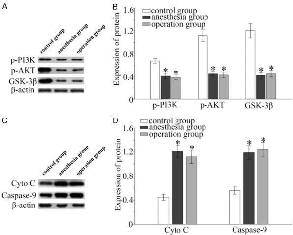
Effects of anesthesia and surgery on protein expressions of key genes in PI3K/AKT/GSK-3β pathway in rat hippocampus
Downregulated p-PI3K, p-AKT, and GSK-3β, as
well as upregulated Cyto-C and caspase-9 were found in the anesthesia group and splenecto-my group after the procedure by Western blot (Figure 4, P<0.05). Additionally, no significant
differences in the abovementioned gene ex-
age. At the cellular level, expression levels of neurotransmitters are remarkably downregu-lated, including cetylcholine (Ach), dopamine, norepinephrine, tyrosine and 5-serotonin. In contrast, activities of neurotransmitter decom-posing enzymes such as monoamine oxidase and catechol-O-methyltransferase are enhan- ced, further downregulating the intracranial neurotransmitters. In addition, slower compen-satory production of receptor speed and
reduc-tion of receptor affinity for neurotransmitter
[image:5.612.89.387.70.309.2]molecules are accompanied. AD is the most severe manifestation of senile mental disor-ders. POCD is very similar to that of the early stage of AD. Some studies considered that POCD may be the manifestation of early stages of AD [20], or POCD triggers pathophysiological process of AD [8]. Our Morris water maze exper-iments suggested that the learning and spatial abilities in rats of anesthesia group and sple-nectomy group were worse than those of sh- am group. The data indicated that anesthesia severely affects the cognitive and memory abili-ties. Abundant research has proven that hippo-campus dysfunction could lead to learning and Figure 4. Effects of anesthesia and surgery on protein expressions of key genes
in PI3K/AKT/GSK-3β pathway in rat hippocampus. A. Protein levels of p-PI3K, p-AKT, and GSK-3β in a sham group, a anesthesia group and a splenectomy group. B. Comparison of protein levels of p-PI3K, p-AKT, and GSK-3β in a sham
memory impairments, as well as decreased spatial orientation [21, 22].
PI3K/Akt pathway is widely present in cells and is important for membrane receptor transduc-tion. PI3K/Akt pathway is capable of promoting cell proliferation, differentiation, and survival by regulating cell cycle and apoptosis-related proteins.
Current studies have shown that PI3K/AKT pathway is widely expressed in the central ner-vous system and its neuroprotection role has been well studied. Dudek et al. [23] found that PI3K/Akt pathway can inhibit neuronal apopto-sis [24]. Other experiments have demonstrated that Akt can inhibit neuronal apoptosis by pr- omoting CREB activation and up-regulating Bcl-2 expression [25, 26]. In addition to its neu-roprotective effects in acute cerebral ischemia, PI3K/Akt pathway also exerts an essential role in brain trauma and chronic neurodegenerative diseases, such as AD. Traditional Chinese med-icine Herba Rhodiolae relieves cerebral edema and inhibits the occurrence of cortical neuron apoptosis through activating PI3K/Akt pathway [27, 28]. In animal models of AD, activation of PI3K/Akt pathway can antagonize the
neuro-toxic effects of Aβ and improve cognitive func -tion in rats [29-31]. Akt is activated by PI3K and regulates cell apoptosis through multiple path-ways. Among them, Akt could directly inhibits GSK-3 activity, thereafter inhibiting cell apopto-sis [32, 33]. In the present study, protein levels
of p-PI3K, p-Akt, and GSK-3β in rat hippocam -pus were remarkably downregulated in anes-thesia group and splenectomy group compared with those of sham group. However, expression levels of Cyto C and caspase-9 were upregulat-ed, indicating that the inhibition of PI3K/AKT/
GSK-3β pathway exerts a crucial role in POCD
development.
Conclusions
PI3K/Akt/GSK-3β pathway inhibits the
pos-toperative cognitive dysfunction in elderly rats after anesthesia and splenectomy.
Disclosure of conflict of interest
None.
Address correspondence to: Li Shen, Department of Oncology, Zhangjiagang Ao Yang Hospital, Zhang- jiagang 215600, Jiangsu Province, China. Tel: 86-
013915710816; E-mail: [email protected]; Hai- yan Sun, Department of Anesthesiology, Zhangjia-
gang TCM Hospital Affiliated to Nanjing University
of Chinese Medicine, Zhangjiagang 215600, Jiang- su Province, China. Tel: 86015895678696; E-mail: [email protected]
References
[1] Feinkohl I, Winterer G, Spies CD and Pischon T. Cognitive reserve and the risk of postoperative cognitive dysfunction. Dtsch Arztebl Int 2017; 114: 110-117.
[2] He X, Wen LJ, Cui C, Li DR and Teng JF. The
significance of S100beta protein on postoper -ative cognitive dysfunction in patients who derwent single valve replacement surgery un-der general anesthesia. Eur Rev Med Pharma- col Sci 2017; 21: 2192-2198.
[3] Chandrasekhar R, Ely EW and Patel MB. Chal-lenges with postoperative cognitive impair-ment research. JAMA Surg 2019; [Epub ahead of print].
[4] Chi YL, Li ZS, Lin CS, Wang Q and Zhou YK. Evaluation of the postoperative cognitive dys-function in elderly patients with general anes-thesia. Eur Rev Med Pharmacol Sci 2017; 21: 1346-1354.
[5] Lyketsos CG, Toone L, Tschanz J, Corcoran C, Norton M, Zandi P, Munger R, Breitner JC and Welsh-Bohmer K. A population-based study of the association between coronary artery by-pass graft surgery (CABG) and cognitive de-cline: the Cache County study. Int J Geriatr Psychiatry 2006; 21: 509-518.
[6] Petersen RC, Doody R, Kurz A, Mohs RC, Morris JC, Rabins PV, Ritchie K, Rossor M, Thal L and Winblad B. Current concepts in mild cognitive impairment. Arch Neurol 2001; 58: 1985-1992.
[7] Miller D, Lewis SR, Pritchard MW,
Schofield-Robinson OJ, Shelton CL, Alderson P and Smith AF. Intravenous versus inhalational mainte-nance of anaesthesia for postoperative cogni-tive outcomes in elderly people undergoing non-cardiac surgery. Cochrane Database Syst Rev 2018; 8: D12317.
[8] Arora SS, Gooch JL and Garcia PS. Postopera-tive cogniPostopera-tive dysfunction, Alzheimer’s dis-ease, and anesthesia. Int J Neurosci 2014; 124: 236-242.
[23] Dudek H, Datta SR, Franke TF, Birnbaum MJ, Yao R, Cooper GM, Segal RA, Kaplan DR and Greenberg ME. Regulation of neuronal survival by the serine-threonine protein kinase Akt. Sci-ence 1997; 275: 661-665.
[24] Crowder RJ and Freeman RS. Phosphatidylino-sitol 3-kinase and Akt protein kinase are
nec-essary and sufficient for the survival of nerve
growth factor-dependent sympathetic neurons. J Neurosci 1998; 18: 2933-2943.
[25] Du K and Montminy M. CREB is a regulatory target for the protein kinase Akt/PKB. J Biol Chem 1998; 273: 32377-32379.
[26] Pugazhenthi S, Nesterova A, Sable C, Heiden-reich KA, Boxer LM, Heasley LE and Reusch JE. Akt/protein kinase B up-regulates Bcl-2 ex-pression through cAMP-response element-bin- ding protein. J Biol Chem 2000; 275: 10761-10766.
[27] Chen SF, Tsai HJ, Hung TH, Chen CC, Lee CY, Wu CH, Wang PY and Liao NC. Salidroside im-proves behavioral and histological outcomes and reduces apoptosis via PI3K/Akt signaling after experimental traumatic brain injury. PLoS One 2012; 7: e45763.
[28] Zhi WH, Zeng YY, Lu ZH, Qu WJ, Chen WX, Chen L and Chen L. Simvastatin exerts antiamnesic effect in Abeta25-35-injected mice. CNS Neu-rosci Ther 2014; 20: 218-226.
[29] Shang YC, Chong ZZ, Wang S and Maiese K. Prevention of beta-amyloid degeneration of microglia by erythropoietin depends on Wnt1, the PI 3-K/mTOR pathway, Bad, and Bcl-xL. Ag-ing (Albany NY) 2012; 4: 187-201.
[30] Lee ST, Chu K, Park JE, Jung KH, Jeon D, Lim JY, Lee SK, Kim M and Roh JK. Erythropoietin improves memory function with reducing en-dothelial dysfunction and amyloid-beta burden in Alzheimer’s disease models. J Neurochem 2012; 120: 115-124.
[31] Echeverria V, Zeitlin R, Burgess S, Patel S, Bar-man A, Thakur G, Mamcarz M, Wang L, Sattelle DB, Kirschner DA, Mori T, Leblanc RM, Prabha-kar R and Arendash GW. Cotinine reduces am-yloid-beta aggregation and improves memory in Alzheimer’s disease mice. J Alzheimers Dis 2011; 24: 817-835.
[32] Chen Z, Yang L, Liu Y, Tang A, Li X, Zhang J and Yang Z. LY294002 and rapamycin promote coxsackievirus-induced cytopathic effect and apoptosis via inhibition of PI3K/AKT/mTOR signaling pathway. Mol Cell Biochem 2014; 385: 169-177.
[33] Song G, Ouyang G and Bao S. The activation of Akt/PKB signaling pathway and cell survival. J Cell Mol Med 2005; 9: 59-71.
[10] Monk TG and Price CC. Postoperative cognitive disorders. Curr Opin Crit Care 2011; 17: 376-381.
[11] Newman S, Stygall J, Hirani S, Shaefi S and
Maze M. Postoperative cognitive dysfunction after noncardiac surgery: a systematic review. Anesthesiology 2007; 106: 572-590.
[12] Eichenbaum H. The hippocampus and declara-tive memory: cognideclara-tive mechanisms and neu-ral codes. Behav Brain Res 2001; 127: 199-207.
[13] Bedford PD. Adverse cerebral effects of anaes-thesia on old people. Lancet 1955; 269: 259-263.
[14] Lewis MC, Nevo I, Paniagua MA, Ben-Ari A, Pretto E, Eisdorfer S, Davidson E, Matot I and Eisdorfer C. Uncomplicated general anesthe-sia in the elderly results in cognitive decline: does cognitive decline predict morbidity and mortality? Med Hypotheses 2007; 68: 484-492.
[15] Jie P, Hong Z, Tian Y, Li Y, Lin L, Zhou L, Du Y, Chen L and Chen L. Activation of transient re-ceptor potential vanilloid 4 induces apoptosis in hippocampus through downregulating PI3K/ Akt and upregulating p38 MAPK signaling pathways. Cell Death Dis 2015; 6: e1775. [16] Wagner S, Quente J, Staedtler S, Koch K,
Rich-ter-Schmidinger T, Kornhuber J, Ihmsen H and Schuettler J. A high risk of sleep apnea is as-sociated with less postoperative cognitive dys-function after intravenous anesthesia: results of an observational pilot study. BMC Anesthe-siol 2018; 18: 139.
[17] Maitra S, Baidya DK, Pawar DK, Arora MK and Khanna P. Epidural anesthesia and analgesia in the neonate: a review of current evidences. J Anesth 2014; 28: 768-779.
[18] Gong GL, Liu B, Wu JX, Li JY, Shu BQ and You ZJ. Postoperative cognitive dysfunction in-duced by different surgical methods and its risk factors. Am Surg 2018; 84: 1531-1537. [19] Alalawi R and Yasmeen N. Postoperative
cogni-tive dysfunction in the elderly: a review
com-paring the effects of desflurane and sevflu -rane. J Perianesth Nurs 2018; 33: 732-740. [20] Vanderweyde T, Bednar MM, Forman SA and
Wolozin B. Iatrogenic risk factors for Alzhei- mer’s disease: surgery and anesthesia. J Al-zheimers Dis 2010; 22 Suppl 3: 91-104. [21] Ribak CE, Nitsch R and Seress L. Proportion of
parvalbumin-positive basket cells in the GAB-Aergic innervation of pyramidal and granule cells of the rat hippocampal formation. J Comp Neurol 1990; 300: 449-461.