LEARNING TO FOCUS AND FOCUSING TO LEARN:
MORE THAN A CORTICAL TRICK
Sandeep S. Dhawan
A Thesis Submitted for the Degree of PhD
at the
University of St Andrews
2018
Full metadata for this thesis is available in
St Andrews Research Repository
at:
http://research-repository.st-andrews.ac.uk/
Please use this identifier to cite or link to this thesis:
http://hdl.handle.net/10023/15883
This item is protected by original copyright
This item is licensed under a
Creative Commons Licence
Learning to focus and focusing to learn: more than
a cortical trick
Sandeep S. Dhawan
This thesis is submitted in partial fulfilment for the degree of
Doctor of Philosophy (PhD)
at the University of St Andrews
ii
Candidate's declaration
I, Sandeep Sonny Dhawan, do hereby certify that this thesis, submitted for the degree of PhD, which is approximately 64,000 words in length, has been written by me, and that it is the record of work carried out by me, or principally by myself in collaboration with others as acknowledged, and that it has not been submitted in any previous application for any degree.
I was admitted as a research student at the University of St Andrews in September 2013.
I received funding from an organisation or institution and have acknowledged the funder(s) in the full text of my thesis.
Date Signature of candidate
Supervisor's declaration
I hereby certify that the candidate has fulfilled the conditions of the Resolution and Regulations appropriate for the degree of PhD in the University of St Andrews and that the candidate is qualified to submit this thesis in application for that degree.
Date Signature of supervisor
Permission for publication
iii
I, Sandeep Sonny Dhawan, confirm that my thesis does not contain any third-party material that requires copyright clearance.
The following is an agreed request by candidate and supervisor regarding the publication of this thesis:
Printed copy
No embargo on print copy.
Electronic copy
No embargo on electronic copy.
Date Signature of candidate
iv
Underpinning Research Data or Digital Outputs
Candidate's declaration
I, Sandeep Sonny Dhawan, understand that by declaring that I have original research data or digital outputs, I should make every effort in meeting the University's and research funders' requirements on the deposit and sharing of research data or research digital outputs.
Date Signature of candidate
Permission for publication of underpinning research data or digital outputs
We understand that for any original research data or digital outputs which are deposited, we are giving permission for them to be made available for use in accordance with the requirements of the University and research funders, for the time being in force.
We also understand that the title and the description will be published, and that the underpinning research data or digital outputs will be electronically accessible for use in accordance with the license specified at the point of deposit, unless exempt by award of an embargo as requested below.
The following is an agreed request by candidate and supervisor regarding the publication of underpinning research data or digital outputs:
No embargo on underpinning research data or digital outputs. Date Signature of candidate
v
Table of Contents
Acknowledgements ... xi
Abstract ... xii
Abbreviations ... xiii
Chapter 1
General Introduction ... 16
1.1 Impairments in executive functioning ... 19
1.2 Neuropsychological tests of executive function: sorting and shifting ... 21
1.3 Attentional set ... 25
1.4 The Basal Ganglia ... 32
1.5 Connections of the STN and ZI ... 33
1.6 Functional division of the STN and ZI ... 36
1.7 Neurochemistry of the STN and ZI ... 38
1.7.1 Glutamate ... 38
1.7.2 Dopamine ... 42
1.7.3 GABA ... 45
1.7.4 Serotonin ... 47
1.7.5 Neurochemical pathways and HFS ... 48
1.8 The STN and cognition ... 52
1.8.1 Visuo-spatial attention ... 53
1.8.2 Decision making and risky behaviours ... 58
1.8.3 Learning and memory ... 63
vi
1.9 Thesis overview ... 70
Chapter 2
General Methods... 72
2.1 Animals ... 73
2.2 Apparatus ... 74
2.3 Surgery ... 76
2.3.1 Neurotoxin ... 76
2.3.2 Injection materials ... 76
2.3.3 Surgical procedure ... 76
2.4 The rodent attentional set-shifting task ... 78
2.4.1 Training ... 78
2.4.2. Testing on the ‘standard 7-stage task’... 79
2.5 Histology ... 80
2.6 Statistical analysis ... 82
Chapter 3
Exploring set-formation with the 11-stage task ... 83
3.1 Introduction ... 84
3.2 Methods ... 87
3.2.1 Animals ... 87
3.2.2 Apparatus ... 87
3.2.3 Surgery ... 87
3.2.4 Behavioural training ... 87
3.2.5 Behavioural testing ... 87
3.2.6 Repeat testing ... 90
vii
3.2.8 Histology ... 91
3.3 Results ... 93
3.3.1 Histology ... 93
3.3.2 Study I: Examining set-formation with the 7-stage task ... 95
3.3.3 Study II: Investigating set-formation with the 11-stage task ... 97
3.4 Discussion ... 101
3.4.1 Limitations and considerations ... 101
3.4.2 Conclusions ... 104
Chapter 4
Using a palatable jelly tablet to deliver cognitive-enhancing drugs in an
‘early/late probe’ task ... 105
4.1 Experiment I ... 106
4.1.1 Introduction ... 106
4.1.2 Methods ... 116
4.1.3 Results ... 120
4.1.4 Discussion ... 122
4.2 Experiment II ... 123
4.2.1 Introduction ... 123
4.2.2 Methods ... 124
4.2.3 Results ... 128
4.2.4 Discussion ... 132
Chapter 5
Designer receptors transiently inhibit neuronal functioning in the STN
... 138
viii
5.1.1 G protein-coupled receptors (GPCRs) ... 139
5.1.2 Adeno-associated viral vectors ... 142
5.1.3 Serotype ... 143
5.1.4 Promoter ... 143
5.1.5 Efficacy of DREADDs inhibition ... 145
5.1.6 Clozapine N-oxide... 146
5.1.7 Fluorescent proteins ... 147
5.1.8 Current experiment ... 147
5.2 Materials and methods ... 149
5.2.1 Animals ... 149
5.2.2 Surgery ... 149
5.2.3 Apparatus ... 150
5.2.4 Habituation ... 150
5.2.5 Behavioural testing ... 150
5.2.6 Histology ... 151
5.2.7 Data analysis ... 152
5.3 Results ... 153
5.3.1 Study I ... 153
5.3.2 Study II ... 154
5.4 Discussion ... 160
5.4.1 Conclusions ... 160
Chapter 6
Pharmacosynthetic inhibition of the STN and ZI/LHA-area impairs the
formation of attentional set ... 161
ix
6.2 Materials and methods ... 164
6.2.1 Animals ... 164
6.2.2 Apparatus ... 164
6.2.3 Surgery ... 164
6.2.4 Behavioural training ... 164
6.2.5 Behavioural testing ... 164
6.2.6 Histology ... 165
6.2.7 Data analysis ... 167
6.3 Results ... 168
6.3.1 Histology ... 168
6.3.2 Baseline assessment using the standard 7-stage task ... 177
6.3.3 CNO-treatment impairs set-formation on the 11-stage task ... 178
6.4 Discussion ... 193
6.4.1 Memory impairment vs attentional selectivity... 193
6.4.2 Evaluating DREADDs technology ... 195
6.4.3 Conclusions ... 198
Chapter 7
Examining the relationship between associative and attentional
processes with the Overtraining Reversal Effect ... 200
7.1.1 Introduction ... 201
7.1 Experiment I ... 203
7.1.2 Materials and methods ... 203
7.1.3 Results ... 205
7.1.4 Conclusion ... 206
x
7.2.2 Materials and methods ... 207
7.2.3 Results ... 208
7.2.4 Discussion ... 220
Chapter 8
General Discussion ... 224
8.1 Findings ... 225
8.2 The role of the STN in attentional set-formation ... 229
8.3 Why the STN? ... 235
8.4 Reconciling attention and associability ... 239
8.5 Limitations: persisting with ibotenic acid lesions ... 243
8.6 Conclusions ... 245
xi
Acknowledgements
I am incredibly thankful to a number of people who have coached me, patiently listened to my ideas, and believed in my abilities throughout this four-year journey. I am indebted to my supervisor, Prof. Verity Brown, for her perceptive insight, expertise in the field, and for her great sense of humour. Thanks to Verity’s close mentorship, I emerge this doctoral experience as a more capable individual. I owe immeasurable gratitude to Dr David Tait: without his guidance and patience I would have been lost from the very start. Dave’s advice, support, technical instruction, and impromptu ‘English’ lessons made the lab an informed yet enjoyable place to work; I was extremely fortunate to have such a helpful second supervisor.
I have had the privilege and pleasure to work alongside several members of academic staff, and owe many thanks to Paul Gardner and Drs Eric Bowman, Akira O’Connor, and James Ainge, for providing an enriching environment in which to learn. For their hard work and patience I owe thanks to the members of St Mary’s Animal Unit: Mary, Wendy, Trevor, Mike, Louise, Ros, Jill, Isobel, and Jerico, whose effort made data collection possible. I would also like to thank the SH students: James, Emily and Ellen; along with Drs Christoffer Bundgaard and Shuang Xia for their patient contribution. I am indebted to Dr Andy Mackenzie whose friendship and help I will never forget, and to Charles, in whose company I found respite from PhD work in the form of Playstation gaming sessions. I am also thankful to the members of the Lilac room, who remained bright despite being in the basement, and Alonzo and the rest of the Brown lab to bounce ideas off of.
xii
Abstract
The consequence of many psychiatric and neurodegenerative disorders, such as Parkinson’s disease and schizophrenia, is an impairment in ‘executive functioning’; an umbrella term for several cognitive processes, including the focussing and shifting of attention and the inhibition of responding. The ability to form an ‘attentional set’ involves learning to discriminate qualities of a multidimensional cue, and to subsequently learn which quality is relevant, and therefore predictive of reward. According to recent research, the subthalamic nucleus (STN) and possibly the adjacent zona incerta (ZI) may mediate the formation of attentional set. Dysregulation of the STN as a result of Parkinson’s disease contributes to characteristic motor symptoms, and whilst deep-brain stimulation of this region may treat gross motor impairments, it may also impair cognition. The work in this thesis aimed to expand our understanding of the mechanisms of attentional set-formation, and the role of the STN in this process.
xiii
Abbreviations
5CSRTT 5-Choice Serial Reaction Time Task 5-HT 5-hydroxytryptomine
6-OHDA 6-hydroxydopamine AAV Adeno-associated virus
ADHD Attention-deficit/hyperactivity disorder ADS Antibody diluting solution
AMPA α-amino-3-hydroxy-5-methyl-4-isoxazolepropionic acid ANOVA Analysis of variance
ASST Attentional set-shifting task BG Basal ganglia
CaMKII Ca2+/calmodulin-dependent protein kinase II cAMP Cyclic adenosine monophosphate
CANTAB Cambridge Neuropsychological Automated Test Battery CD Compound discrimination
CeA Central nucleus of the Amygdala CNO Clozapine n-oxide
CRT Choice Reaction Time CT Circadian time
cZI Caudal Zona Incerta
DAB Sigma Fast 3.3-Diaminobenzidine DAT Dopamine transporter
DBS Deep-brain stimulation
dlPFC Dorso-lateral prefrontal cortex DMS Dorsomedial striatum
DREADDs Designer Receptors Exclusively Activated by Designer Drugs DRN Dorsal Raphe Nuclei
xiv EEG Electroencephalogram
Ef1a Human elongation factor-1-alpha EP Entopeduncular nucleus
FF Fields of Forel GABA γ-aminobutyric acid
GAD Glutamic acid decarboxylase GFAP Glial-fibrillary acidic protein GFP Green fluorescent protein
GIRK G-protein inward-rectifying potassium channel Glu Glutamate
GPe External globus pallidus GPi Internal globus pallidus
HA Human influenza haemagglutinin HFS High-frequency stimulation hsyn Human synapsin
ID Intradimensional acquisition stage IGT Iowa Gambling Task
LHA Lateral hypothalamic area LMA Locomotor activity LTP Long-term potentiation
mGluR Metabotropic glutamate receptor MGP Medial globus pallidus
MPEP 2-methyl-6-(phenylethynyl)-pyridine mPFC Medial prefrontal cortex
MPTP 1-methyl-4-phenyl-1,2,3,6-tetrahydropyridine MSNs Medium spiny neurons
NA Noradrenaline
NCD Novel compound discrimination NMDA N-methyl-D-aspartate
xv ORE Overtraining Reversal Effect
ORG ORG49209 OT Overtraining
PAM Positive allosteric modulator PB Phosphate buffer
PBS Phosphate-buffered saline PFC Prefrontal cortex
PSD Postsynaptic density Psth Posterior subthalamic area REV Reversal learning stage rGT Rat Gambling Task
RNAP Ribonucleic acid polymerase RT Reaction time
SD Simple discrimination SEM Standard error of the mean SNc Substantia nigra compacta SNr Substantia nigra reticulate SRT Simple Reaction Time SSRT Stop-signal reaction time STN Subthalamic nucleus
WCST Wisconsin Card-Sorting Task
WGTA Wisconsin General Testing Apparatus ZID Dorsal zona incerta
16
Chapter 1
General Introduction
17
It was the summer of 2014. As a first-year PhD student, I eagerly anticipated my first summer ‘vacation’ in picturesque Scotland, and decided to hire a car for the purpose of driving to the Highlands. I ventured westward and was staying the night in Ballachulish, near Glencoe, which has been lauded for its striking, natural beauty. However, there was an ‘obstacle’ which loomed over me and required addressing before setting off from St Andrews – as a Canadian who has never driven in the UK before, I have never operated a right-hand drive car.
I hesitantly departed from the east coast with this consideration in mind, and being a motoring enthusiast I felt confident in my ability to not become ‘one with nature’ by ending up in a ‘loch’. As the journey progressed, I gradually became more accustomed to driving on the left side of the road, whilst concurrently operating the gear stick with my left hand. I also kept a watchful eye out for signs for the A85, to guide me toward the Trossachs National park, whilst equally ignoring signs that would take me north, into the Cairngorms National park. Despite not having driven since leaving Canada over a year prior to this trip, I managed to arrive at my destination unscathed, and fortunately, so did the hired car. Whilst this adaptation in driving behaviour does not seem overly arduous, there existed a myriad of cognitive processes which facilitated the journey, each intersecting with harmonious complexity. In order to make this cross-Scotland expedition, I needed to:
1. remember how to operate a car, including mechanical skills such as operating a clutch pedal
2. attend to visual cues in my environment consistent with my goal, such as directional signs, and signs indicating the speed limit
3. retain navigational directions for my destination, and continually update these directions with passing road signs
4. extrapolate relevant memories from navigating Canadian roads to Scottish roads (i.e., landmark X is roughly 10 miles from the upcoming turn onto road Y)
18
6. manipulate and apply these rules to driving in the UK (look right, then left when proceeding)
7. respond flexibly to sudden changes in my internal (e.g., sudden motion sickness) or external (e.g., deer in the road) environment
8. avoid distraction from extraneous activities in my environment (e.g., a kilted bagpiper on the roadside)
The listed steps illustrate fundamental cognitive requirements for everyday life, including the ability to identify goals and organise them into a plan, retain information in memory, access and integrate relevant memories with current environmental information, all whilst remaining flexible to changes in one’s environment. These processes exemplify what is known as ‘executive control’; an umbrella term used to describe cognitive functions such as working memory, planning, mental flexibility, and the initiation or inhibition of action (for review see Chan, Shum, Toulopoulou, & Chen, 2008). Inhibition of action can be further divided based on whether the cognitive demands arise from a required change in the internal (‘self-control’: resisting temptation or impulsive action) or external (‘interference control’: selectively attending to relevant information/inhibiting irrelevant information) environment (Diamond, 2013). As in my trip to Ballachulish, the roles of many executive functions intersect, or are dependent on one another, which gives rise to whether a ‘central executive’ or an attentional-controlling system may govern executive resources (Atkinson & Shiffrin, 1971; but see: Baddeley & Hitch, 1974).
19
reconcile competing evidence for non-memory executive tasks, along with selective memory impairments in one sensory modality and not another (e.g., auditory and not vision; see Warrington & Shallice, 1969). Baddeley & Hitch (ibid) proposed a ‘working
memory’ system which simultaneously provides storage and processing of information via the central executive and two slave systems: the visuospatial sketchpad (which manipulates visual images), and the phonological loop (which stores and rehearses speech-based information; Baddeley, 1992; 1996).
Research attempting to identify the neurological substrate of the central executive has revealed that the prefrontal cortex (PFC) is the most likely contender for this role (Duncan, 2001; Miller & Cohen, 2001). Evidence which spurred early experimental research mostly stemmed from observations of aberrant behaviour after some form of trauma, irrespective of direct aetiology (i.e., tumour, injury, cerebrovascular occlusion, etc.). Increased admission rates of brain-damaged patients – typically to the frontal area – during World War II (Goldstein & Scheerer, 1941), and in some case studies, as early as the end of World War I (Gelb & Goldstein, 1920, reviewed in Goldstein & Scheerer, 1941), present as some of the earliest formalised research of executive functioning impairment. Whilst this exploration of covert cognition by examining overt behaviour paved the road for researchers in the generations to come, these experiments were predominantly qualitative case studies, with a general diagnosis of “dementia” or “amnesia”, and in the case of the World War veterans, symptoms resembling “dementia praecox” (i.e., schizophrenia; ibid). Based on this account, there appeared to be a functionally similar consequence of frontal brain damage and psychiatric symptoms.
1.1 Impairments in executive functioning
20
reduction of volume is the PFC (see: Takayanagi et al., 2010), resulting in its dysfunction, and likely contributing to the observed cognitive symptoms (Meyer-lindenberg et al., 2005). Furthermore, aberrant PFC morphology can also be used as a predictor for symptom onset, and potential development of schizophrenia amongst high-risk groups (Chakirova et al., 2010). In addition to schizophrenia, PFC dysregulation has been implicated in a plethora of psychiatric illnesses and conditions resulting if a variety of executive impairments, including attention-deficit/hyperactivity disorder (ADHD) (Bush, Valera, & Seidman, 2005), depression (Mann et al., 1996; but see: Stanley, Virgilio, & Gershon, 1982), Alzheimer’s disease (Donix, Small, & Bookheimer, 2012), frontotemporal dementia (Rohrer & Warren, 2011), Parkinson’s disease (Taylor, Saint-Cyr, & Lang, 1986), along with participating in a reward circuit facilitating drug abuse (Volkow & Fowler, 2000). In patients PFC tumours, the degree of insult varies by case, which creates heterogeneous symptomology. For example, in one case a patient scored highly in language ability, memory proficiency, visual perception, ethical reasoning and computational calculation, but was impaired in decision-making, and despite being able to solve decision-making problems in abstract circumstances (i.e., in a test or battery), the patient could not apply problem-solving solutions to ‘real-life’ situations (Eslinger & Damasio, 1985).
21
dysfunction of the caudate nucleus of the dorsal striatum (Bush et al., 2005), emphasising the importance of the fronto-striatal pathway, and more broadly the fronto-BG circuitry, in the maintenance of executive functioning.
Given this intimate connectivity, it is essential to investigate how the subcortex contributes to the maintenance of ‘processes’ typically considered executive functions. These contributory processes can be isolated and studied across a range of species to better understand how psychological concepts (e.g., working memory, inhibition, etc.) are influenced by neural substrates. It is therefore pertinent to explore how quantifiable neuropsychological tasks in humans, non-human primates and rodents have developed to probe such processes in order to shift away from qualitative case studies.
1.2 Neuropsychological tests of executive function: sorting and
shifting
22
strategies, and instead perseverated with an existing one. Despite this information, Weigl’s task still presented with shortcomings, especially since diverse sorting strategies were admissible owing to many stimulus shapes (e.g., sorting varied quadrilaterals separately)
(ibid: experiment 5). In order to address this, and in an effort to move towards examining
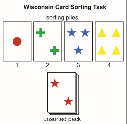
cognitive performance, rather than a simple diagnosis, the Wisconsin Card Sorting Task (WCST) was developed (Berg, 1948).
The WCST, which is still widely used today, presents subjects with a deck of cards, illustrated with a number of coloured shapes, and the subject must learn – through trial and error – the correct strategy for sorting the cards either based on the number of shapes on the card, the colour of the shapes, or the shapes themselves. The subject is requested to proceed through a deck of cards and asked to sort each ‘response card’ in to one of the four ‘stimulus card’ piles in front of them, and sorting of a ‘response card’ is followed by feedback indicating whether their selection was correct or incorrect (figure 1.1). Following attainment of learning criterion (i.e., five consecutive correct responses in Berg, 1948), the experimenter will ‘shift’ the sorting strategy to a novel one, which provides a measure of cognitive flexibility, along with a quantitative index for number of errors, which previous sorting tasks failed to measure (Grant & Berg, 1948). Healthy control participants exhibit a behavioural cost (i.e., an increase in trials and errors) when the strategy is shifted, however subsequent experiments revealed that this ‘cost’ was exacerbated in groups with executive functioning impairment, including patients with dorsolateral prefrontal cortex (dlPFC) damage (Milner, 1963), age-related cognitive decline (Ridderinkhof, Span, & Molen, 2002), schizophrenia (Nieuwenstein, Aleman, & de Haan, 2001), ADHD (Romine et al., 2004), along with neurodegenerative diseases such as Alzheimer’s disease (Nagahama, Okina, Suzuki, Nabatame, & Matsuda, 2005) and Parkinson’s disease (Paolo, Triister, Axelrod, & Koller, 1995; for review see: Brown & Tait, 2016).
23
[image:24.612.87.527.168.598.2]latter, as the name implied, required rats to jump from a small, unstable elevated platform towards one of two cardboard cut-outs differing in visual pattern, such as shape (e.g., horizontal line, triangle) and ‘brightness’ (e.g., black or white), with one stimulus pairing
24
(e.g., black triangle) being correct, whilst the other pairing (e.g., white horizontal line) was incorrect. The correct stimulus could be easily folded, allowing entry into a reward chamber with a larger platform, whilst the incorrect stimulus was immovable; overall, rats learned to discriminate between the visual patterns on the stimuli cards fairly quickly (ibid). Jacobson subsequently adapted Lashley’s procedure for work with monkeys, in which subjects were trained to retrieve food by displacing a visual stimulus card to gain access to a food well (Jacobson, 1936), which ultimately inspired the Wisconsin General Testing Apparatus (WGTA), and is still used in both monkey and human research to date (Harlow & Bromer, 1938). With this task, Harlow critiqued ‘rat psychologists’ for not focussing on how we “learn to learn”; an effect which he observed in primates and referred to as “learning set”, and can be regarded as a practice effect of sorts, arising from consecutive discrimination learning/reversal learning (Harlow, 1949).
At the same time, Lawrence investigated the effects of ‘transferring’ stimulus aspects between discrimination learning trials in rats (1949). In his experiment, rats were trained in a ‘two-choice’ (either stimulus A or B) apparatus with a ‘waiting area’ placed in front to two goal compartments, which extended linearly with a reward found at each end. The apparatus was modified such that two “stimulus dimensions” were presented in compound, with one dimension predictive of reward (‘relevant’), and the other was non-predictive (‘irrelevant’). For example, for one group of rats, the brightness of the goal compartment (black vs white paint) may be relevant, whilst the width of the compartment (or the texture of the floor) was irrelevant for obtaining reward. Following training, rats completed a testing phase in a ‘T-Maze’ with the same stimulus dimensions present, however the relevancy of the stimulus dimension was switched for half of the rats. Lawrence found that rats completed the testing phase with fewer errors if the relevant dimension stimuli were the same as the training phase (“positive transfer”), compared to rats that were tested on the previously irrelevant dimension stimuli (“negative transfer”;
25
To reconcile Lawrence’s observations, Sutherland & Mackintosh (1971) proposed a ‘two-stage’ theory of discrimination learning, which assumed that animals must learn to attend to a relevant stimulus dimension (i.e., shapes) and learn to attach the correct responses to stimuli (i.e., approach triangle; avoid square). The authors posited that stimulus input is fed into a number of ‘analysers’, which evaluate this input along particular dimensions. These analysers contain different possible outputs, which form an attachment to a given response (i.e., approach or avoid; see figure 1.2). For example, during a task in which the ‘shape’ dimension is relevant (triangle=correct; rectangle=incorrect), and the ‘brightness’ is irrelevant, the rat would receive a reward for selecting a black triangle (vs a white square). This will strengthen the ‘shapes analyser’ since the output of triangle formed an ‘approach’ response attachment (whilst square formed an avoidance response attachment). Furthermore, during this trial the ‘brightness analyser’ would also be strengthened due to partial reinforcement (i.e., the output of ‘black’ would also form an
approach response attachment); however in a subsequent trial, in which a white triangle may be presented, this brightness analyser would lose strength, upon selection of the correct stimulus. The authors’ anthropomorphised process of strengthening or ‘switching in’ analysers forms the basis for attentional set-formation; by focussing attentional resources toward a particular stimulus dimension, novel learning within this dimension is enhanced; this process can also be likened to Harlow’s process of ‘learning to learn’ (Harlow, 1949; Sutherland & Mackintosh, 1971).
1.3 Attentional set
26
[image:27.612.94.518.137.471.2]to anticipate for future events, limiting the range of available options, and increasing the speed of processing (Brown & Tait, 2010). Consequently, when faced with an unexpected
Figure 1.2: Diagram of Sutherland & Mackintosh’s two-stage theory of learning; stimulus input is fed through ‘analysers’, which are strengthened or weakened by the response attachment formed with their outputs; solid lines indicate learned response attachments; hashed lines indicate other possible response attachments; B designates black; W, white; T, triangle; S, square; L, left; R, right; adapted from Sutherland & Mackintosh (1971).
27
than acquiring an ID discrimination; the increased learning requirements arise as a result of a predisposition to selectively attend to one stimulus dimension over another. This consideration is referred to as the “Einstellung effect” (see Luchins, 1942), which posits that novel learning is retarded by the erroneous application of a previous rule or principle. It thus follows that in the absence of an attentional set, there would be no cost of learning an ED acquisition; however, in the absence of set, there would now be an existent deficit during ID learning, when an attentional bias would have facilitated novel learning.
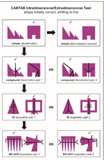
A measure of set-shifting performance presents as a valuable index of several cognitive processes for clinical and experimental neuropsychology, and therefore various tasks have been developed in both humans and animals. These attentional set-shifting tasks employ a ‘total change’ design (Slamecka, 1968), which presents a series of discriminations, including stages in which completely novel exemplars are introduced, along with reversal learning stages. These total change design tasks can be contrasted to ‘rule change’ set-shifting tasks’ (otherwise known as ‘rule-set-shifting tasks) (Settlage, Butler, & Odoi, 1956), such as the WCST, which maintain the same stimuli throughout the task. This thesis will exclusively focus on a rodent attentional set-shifting task of total change (Birrell & Brown, 2000), which owes its conception to the Cambridge Neuropsychological Automated Test Battery (CANTAB) (Roberts, Robbins, & Everitt, 1988; Sahakian & Owen, 1992).
28
[image:29.612.141.474.81.593.2]29
flexibility amongst brain-damaged patients and those suffering from mental illness (Lawrence, Sahakian, & Robbins, 1998; Owen et al., 1991; 1993). Excitotoxic lesion studies in monkeys have provided invaluable findings regarding the functional architecture of the PFC (Dias, Robbins, & Roberts, 1996a; Dias et al., 1996b; Roberts, Robbins, Everitt, & Muir, 1992); however the monkey task reportedly takes several weeks to complete (9-11 weeks in Roberts et al., 1992), which complicates the dosing schedule for pharmacological studies, along with increased odds of neuroplasticity throughout the post-surgery test period. To account for this, the CANTAB ID/ED task was adapted for use in rats.
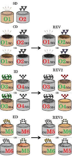
It is well-documented that rats are not particularly visual animals, and possess poor visual acuity for detail (Birch & Jacobs, 1979) and being nocturnal, rats typically flee from brighter environments. Instead, rats rely on their highly attuned sense of smell and touch as primary investigative senses. Whilst previous discrimination learning tasks have employed visual stimuli (Lawrence, 1949; Mackintosh & Little, 1969), training and testing usually spanned hundreds of trials (Bussey, Muir, Everitt, & Robbins, 1997) for rats to attain criterion learning, providing little refinement from the CANTAB; therefore designing a species-appropriate task, which targets the rats’ natural tendencies was paramount for the rodent ID/ED task. Work by Wood, Dudchenko, & Eichenbaum (1999) found that rats can be trained to dig in small bowls filled with sawdust to obtain a food reward, which suited their natural digging and foraging behaviour. In a follow-up experiment, the authors added various odours to bowls of sand, some of which were predictive of reward in an effort to investigate non-spatial memory in rats (Dudchenko, Wood, & Eichenbaum, 2000). Birrell & Brown (2000) expanded on the fundamental principle of this task, by introducing various digging media, and textured covers for the digging bowls as stimulus dimensions, which could be used alongside odour cues as another dimension.
30
technical refinement to examining set-shifting performance in animals. Furthermore, lesions to the rat medial PFC (mPFC), which is the rodent analogue of the primate lateral PFC, led to comparable behavioural deficits – namely impaired set-shifting performance marked by increased trials at the ED stage, whilst lesions of the rat orbital PFC (OFC; structurally analogous in the primate) matched the impaired reversal learning performance seen in primates (Birrell & Brown, 2000; Brown & Bowman, 2002; Dias, Robbins, & Roberts, 1997; McAlonan & Brown, 2003).
Research by Dias et al. (1996a) posited that lesioning two different regions of the PFC (e.g., lateral vs orbital) produced a ‘double-dissociation’; they evidence that a lesion of the lateral PFC does not disrupt reversal learning, and that the OFC does not likely play a role in attentional set. More broadly, this implied that the mechanisms for reversal learning were different to those involved in attentional set. It is important to note that the OFC-lesioned monkeys in Dias et al. (1996a) took twice as many trials to acquire the ID compared to control and lateral-PFC lesioned animals, yet the data regarding the cost of set-shifting for this group was not reported. The OFC-mediated reversal learning impairment was subsequently replicated in rats (McAlonan & Brown, 2003), and owing to a revised task design in which reversal stages were placed before the ED stage (the reversal stages in Dias were presented after the ED), it was found that lesioned rats failed to present with a ‘positive cost’ of shifting set – in which the ED is greater than the ID – which led McAlonan & Brown to speculate whether an attentional set had even been formed.
31
32
Further research suggested that the impairment in attentional set-formation may involve several neural substrates. It was found that rats with quinolinic acid (QA) lesions of the dorsomedial striatum (DMS) do not present evidence for attentional set-formation, even on the 4-ID task (Lindgren, Wickens, Tait, Brown, & Dunnett, 2013), suggesting that the DMS-mediated set-formation impairment manifests differently than the OFC-mediated impairment. Furthermore, lesions to the non-cholinergic neurons of the rat basal forebrain (Tait & Brown, 2008) may also impair set-formation, as no ID/ED difference was observed, along with replicating the previously observed impairment in reversal learning in monkeys (Roberts et al., 1992); however further research specifically investigating set-formation is needed.
Recent experimental evidence from our lab has suggested that the subthalamic nucleus (STN) and possibly the zona incerta (ZI) may also play a role in the formation of attentional set (Tait, Phillips, Blackwell, & Brown, 2016). The STN and the ZI are anatomically adjacent regions of the BG and are located ventral to the thalamus. Of these two neural substrates, the STN garners considerably more research interest than the ZI. The STN is a key input structure in the BG, responsible for regulation of many components of the BG itself, along with receiving projections from the PFC (Baunez & Lardeux, 2011; Nambu, Tokuno, & Takada, 2002), whereas the ZI, which lies dorsal to the STN and is comprised of a dorsal (ZID) and ventral (ZIV) component, actually remains among some of the least-studied regions in the brain (Urbain & Desche, 2007).
1.4 The Basal Ganglia
33
nigra (SN), in order to provide feedback to the cortex via the ventrolateral thalamus (Alexander, DeLong, & Strick, 1986). Over the next few years, this information was formalised into the first coherent model of BG functioning (figure 1.5a), which included two circuits of motor control – the ‘direct’ and ‘indirect’ pathways (Albin, Young, & Penney, 1989; DeLong, 1990; Penney & Young, 1986). Both pathways originate from the striatum, with the ‘direct’ pathway resulting in an increase in thalamo-cortical activity, and the ‘indirect’ pathway, a decrease in thalamo-cortical activity. This created a “rate model” of BG functioning, in which the ‘direct’ pathway facilitated movement, and the ‘indirect’ pathway inhibited/stopped movement. In the ‘direct’ pathway, medium spiny neurons (MSNs) of the striatum express D1-type dopamine receptors, which exert tonic, inhibitory
control over the SN-reticulata (SNr) and internal globus pallidus (GPi), which are now inhibited, and therefore their projection to the thalamus results in increased thalamo-cortical activity (Albin et al., 1989). In the ‘indirect’ pathway, MSNs expressing D2-type receptors
from the striatum inhibit the external globus pallidus (GPe), which attenuates the GPe’s inhibitory effect on the STN, which in turn stimulates the SNr and GPi, resulting in decreased thalamo-cortical activity (ibid). The modulation of striatal dopamine is crucial for movement, and as expected dopaminergic depletion – the key feature of the Parkinsonian state – renders the STN ‘hyperactive’ and consequently enhances activation of the ‘indirect’ pathway (Gerfen et al., 1990; Obeso & Lanciego, 2011). In turn, silencing the STN via deep-brain stimulation (DBS) has become a popular therapeutic option for amelioration of motor symptoms of Parkinson’s disease (Benabid et al., 1994).
1.5 Connections of the STN and ZI
34
Takada, Inase, & Tokuno, 1996; 1997), yet there was additionally evidence from tracing studies that showed that frontal ‘cognitive’ areas, such as the mPFC and OFC, directly project to the STN as well (Janssen, Visser-Vandewalle, & Temel, 2010). It is suggested that the fronto-subthalamo pathway operates in competition with the ‘direct’ pathway (Leblois, Boraud, Meissner, Bergman, & Hansel, 2006), and that this pathway is fast-acting, compared to the slower striatal pathways (Isoda & Hikosaka, 2008; Nambu, 2004), which may work to suppress activation of the ‘direct’ pathway (Aron & Poldrack, 2006a). This connection, coupled with the finding that the STN also has a direct output projection to the ventral thalamus (Rico et al., 2010) – therefore bypassing the pallidum – likely suggests that the STN is crucial in the rapid control and selection of responses.
The STN may also play a modulatory role in inter-BG circuitry, and the identification of an STN-GPe-GPi ‘microcircuit’ also ascribed a new role to the GPe for controlling BG output activity, rather than merely being a ‘go-through’ station in an ‘indirect’ pathway (Obeso, Rodriguez-oroz, Blesa, & Guridi, 2006). The reciprocal connection between the STN and the GPe normally exhibits weakly correlated, irregular activity (Urbain et al., 2000); however, following dopamine depletion in Parkinson’s disease, this reciprocal connection exhibits highly correlated, rhythmic bursting activity (Bergman, Wichmann, Karmon & De Long, 1994), which likely contributes to the increased downstream inhibition of the thalamocortical neurons, which may eventually express as akinesia and rigidity.
In light of these new connections and updated roles for functioning, the view that the BG operates as an ‘on-off’ pathway for action selection is outdated; current modelling suggests that the BG operates via a series of parallel, largely segregated re-entry loops or channels (see Redgrave, Vautrelle, & Reynolds, 2011), which receive input from functionally segregated areas of the cerebral cortex (i.e., limbic, associative, sensory and motor; for review see Mcgeorge & Faull, 1989) in concert with the dense, and intimately interconnected inter-BG modulatory networks (i.e., the STN-GPe-GPi microcircuit), which contribute to the shaping of output behaviour.
Figure 1.5: a) An illustration of the classic Albin-DeLong model of the Basal Ganglia; red lines indicates excitatory projections, blue lines inhibitory, and green lines dopaminergic. Solid arrows indicate the ‘direct’ pathway, and hashed lines the ‘indirect’ pathway, in which the STN was seen as a relay centre; b) A revised model of Basal Ganglia connections based on recent findings, illustrating a revised, and increasingly complex, role for STN functioning (simplified from Obeso and Lanciego, 2011).
36
each subdivision gives rise to a distinct pattern of efferent projections (for review see Romanowski, Mitchell & Crossman, 1985; also Watanabe & Kawana, 1982). Early research has demonstrated that the rat ZI receives afferent inputs from the thalamus (Swanson, Cowan & Jones, 1974), hypothalamus (Krieger, Conrad & Pfaff, 1979), brain stem (Bowsher, 1975) and the cerebellum (Faull & Carman, 1978). More recent research has determined that projections originating in the cerebral cortex terminate in the ZI, and that the ZI and the STN may work together to process this cortical information, as it was found that cortical representations in these regions have highly overlapped borders, primarily from the layer V neurons of the cortex, but also from areas implicated in higher cognitive functioning, such as the OFC and mPFC (Kita, Osten, & Kita, 2014). Recent research has also evidenced that the ZI projections to cortical layer I of the neonatal rodent brain are crucial during development and maturation, as disrupting this pathway during the first postnatal week increases the likelihood of epileptiform activity in the adult brain (Chen & Kriegstein, 2015).
The ZI also projects to – in some cases reciprocally – and exhibits predominantly inhibitory influence to several key areas of the brain, including the cortex, BG, brainstem, thalamus and spinal cord (for review see Mitrofanis, 2005; Power, Kolmac & Mitrofanis, 1999). The inhibitory projection to the dorsal thalamus (Power, Kolmac, & Mitrofanis, 1999), exerts a powerful influence on the firing properties of thalamic neurons (Bartho et al., 2002). These projections mostly originate in the ZIV – which lies adjacent to the dorsal STN – and plays a role in vibrissal sensorimotor functioning (Shaw, Liao, Chen, Huang, & Lin, 2013; Urbain & Desche, 2007), and furthermore, this thalamic projection also extends into the whisker representation regions of the primary somatosensory cortex (Nicolelis, Chapin, & Lin, 1995), ultimately forming a reciprocal connection with the ZI.
1.6 Functional division of the STN and ZI
37
Both the primate and rodent STN receives BG input via the GPe, yet the STN also receives direct afferent input from the cortex, which may project differentially within the nucleus itself, and by species.
In the primate, three functional divisions of the STN have been circumscribed: a dorsolateral ‘motor’ part, a medial ‘limbic’ part, and a ventrolateral ‘associative’ part (Parent & Hazrati, 1995). The dorsolateral section projects to sensorimotor territories in the putamen and GPe, the medial section projects to limbic regions of the ventral GPi, whilst the ventrolateral associative (cognitive) regions project to the GPi/SNr and the caudate nucleus (Joel & Weiner, 1997; Parent & Hazrati, 1995). Advancement in anterograde tracing technology has revealed a considerable convergence of projections from functionally diverse cortical areas into the primate STN, creating potentially important interfaces between terminal fields, which appear to overlap rather than be anatomically distinct (Haynes & Haber, 2013). For example, limbic projections from the cortex to the medial STN also extend into the adjacent lateral hypothalamus (LH), and that the processing of limbic information is performed by both regions, suggesting that the lateral LH might be considered the limbic cone of the STN (ibid; Berthoud & Munzberg, 2011).
38
STN’s contribution to cognitive functioning, including the experiments in the current thesis, would benefit by targeting the frontal connections of the medial STN.
Research evidencing a functional division of the ZI is sparse, yet anatomical tracing studies have shown that the ZI receives input from the cortex, BG, brainstem, and spinal cord, leading to partially overlapping regions which contribute differential effects on behaviour, including controlling visceral activity, influencing arousal, orienting visual attention, and maintaining posture and locomotion (Lin, Nicolelis, Schneider, & Chapin, 1990; Mitrofanis, 2005). For example, lesioning or stimulating the rostral ZI (and no other region) will decrease food/water intake, changing ingestive activity (Tonelli & Chiaraviglio, 1995); however the same procedure when applied to the caudal ZI induces changes in posture and locomotion (Edwards & Isaacs, 1991).
Most of the rostral ZI and the ZID receive limbic projections from the cingulate, and the majority of the ZIV and caudal ZI receive projections from the somatosensory cortex (ibid). In addition to a rostral-caudal and dorsal-ventral regional division of functioning, the medial third of the ZI contain projections from the dorsomedial frontal and primary motor cortices; projections which also extend into the adjacent LH (ibid; Lin et al., 1990). In addition to the identified subdivisions, it is worth noting that this ‘subthalamic region’, including the STN, ZI, and LHA, with converging and overlapping cortical projections from the mPFC and OFC, likely works in concert to some degree to shape output behaviour (Kita et al., 2014).
1.7 Neurochemistry of the STN and ZI
1.7.1 Glutamate
Considerable research has shown that the dominant postsynaptic receptor in the STN is the glutamate receptor (for review see Clarke & Bolam, 1998). Pioneering electrophysiology research by Robledo & Féger (1990) demonstrated that the STN exerts an excitatory effect on its efferent structures, and that furthermore, chemical blocking of STN neuronal activity with muscimol [a γ-aminobutyric acid receptor-type A (GABAA)
39
presented new evidence for glutamatergic transmission in subthalamic projections. Subsequent neurophysiological tracing research spearheaded by Atsushi Nambu spanning nearly a decade (1996; 1997; 2000; 2002) built on the seminal findings posited by Hartmann-von Monakow et al. (1978), which originally identified that the STN receives direct excitatory cortical projections from wide-spanning areas of the frontal cortex, including the supplementary motor area (Nambu et al., 1996) and premotor area (Nambu et al., 1997), along with glutamatergic projections from the thalamus and brainstem (Joel & Wiener, 1997; Mathai & Smith, 2011). The ‘hyperdirect’ pathway is the chief excitatory (glutamatergic) input for the STN, and work by Nambu et al. (2000) found that stimulation of the primary motor and somatosensory cortices via implanted electrodes in monkeys resulted in a pattern of early, short-latency excitation, followed by a late excitation in the STN, GPe and GPi, in which the early excitation of the STN preceded that of the early excitation for the GPe/GPi. Similar to Robledo & Féger (1990), STN-muscimol treatment in Nambu et al. (2000) abolished both the early and late excitation events in the measured GP neurons following cortical stimulation. Building on Robledo & Féger (1990), Nambu (2000) found that injections of N-methyl-D-aspartate (NMDA) receptor antagonists, and
not α-amino-3-hydroxy-5-methyl-4-isoxazolepropionic acid (AMPA) and kainate receptor antagonists into the STN attenuated the early and late excitations found in the GP, which suggests that the ‘hyperdirect’ pathway (cortico-subthalamic) is largely mediated by NMDA receptors. These fast-acting ionotropic receptors (compared to slower-acting metabotropic receptors) convey motor-related cortical information to the GP via the STN, with shorter conduction time than the effects conveyed through the striatum (Nambu et al., 2000).
Of the NMDA receptor, the NR1-type receptor is the most prevalent, and despite
NMDA being the most prominent ionotropic glutamate receptor type in the STN, and despite the findings in Nambu et al. (2000), there also exists evidence that AMPA may play a role in modulating STN activity during a state of nigrostriatal dopamine denervation (i.e.,
40
from the fact that AMPA mediates faster neurotransmission than NMDA (for review see: Ozawa, Kamiya & Tsuzuki, 1998), along with cascades of compensatory neurotransmission within the BG during a dopamine-denervated state (see section 1.7.2; for review see: Galvan, Kuwajima & Smith, 2006). Furthermore, Mouroux & Féger (1993) demonstrated that the expected pattern of STN excitation following electrophysiological stimulation of the parafascicular nucleus of the thalamus was silenced following both NMDA- and AMPA-antagonism, suggesting that both of these ionotropic receptors contributed to STN excitation.
In addition to the ionotropic glutamate receptors of the STN, there exists a population of metabotropic glutamate receptors (mGluRs); these are mostly asymmetrical and colocalised on a single synapse with the ionotropic receptors (Porter, Greene, Higgins, & Greenamyre, 1994). Whilst the ionotropic receptors contribute to fast-acting, excitatory behaviour, the slower-acting mGluRs regulate sustained depolarisation (Ozawa, Kamiya & Tsuzuki, 1998); the colocalised organisation of these two receptor types leads to complex glutamate signalling in the STN (Clarke & Bolam, 1998). There are three distinct families of mGluR (Group I, II, & III) each differing based on their mechanism of action and cellular response outcome; for example, activation of Group I mGluRs – consisting of mGluR1 and mGluR5 – leads to a net excitatory response by inducing a mobilisation of
intracellular Ca2+, along with increasing NMDA (Kuwajima, Hall, Aiba, & Smith, 2004). Conversely, Group II mGluRs, which include mGluR2 and mGluR3, are negatively coupled
to adenylyl cyclase, and yield an inhibitory effect on signal transduction (Conn & Pin, 1997; Kuwajima et al., 2004).
In vitro electrophysiology studies have determined that Group I (both mGluR1 and
mGluR5) receptor types are found in the STN of the rodent (Awad, Hubert, Smith, Levey,
& Conn, 2000) and non-human primate (Clarke & Bolam, 1998; Kuwajima et al., 2004; Wang, Ong, Lee, & Huganir, 2000), and that these receptors are mostly localised on postsynaptic dendrites of the STN, and may contribute to an important role in net excitatory drive to several key areas of the BG, including the GPe and GPi, along with the thalamus (Galvan Kuwajima & Smith, 2006; Obeso & Lanciego, 2011). Group II (both mGluR2 and
41
STN terminals; the activation of these receptors inhibits excitatory transmission at STN synapses (Bradley et al., 2000). However, both mGluR2 and mGluR3 can also be found
postsynaptically, and it is postulated that this distribution may aid in regulating output activity of the STN (Clarke & Bolam, 1998; Tamaru, Nomura, Mizuno, & Shigemoto, 2001; Wang et al., 2000).
The usually regular, tonic activity of glutamatergic neurons in the STN (Bevan & Wilson, 1999; Nakanishi, Kita, & Kitai, 1987) is rendered irregular in the Parkinsonian brain, with bursts of high-frequency spikes; this dysregulation appears to be essential for clinical manifestation of the disease (Porter et al., 1994; Tai et al., 2003; Vila et al., 1999). Furthermore, 6-hydroxydopamine (6-OHDA) lesions of the SNc – leading to striatal dopamine depletion – serves as a Parkinsonian disease model, partly by increasing glutamatergic output activity from the STN (for review see Henderson & Dunnett, 1998). During this state of glutamate hyperactivity (i.e., Parkinson’s disease), application of an
mGluR5 antagonist (2-methyl-6-(phenylethynyl)-pyridine; MPEP), has been shown to
regulate activity, resulting in an attenuation of severe motor and sensorimotor asymmetries (Phillips, Lam, Ackerson, & Maidment, 2006).
42
possible that the cells labelled downstream in the SN and EP do not fully represent the breadth of connections of the ZI, whereas Nicolelis, Chapin & Lin, 1995 used retrograde tracers from the primary somatosensory cortex to map out subdivisions within the ZI. It is clear that glutamate signalling in the ZI influences output behaviour, as infusions of both AMPA or kainic acid agonists (but not NMDA) directly to the medial or rostral ZI induce a marked stimulation of locomotor activity, along with a postural change, and that furthermore, administration of the respective antagonists resulted in an inhibition of locomotor activity (Supko, Uretsky & Wallace, 1991). These excitatory projections place the ZI in a position to induce a modulatory effect on BG and cortical activity, and merit further investigation in an effort to disseminate the subpopulations of neurotransmitters within this area.
1.7.2 Dopamine
It has been documented through retrograde tracing and immunohistochemistry studies that the rat STN receives dopaminergic projections from the SNc and the ventral tegmental area (VTA) (Brown & Wolfson, 1978; Brown et al., 1979; Campbell et al., 1985; Hassani, Francois, Yelnik, & Feger, 1997), including sparse arborisations via the striatum (Gauthier, Parent, & Levesque, 1999). This dopaminergic projection has also been identified in the cat (Meibach & Katzman, 1979; Rinvik et al., 1979), and the primate (Rinvik et al., 1979; Galvan et al., 2014) STN; however there is less robust evidence for this in the human brain, with weak, albeit specific binding for D1-type dopamine receptors
(Augood, Hollingsworth, Standaert, Emson, & Penney, 2000). Indeed several studies have documented that the D1, D2, D3, and D5 receptor messenger RNAs and binding sites were
present in the rodent STN (Flores et al., 1999; Svenningsson &Le Moine, 2002; Baufreton et al., 2003), highlighting a potentially diverse profile of dopamine modulation within the nucleus.
Functionally, earlier in vivo research suggested that dopamine largely had an inhibitory effect on STN neurons via D1 and D2 dopamine receptors (Campbell et al., 1985;
Hassani & Féger, 1999), whilst another group of researchers found that D1 agonism had a
43
D5) increases burst firing in subthalamic neurons, therefore shaping neuronal activity
(Baufreton et al., 2003). There is also evidence that activation of D2-like receptors are
implicated in excitatory synaptic transmission, with a different mechanism of action compared with D1-like receptors (Zhu, Shen & Johnson, 2002; Floran, Floran, Erlij &
Aceves, 2004; Galvan et al., 2014). These D2-like receptors influence firing either
presynaptically or postsynaptically: binding with D2 and D3 receptors reduces resting K+
conductance, and thus promotes firing (presynaptic; Zhu, Shen & Johnson, 2002; Baufreton & Bevan, 2008), whereas selective D4 agonism decreases GABA release to the STN (see
section 1.7.3), thus reducing inhibition of the nucleus (postsynaptic: Shen & Johnson, 2003; Floran, Floran, Erlij & Aceves, 2004). Furthermore, the fact that an inward current induced by dopamine superfusion (in vitro) was observed, despite the application of tetrodotoxin (a sodium channel blocker) and ionotropic glutamate receptor antagonists [both AMPA/KA antagonists: (±)-sulpiride and 6-cyano-7-nitroquinoxaline-2,3-dione (CNQX) and NMDA: 2-amino-5-phosphonopentanoic acid (AP5)], suggests that dopamine exerts a direct action on postsynaptic STN neurons (Shen & Johnson, 2003).
44
D2 and D3 receptor levels in the nucleus accumbens (Carcenac et al., 2015). This may
partially explain the therapeutic efficacy of STN-HFS to treat motor symptoms of Parkinson’s disease (see section 1.7.5).
Functionally, in vivo studies have found that dopaminergic inputs to the STN may be crucial in regulating movement, as the application of apomorphine (dopamine agonist) induced an increase in abnormal, nondirected orofacial movements; this behaviour may largely be driven by D1-like receptors, since D1-like and not D2-like receptor antagonists
blocked the expression of this behaviour (Parry et al., 1994). In the absence of a dopamine agonist, chemical antagonism of the D1-like receptors in the STN results in catalepsy in rats
(Hauber, 1998). These results support the assertion that dopamine plays a prominent role in the regulation of motor functions, and excitatory drive in the STN.
Within the ZI, a sparse population of dopaminergic neurons have been exclusively identified in the rostral sector, and are found in the medial region (both dorsal and ventral) in both the cat and the rat; these cells comprise roughly 15% of the total number of cells within this region (Wagner, Eaton, Moore & Lookingland, 1995; Cheung, Ballew, Moore, & Lookingland, 1998; Kolmac & Mitrofanis, 1999; Mitrofanis, 2005). These sparse dopaminergic cells have been shown to project to a variety of brain areas, including ipsilateral projections to the adjacent lateral hypothalamus – which contains a large, densely populated group of dopaminergic cells – neighbouring regions such as lateral preoptic area and the limbic structures at the diencephalic-telencephalic juncture, along with more distal regions such as the central nucleus of the amygdala, horizontal diagonal band of Broca, and the paraventricular nucleus (Wagner et al., 1995; Cheung, Ballew, Moore, & Lookingland, 1998; Kolmac & Mitrofanis, 1999).
Research regarding the role of these neurons in behaviour is still limited, with earlier work evidencing that direct injection of a D1-receptor agonist to the ZI had a
stimulatory control on the release of lutenising hormone and occurrence of ovulation, and selective D1-receptor antagonism inhibited ovulation; conversely chemical manipulation of
the D2-receptor had no effect on sexual cycles (James, Mackenzie, Tuohy-Jones & Wilson,
45
whilst a D1-receptor agonist had no effect, suggesting that D2 receptors may modulate
ingestive behaviour.
1.7.3 GABA
The principal inhibitory neurotransmitter, GABA, exerts its effects via two ligand-gated channels (GABAA and GABAC) and one regulated via G-protein (GABAB) (Jones et
al., 1998). An important inhibitory input to the STN originates from the GPe, which uses both GABAA and GABAB to regulate STN output activity, and has been found in both
monkeys (Galvan, Charara, Levey, & Smith, 2004) and rats (Bell, Churchill, & Kalivas, 1995). GABAergic boutons from the GPe form symmetric synapses on the STN, which differs from the asymmetric glutamatergic afferents (Smith, Bevan, Shink, & Bolam, 1998). This inhibitory projection from the GPe is critical in shaping STN output activity via the indirect pathway, as it has also been shown that administration of the GABA antagonist bicuculline significantly increases STN activity (Robledo & Féger, 1990). As outlined in the preceding sections, STN activity is largely shaped by glutamate binding, but may also be influenced by dopamine binding; work by Floran et al. (2004) found that selective D4
receptor agonism inhibited GABA release from the GPe to the STN, which would in turn increase STN activity. In addition to the D4 receptor, Baufreton & Bevan (2008) detailed
that dopamine binding at presynaptic D2-like receptors attenuates GABAergic transmission
(via the GABAA receptor). Parkinson’s disease is marked by a reduction in GABA and
excess glutamate signalling, which contributes to STN hyperactivity, likely resulting from nigrostriatal dopamine denervation (El Arfani et al., 2014).
46
is a reliable, specific marker for GABA neurons (Kaufman, Houser & Tobin, 1991; Gonzales, Kaufman, Tobin & Chesselet, 1991), along with the use of in situ hybridisation histochemistry for GABA transporter 1 (Yasumi et al., 1997). These techniques have been applied to the rat (Oertel & Mugnaini, 1984; Yasumi et al., 1997), monkey (Benson, Isackson, Hendry & Jones, 1991) and human (Nisbet et al., 1996) STN, which revealed few positively labelled cells within the nucleus suggesting that the existence of GABAergic cells in the STN may be minimal. Despite the advancement of these studies, the neuronal nature of these GABAergic cells has not been determined, nor is there a description of the morphological features. In a more recent study, Lévesque & Parent (2005) performed GAD immunohistochemistry on post mortem human brain tissue and revealed that these GABAergic cells comprise 7.5% of the total neuronal population of the STN, and are more abundant in the caudal-ventral-medial section of the nucleus, which is largely implicated in limbic and associative functioning.
GABAergic cells are the most populous neurotransmitter founding in the ZI, and are most densely located in the ZIV, but can be found throughout the entire nucleus (Nicolelis
et al., 1995; Mitrofanis, 2005; Park, Hoffman, & Keller, 2014). Furthermore, the
projections originating in the ZI to the cortex (incertocortical projection) and the dorsal thalamus (incertothalamic projection) are also GABAergic, and in the case of the thalamus, the ZI provides a significant inhibitory influence via the GABAA and GABAB receptor on
47
cytoarchitectonic subdivisions of the ZI, the predominant distribution of GABAergic cells in the ZIV overlap with the glutamatergic cells in the ZID, and it has been speculated whether this inhibitory ventral region and excitatory dorsal region may divide ZI function (Mitrofanis, 2005).
1.7.4 Serotonin
Early tracing studies found a distribution of serotonin (hydroxytryptomine; 5-HT)-immunoreactive nerve fibres in the monkey and rat STN, mostly in the ventral and medial regions of the nucleus, and were more likely found in the caudal sections in the rat (Mori, Takino, Yamada, & Sano, 1985). Recent research has identified that these projections originate in the dorsal raphe nuclei (DRN) and that there are several types of 5-HT receptor in the STN, including evidence of 5-5-HT1A, 5-HT1B, 5-HT2C and 5-HT4
receptors, although only a sparse number of axon terminals were observed in the STN (for review see Reznitsky, Plenge, & Hay-schmidt, 2016; see also Stanford, Kantaria, Chahal, Loucif, & Wilson, 2005).
The effects of 5-HT in the STN can result in two distinct effects on the same neuron: excitation is mediated by the 5-HT2C and 5-HT4 receptors, whilst inhibition is
mediated by the 5-HT1A/B receptor (Stanford, Kantaria, Chahal, Loucif, & Wilson, 2005).
Furthermore, application of exogenous 5-HT or a non-specific 5-HT2 agonist increases
STN neuron firing rate, without changing STN firing pattern, and that these excitations were reduced by 5-HT2C and 5-HT4 receptor antagonists (Stanford, Kantaria, Chahal,
Loucif, & Wilson, 2005; Xiang, Wang, & Kitai, 2005). Application of 5-HT elicits a biphasic response on STN neurons – an initial excitation event, followed by inhibition – which were maintained even after application of picrotoxin (a GABAA antagonist) and
CNQX (AMPA/KA antagonist); however application of a 5-HT1A antagonist blocked this
inhibitory event, which suggested that although HT receptors are sparse in the STN, 5-HT-mediated excitation and inhibition events are separate entities that arise from direct postsynaptic receptor mediated effects (Stanford, Kantaria, Chahal, Loucif, & Wilson, 2005). It has also been shown that activation of the 5-HT1B receptor inhibits
48
(100µM) of 5-HT was required to inhibit the GABA-mediated inhibitory postsynaptic currents to a comparable extent (Shen & Johnson, 2008). These findings suggest that 5-HT, similar to dopamine, may exercise a regulatory role in controlling STN output activity.
In the rat ZI, serotonin-immunoreactive cells are sparsely found throughout all regions, with no particular area of concentration, making up around 2% of the total cell population in the dorsal, ventral and caudal sectors and around 5% of the total cell population in the rostral sector (Kolmac & Mitrofanis, 1999). In the ZI, 5-HT plays a role in neuroendocrine function (see Kordon et al., 1980), and it is thought that the serotonergic projections from the medial ZI may modulate prolactin and gonadotrophin secretion (Bosler, Joh, & Beaudet, 1984). Further, and more recent, research found additional evidence that 5-HT in the ZI is implicated in neuroendocrine function: application of 5-HT directly to the ZI inhibits the secretion of lutenising hormone, which in the ZI is mediated by 5-HT7 receptor activation (Siddiqui, Abu-amara, Aldairy, Hagan, & Wilson, 2004).
1.7.5 Neurochemical pathways and HFS