R E S E A R C H
Open Access
Sulforaphane rescues amyloid-
β
peptide-mediated decrease in MerTK expression
through its anti-inflammatory effect in
human THP-1 macrophages
Kyoung A. Jhang
1, Jin-Sun Park
2, Hee-Sun Kim
2*and Young Hae Chong
1*Abstract
Background:Mer tyrosine kinase (MerTK) activity necessary for amyloid-stimulated phagocytosis strongly implicates that MerTK dysregulation might contribute to chronic inflammation implicated in Alzheimer’s disease (AD) pathology. However, the precise mechanism involved in the regulation of MerTK expression by amyloid-β(Aβ) in proinflammatory environment has not yet been ascertained.
Methods:The objective of this study was to determine the underlying mechanism involved in Aβ-mediated decrease in MerTK expression through Aβ-mediated regulation of MerTK expression and its modulation by sulforaphane in human THP-1 macrophages challenged with Aβ1-42. We used protein preparation, Ca2+influx fluorescence imaging, nuclear fractionation, Western blotting techniques, and small interfering RNA (siRNA) knockdown to perform our study. Results:Aβ1-42 elicited a marked decrease in MerTK expression along with increased intracellular Ca2+level and induction of proinflammatory cytokines such as IL-1βand TNF-α. Ionomycin A and thapsigargin also increased intracellular Ca2+levels and production of IL-1βand TNF-α, mimicking the effect of Aβ1-42. In contrast, the Aβ1-42-evoked responses were attenuated by depletion of Ca2+with ethylene glycol tetraacetic acid. Furthermore, recombinant IL-1βor TNF-αelicited a decrease in MerTK expression. However, immunodepletion of IL-1βor TNF-αwith neutralizing antibodies significantly inhibited Aβ1-42-mediated downregulation of MerTK expression. Notably, sulforaphane treatment potently inhibited Aβ1-42-induced intracellular Ca2+level and rescued the decrease in MerTK expression by blocking nuclear factor-κB (NF-κB) nuclear translocation, thereby decreasing IL-1βand TNF-αproduction upon Aβ1-42 stimulation. Such adverse effects of sulforaphane were replicated by BAY 11-7082, a NF-κB inhibitor. Moreover, sulforaphane’s anti-inflammatory effects on Aβ1-42-induced production of IL-1βand TNF-αwere significantly diminished by siRNA-mediated knockdown of MerTK, confirming a critical role of MerTK in suppressing Aβ1-42-induced innate immune response. Conclusion:These findings implicate that targeting of MerTK with phytochemical sulforaphane as a mechanism for preventing Aβ1-42-induced neuroinflammation has potential to be applied in AD therapeutics.
Keywords:Alzheimer’s disease, Aβ1-42, MerTK, Sulforaphane, Innate immune response
* Correspondence:[email protected];[email protected]
2Department of Molecular Medicine, Tissue Injury Defense Research Center,
School of Medicine, Ewha Womans University, Seoul 158-710, Republic of Korea
1Department of Microbiology, Division of Molecular Biology and
Neuroscience, School of Medicine, Ewha Medical Research Institute, Ewha Womans University, Seoul 158-710, Republic of Korea
Background
Alzheimer’s disease (AD), a progressive age-related disease, is the most common neurodegenerative disorder primarily affecting the elderly population over the age of 60 years. Amyloid-β (Aβ) deposition, neurofibrillary tangle forma-tion, and neuroinflammation are major pathogenic mecha-nisms that lead to neocortical and hippocampal atrophy, memory dysfunction, and cognition decline in AD patients [1]. Chronic neuroinflammation including reactive astro-cytes, activated microglia, and enhanced cytokine load elicited by the accumulation and subsequent deposition of Aβ within the brain plays a critical role in the initiation and progression of AD [2, 3]. Its important role in AD has been highlighted by a succession of genetic studies identifying numerous immune mediators such as trig-gering receptor expressed on myeloid cells (TREM) and others linked to elevated AD risk [4, 5]. Up to date, available therapeutic agents are only able to slow down the progression of AD with limited benefits.
Tyro3, Axl, and Mer tyrosine kinase (MerTK) (TAM) family receptor tyrosine kinases (RTKs) together with their ligands Gas6 and PROS1 are negative feedback regulators that can reduce inflammation in tissue mac-rophages [6, 7]. Aberrant expression and dysregulated activation of TAM family members have demonstrated that TAM receptors are both controllers of microglial physiology and potential targets for therapeutic interven-tion for a variety of central nervous system (CNS)-related disorders, including AD [8, 9]. In fact, earlier report has described that Gas6, a ligand of TAM receptors, has a protective role in Aβ-induced apoptosis [10]. Tyro3 over-expression in HEK293 cells stably expressing a mutant amyloid precursor protein has been linked to decreased Aβ accumulation while Tyro3 partial knockdown in vivo is associated with increased amyloid plaque formation [11]. Besides, TAM receptors play pivotal roles in adult hippocampal neurogenesis. The loss of these receptors can cause comprised neurogenesis in the dentate gyrus of adult hippocampus [12]. Notably, a recent study has demonstrated that induction of MerTK expression in plaque-associated macrophages consequently licensed their phagocytic activity and promoted plaque clearance in murine models of AD [13]. This strongly suggests that Mer receptors play a vital role in rapid reduction of plaque burden. In addition, MerTK activity necessary for amyloid-stimulated phagocytosis strongly implicates that MerTK dysregulation might contribute to chronic inflammation indicated in AD pathology. However, the precise mechanism involved in the regulation of MerTK expression by Aβ1-42 in proinflammatory environment has not yet been ascertained.
Among naturally occurring dietary phytochemicals, isothiocyanate sulforaphane derived from cruciferous vegetables such as broccoli has received considerable
attention as an alternative candidate for AD therapy due to its safety, efficacy, and blood–brain barrier penetration [14]. Indeed, sulforaphane can ameliorate the cognitive function of Aβ-induced AD acute mouse models [15] and decreased locomotor activity in mice with AD-like lesions [16]. Moreover, sulforaphane protects the brain from Aβ -induced oxidative cell death via activating nuclear factor erythroid 2-related factor 2 (NRF2) signaling cascade [17] which induces cytoprotective proteins including heme oxygenase-1 in the CNS [14]. Recently, we have reported that sulforaphane possesses anti-inflammatory activity against Aβpeptide via signal transducer and activator of transcription-1 (STAT-1) dephosphorylation and activation of NRF2/HO-1 cascade in human THP-1 macrophages [18]. Nonetheless, direct evidence indicating that sulfo-raphane can regulate Aβ1-42-induced effect on MerTK expression during inflammatory responses has not been reported. In addition, the potential mechanism of sulfo-raphane involved in the modulation of MerTK expression in human microglia-like THP-1 cells is currently unclear.
Therefore, the objective of this study was to determine the underlying mechanism involved in the Aβ-mediated regulation of MerTK expression and its modulation by sulforaphane in human THP-1 macrophages in vitro. Results of the present study indicated that Aβ1-42 could downregulate the level of MerTK protein via increasing intracellular Ca2+ level and NF-κB activation, thereby overproducing interleukin 1β(IL-1β) and tumor necrosis factor-α (TNF-α), which could act as negative feedback regulators of MerTK expression. Notably, these effects of Aβ1-42 can be significantly reversed by sulforaphane in human THP-1 macrophages. Moreover, small interfering RNA (siRNA)-mediated knockdown of MerTK diminished sulforaphane’s anti-inflammatory effect on Aβ1-42-mediated induction of IL-1βand TNF-α, implicating a critical role of MerTK in the negative regulation of Aβ1-42-induced innate immune response. Collectively, these findings implicate that targeting of MerTK with phytochemical sulforaphane as a mechanism for preventing Aβ1-42-induced neuroinflamma-tion has potential to be applied in AD treatment strategies.
Methods
Materials
obtained from Jackson ImmunoResearch (West Grove, PA, USA). Sulforaphane and BAY 11-7082 were also acquired from Abcam. Pre-immune IgG was obtained from BioLegend (San Diego, CA, USA). Recombinant proteins of human TNF-α, IL-1β, and monocyte chemo-attractant protein-1 (MCP-1) were purchased from R&D Systems (Minneapolis, MN, USA). Actinomycin D (inhibitor of de novo mRNA expression) and cycloheximide (inhibitor of protein synthesis) were obtained from Calbiochem (La Jolla, CA, USA). Ionomycin (Ca2+ ionophore) and thapsigargin (an endoplasmic reticulum Ca2+ pump inhibitor) were acquired from Sigma-Aldrich (St. Louis, MO, USA). Anti-β-actin antibody and other chemicals were also acquired from Sigma-Aldrich.
Preparation of Aβpeptides
Aβ1-42 peptide was purchased from American Peptides (Sunnyvale, CA, USA) and prepared before use as described previously [19, 20]. Aβ1-42 peptide was dissolved in dimethyl sulfoxide at 5μM to be diluted to 250 μM in double-distilled water before experiments. This preparation mostly contained a monomeric form of Aβ1-42 with very small amounts of dimers and larger oligomers up to 6-mers [21].
Differentiation of human microglia-like THP-1 cells Human monocytic cell line THP-1 was obtained from American Type Culture Collection (ATCC, Rockville, MD, USA) and maintained in RPMI 1640 containing 10% heat-inactivated fetal calf serum as described previously [21, 22]. THP-1 has been widely used as a model of human monocytes/macrophages or microglia due to its functional and morphological similarities, including its capacity for signal transduction pathways as well as its functional differences in distinct species [22]. Human monocyte-derived macrophages share many phenotypic and functional features with human microglial cells (so-called brain macrophages). Thus, all experiments required THP-1 cells to be differentiated to explore substantial changes in responsiveness during differenti-ation from monocyte to macrophage. THP-1 cells (105 cells/mL) were seeded into 96-well culture plates and incubated with 20 nM phorbol 12-myristate 13-acetate (PMA) for 48 h to become adherent to plastic culture dish and develop morphology of differentiated macrophages most closely resembling microglia as described previously [21,22].
Experimental treatment
After washing, adherent THP-1 macrophages were incu-bated at 37 °C with serum-free RPMI supplemented with 0.5% glucose for 1 h before stimulation by adding Aβ 1-42 peptide in the presence or absence of sulforaphane (5μM). In some experiments, cells were incubated with
ionomycin A or thapsigargin to determine the effect of in-creased intracellular Ca2+level. To examine the relationship between proinflammatory cytokines and MerTK expression, THP-1 cells were also exposed to recombinant TNF-α, IL-1β, or MCP-1. All concentrations were selected based on the maximal effect of the drug on its specified target. Vehicles were treated identically without Aβ1-42 or pharma-cological agents. After stimulation with Aβ1-42 and/or specific agent for an appropriate time, total cell lysates and supernatants were prepared and stored at−20 °C until use for Western blot analysis as described below. Concentra-tions of human IL-1βor TNF-αin culture media were also analyzed as described below.
Calcium imaging and fluorescence measurements
To visualize intracellular steady-state Ca2+ levels, THP-1 cells were stained by adding Fluo-3A in its acetoxymethyl ester form (Fluo-3AM) to culture media at a final concen-tration of 5μg/mL throughout Aβ1-42 or vehicle treatment as described previously [20]. Ca2+ influx fluorescence images were captured after the indicated treatment. Images were recorded using an inverted microscope (Nikon Eclipse TE300) and analyzed with ImageJ program. An increase of intracellular Ca2+level in different cultures was expressed in fold compared to that in vehicle-treated control for each individual experiment.
Nuclear fractionation
Cells were harvested and lysed on ice in 100μL of lysis buffer A (10 mM Tris-HEPES (pH 7.9), 10 mM KCl, 1.5 mM MgCl2, 0.1 mM EDTA, 0.5 mM dithiothreitol,
0.5 mM phenylmethylsulfonyl fluoride, and 1% NP-40) for 4 min as described previously [19]. After centrifuga-tion at 3000g for 10 min, cell pellet was resuspended in 50 μL of extraction buffer B (20 mM HEPES (pH 7.9), 20% glycerol, 1.5 mM MgCl2, 1 mM EDTA, 0.5 mM
dithiothreitol, and 0.5 mM phenylmethylsulfonyl fluoride), incubated on ice for 30 min, and centrifuged at 13,000g for 5 min. Nuclear proteins were stored at −70 °C after determining protein concentration. Nuclear fractions were then subjected to Western blot analysis.
siRNA studies
Electrophoresis and Western blotting
Immunoblotting was conducted as described previously [19,20]. Briefly, equal quantities of sample proteins were subjected to 11% sodium dodecyl sulfate-polyacrylamide gel electrophoresis (SDS-PAGE), transferred to polyvinylidene difluoride membranes (GE Healthcare, Buckinghamshire, UK), blocked with 3% milk in Tris-buffered saline-Tween for 0.5 h, and probed with primary antibody diluted with 1% milk and incubated at 4 °C overnight. After incubating with horseradish peroxidase-conjugated secondary antibodies (Jackson ImmunoResearch), signals were acquired with an enhanced chemiluminescence system. Densitometric values were normalized against levels ofβ-actin.
ELISA
Differentiated THP-1 cells were treated with a variety of stimuli as indicated, and concentrations of human IL-1βor TNF-α in culture media were evaluated with sandwich ELISA kits (BD Biosciences) in accordance with the manu-facturer’s recommendations. Standard curves were obtained using recombinant human IL-1βor TNF-α.
Statistical analyses
Differences between groups were evaluated for statistical significance using one-way ANOVA and Student’sttest.
Null hypotheses of no difference were rejected ifpvalue was less than 0.05.
Results
Aβ1-42 treatment reduces MerTK expression in human THP-1 macrophages
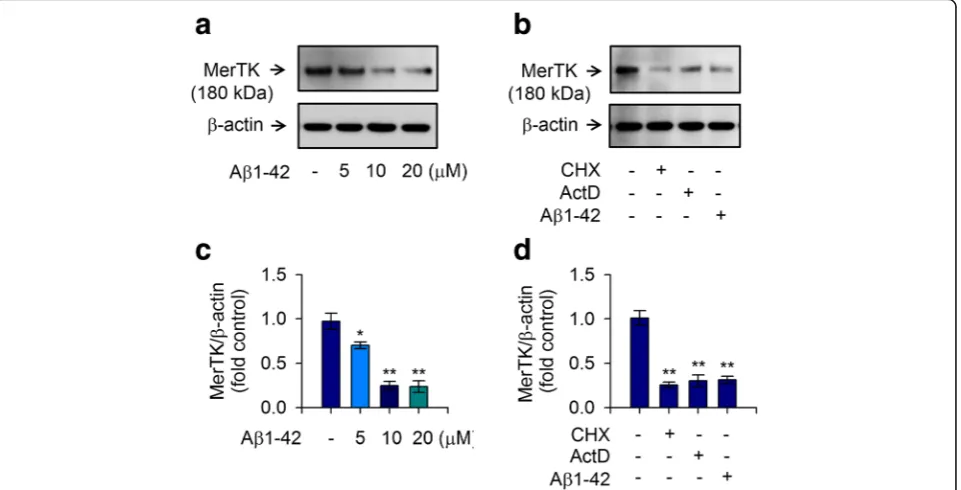
To clarify the pathological mechanism related to MerTK in AD, we measured the expression level of MerTK in response to stimulation by Aβ1-42 in human THP-1 macrophages. We treated cultured THP-1 macrophages with Aβ1-42 for 16 h and found that treated THP-1 cells expressed lower levels of MerTK protein than naive cells in a dose-dependent manner. Significant reduction in MerTK protein level was found after treatment with 5 μM of Aβ1-42. MerTK protein level was further decreased after treatment with 10μM Aβ1-42 (Fig.1a,c). Since a very similar level of reduction in MerTK protein level was obtained after treatment with 20μM of Aβ1-42 compared to that after treatment with 10μM of Aβ1-42, the lower concentration (10μM) of Aβ1-42 was used for the following experiments. Notably, MerTK protein was consistently reduced when de novo mRNA expression and protein synthesis were inhibited by actinomycin D and cycloheximide, respectively (Fig.1b,d). Our data confirmed that Aβ1-42 elicited a significant decrease of MerTK
Fig. 1MerTK expression in human THP-1 macrophages is decreased by treatment with Aβ1-42. To measure MerTK expression in response to Aβ1-42 stimulation, THP-1 cells were incubated with either the vehicle only (−) or increasing amounts of Aβ1-42 for 8 h in serum-free RPMI 1640 medium supplemented with glucose (0.5%).aTotal cell lysates were examined for MerTK protein via immunoblot. Aβ1-42 decreased MerTK in a dose-dependent manner.bTHP-1 cells were also incubated with cycloheximide (CHX, 2μM), actinomycin D (ActD, 100 nM), or Aβ1-42 (10μM) for 8 h. Western blot analyses were conducted to determine the effect of actinomycin D or cycloheximide on MerTK expression. Levels ofβ-actin were examined to ensure equal amounts of total protein as a loading control. Results are representative of three independent experiments.c,d
expression in human THP-1 macrophages in a dose-dependent manner and the reduction of MerTK expression was at both transcriptional and translational levels.
An increase of intracellular Ca2+level upon Aβ1-42 stimulation is responsible for the reduction in MerTK expression
The increase of intracellular Ca2+ level may initiate inflammatory response in activated microglia. We have observed that Aβ1-42 can extensively increase intracellular levels of Ca2+ in murine microglial BV2 cells [23] and THP-1 monocytes [20] using the Fluo-3AM method. Thus, we investigated the role of intracellular Ca2+level in the reduction of MerTK protein level elicited by Aβ1-42 insult. Our results showed that 10 μM Aβ1-42 significantly increased intracellular Ca2+levels in THP-1 macrophages using the Fluo-3AM method despite the low magnitude of intracellular Ca2+level (Fig.2a,b). Furthermore, treatment of THP-1 cells with ionomycin A (Ca2+ionophore) which induced [Ca2+]i elevation and increased intracellular
Ca2+ concentration (Fig. 2, b) induced Aβ1-42-evoked response (decreasing the level of MerTK protein) (Fig.2,d). Thapsigargin, an endoplasmic reticulum Ca2+pump inhibi-tor, also induced an increase of intracellular Ca2+ level, mimicking Aβ1-42-evoked effects (Fig.2a–d). However, the Aβ1-42-evoked response was significantly attenuated
by depletion of Ca2+ with ethylene glycol tetraacetic acid (EGTA) (Fig.2e,f). Taken together, these findings suggest that increased intracellular Ca2+is required for decreased MerTK expression in response to Aβ1-42 in human THP-1 macrophages.
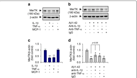
Aβ1-42-mediated decrease of MerTK protein level is associated with Ca2+-dependent hypersecretion of IL-1β and TNF-αthat could negatively regulate MerTK expression Expression of MerTK is prominently upregulated in immunosuppressive environments whereas Axl levels are increased by proinflammatory factors [24]. Thus, we measured the production of proinflammatory cytokines upon stimulation with Aβ1-42 in human THP-1 macro-phages. Expression levels of TNF-α and IL-1β were greatly increased in response to treatment with Aβ1-42 in THP-1 macrophages (Fig.3a,b). Notably, levels of these cytokines were also increased in THP-1 cells treated with either ionomycin A or thapsigargin (Fig.3,b). In contrast, the Aβ1-42-mediated increase of IL-1β and TNF-α was attenuated by depletion of Ca2+ with EGTA (Fig.3c, d). To further examine the relationship between the marked decrease in MerTK protein level and the excessive production of these cytokines, THP-1 cells were exposed to recombinant TNF-αor IL-1β. As shown in Fig.4, both TNF-α and IL-1β potently decreased MerTK protein
levels whereas MCP-1 did not alter MerTK protein expression (Fig. 4a, c). Moreover, neutralizing antibodies against IL-1βor TNF-αsignificantly inhibited the Aβ 1-42-mediated reduction of MerTK protein level (Fig. 4b, d). These observations indicate that Aβ1-42 treatment induced a decrease of MerTK protein level via increasing intracellular Ca2+levels as shown in Figs. 2and3, conse-quently resulting in excessive secretion of proinflammatory cytokines such as IL-1βand TNF-α, which could act as negative feedback regulators of MerTK expression in human THP-1 macrophages.
Sulforaphane attenuates Aβ1-42-induced MerTK reduction through inhibiting intracellular Ca2+overload and NF-κB signaling
Despite recent growing evidence suggesting the bene-ficial effect of sulforaphane on Aβ pathology of AD [16, 18, 25, 26], the effect of sulforaphane on MerTK expression upon Aβ1-42 stimulation and the under-lying signaling pathway remain unclear. Thus, we determined whether sulforaphane could decrease the
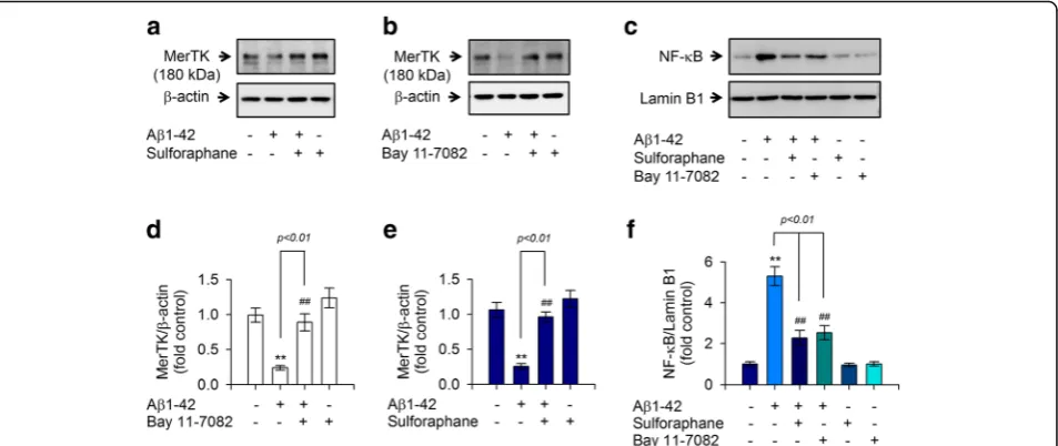
production of these proinflammatory cytokines and attenuate Aβ1-42-mediated reduction of MerTK protein level under the same experimental paradigm. Results showed that sulforaphane potently inhibited intracellular Ca2+levels (Fig.5a) and the reduction of MerTK expression was provoked by Aβ1-42 stimulation (Fig.6a,d). Further-more, sulforaphane significantly reduced the hypersecretion of both IL-1βand TNF-αin Aβ1-42-treated THP-1 macro-phages (Fig.5b,c). Subsequent mechanistic studies demon-strated that application of sulforaphane resulted in a significant decrease in nuclear translocation of NF-κB induced by Aβ1-42 insult (Fig. 6c, f). Consistently, BAY 11-7082, a pharmacological NF-κB inhibitor mimicking the effect of sulforaphane mentioned earlier, reduced the nuclear translocation of NF-κB (Fig. 6c, f) and decreased the reduction of MerTK protein level in Aβ1-42-treated cells (Fig.6b,e), thereby inhibiting the production of both IL-1βand TNF-α(Fig.5b,c). These results confirmed that sulforaphane could inhibit NF-κB signaling and restore MerTK levels, ultimately decreasing IL-1βand TNF-α pro-duction in human THP-1 macrophages exposed to Aβ1-42.
Fig. 3Ionomycin A or thapsigargin induced excessive production of proinflammatory cytokines IL-1βand TNF-αin comparison with treatment with Aβ1-42.a,bTHP-1 cells were incubated with either the vehicle only (−), ionomycin A (4μM), thapsigargin (2μM), or Aβ1-42 (10μM) for 16 h.c,d
MerTK siRNA reduces anti-inflammatory activities of sulforaphane against Aβ1-42-induced innate immune response
Recent reports have suggested that depletion of MerTK which is indispensable for negative regulation of innate immune response [6, 7] may contribute to chronic inflammation implicated in AD pathology [13]. Since sulforaphane could restore an Aβ1-42-induced decrease
of MerTK, next, we evaluated if this increase of MerTK was responsible for sulforaphane’s anti-inflammatory activities against excessive production of IL-1βor TNF-α upon Aβ1-42 stimulation. As shown in Fig.7, depletion of MerTK with siRNA significantly diminished sulforaphane’s anti-inflammatory effects on Aβ1-42-induced production of proinflammatory cytokines, thus increasing the release of IL-1β and TNF-α. However, control siRNA had little
Fig. 4IL-1βand TNF-αdecreased MerTK expression whereas neutralizing antibodies against IL-1βand TNF-αinhibited Aβ1-42-mediated reduction in MerTK expression.aTHP-1 cells were incubated with either the vehicle only (−), IL-1β, TNF-α, or MCP-1 (10 ng/mL each) for 8 h.bTHP-1 cells were exposed to Aβ1-42 (10μM) for 8 h in the presence or absence of neutralizing anti-IL-1β, anti-TNF-αantibodies, or preimmune IgG (0.1μg/mL each). Total cell lysates were examined for MerTK protein via immunoblot as described in Fig.1. Results are representative of three independent experiments.
c,dDensitometric quantification of analyses ofaandbshowing levels of MerTK protein. All data are presented as means ± SEM (n= 3–5). **P< 0.01, versus vehicle-treated samples;#P< 0.05, compared to Aβ1-42-treated samples. Aβ, amyloid-β; IgG, immunoglobulin G; IL-1β, interleukin 1-β; MCP-1, monocyte chemoattractant protein-1; MerTK, Mer tyrosine kinase; TNF-α, tumor necrosis factor-α
effect (Fig. 7a, b). These results demonstrated that depletion of MerTK could significantly relieve sulfora-phane’s anti-inflammatory activity against Aβ1-42 and consequently restore Aβ1-42-elicited production of proinflammatory cytokines such as IL-1βand TNF-αin human THP-1 macrophages.
Discussion
Results of the present study indicated that Aβ1-42 elicited a significant decrease in MerTK expression accompanied by increased intracellular Ca2+level, thereby overproducing IL-1β and TNF-α in human THP-1
macrophages. These effects were mimicked by ionomycin A or thapsigargin and attenuated by depletion of Ca2+ with EGTA. Interestingly, recombinant IL-1βor TNF-α potently decreased MerTK expression whereas immuno-depletion of IL-1βor TNF-αwith neutralizing antibodies significantly rescued the decrease in MerTK protein level provoked by Aβ1-42 treatment, implicating a negative feedback regulation of MerTK expression by IL-1β and TNF-α. Notably, sulforaphane inhibited Aβ1-42-induced downregulation of MerTK expression by decreasing the overload of intracellular Ca2+ level, ultimately inhibiting IL-1β and TNF-α production. A subsequent mechanistic
Fig. 6Sulforaphane rescued an Aβ1-42-mediated decrease of MerTK expression through inhibiting NF-κB nuclear translocation.a,bTHP-1 cells were pretreated withasulforaphane (5μM) orbBAY 11-7082 (20μM) for 30 min and then exposed to Aβ1-42 for 8 h. MerTK protein in cell lysates was measured as described above.cTHP-1 cells were pretreated with sulforaphane (5μM) or BAY 11-7082 (20μM) for 30 min and then exposed to Aβ1-42 for 8 h. NF-κB protein in nuclear extracts was measured by Western blotting and normalized against the level of lamin B1.d–fDensitometric analysis ofa–c. Values are expressed as means ± SEM of triplicate experiments. **P< 0.01, compared to vehicle-treated samples;##P< 0.01, compared to Aβ1-42-treated samples. Aβ, amyloid-β; MerTK, Mer tyrosine kinas; NF-κB, nuclear factor kappa B
Fig. 7MerTK silencing using siRNA decreased anti-inflammatory activities of sulforaphane against Aβ1-42-induced production of IL-1βand TNF-α.
study revealed that inhibition of NF-κB signaling was involved in the anti-inflammatory effect of sulforaphane as BAY 11-7082, a NF-κB inhibitor, mimicked the aforemen-tioned effect of sulforaphane. Moreover, siRNA-mediated knockdown of MerTK diminished the anti-inflammatory effect of sulforaphane against Aβ1-42, consequently resulting in increased production of IL-1β and TNF-α. These findings suggest that MerTK is indispensable for the negative regulation of innate immune response provoked by Aβ1-42 in human macrophages. Moreover, the anti-inflammatory potential of sulforaphane through inhibiting an Aβ1-42-mediated decrease in MerTK pro-tein level strongly supports the notion that sulforaphane may serve as a potential nutraceutical agent for AD.
Results from the present study clearly provide direct evidences that negative modulation of MerTK expression by Aβ1-42 occurs through increased intracellular Ca2+ levels, subsequently leading to hypersecretion of IL-1βand TNF-αin human THP-1 macrophages. First, ionomycin A (Ca2+ ionophore) and thapsigargin (an endoplasmic reticulum Ca2+pump inhibitor) increase the intracellular Ca2+level and decrease the level of MerTK protein while they increase the production of IL-1βand TNF-α. Second, these Aβ1-42-evoked responses were attenuated by depletion of Ca2+with EGTA. These observations indi-cate that an increase of intracellular Ca2+levels resulted in a decrease of MerTK protein level which ultimately released excessive IL-1β and TNF-α upon exposure to Aβ1-42. Thus, this is the first report showing that Aβ1-42 can decrease MerTK expression through intracellular Ca2
+
overload, leading to the excessive production of IL-1β and TNF-αin human macrophages. Interestingly, recom-binant IL-1β or TNF-α treatment elicited a decrease in MerTK protein level. Furthermore, immunodepletion of IL-1βor TNF-α with neutralizing antibodies significantly inhibited an Aβ1-42-mediated decrease in MerTK protein level. These findings implicate a negative feedback regula-tion of MerTK expression by IL-1βand TNF-αin human THP-1 macrophages. Our observation is consistent at least in part with a previous report demonstrating that MerTK is induced in immunosuppressive environments whereas Axl levels are increased by proinflammatory factors [24]. Interestingly, a more recent study has demonstrated that MerTK is downregulated in classic-ally activated microglial cells while it is one of the proteins mostly upregulated in TGF-β-treated microglia cells [27].
Both MerTK and Axl receptors as markers of alternative activation for anti-inflammatory actions in macrophages can act as negative feedback regulators to reduce inflam-mation. They are responsible for promoting tissue repair and phagocytosis in tissue macrophages [6, 7, 28]. In particular, roles for MerTK in mediating phagocytosis and clearance of apoptotic cells have been described in
MerTK−/− macrophages [29] via tethering apoptotic cells to macrophage surface and driving their subsequent internalization [30]. Nevertheless, a previous study has demonstrated that abundant inflammatory macrophages are principally associated with extracellular deposits of amyloid in the AD brain. These macrophages are unable to efficiently phagocytose or clear plaques from the brain [2]. More studies have also reported that the interaction of macrophages with plaques elicits the secretion of an array of inflammatory cytokines that can directly suppress phagocytosis [31]. In addition, the presence of amyloid acts to suppress macrophage phagocytic function [32]. In this regard, our results clearly indicate that the interaction of infiltrating macrophages upon exposure to amyloid could directly suppress MerTK protein level, which can permit excessive secretion of proinflammatory cytokines such as IL-1βand TNF-α, consequently resulting in phago-cytically inactive macrophages. Furthermore, our data dem-onstrating a negative feedback regulation of the MerTK expression by IL-1βand TNF-α excessively induced upon Aβ1-42 exposure might explain why inflammatory macro-phages fail to effectively phagocytize amyloid deposits, lead-ing to progressive accumulation of plaques. Consistently, a recent study has demonstrated that induction of phagocytic receptor MerTK can enhance phagocytic capacity and reduce plaque burden in murine models of AD [13], strongly supporting results of our study. It is also important to note that MerTK plays a critical role in adult neurogenesis [12]. In addition, adult mice deficient in microglial MerTK have exhibited marked accumulation of apoptotic cells in neurogenic regions of the CNS [9].
It has been reported that sulforaphane can suppress lipopolysaccharide (LPS)-induced IL-1βsecretion via inhi-biting transcriptional activity of NF-κB [34]. In addition, sulforaphane can suppress LPS-induced inflammation in mouse peritoneal macrophages through NRF2-dependent pathway [35]. Our recent study has also shown that sulfo-raphane possesses anti-inflammatory effects against Aβ 1-42 through STAT-1 dephosphorylation and activation of NRF2/HO-1 cascade in human THP-1 macrophages [18]. These findings together imply that the anti-inflammatory effect of sulforaphane is likely due to its activities against various molecular targets, some of which might be inter-dependent. Therefore, more work is required to elucidate the mechanisms underlying the intricate signaling crosstalk between NF-κB inactivation and NRF2/HO-1 activation. Importantly, anti-inflammatory activities of sulforaphane at a multitude of molecular targets would be beneficial to the exploitation of multitarget-directed drugs to control AD [36], given that AD is a multifactorial disease, of which approximately 95% occurs sporadically with unknown origin while 5% may be due to familial AD [37].
Notably, our finding that depletion of MerTK with siRNA significantly suppressed sulforaphane’s anti-inflammatory activities against Aβ1-42 clearly implicates that MerTK plays a pivotal role in the negative regulation of innate immune response. Our recent study has demonstrated that inhibiting MerTK kinase can enhance inflammatory responses in LPS-induced acute lung injury [38]. We have also observed that restoring MerTK protein expression by treatment with TNF-α processing inhibitor-0 (TAPI-0) can efficiently prevent the inflammatory cascade during acute lung injury [39]. Moreover, an earlier study has shown that a lack of Mer inhibitory signal inmerKD mice will lead to elevated and prolonged NF-κB activation, causing excess macrophage activation and TNF-α produc-tion [40]. Taken together, these observations suggest that MerTK is indispensable for the negative regulation of innate immune response provoked by Aβ1-42. In particular, MerTK depletion could contribute to a decrease of Aβ phagocytic activity [13], increasing Aβ accumulation and chronic inflammation implicated in AD pathology. Further study remains to present the Aβ phagocytosis following MerTK depletion in the macrophages/microglia with sulfo-raphane to provide important molecular insights into AD and sulforaphane’s therapeutic potential.
Conclusion
The present study clearly demonstrated that Aβ1-42 could increase in intracellular Ca2+level and NF-κB acti-vation which potently decreased MerTK expression in human THP-1 macrophages, consequently overprodu-cing IL-1β and TNF-α. Furthermore, Aβ1-42-induced IL-1β and TNF-α could decrease MerTK protein level, implicating a negative feedback regulation of MerTK
expression by IL-1βand TNF-αas summarized in Fig.8. Notably, sulforaphane significantly rescued an Aβ 1-42-mediated decrease of MerTK protein level through mecha-nisms involving decreases of Ca2+overloading and NF-κB activation, thereby inhibiting the excessive production of IL-1βand TNF-α, at least in part. Therefore, these findings together implicate that targeting of MerTK with phyto-chemical sulforaphane as a mechanism for preventing Aβ1-42-induced neuroinflammation might have therapeutic potential for AD management, although the relevance of these in vivo findings remains to be clearly elucidated.
Fig. 8Proposed mechanisms for beneficial effects of sulforaphane against decreased MerTK expression following Aβ1-42 insult in human THP-1 macrophages. Sulforaphane attenuates Aβ1-42-induced MerTK reduction through inhibiting intracellular Ca2+overload and NF-κB signaling as replicated by EGTA and BAY 11-7082. Consequently, sulforaphane rescues a decrease of MerTK expression following Aβ1-42 stimulation, thereby inhibiting overproduction of IL-1βand TNF-α. Interestingly,Αβ1-42-induced IL-1βand TNF-αcould act as negative feedback regulators of MerTK expression as confirmed with neutralizing antibodies against IL-1βor TNF-α, implicating that MerTK downregulation and induction of IL-1βand TNF-αby Aβ1-42 stimulation are interdependent. Depletion of MerTK with siRNA significantly suppressed the sulforaphane’s anti-inflammatory activities against Aβ1-42, implicating a pivotal role of MerTK for the negative regulation of the innate immune response elicited by Aβ1-42 in human
Abbreviations
Aβ:Amyloid-β; ActD: Actinomycin D; AD: Alzheimer’s disease; CHX: Cycloheximide; CNS: Central nervous system; EGTA: Ethylene glycol tetraacetic acid; ELISA: Enzyme-linked immunosorbent assay;
IgG: Immunoglobulin G; IL-1β: Interleukin 1β; MCP-1: Monocyte
chemoattractant protein-1; MerTK: Mer tyrosine kinase; NF-κB: Nuclear factor kappa B; NRF2: Nuclear factor erythroid 2-related factor 2; RTKs: Receptor tyrosine kinases; SDS-PAGE: Sodium dodecyl sulfate polyacrylamide gel electrophoresis; SEM: Standard error of the mean; siRNA: Small interfering RNA; STAT-1: Signal transducer and activator of transcription-1; TAPI-0: TNF-α processing inhibitor-0; TNF-α: Tumor necrosis factor-α
Acknowledgements
Not applicable.
Funding
This work was supported by the National Research Foundation of Korea (NRF) grant funded by the Korean government (MSIT) (2010-0027945).
Availability of data and materials
The datasets used and/or analyzed during the current study are available from the corresponding author on reasonable request.
Authors’contributions
The work presented here was carried out in collaboration between all authors. KJ carried out most of the laboratory experiments, analyzed the data, and interpreted the results. JP helped in the preparation of the manuscript. HK and YC conceived of the idea for the study, helped in designing the methods and experiments, and critically supervised the complete study. All the authors read and approved the final revised manuscript.
Ethics approval and consent to participate
Not applicable.
Consent for publication
Not applicable.
Competing interests
The authors declare that they have no competing interests.
Publisher’s Note
Springer Nature remains neutral with regard to jurisdictional claims in published maps and institutional affiliations.
Received: 8 December 2017 Accepted: 1 March 2018
References
1. Selkoe DJ. The therapeutics of Alzheimer’s disease: where we stand and where we are heading. Ann Neurol. 2013;74:328–36.
2. Akiyama H, Barger S, Barnum S, Bradt B, Bauer J, Cole GM, Cooper NR, Eikelenboom P, Emmerling M, Fiebich BL, Finch CE, Frautschy S, Griffin WS, Hampel H, Hull M, Landreth G, Lue L, Mrak R, Mackenzie IR, McGeer PL, et al. Inflammation and Alzheimer’s disease. Neurobiol Aging. 2000;21:383–421. 3. Eikelenboom P, Veerhuis R, van Exel E, Hoozemans JJ, Rozemuller AJ, van
Gool WA. The early involvement of the innate immunity in the pathogenesis of late-onset Alzheimer’s disease: neuropathological, epidemiological and genetic evidence. Curr Alzheimer Res. 2011;8:142–50. 4. Cuyvers E, Bettens K, Philtjens S, Van Langenhove T, Gijselinck I, van der Zee
J, Engelborghs S, Vandenbulcke M, Van Dongen J, Geerts N, Maes G, Mattheijssens M, Peeters K, Cras P, Vandenberghe R, De Deyn PP, Van Broeckhoven C, Cruts M, Sleegers K. Investigating the role of rare heterozygous TREM2 variants in Alzheimer’s disease and frontotemporal dementia. Neurobiol Aging. 2014;35:726.e11–9.
5. Lambert JC, Ibrahim-Verbaas CA, Harold D, Naj AC, Sims R, Bellenguez C, DeStafano AL, Bis JC, Beecham GW, Grenier-Boley B, Russo G, Thorton-Wells TA, Jones N, Smith AV, Chouraki V, Thomas C, Ikram MA, Zelenika D, Vardarajan BN, Kamatani Y, et al. Meta-analysis of 74,046 individuals identifies 11 new susceptibility loci for Alzheimer’s disease. Nat Genet. 2013;45:1452–8. 6. Rothlin CV, Ghosh S, Zuniga EI, Oldstone MB, Lemke G. TAM receptors are
pleiotropic inhibitors of the innate immune response. Cell. 2007;131:1124–36.
7. Rothlin CV, Carrera-Silva EA, Bosurgi L, Ghosh S. TAM receptor signaling in immune homeostasis. Annu Rev Immunol. 2015;33:355–91.
8. Pierce AM, Keating AK. TAM receptor tyrosine kinases: expression, disease and oncogenesis in the central nervous system. Brain Res. 2014;1542:206–20. 9. Fourgeaud L, Través PG, Tufail Y, Leal-Bailey H, Lew ED, Burrola PG, Callaway
P, Zagórska A, Rothlin CV, Nimmerjahn A, Lemke G. TAM receptors regulate multiple features of microglial physiology. Nature. 2016;532:240–4. 10. Yagami T, Ueda K, Asakura K, Sakaeda T, Nakazato H, Kuroda T, Hata S,
Sakaguchi G, Itoh N, Nakano T, Kambayashi Y, Tsuzuki H. Gas6 rescues cortical neurons from amyloidβprotein-induced apoptosis. Neuropharmacology. 2002;43:1289–96.
11. Zheng Y, Wang Q, Xiao B, Lu Q, Wang Y, Wang X. Involvement of receptor tyrosine kinase Tyro3 in amyloidogenic APP processing and beta-amyloid deposition in Alzheimer’s disease models. PLoS One. 2012;7:e39035. 12. Ji R, Meng L, Li Q, Lu Q. TAM receptor deficiency affects adult hippocampal
neurogenesis. Metab Brain Dis. 2015;30:633–44.
13. Savage JC, Jay T, Goduni E, Quigley C, Mariani MM, Malm T, Ransohoff RM, Lamb BT, Landreth GE. Nuclear receptors license phagocytosis by trem2+ myeloid cells in mouse models of Alzheimer’s disease. J Neurosci. 2015;35: 6532–43.
14. Alfieri A, Srivastava S, Siow RC, Cash D, Modo M, Duchen MR, Fraser PA, Williams SC, Mann GE. Sulforaphane preconditioning of the Nrf2/HO-1 defense pathway protects the cerebral vasculature against blood-brain barrier disruption and neurological deficits in stroke. Free Radic Biol Med. 2013;65:1012–22.
15. Kim HV, Kim HY, Ehrlich HY, Choi SY, Kim DJ, Kim Y. Amelioration of Alzheimer’s disease by neuroprotective effect of sulforaphane in animal model. Amyloid. 2013;20:7–12.
16. Zhang J, Zhang R, Zhan Z, Li X, Zhou F, Xing A, Jiang C, Chen Y, An L. Beneficial effects of sulforaphane treatment in Alzheimer’s disease may be mediated through reduced HDAC1/3 and increased P75NTR expression. Front Aging Neurosci. 2017;9:121.
17. Lee C, Park GH, Lee SR, Jang JH. Attenuation ofβ-amyloid-induced oxidative cell death by sulforaphane via activation of NF-E2-related factor 2. Oxidative Med Cell Longev. 2013;2013:313510.
18. An YW, Jhang KA, Woo SY, Kang JL, Chong YH. Sulforaphane exerts its anti-inflammatory effect against amyloid-βpeptide via STAT-1
dephosphorylation and activation of Nrf2/HO-1 cascade in human THP-1 macrophages. Neurobiol Aging. 2016;38:1–10.
19. Jhang KA, Lee EO, Kim HS, Chong YH. Norepinephrine provides short-term neuroprotection against Aβ1-42 by reducing oxidative stress independent of Nrf2 activation. Neurobiol Aging. 2014;35:2465–73.
20. Lee EO, Yang JH, Chang KA, Suh YH, Chong YH. Amyloid-βpeptide induced extracellular S100A9 depletion is associated with decrease of antimicrobial peptide activity in human THP-1 monocytes. J Neuroinflammation. 2013;10:68. 21. Lee EO, Kang JL, Chong YH. The amyloid-beta peptide suppresses
transforming growth factor-beta1-induced matrix metalloproteinase-2 production via Smad7 expression in human monocytic THP-1 cells. J Biol Chem. 2005;280:7845–53.
22. Yang JH, Lee EO, Kim SE, Suh YH, Chong YH. Norepinephrine differentially modulates the innate inflammatory response provoked by amyloid-β peptide via action atβ-adrenoceptors and activation of cAMP/PKA pathway in human THP-1 macrophages. Exp Neurol. 2012;236:199–206.
23. Ha TY, Chang KA, Kim J, Kim HS, Kim S, Chong YH, Suh YH. S100a9 knockdown decreases the memory impairment and the neuropathology in Tg2576 mice, AD animal model. PLoS One. 2010;5:e8840.
24. Zagórska A, Través PG, Lew ED, Dransfield I, Lemke G. Diversification of TAM receptor tyrosine kinase function. Nat Immunol. 2014;15:920–8.
25. Tarozzi A, Angeloni C, Malaguti M, Morroni F, Hrelia S, Hrelia P. Sulforaphane as a potential protective phytochemical against neurodegenerative diseases. Oxidative Med Cell Longev. 2013;2013:415078.
26. Zhang R, Miao QW, Zhu CX, Zhao Y, Liu L, Yang J, An L. Sulforaphane ameliorates neurobehavioral deficits and protects the brain from amyloidβ deposits and peroxidation in mice with Alzheimer-like lesions. Am J Alzheimers Dis Other Demen. 2015;30:183–91.
27. Healy LM, Perron G, Won SY, Michell-Robinson MA, Rezk A, Ludwin SK, Moore CS, Hall JA, Bar-Or A, Antel JP. MerTK is a functional regulator of myelin phagocytosis by human myeloid cells. J Immunol. 2016;196:3375–84. 28. Zizzo G, Hilliard BA, Monestier M, Cohel PL. Efficient clearance of early
29. Scott RS, McMahon EJ, Pop SM, Reap EA, Caricchio R, Cohen PL, Earp HS, Matsushima GK. Phagocytosis and clearance of apoptotic cells is mediated by MER. Nature. 2001;411:207–11.
30. Dransfield I, Zagórska A, Lew ED, Michail K, Lemke G. Mer receptor tyrosine kinase mediated both tethering and phagocytosis of apoptotic cells. Cell Death Dis. 2015;6:e1646.
31. Koenigsknecht-Talboo J, Landreth GE. Microglial phagocytosis induced by fibrillar beta-amyloid and IgGs are differentially regulated by
proinflammatory cytokines. J Neurosci. 2005;25:8240–9.
32. Krabbe G, Halle A, Matyash V, Rinnenthal JL, Eom GD, Bernhardt U, Miller KR, Prokop S, Kettenmann H, Heppner FL. Functional impairment of microglia coincides with beta-amyloid deposition in mice with Alzheimer-like pathology. PLoS One. 2013;8:e60921.
33. Feng S, Xu Z, Wang F, Yang T, Liu W, Deng Y, Xu B. Sulforaphane prevents methylmercury-induced oxidative damage and excitotoxicity through activation of the Nrf2-ARE pathway. Mol Neurobiol. 2017;54:375–91. 34. Folkard DL, Melchini A, Traka MH, Al-Bakheit A, Saha S, Mulholland F,
Watson A, Mithen RF. Suppression of LPS-induced transcription and cytokine secretion by the dietary isothiocyanate sulforaphane. Mol Nutr Food Res. 2014;58:2286–96.
35. Lin W, Wu RT, Wu T, Khor TO. Sulforaphane suppressed LPS-induced inflammation in mouse peritoneal macrophages through Nrf2 dependent pathway. Biochem Pharmacol. 2008;76:967–73.
36. Dias KS, Viegas C Jr. Multi-target directed drugs: a modern approach for design of new drugs for the treatment of Alzheimer’s disease. Curr Neuropharmacol. 2014;12:239–55.
37. Danborg PB, Simonsen AH, Waldemar G, Heegaard NH. The potential of microRNAs as biofluid markers of neurodegenerative diseases—a systematic review. Biomarkers. 2014;19:259–68.
38. Lee YJ, Han JY, Byun J, Park HJ, Park EM, Chong YH, Cho MS, Kang JL. Inhibiting Mer receptor tyrosine kinase suppresses STAT1, SOCS1/3, and
NF-κB activation and enhances inflammatory responses in lipopolysaccharide-induced acute lung injury. J Leukoc Biol. 2012;91:921–32.
39. Choi JY, Park HJ, Lee YJ, Byun J, Youn YS, Choi JH, Woo SY, Kang JL. Upregulation of Mer receptor tyrosine kinase signaling attenuated lipopolysaccharide-induced lung inflammation. J Pharmacol Exp Ther. 2013; 344:447–58.
40. Camenisch T, Koller BH, Earp HS, Matsushima GK. A novel receptor tyrosine kinase, Mer, inhibits TNF-alpha production and lipopolysaccharide-induced endotoxic shock. J Immunol. 1999;162:3498–503.
• We accept pre-submission inquiries
• Our selector tool helps you to find the most relevant journal
• We provide round the clock customer support
• Convenient online submission
• Thorough peer review
• Inclusion in PubMed and all major indexing services
• Maximum visibility for your research
Submit your manuscript at www.biomedcentral.com/submit