1Dr. Lipika Boruah P.T *2Dr. Abhijit Dutta P.T 3Dr. Pritam Deka P.T 4Dr. Jeutishree Roy P.T
CORRESPONDING AUTHOR
*2Dr. Abhijit Dutta P.T
Associate Prof. cum HOD Dept. of Physiotherapy
Assam down town University, Guwahati, Assam.
Int J Physiother. Vol 2(5), 811-818, October (2015) ISSN: 2348 - 8336
ABSTRACT
Background: Joint mobilization is an effective intervention for adhesive capsulitis. Scapular Mobilization in shoulder adhesive capsulitis is used to decrease intra articular pressure by increasing mobility of the joint capsule and its surrounding soft tissue that results in a reduction of pain and increase range of motion and shoulder function. At the same time the use of mobilization with movement (MWM) for peripheral joints was also used clinically. This technique combines a sustained application of a manual technique ‘gliding’ force to a joint with concurrent physiologic motion of joint, either actively performed by the subject or passively performed by the therapist. So far there is no study which is done on comparison between both of these techniques. The aim of the study is to find out whether the scapular mobilization or mobilization with movement technique improve gleno-humeral range of motion and reduce pain in patients with shoulder adhesive capsulitis.
Methods: 50 subjects with adhesive capsulitis were randomly divided in to two groups and one group was treated with mobilization with movement and another group treated with scapular mobilization technique. Each group consist 25 patients. Both groups were given hot packs and pendular exercises as conventional therapy procedures. Treatment was given 5 days a week for 3 weeks. Restricted joint range of motion and severity of pain were measured before and after treatment completion by using goniometer and SPADI pain score respectively.
Result: Results of the present study revealed that there was a significant difference in SPADI pain score(%), AROM-GH-Flexion and AROM-GH-External rotation who were treated in group A(MWM) with mean being 44.00, 102.24 and 46.08 respectively compared to group B (SM) with mean being 54.00, 81.00 and 35.84 in 3 weeks. Comparisons between these three parameters used in two treatment techniques were extremely significant (p= 0.000 for all).
Conclusion: On the basis of the results, it can be concluded that, the present study provided evidence to support the use of physical therapy regimen for shoulder adhesive capsulitis in the form of mobilization with movement and scapular mobilization in reduction of pain and improvement of glenohumeral range of motion in terms of SPADI pain scale(%) and AROM respectively. In addition results support that mobilization with movement showed better result as compared to scapular mobilization in 3 weeks.
Keywords: Adhesive Capsulitis, Movement with Mobilization, Shoulder, Mobilizations.
DOI: 10.15621/ijphy/2015/v2i5/78239
1Physiotherapist, Sun Valley Hospital,
Guwahati, Assam.
3Assistant Prof., Dept of Physiotherapy
Assam down town University, Guwahati, Assam
4Physiotherapist, Astitva Multispeciality
Therapy center, Guwahati, Assam.
Received 28th August 2015, revised 24th September 2015, accepted 30th September 2015
INTRODUCTION
Subjects with the diagnosis of adhesive capsulitis are commonly seen in the physiotherapy department. Adhesive capsulitis of the shoulder is a very unique entity in that it is the only joint in the body that is affected by this type of disease process.1 It is a very common but poorly
understood condition causing pain and loss of range of motion in the shoulder. Though it is often self-limited, can persist for years and may never fully resolve.2
Adhesive capsulitis is also known as frozen shoulder and this condition was termed “peri-arthritis scapula humerale” by Duplay in 1896. Codman, in 1934 stated the diagnosis of “frozen shoulder” as a condition characterized by pain and reduce range of motion in affected shoulder. Naviaser, in the prearthroscopic era subsequently used the term “Adhesive capsulitis” to describe the findings of chronic inflammation and fibrosis of the joint capsule, although arthroscopic examination would support the term “fibrotic capsulitis” with the absence of adhesion.3 It is often quoted by
many studies to affect approximately 2% to 5% of general population. It most commonly affected women aged between 40 and 60 years.3 The
non-dominant shoulder is slightly more likely to be affected within 5 years.4
Several authors have proposed that impaired shoulder movement in adhesive capsulitis is related to shoulder capsule adhesion, contracted soft tissues, and adherent axillary recess. The adherent axillary recess hinders humeral head mobility, resulting in diminished mobility of the shoulder.5
Adhesive capsulitis is mainly classified into two types- primary and secondary. Primary or idiopathic adhesive capsulitis occurs spontaneously without a specific precipitating event. It may results from a chronic inflammatory response with fibroblastic proliferation which may actually be an abnormal response from immune system. Secondary adhesive capsulitis occurs after a shoulder injury or surgery or may be associated with another condition such as diabetes, rotator cuff injury, autoimmune disease, cerebovascular or cardiovascular diseases.6
The common capsular pattern of limitation in adhesive capsulitis is diminishing motion with external shoulder rotation being the most limited, followed closely by shoulder abduction and internal rotation.7 The loss of passive external
rotation is the single most important finding on physical examination which helps to differentiate the diagnosis from a rotator cuff problem. The
diagnosis is confirmed when radiographic studies are normal.8
The joint is generally considered to have 1200 of
flexion and about 500 of extension. The
internal/external rotation range varies with position. At neutral position 900 of GH abduction
can be obtained. The ROM for abduction of the GH joint are reported to be anywhere from 900 to 1200.
The scapular movement combined with the humeral movement results a maximum range of elevation to 1800 and in an overall ratio of 20 of GH
to 10 of scapulothoracic motion. The combination
of concomitant GH and ST motion is commonly referred to as scapulohumeral rhythm.9
Multiple interventions have been described for the treatment of adhesive capsulitis such as corticosteroid injections, surgery, patient education, modalities (TENS, ultrasound, short wave diathermy), cryotherapy, moist heat, joint mobilizations, transitional manipulation under anesthesia, stretching exercises, pendular exercise, scapula thoracic strengthening exercises etc.10
Joint mobilization is an effective intervention for adhesive capsulitis. Manual therapy in shoulder adhesive capsulitis is used to decrease intra articular pressure by increasing mobility of the joint capsule and its surrounding soft tissue that results in a reduction of pain and increase range of motion and shoulder function.11 Mobilization
techniques improve the normal extensibility of the shoulder capsule and stretch the tightened soft tissues to induce beneficial effects.
The use of mobilization with movement (MWM) for peripheral joints was developed by Mulligan. This technique combines a sustained application of a manual technique ‘gliding’ force to a joint with concurrent physiologic (osteo-kinematic) motion of joint, either actively performed by the subject or passively performed by the therapist. The intent of MWM is to restore pain free motion at joints that have painful limitation of range of movement. So far there is no study which is done on comparison between both of these techniques. Several studies which were done on mobilization with movement have shown improvement in 3 weeks and in some studies which were done on scapular mobilization, the study periods are varied. So, this comparative study is needed to see which technique give the better result in lesser period i.e. in 3 weeks to reduce pain and improve gleno-humeral range of motion in adhesive capsulitis of shoulder.
MATERIALS AND METHOD
study design. All the subjects were diagnosed as adhesive capsulitis of shoulder by orthopedic doctors. All the subjects included in to study were diagnosed with stage II Unilateral adhesive capsulitis (3-9 months)aged between 45-55 years. Patients with 50% loss of passive ROM of the shoulder joint comparing to the non-affected side in at least 2 movements (forward flexion, abduction or external rotation in zero degree of abduction) were selected.12 All the Patients are able
to comprehend command and willing to participate in the study. History of previous shoulder surgeries to the affected shoulder, Presence of rheumatoid arthritis, rotator cuffs rupture or tendon calcification, Presence of any neurological diseases and sensory deficit, Presence of any high risk medical conditions were considered as exclusion criteria. Duration of the study is of 3 weeks and data was collected on first day and on 21st days.
Outcome measures are shoulder range of motion 13
and SPADI pain scale.14
Procedure:
25 subjects were assigned in two groups randomly. GROUP A
Subjects were treated with hot packs, mulligan’s mobilization with movement and pendular exercises. Mobilization with movement were given 2 sets of 10 repetition; 5 sessions in a week for 3 weeks.
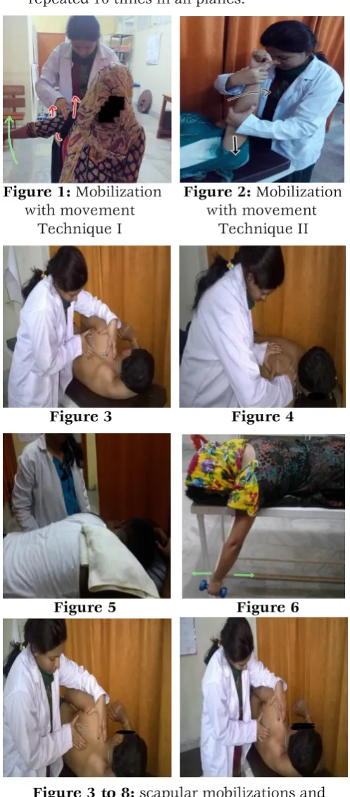
MOBILIZATION WITH MOVEMENT (MWM):- techniques of mobilization with movement technique was performed on the involved shoulder as described by Mulligan.15
Therapist stood behind the seated subject and placed a belt around her hips and the subject’s shoulder proximal to joint. Now by placing a hand on the scapula for fixation leans back in such a way as to glide the humeral head back obliquely in the treatment plane, the free hand secured the belt and prevents slipping. Maintaining the glide, subject was instructed to perform active slow movement to end of the pain free range and released after returning to the starting position. Two sets of 10 repetitions were performed.
Therapist stood at the head end of the patient’s bed and grasped the humerus with one hand the forearm with the other on the affected side, now pushing down along the shaft of the humerus, patient was asked to raise his arm till the end of the pain free range and released after returning to the starting position. Two sets of 10 repetitions were performed.
GROUP B
Subjects were treated with hot packs, scapular mobilization and pendular exercises. Hot packs were used to both Group A and Group B prior to the treatments. (1 hour session)
Scapular mobilizations were given 2 sets of 10 repetitions with a rest interval of 30 seconds between sets5 sessions in a week for 3 weeks.Hot packs applied on the target shoulder for 20 minutes prior to the mobilization.Pendular exercises given for 3 times daily for 2-3 minutes.
SCAPULAR MOBILIZATION (SM):-
Scapula superior glide.
- Patient was lying on his unaffected side, the therapist placing the index finger of one hand under the medial scapular border of the affected side, the other hand grasping the superior border of the scapula. The scapula was moved superiorly for superior glide.
Scapula inferior glide.
- Patient was lying on his unaffected side, the therapist placing the index finger of one hand under the medial scapular border of the affected side, the other hand grasping the superior border of the scapula. The scapula was moved inferiorly for inferior glide.
Scapula upward rotation.
- Scapular upward rotation was performed with the patient in side lying on unaffected side, the therapist was placed her index finger of one hand under the medial border of the scapula and other hand grasping the superior border of scapula and then scapula was rotated upwardly.
Scapula downward rotation.
- Scapular upward rotation was performed with the patient in side lying on unaffected side, the therapist was placed her index finger of one hand under the medial border of the scapula and other hand grasping the superior border of scapula and then scapula was rotated downwardly.
Scapula distraction.
- The patient was lying prone. The therapist was put the ulnar fingers under the medial scapular border and distracted the scapula from the thorax(scapular tilt).
CONVENTIONAL THERAPY:-
Hot packs was wrapped in toweling well place on the target shoulder for 20 minutes for both groups prior to mobilization.
movements in all planes by lying prone with effected limb hanging out of plinth. This was repeated 10 times in all planes.
Figure 1: Mobilization Figure 2: Mobilization with movement with movement Technique I Technique II
Figure 3 Figure 4
Figure 5 Figure 6
Figure 3 to 8: scapular mobilizations and pendular exercises.
DATA ANALYSIS
All analysis were obtained using PASW version 21.0. Demographic data of patient including sex, age, SPADI pain score, AROM - GH Flexion and AROM-GH External rotation were descriptively summarized. An alpha-level of 0.05 was used to determine statistical significance. Paired t-test was performed to find effectiveness of MWM and SM in reducing pain and improving glenohumeral flexion and external rotation range of motion in adhesive capsulitis of shoulder. Independent sample t-test was carried out to compare MWM and SM.
In this study 50 subjects were conveniently selected, and then were allocated in group A and B. There were 18 males and 32 females with a mean age of 49.76 + 3.49 in group A and a mean age of 50.04 + 3.34 in group B. demographic information of the study population is outlined in the table below.
Group A
Age (Mean + SD) 49.76 + 3.49
Gender (Male : Female) 7 : 18
Group B
Age (Mean + SD) 50.04 + 3.34
Gender (Male : Female) 11 : 14
Table 1: Demographic information of the study population
Graph 1: Mean age of subjects of group A and group B
COMPARISION OF OUTCOME MEASURES Within group analysis-SPADI pain score (%)
Outcome
Measure Group Day Mean + S.D. Paired ‘t’ test Comment
SPADI pain score
Group A Day 0 64.48 + 6.46 0.000 Significant difference between
day 0 and day 21.
Day 21 44.00 + 4.62
Group B Day 0 64.64 + 5.76 0.000 Significant difference between
day 0 and day 21
Day 21 54.00 + 4.58
Table 2: Group analysis within groups of Group A and Group B for SPADI pain scale 42
44 46 48 50 52 54
Group A Group B
M
e
an
A
ge
(year
s)
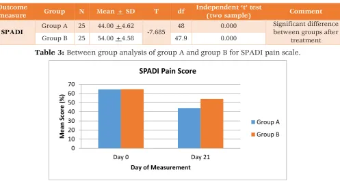
The average pre-interventional i.e. day 0 SPADI pain score (%) in the group A was 64.48 + 6.46 and in the group B was 64.64 + 5.76 where as post-interventional i.e. day 21 the scores reduced to 44.00 + 4.62 in the group A and 54.00 + 4.58 in the
group B. Changes in the SPADI pain subscale revealed highly statistically significant reduction in pain post-interventional for both the groups (p). This was done using paired‘t’ test.
Between group analysis- SPADI pain score (%)
Outcome
measure Group N Mean + SD T df
Independent ‘t’ test
(two sample) Comment
SPADI Group A 25 44.00 +4.62 -7.685 48 0.000
Significant difference between groups after
treatment
Group B 25 54.00 +4.58 47.9 0.000
Table 3: Between group analysis of group A and group B for SPADI pain scale.
Graph 2: Between groups mean difference on SPADI pain scale before and after treatment. Within group analysis: Active ROM – GH Flexion (degree) by Goniometer
Outcome
Measure Group Day Mean + S.D. Paired t test Comment
AROM – GH Flexion (degree)
Group A Day 0 56.28 + 6.55 0.000 Significant difference between
day 0 and day 21.
Day 21 102.24 + 10.26
Group B Day 0 54.92 + 5.94 0.000 Significant difference between
day 0 and day 21
Day 21 81.00 + 8.97
Table 4: Within group analysis of group A & group B for AROM-GH-Flexion The average pre-interventional i.e. day 0 AROM-
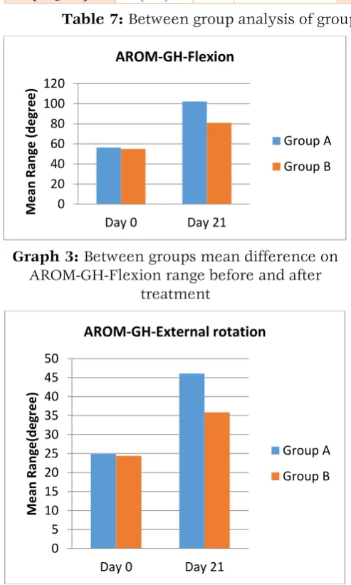
GH flexion (degree) in the group A was 56.28 + 6.55 and in the group B was 54.92 + 5.94 where as post-interventional i.e. day 21 the flexion range increased to 102.24 + 10.26 in the group A and
81.00 + 8.97 in the group B. Changes in the active GH flexion range of motion revealed highly statistically significant improvement in Flexion ROM post-interventional for both the groups (p). This was done using paired‘t’ test.
Between group analysis: Active ROM – GH Flexion (degree) by Goniometer
Outcome
measure Group N Mean + SD t Df
Independent ‘t’ test (two
sample)
Comment
AROM – GH Flexion
(degree)
Group A
(MWM) 25 102.24 + 10.26
7.790
48 0.000 Significant difference
between groups after treatment Group B
(SM) 25 81.00 + 8.97 47.168 0.000
Table 5: Between group analysis of group A & group B for AROM-GH-Flexion. The between group analysis for goniometric
measurement of AROM of GH Flexion was done using Independent ‘t’ test showed statistically very
significant GH Flexion range of motion in the group A (MWM) as compared to the group B (SM). 0
10 20 30 40 50 60 70
Day 0 Day 21
M
e
an
S
co
re
(
%
)
Day of Measurement
SPADI Pain Score
Group A
Within group analysis: AROM – GH External rotation (degree) using Goniometer
Outcome Measure Group Day Mean + S.D. Paired
‘t’ test Comment
AROM – GH External rotation
(degree)
Group A Day 0 24.88 + 4.73 0.000 Significant difference between
day 0 and day 21.
Day 21 46.08 + 7.90
Group B Day 0 24.40 + 5.63 0.000 Significant difference between
day 0 and day 21
Day 21 35.84 + 6.34
Table 6: Within group analysis of group A & group B for AROM-GH-External rotation. The average pre-interventional i.e. day 0 AROM-
GH External rotation (degree) in the group A was 24.88 + 4.73 and in the group B was 24.40 + 5.63 where as post-interventional i.e. day 21 the external rotation range increased to 46.08 + 7.90 in the group A and 35.84 + 6.34 in the group B.
Changes in the active GH external rotation range of motion revealed highly statistically significant improvement in External rotation ROM post-interventional for both the groups (p). This was done using paired‘t’ test.
Between group analysis- AROM – GH External rotation (degree) using Goniometer
Outcome
measure Group N Mean + SD t df
Independent ‘t’
test (two sample) Comment
AROM – GH External
rotation (degree)
Group A
(MWM) 25 46.08 + 7.90
5.054
48 0.000 Significant
difference between groups after
treatment Group B
(SM) 25 35.84 + 6.34 45.826 0.000
Table 7: Between group analysis of group A & group B for AROM-GH-External rotation
Graph 3: Between groups mean difference on AROM-GH-Flexion range before and after
treatment
Graph 4: Between groups mean difference on AROM-GH-External rotation before and after
treatment.
RESULTS
After the intervention considerable change was observed in the mean values of group A and group B from that of the baseline value (SPADI pain scale and AROM). Statistical analysis revealed a significant reduction of pain and improvement of ROM in both groups.
Paired‘t’ test was performed to see significant different in SPADI pain scale (%) before and after the treatment in both groups. It was found in group A, t= 20.579, which is highly significant (p= 0.000) and we can say that there is remarkable reduction of pain in the SPADI pain subscale after applying MWM. The value of ‘t’ to find the difference in SPADI pain scale(%) before and after treatment in group B is 16.902, this value is highly significant(p=0.000). it has been found that reduction of pain in the SPADI pain subscale after applying SM.
Statistical analysis revealed a significant improvement in goniometric measurement of AROM of GH flexion and external rotation range (degree) in both groups. In group A (MWM), value of‘t’ for AROM-GH-flexion and External rotation are -28.660 and -12.639 respectively. We can thereby say that ROM increased significantly after applying MWM (p= 0.000). For group B, after applying SM, the values of t= -15.991 (AROM-GH-Flexion) and t= -14.807 (AROM-GH-External rotation), which are highly significant (p= 0.000). 0
20 40 60 80 100 120
Day 0 Day 21
M
e
an
R
an
ge
(
d
e
gr
e
e
)
AROM-GH-Flexion
Group A
Group B
0 5 10 15 20 25 30 35 40 45 50
Day 0 Day 21
M
e
an
R
an
ge
(d
e
gr
e
e
)
AROM-GH-External rotation
Group A
it also shows improvement of active glenohumeral flexion and ER range after applying SM.
Results of the present study revealed that there was a considerable effects of MWM (group A) in reduction of pain and improve GH range of motion as compared to SM (group B) while using Independent ‘t’ test for between group analysis. Results of the present study revealed that there was a significant difference in SPADI pain score(%), AROM-GH-Flexion and AROM-GH-External rotation who were treated in group A(MWM) with mean being 44.00, 102.24 and 46.08 respectively compared to group B (SM) with mean being 54.00, 81.00 and 35.84 in 3 weeks. . Comparisons between these three parameters used in two treatment techniques were extremely significant (p= 0.000 for all).
DISCUSSION
The comparative study was conducted to study the effectiveness of MWM and SM techniques in the treatment of adhesive capsulitis for three weeks in terms of reduction of pain using SPADI pain subscale and improvement in GH range of motion using universal goniometer. It was also intended to compare the effectiveness of SM versus MWM techniques. Hot packs and pendulum exercises were common treatment in both groups.
The age distribution of the groups showed the homogenecity of subjects. The results from the statistical analysis of the study supported alternative hypothesis which stated that there was significant difference between the effectiveness of SM versus MWM technique to reduce pain and improve GH range of motion in adhesive capsulitis of shoulder.
In a study the Intra-articular triamcinolone injection and physiotherapy singly or combined in the treatment of adhesive capsulitis of shoulder. 80 patients were randomly assigned into one of the 4 groups. The outcome measures taken were shoulder disability questionnaire, VAS, range of passive external rotation. They concluded the study as corticosteroid injection was effective in improving shoulder related disability and physiotherapy is improving ROM in external rotation in adhesive capsulitis.16, 17, 18 Even though
some studies are saying the equal importance of anterior and posterior mobilizations few studies supporting the benefits of posterior mobilizations. Andrea J. Johnson, et al (2007), conducted a randomized clinical trial to compare the effectiveness of anterior versus posterior mobilization procedure to improve shoulder external rotation range of motion in patient with adhesive capsulitis. They found that both
techniques had a significant decrease in pain. But a posteriorly directed joint mobilization technique was more effective than an anteriorly directed mobilization technique for improve ER range in shoulder adhesive capsulitis.19 application of heat
may be a cause to get desired range of motion iafter treatment. Manska &Prohaska (2010) mentioned that during the second stage of adhesive capsulitis the joint end feel become capsular in nature. Moist heat may be helpful and the patient can generally tolerate a more aggressive stretching and mobilization.20
Jonathan Cluett (2010) in his article on frozen shoulder treatment has mentioned application of moist heat to the shoulder can help to loosen the joint and provide relief of pain. Moist heat can be applied to the shoulder prior to the treatment protocol to gain better results in terms of reducing pain in frozen shoulder as it improves blood circulation to the local area thereby enhancing waste product removal from the soft tissues, by means of vasodilation.21 Hot pack is common
modality in both groups. It has analgesic effects. The heat reduces the viscosity of collagen, increases tissue extensibility and makes connective tissue less resistant to active or passive stretching. Thus heating before passive joint mobilization helps effective application of techniques. For regular maintenance pendulum exercises were given to both groups.
The mean values of data from present study indicate that both SM and MWM technique could be beneficial in the treatment of adhesive capsulitis of shoulder. There was statistically significant difference in the SPADI pain score and active GH flexion and ER range in both group A and group B from ‘day 0’ to ‘day 21’. The between group comparison showed statistically significant reduction of pain on SPADI pain subscale and improvement of GH-AROM in group A (MWM) as compare to group B (SM).
Shrivastava, et al(2011) conducted a randomized controlled study to compare the effectiveness of Mulligan’s mobilization with Maitland’s mobilization techniques in frozen shoulder rehabilitation and they found that Mulligan’s mobilization technique is better than Maitland in terms of improvement in the range of extension while remaining range were similarly improved by both techniques.22, 23
pain score(%) and goniometric measurements of GH flexion and external rotation AROM in 3 weeks.
CONCLUSION
On the basis of the results, it can be concluded that, the present study provided evidence to support the use of physical therapy regimen for shoulder adhesive capsulitis in the form of mobilization with movement and scapular mobilization in reduction of pain and improvement of glenohumeral range of motion in terms of SPADI pain scale(%) and AROM (goniometer) respectively. In addition results support that mobilization with movement showed better result as compared to scapular mobilization in 3 weeks.
Hence Mulligan’s MWM technique could be used effectively to reduce pain and improve GH range of motion in shoulder adhesive capsulitis.
REFERENCES
1. James P. Tasto, et al. Adhesive capsulitis. J Sports Med. Arthro Rev. 2007;15(4);216-221. 2. Anthony Ewald. Adhesive capsulitis: a review.
J Am Fam. Physician.2011;15:83(4) :417-422. 3. William J. Warrender, et al. Current review of
adhesive capsulitis. J shoulder Elbow surg.2011; 20(3):502-14.
4. Rizk TE, Pinab RS.Frozen shoulder; Semin Arthritis Rehum. 1982 ;11(4):440-52.
5. Jing-Ian Yang, et.al. Mobilization techniques in subjects with frozen shoulder syndrome, randomized multiple treatment trial. J. phys ther. 2007;87(10):1307-1315.
6. Phil Page & Andre Labbe. Adhesive capsulitis: use the evidence to integrate your interventions. N. Am J. sports phys. Ther. 2010; 5(4):266-273.
7. David J Magee. Orthopedic physical assessment.5th edition;2007.
8. S. Brent Brotzman & Kenin E. Wilk.Clinical orthopaedic Rehabilitation.2nd ed;2003.
9. Pamela K. Levangie & Cynthia C. Norkin. Joint structure and function: a comprehensive analysis. 5th ed; 2011.
10.Martin J. Kelley et al. Shoulder pain and mobility deficits.-adhesive capsulitis. J. orthop sports phys. Ther. 2013; 43(5); A1-A31.
11.Mehta Bryna Pankaj, et al. Comparative effect of Gong’s mobilization versus Mulligan’s mobilization on pain and shoulder abduction
mobility in frozen shoulder, International J. phys. Ther. 2013; 1(5): 227-37.
12.Jing Ian Yang, et al. Effectiveness of the ERM and scapular mobilization approach in a subgroup of subjects with frozen shoulder syndrome- a randomized controlled trial. J. Manual therapy. 2012;17(1);47-52.
13.Morey J Kolber & Willium J Hanney. The reliability and concurrent validity of shoulder mobility measurement using a digital inclinometer and goniometer. Int. J. Sports. Phys. Ther. 2012;7(3):306-313.
14.Paul, et al. A comparative study of four shoulder specific questionnaires in primary care. Ann Rheum. Dis. 2004;63(10);1293-1299. 15.Mulligan BR. Manual therapy: ‘NAGS’, ‘SNAGS’,
‘MWM’, etc. 5th ed;2006.
16.Ryans A. Montgomery, R. Galway. A randomized controlled trial of intraarticular triamcinolone and for physiotherapy in shoulder adhesive capsulitis. Rheumatology 2005; 44(4):529-535.
17.Riddle DL, et al. Goniometric reliability in a clinical setting; shoulder managements. Phys. Ther. Journal.1987;67(5);668-73.
18.Cynthia C norkin & D Joyce White- Measurement of joint Motion: a guide to goniometry-4th ed.- Jaypee-FA Davis company.
Chapter 1,2;pp 3-29.
19.Andrea J Johnson, et al. The effect of anterior versus posterior glide joint mobilization on external rotation range of motion in patients with shoulder adhesive capsulitis. J of orthopaedic & sports phys. Ther. 2007; 37(3): 88-99.
20.Robert C Manaska & Daniel Prohaska. Clinical commentary and literature review: diagnosis, conservative and surgical management of adhesive capsulitis. J British Elbow and Shoulder Society. 2010;2(4):238-254.
21.Jonathan Cluett. Orthopedic guide. Treatment of frozen shoulder. March 30’2010.
22.Shrivastava, et al. Randomized controlled study of Mulligan’s versus Maitland mobilization technique in adhesive capsulitis of shoulder joints- Indian Journal of physiotherapy & occupational therapy.2011;5(4):12-15.
23.Brian Mulligan The painful dysfunctional shoulder: A new treatment approach using Mobilization with Movement. NZ J of physiotherapy. 2003,31(3):140-142.
Citation
Lipika Boruah, Abhijit Dutta, Pritam Deka, & Jeutishree Roy. (2015). TO STUDY THE EFFECT OF SCAPULAR MOBILIZATION VERSUS MOBILIZATION WITH MOVEMENT TO REDUCE PAIN AND IMPROVE GLENO-HUMERAL RANGE OF MOTION IN ADHESIVE CAPSULITIS OF SHOULDER: A COMPARATIVE STUDY.