28 | P a g e
International Journal of Pharmaceutical
Development & Technology
e ISSN - 2248 - 910X
www.ijpdt.com
Print ISSN - 2248 - 9096STANDARDIZATION OF PROCESS PARAMETERS INVOLVED
ERYTHROMYCIN MICROSPONGES BY QUASSI EMULSION
SOLVENT DIFFUSION METHOD
Ravi R and Senthil Kumar SK
Bharathi College of Pharmacy, Bharathi Nagara, Karnataka-571422, India.
ABSTRACT
The aim of present work is to prepare microsponges of Erythromycin using Ethyl cellulose as polymer. Erythromycin has bacteriostatic activity which inhibits growth of bacteria. They mainly act by binding to the 50s subunits of bacteria, 70s r-RNA complex, and protein synthesis. Erythromycin is also used topically to treat acne. They are used to treat moderate to severe inflammatory acnes or acne that isn’t getting better with other treatments. Erythromycin works to treat acne by reducing the amount of acne causing bacteria called “propionibacteria” acnes on the skin, it also lessens inflammation and redness. Erythromycin is easily inactivated by the gastric environment and produce gastric disturbances such as diarhoea, nausea, abdominal pain and vomiting. Erythromycin microsponges were prepared using quassi emulsion solvent diffusion method. In order to standardize the microsponge formulation, factors affecting the physical properties of microsponges were determined. The SEM and DSC studies were carried out to study shape, morphology of microsponges and thermal analysis respectively Thus it was concluded that erythromycin can be formulated as microsponge gel that can release the drug up to 8hrs with reduced side effects.
Keywords: Acne, Erthromycin, Microsponges, Gel.
INTRODUCTION
Microsponge Drug Delivery System
In recent years, there has been a considerable emphasis given to the development of novel microsponge based on the drug delivery systems, in order to modify and control the release behaviour of the drugs. By incorporation into a carrier system, it is possible to alter the therapeutic index and duration of the activity of drugs [1].
Microsponges are microscopic spheres capable of absorbing skin secretions, therefore reducing oiliness from the skin. Spherical particles composed of clusters of even tinier spheres are capable of holding four times their weight in skin secretions. Microsponge particles are extremely small, inert, indestructible spheres that do not pass through the skin. Rather, they collect in the tiny nooks and crannies of the skin and slowly release the entrapped drug, as the skin needs it. The microsponge system can prevent excessive accumulation of ingredients within the epidermis and the dermis. Potentially, the microsponge system can significantly reduce the irritation of effective drugs without reducing their efficacy. The empty spheres are then washed away with the next cleansing.The size of the microsponges can be varied, usually from 5 - 300 μm in diameter,
depending upon the degree of smoothness or after-feel required for the end formula. Although the microsponge size may vary, a typical 25 μm sphere can have up to 250000 pores and an internal pore structure equivalent to 10 ft in length, providing a total pore volume of about 1 ml/g. This resulted in a large reservoir within each microsponge, which can be loaded with up to its own weight of active agent. The microsponge particles themselves are too large to be absorbed into the skin and this adds a measure of safety to these microsponge materials [2].
The drugs which have been explored in microsponge delivery system are namely, Ibuprofen, Benzoylperoxide, Mupirocin, Ketoconazole, Ketoprofen [3,4,6-9].
In this study an attempt is made to combine the beneficial effects of microsponges along with the gel formulation. The study focuses on development of microsponge system of erythromycin, which is further incorporated into a gel for topical treatment of acne.
MATERIALS AND METHODS Materials
29 | P a g e
Erythromycin was received as gift sample from Micro Labs pharmaceutical Pvt. Ltd. Bangalore (India), Ethyl cellulose, ethanol, methanol, dichloromethane, Carbopol 450, was obtained from SD fine chemicals Ltd., Mumbai (India). All other chemical and reagents were of analytical garde.
Preparation of Microsponges
Formulation of Microsponges of Erythromycin
Microsponges were prepared by Quasi emulsion solvent diffusion technique which requires two immiscible phases, internal and external phase with a surfactant which aids formation of an emulsion by reducing the interfacial tension.
Method of preparation of Microsponges
The required amount of erythromycin and ethyl cellulose were weighed accurately and dissolved in 20ml of DCM: Methanol (1:1) under sonication, this constitutes the internal phase. The surfactant PVA was weighed accurately and dissolved in distilled water at 600 C, which is the external phase. The external phase was allowed to cool to attain room temperature. The internal phase was taken in a burette and added drop wise to the external phase. During addition the emulsion was stirredusing a Remi mixer at 800 rpm. Mixing was continued for about 2hrs to achieve complete diffusion of the external phase. The microsponges formed were filtered and dried in hot air oven at 400 C for a period of 12 hrs [5-9].
1.Standardization of formulation and process parameters
In order to obtain the most satisfactory microsponge formulation, different formulation parameters such as concentration of retardant material, surfactant, and volume of external phase were standardized. The effect of stirring speed was also studied.
2.Standardization of concentration of retardant material
Microsponges were prepared with varying concentration of ethyl cellulose (1%, 2%, 3%, 4%, 5% and 6%) in the internal phase, while other formulation parameters were kept constant. The resulted microsponges were washed with 25ml of distilled water. The minimum concentration of retardant material required for the formation of microsponges was determined by observing the microsponges for their physical characteristics (Table 1).
3.Standardization of surfactant concentration
In order to know the optimum concentration of surfactant required for the formation of microsponges different concentration of polyvinyl alcohol i.e., 0.25%, 0.5 %, 0.75%, 1.0%, 1.5% and 2% w/v of the external phase were used. Polymer concentration was 3% and stirring speed was 800 rpm. The microsponges formed were observed for their physical characteristics (Table 2).
4.Standardization of volume of external phase
Different volumes of external phase viz., 30 ml, 60 ml, 90 ml and 120 ml were used in order to standardize the volume of external phase. During this study, the polymer concentration was 3%, volume of internal phase was 20 ml with ratio of Dichloromethane: Methanol being 1:1 and stirring speed was 800 rpm. The surfactant concentration was 0.75% w/v (Table 3). The product was evaluated for physical characteristics, particle size and free drug content.
5.Effect of stirring speed on the formation of microsponges
In order to evaluate the effect of stirring speed on the formation of microsponges, micro particles were prepared with different RPM of 500, 800, 1200 and 1500 keeping all the formulation parameters constant as shown in table 4.
6.Evaluation of Drug Loaded Microsponges
The prepared microsponges were evaluated for the following parameters
a) Drug content
Microsponges equivalent to 100 mg of erythromycin were dissolved and made up to the mark in 100 ml volumetric flask with methanol, further 10 ml was diluted to 100 ml with methanol and the final dilution were made using distilled water to get a concentration within Beer’s range. The absorbance was measured spectrophotometrically at 264 nm using blank microsponges treated in the same manner as sample.
b) Drug loading efficiency
The loading efficiency (%) was calculated according to the following equation [7].
c) Percentage yield
The percentage yield of the microsponge was determined by calculating accurately the initial weight of the raw materials and the last weight of the microsponge obtained [7].
Practical weight of microsponges Percentage yield = X 100 Theoretical wei
d)Particle size and size distribution
The particle size was determined using an optical microscope. The microscope was fitted with a stage micrometer to calibrate the eyepiece micrometer.
Calibration of the eyepiece micrometer
One division of the stage micrometer = 0.01mm = 10μm C = (SM X 100) / EM
Where C = correction factor
30 | P a g e
The particle diameters of around 100 microsponges were measured at random with optical microscope.
The average particle size was determined using the equation D (mean) = nd / n
Where n = No of microspheres observed, d = mean size range.
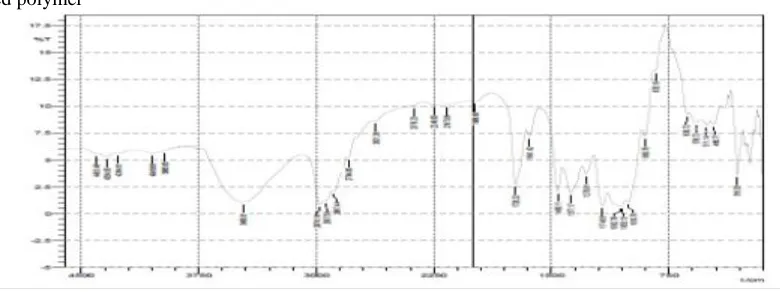
7.Compatibility Studies a)FTIR studies
Instrument used was Shimadzu FTIR-8700 spectrophotometer. In this study, potassium bromide disc method was employed. IR study of pure drug, physical mixtures and polymer were done. The powdered sample was intimately mixed with dry powdered potassium bromide. The mixture was then compressed into transparent disc under high pressure using special dies. The disc was placed in IR spectrophotometer using sample holder and spectrum was recorded from 4000 to 500 cm-1 [7].
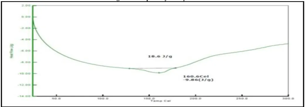
b)DSC studies
Differential scanning calorimetry studies were conducted using DSC Q2000. Sample was weighed
(8.00-10.00 ± 0.5 mg) and placed in sealed aluminium pans. The coolant was liquid nitrogen. The samples were scanned at 100 ˚C/ min from 200 to 2500
C. DSC thermo grams of pure erythromycin, physical mixtures and polymer were taken [11-13].
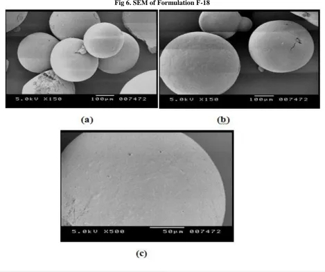
c) (SEM) Scanning electron microscopy
Scanning electron microscopy (SEM) is an electron optical imaging technique that provides photographic images and elemental information. SEM is useful for characterizing the morphology and size of microscopic specimens with particle size as low as 10-10 to 10-12 grams. The sample was placed in an evacuated chamber and scanned in a controlled pattern by an electron beam. Interaction of the electron beam with the specimen produces a variety of physical phenomena that, when detected, are used to form images and provide elemental information about the specimens. Scanning electron microscopy (JSM 840 A) was used to study the surface morphology of the microsphere of formulation I. The samples were analyzed after they were gold sputtered using 25 nm gold film thickness [7-10].
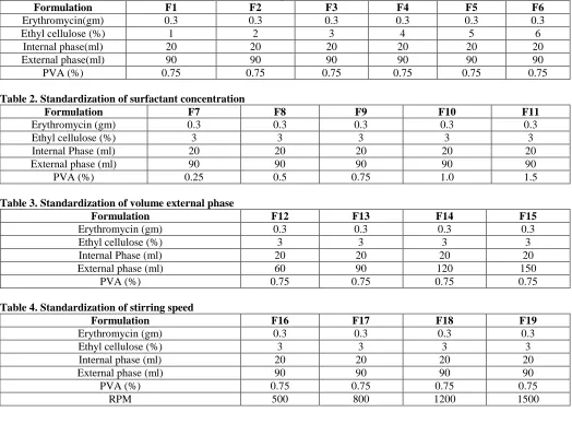
Table 1. Concentration of retardant material in the internal phase
Formulation F1 F2 F3 F4 F5 F6
Erythromycin(gm) 0.3 0.3 0.3 0.3 0.3 0.3
Ethyl cellulose (%) 1 2 3 4 5 6
Internal phase(ml) 20 20 20 20 20 20
External phase(ml) 90 90 90 90 90 90
PVA (%) 0.75 0.75 0.75 0.75 0.75 0.75
Table 2. Standardization of surfactant concentration
Formulation F7 F8 F9 F10 F11
Erythromycin (gm) 0.3 0.3 0.3 0.3 0.3
Ethyl cellulose (%) 3 3 3 3 3
Internal Phase (ml) 20 20 20 20 20
External phase (ml) 90 90 90 90 90
PVA (%) 0.25 0.5 0.75 1.0 1.5
Table 3. Standardization of volume external phase
Formulation F12 F13 F14 F15
Erythromycin (gm) 0.3 0.3 0.3 0.3
Ethyl cellulose (%) 3 3 3 3
Internal Phase (ml) 20 20 20 20
External phase (ml) 60 90 120 150
PVA (%) 0.75 0.75 0.75 0.75
Table 4. Standardization of stirring speed
Formulation F16 F17 F18 F19
Erythromycin (gm) 0.3 0.3 0.3 0.3
Ethyl cellulose (%) 3 3 3 3
Internal phase (ml) 20 20 20 20
External phase (ml) 90 90 90 90
PVA (%) 0.75 0.75 0.75 0.75
31 | P a g e
Table 5. Effect of concentration of ethyl cellulose on the internal phase
Sl. No Formulations Formation of
microsponges Physical appearance Particle size in µm Production yield (%)
1 F1 + Irregular 150-210±7.35 64.4
2 F2 + Spherical but collapses
after some time 210-260±6.95 72.4
3 F3 + Spherical 260-375±4.82 80.3
4 F4 + Spherical 360-435±3.78 84.7
5 F5 + Spherical rigid 420-470±3.76 88.5
6 F6 + Spherical rigid 470-570±2.67 90.5
+ indicates formation of microsponges.
4) Effect of surfactant concentration Table 6. Effect of surfactant concentration
Sl No Formulations Physical appearance Mean particle size (µm) Characteristics of emulsion
1 F7 Irregular 170- 220 ±7.35 Irregular
2 F8 Uniform 210- 280 ±6.91 Uniform
3 F9 Uniform 290- 350±3.76 Uniform
4 F10 Irregular 350- 440±2.48 Foaming
5 F11 Irregular 425- 480±3.61 Foaming
5) Effect of volume of external phase on microsponges Table 7. Effect of volume of external phase
Sl. no Formulations Physical appearance Particle size in µm Drug content (%) % entrapment
1 F12 Irregular, non-uniform 170-260±4.56µm 76.8 67.33 2 F13 Spherical, uniform 280-360±3.82µm 89.3 84.77 3 F14 Spherical, uniform 380-460±4.39µm 89.2 82.55 4 F15 Spherical, uniform 490-580±5.67µm 78.2 72.63
6) Effect of process parameters
a) Effect of stirring speed on formation of microsponges Table 8. Effect of stirring speed
Sl. No Formulations Physical appearance of microsponges
Mean particle size
(µm) Drug content
% Entrapment
1 F16 No microsponges were
formed - - -
2 F17 Uniform 290-430±3.68 75.49 67.33
3 F18 Uniform 170-250±4.34 91.33 88.77
4 F19 Uniform 100-180±2.75 72.41 65.34
RESULTS
Fig 1. FTIR of physical mixture of pure erythromycin, ethyl Cellulose and PVA
32 | P a g e
2) DSC studies
DSC studies were carried out for pure erythromycin and physical mixture of drug with Ethyl Cellulose, PVA.
Fig 2. DSC of pure erythromycin
3) FORMULATION OF ERYTHROMYCIN MICROSPONGES
a) Effect of formulation parameters
i) Concentration of retardant material in the internal phase
Fig 3. DSC of physical mixture of erythromycin, ethyl cellulose and PVA
7) FTIR STUDIES
33 | P a g e
8)DSC STUDIES
Fig 5. DSC studies of Formulation F-18
9) Morphological studies of microsponges a) SEM studies
(a)and (b) representing microsponges are spherical in shape (c) Representing microsponges are porous and spongy in nature.
34 | P a g e
DISCUSSION AND CONCLUSION
The present study was to formulate and evaluate the microsponge for topical sustained drug delivery of erythromycin for extended release. The study design focused on standardization of process parameters like standardization of concentration of retardant material table no (1), concentration of surfactant table no (2), volume of external phase table no (3) and the effect of stirring speed table no (4) involved in the preparation of erythromycin microsponges by quassi-emulsion solvent diffusion method. Ethyl cellulose was used as a polymer, poly vinyl alcohol as a surfactant, methanol and dichloromethane as solvents in the internal phase, water as external phase. By compatibility studies it was found there was no interaction between the drug and excipients. The best standardized F18
formulation showed good loading efficiency, production yield, particle size, drug content. The SEM results showed that the microsponges were spherical and porous in nature.
Thus it was concluded that the selected anti-acne drug can be developed into microsponges and further can be incorporated into gel for topical application.
ACKNOWLEDGEMENTS
I wish to thank Micro pharmaceutical Ltd Bangalore for providing gift sample of Erythromycin drug and Dr. S.K. Senthil kumar HOD of pharmaceutics Dept, Dr. T. Tamiz Mani Principal of Bharathi College of pharmacy, Bharathi Nagar, Mandya, for providing necessary facilities.
REFERENCES
1. Lin HS, et al. Biopharmaceutics of 13-cis-retinoic acid (isotretinoin) formulated with modified β-cyclodextrins. Int J Pharm, 341, 2007, 238-45.
2. Kaity S, Maiti S, Ghosh AK, Pal D, Ghosh A and Banerjee S. Microsponges: A novel strategy for drug delivery system. J. Adv. Pharm.Tech. Res, 1(3), 2010, 283-90.
3. Kawashima Y, Niwa T, Handa T, Takeuchi H, Iwamoto T, Itoh K. Preparation of controlled-release microspheres of ibuprofen with acrylic polymer by a novel quasi-emulsion solvent diffusion method. J Pharm Sci, 78, 1989, 68-72.
4. Rigopoulos D, Larios G, Katsambas AD. The role of Isotretinoin in acne therapy: why not as first-line therapy? Facts and controversies. Clin Dermatol, 28, 2010, 24–30.
5. Archana J et al. Retinoids-An overview of clinical applications in dermatology.J. Pharm. Sci. & Res, 2 (7), 2010, 376-83.
6. Amrutiya N, Bajaj A, Madan M. Development of Microsponges for Topical Delivery of Mupirocin. AAPS PharmSciTech, 10(2), 2009, 402-9.
7. Jelvehgari M, Siahi SMR, Azarmi S, Martin GP, Nokhodchi A. The microsponge delivery system of benzoyl peroxide: preparation, characterization and release studies. Int J Pharm, 308, 2006, 124-32.
8. Saboji JK, Manvi FV, Gadad AP and Patel BD. Formulation and evaluation of Ketoconazole microsponge gel by quassi emulsion solvent diffusion. J. Cell Tissue Research, 11(1), 2011, 2691-96.
9. Mishra MK, Shikhri M, Sharma R and Goojar MP. Optimization, formulation development and characterization of Eudragit RS 100 loaded microsponges and subsequent colonic delivery.Int J Drug Dis Herbal Res, 1(1), 2011, 8-13. 10. Comoglu T, Gonul N and Baykara T. Preparation and in vitro evaluation of modified release ketoprofen microsponges. Il
Farmaco, 58, 2003, 101-06.
11. Vermeulen B, Remon JP, Nelis H. The formulation and stability of erythromycin–benzoyl peroxide in a topical gel. Int J
Pharm, 178, 1999, 137–41.
12. Korting SM, Mehnert W, Christian H. Lipid nonoparticles for improved topical application of drugs for skin diseases. Adv drug deliv Rev, 59, 2007, 427-43.