
CRYSTAL STRUCTURE OF {[La
2(CNCH
2COO)
6(H
2O)
4]
·
H
2O}
nCOMPLEX
Ana Lazarescu
a*, Elena Melnic
b, Sergiu Shova
a, Victor Kravtsov
b, Constantin Turta
aaInstitute of Chemistry of Academy of Sciences of Moldova, 3, Academiei str., Chisinau MD-2028, Republic of Moldova bInstitute of Applied Physics of Academy of Sciences of Moldova, 5, Academiei str., Chisinau, MD-2028, Republic of Moldova
*e-mail: [email protected]; phone: (+373 22) 73 97 22; fax: (+373 22) 73 99 54
Abstract. The lanthanum(III) cyanoacetate complex of formula {[La2(CNCH2COO)6 (H2O)4]∙H2O}n (1), has been prepared and characterized by X-ray diffraction analysis. Compound crystallizes in the triclinic centrosymmetric space group P-1 (No.2), a=8.997(5) Å, b=9.251(5) Å, c=9.728(5) Å, α=67.849(5)°, β=84.224(5)°, γ=81.351(5)°. Single-crystal X-ray diffraction study reveals that crystals of 1 exhibits one-dimensional coordination polymer structure, which is composed of cyanoacetate bridged lanthanum(III) ions. The O9 coordination surrounding of La cation is completed by oxygen atoms of six carboxylate ligands that are coordinated in the bidentate bridging and tridentate-chelating bridging mode and two water molecules. The cations are bridged in a polymeric chain by four exo-bidentate cyanoacetate ligands or through μ2-O function of two other chelato-bridgind cyanoacetate ligands. The La∙∙∙La separation along the polymer is equal to 4.754(3) Å and 4.608(2) Å.
Keywords: coordination polymers,lanthanum (III), cyanoacetic acid, crystal structure.
Received: November 2014/ Revised fi nal: January 2015/ Accepted: February 2015
Introduction
The synthesis of functional rare-earth coordination polymers with various N- and / or O-donor ligands has attracted increasing interest, due to their various topologies [1-3] and potential applications as optical, electronic and magnetic materials [4-7].
The carboxylate ligands are attractive candidates for construction of the polymeric networks due to their versatile functions as bridges or terminal groups, which lead to generation of dimers [8, 9], 1D chains [10], 2D-layers [11], and 3D- polymeric structures [12].
However, investigation of lanthanide complexes with cyanoacetic acid anion as ligand is very scarce [13,14]. Our previous investigations of 3d and 4f metal complexes with cyanoacetate anions have demonstrated that copper(II) ion has {O4(carbox.)N(CN)} coordination node, while neodymium(III) ion – {O
7
(carbox.)O
2(H²O)}, respectively [15, 16]. The study of magnetic properties of gadolinium(III) cyanoacetate complex has shown that external magnetic fi eld induces two slow relaxation processes, while heat capacity and direct current susceptibility measurements at very low temperatures has indicated strong single ion anisotropy, due to the Kramer’s doublet of the ground state [17].
In continuation of our research on the use of cyanoacetic acid with both O- and N- potential donor atoms as a ligand for 4f cations [16, 17] and d-block transition metal [15], we have synthesized and structurally characterized the {[La2(CNCH2COO)6 (H2O)4]·H2O}n compound by single crystal X-ray method.
Experimental Synthesis
All reagents of analytical grade were commercially purchased and used without further purifi cation. Elemental analyses (C, H, N) were performed on a Elemental Analyzer vario EL(III). The lanthanum content in the complex was determined gravimetrically as stable oxide by precipitating lanthanide (III) ions with ammonium hydroxide [18].
{[La2(CNCH2COO)6 (H2O)4]·H2O}n (1). Solution of cyanoacetic acid (0.51 g, 6 mmol) in water (10 mL) was added to La2O3 (0.32 g, 1 mmol). After stirring the resulting solution was passed through a glass fi lter. The fi ltrate was stored at room temperature for some weeks, whereupon the suitable for X-ray analysis colorless crystals of the product appeared. The crystals were collected by fi ltration, dried under reduced pressure and kept in desiccator. Yield: 52 %. Anal. found (calc.)%: for {C9H12N3O9La}n , C, 23.99 (24.29); H, 2.43 (2.69); N 9.07 (9.45); La, 30.89 (31.22). IR data (Nujol, cm-1): 3545br, 3430br, 3250br, 2265sh, 1705s, 1600vs, 1580s, 1545s, 1410s, 1264m, 1195m, 955w, 930m, 915m, 700m, 570m, 515m.
Physical measurements
IR spectrum of polycrystalline sample was recorded on Perkin Elmer spectrum 100 FT IR Spectrometer in the range of 4000-400 cm-1.
X-ray crystallography
The X-ray data for 1 were collected at room temperature on an Oxford Diffraction Xcalibur diffractometer equipped with CCD area detector and a graphite monochromator utilizing MoKα radiation. The crystals were placed at 40 mm from the CCD detector. The data were processed using the CrysAlis package of Oxford Diffraction [19]. The
using a riding model approximation with Uiso(H) = 1.2Ueq(C), while the hydrogen atoms of water molecules were refi ned with isotropic displacement parameter Uiso (H) = 1.5Ueq(O) using DFIX restrain instruction. The fi gures were produced using MERCURY [21]. The X-ray data and details of the refi nement for 1 are summarized in Table 1, the selected geometric parameters are given in Table 2 and the hydrogen-bonding geometry is given in Table 3. CCDC 1032624 contains the crystallographic data for 1. These data can be obtained free of charge from The Cambridge Crystallographic Data Centre via www.ccdc.cam.ac.uk/data_request/cif.
Таble 1 Crystal and structure refi nement data for 1.
Parameter Value
Compound 1
Empirical formula C9H12N3O9La
Formula weight 445.13
T (K) 293(2) K
Crystal system Triclinic
Space group P-1
Z 2
a, Å 8.997(5)
b, Å 9.251(5)
с, Å 9.728(5)
α, (º) 67.849(5)
, (º) 84.224(5)
γ, (º) 81.351(5)
V, Å3 740.5(7)
Dcalc (g cm -3) 1.996
μ(mm-1) 2.934
F(000) 432
Crystal size (mm) 0.30 x 0.20 x 0.15
θmin, θmax (º) 2.62 - 25.99
Refl ections collected/unique 5555 / 2899
Refl ections with [I > 2σ(I)] 2810
Data/restraints/parameters 2899 / 9 / 217
Goodness-of-fi t (GOF)on F2 1.000
R1, wR2 [I > 2σ(I)] 0.0182, 0.0464
R1, wR2 (all data) 0.0190, 0.0468
Δρmax, Δρmin, (eÅ-3) 0.416, -0.689
Table 2 Selected bond lengths (Å) and angles (º) in coordination metal environment in1.
Bond d, (Å) Bond d, (Å)
La-O(1) 2.511(2) La-O(3) ii 2.804(2)
La-O(2) 2.456(2) La-O(6)i 2.490(2)
La-O(3) 2.525(2) La-O(1w) 2.578(2)
La-O(4) 2.451(2) La-O(2w) 2.667(2)
La-O(5) 2.639(2) -
-Angle ω, (deg) Angle ω, (deg)
O(1)-La-O(2) 117.39(7) O(3)-La-O(3)ii 60.38(6)
O(1)-La-O(3)ii 121.23(5) O(3)-La-O(1w) 74.69(7)
O(1)-La-O(3) 79.09(6) O(3)-La-O(2w) 78.71(6)
O(1)-La-O(4) 75.31(7) O(4)-La-O(3)ii 114.09(6)
O(1)-La-O(5) 138.55(7) O(4)-La-O(5) 77.08(6)
O(1)-La-O(6)i 73.48(7) O(4)-La-O(6)i 117.37(7)
O(1)-La-O(1w) 143.16(6) O(4)-La-O(1w) 138.48(7)
O(1)-La-O(2w) 67.33(6) O(4)-La-O(2w) 68.49(7)
O(2)-La-O(3) 141.97(6) O(5)-La-O(3)ii 47.66(5)
O(2)-La-O(3)ii 120.81(7) O(5)-La-O(6)i 147.68(6)
O(2)-La-O(4) 73.24(7) O(5)-La-O(1w) 75.17(7)
O(2)-La-O(5) 82.64(7) O(5)-La-O(2w) 74.06(6)
O(2)-La-O(6)i 75.29(7) O(6)i -La-O(3)ii 128.54(6)
Continuation of Table 2
Angle ω, (deg) Angle ω, (deg)
O(2)-La-O(2w) 138.57(6) O(1w)-La-O(2w) 130.08(6)
O(3)-La-O(4) 144.14(6) O(1w)-La-O(6)i 75.99(7)
O(3)-La-O(5) 108.01(6) O(2w)-La-O(3)ii 64.55(6)
O(3)-La-O(6)i 77.77(6) O(2w)-La-O(6)i 137.25(6)
Symmetry code: i -x+1, -y, -z+1; ii -x+1, -y+1, -z+1
Table 3 Geometric parameters of hydrogen bonds (Å, deg) in1.
D–H···A d(H···A) d(D···A) Ð(DHA) Symmetry transformation
for acceptor
O1w-H1w1∙∙∙N3 2.28(2) 3.106(4) 168(3) x, y, z
O1w-H2w1∙∙∙O2w 1.92(2) 2.771(3) 161(3) -x+1, -y+1, -z+1
O2w-H2w2∙∙∙O3w 1.90(2) 2.770(3) 164(3) x, y, z
O2w -H1w2∙∙∙O3w 2.02(2) 2.809(3) 153(2) -x+1, -y+1, -z O3w -H1w 3∙∙∙N1 2.29(3) 2.951(4) 136(3) -x+2, -y+1, -z
O3w -H2w3∙∙∙N2 1.95(2) 2.802(3) 176(3) x, y+1, z
Results and discussion
The reaction of lanthanum(III) oxide with cyanoacetic acid in a molar ratio of 1:3 in water solution leads to polymeric complex with the composition {[La2(CNCH2COO)6 (H2O)4]H2O}n (1). The complex is a colorless, air hygroscopic crystalline solid, readily dissolves in water, but is insoluble in organic solvents, such as methanol, acetone and acetonitrile. The IR spectrum shows the absorption band at 2265 cm-1, which is characteristic to the (C≡N) vibrations of the ligand.
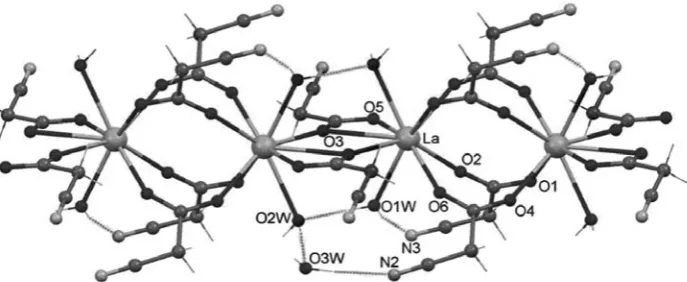
Here we report the results of X-ray study of the lanthanum(III) compound with cyanoacetate ligands, which is a linear polymer of composition {[La2(CNCH2COO)6(H2O)4]H2O}n. Compound crystallizes in the triclinic centrosymmetric space group P-1, a = 8.997(5) Å, b = 9.251(5) Å, c = 9.728(5) Å, α = 67.849(5)°, β = 84.224(5)°, γ = 81.351(5)°, V = 740.5(7) Å3 and is isostructural to the reported earlier Nd analog [16]. The inversion centers related to La cations are linked in 1D structure by carboxylic groups of cyanoacetate ligands, which exhibit different binding modes (Figure 1).
Figure 1. View of polymeric chain.
The La cation is linked to the center of symmetry related neighboring La cations either by four cyanoacetic ligands, acting as syn–syn (c-2-c) bidentate bridges (La–O2, 2.455(2); La–O6, 2.490(2); La–O1’, 2.511(2); La–O4’, 2.450(2) Å), or through two tridentate (32-c) chelate-bridging ligands with the La – O3 bond lengths of 2.524(2) and 2.806(2) Å, respectively.
with the difference in ionic radii of nine coordinated LaIII (1.36 Å) and NdIII (1.30 Å) [22]. The cyano group of the cyanoacetate ligands does not coordinate to the lanthanide cation. The coordinated water molecules are involved in intra- and inter-chain hydrogen bonds (Table 3).
In the crystal, the parallel polymer chains run along a crystallographic axis and are united into a three-dimensional network through a system of hydrogen bonds involving water molecules and nitrogen atoms of cyanoacetic ligands (Figure 2).
Figure 2. Hydrogen bonded parallel polymer chains in the crystal structure of
{[La2(CNCH2COO)6(H2O)4]·H2O}n. Solvent water molecules O3w are shown by bigger radii red spheres.
Conclusions
The lanthanum(III) cyanoacetate polymer of formula {[La2(CNCH2COO)6 (H2O)4]∙2H2O}n (1) has been synthesizedand characterized by single-crystal X-ray diffraction. The complex was found to be isostructural to Nd analog. The cyano-group of the ligand is not coordinated to the lanthanide ion.
References
1. Decadt, R.; Van Hecke, K; Depla, D; Leus, K; Weinberger, D; Van Driessche, I.; Van Der Voort, P; Van Deun, R. Synthesis, crystal structures, and luminescence properties of carboxylate based rare-earth coordination polymers. Inorganic Chemistry, 2012, 51, pp. 11623-11634.
2. Qin, C.; Wang. X.I.; Wang, E.B.; Su, Z.M. A Series of three-dimensional lanthanide coordination polymers with rutile and unprecedented rutile-related topologies. Inorganic Chemistry, 2005, 44, pp. 7122-7129.
3. Gai, Y.-Li; Xiong, K.-C.; Chen, L.; Bu, Y.; Li, X.-J.; Jiang, F.-L.; Hong, M.-C. Visible and NIR Photoluminescence Properties of a Series of Novel Lanthanide–Organic Coordination Polymers Based on Hydroxyquinoline– Carboxylate Ligands. Inorganic Chemistry,2012, 51, pp. 13128-13137.
4. Liu, C.M.; Xiong, M.; Zhang, D.Q.; Du, M.; Zhu, D.B. Two-and three-dimensional lanthanide-organic frameworks constructed using 1-hydro-6-oxopyridine-3-carboxylate and oxalate ligands. Dalton Transactions, 2009, 29, pp. 5666-5672.
5. Hasegawa, Ya.; Nakagawa, T.; Kawai, T. Recent progress of luminescent metal complexes with photochromic units. Coordination Chemistry Review, 2010, 254, pp. 2643-2651.
7. Cunha-Silva, L.; Lima, S.; Ananias, D.; Silva, P.; Mafra, L.; Carlos, I.D.; Pillinger, M.; Valente, A.A.; Almeida Paz, P.A.; Rocha, J. Multi-functional rare-earth hybrid layered networks: photoluminescence and catalysis studies. Journal of Materials Chemistry, 2009, 19, pp. 2618- 2632.
8. Rohde, A.; Urland, W. Synthesis, crystal structures and magnetic behaviour of dimeric and tetrameric gadolinium carboxylates with trichloroacetic acid. Journal of Chemical Society, Dalton Transactions, 2006, 24, pp. 2974-2978. 9. Yi-Shan, S.; Bing, Y.; Zhen-Xia, C. Different crystal structure and photophysical properties of lanthanide complexes
with 5-bromonicotinic acid. Journal of Solid State Chemistry, 2004, 177, pp. 3805-3814.
10. Chen, C.; Zhang, S.-Y.; Song, H.-B.; Shi, W.; Zhao, B.; Cheng, P. One-dimensional lanthanide coordination polymers as promising luminescent materials. Inorganica Chimica Acta, 2009, 362, pp. 2749-2745.
11. Mahata, P.; Ramya, K.V.; Natarajan, S.; Reversible water intercalation accompanied by coordination and color changes in a layered metal-organic framework. Inorganic Chemistry. 2009, 48, pp. 4942-4951.
12. Lu, W.-G.; Jiang, L.; Feng, X.-L.; Lu, T.-B. Three-dimensional lanthanide anionic metal-organic frameworks with tunable luminescent properties induced cation exchange. Inorganic Chemistry, 2009, 48, pp. 6997-6999.
13. Starynowicz P. Structure and electron spectroscopy of potassium triaquadecakis(cyanoacetato)trineodymate hexahydrate. Polish Journal of Chemistry, 1994, 68, pp. 621-629.
14. Starynowicz, P. Catena-Poly[{aqua(cyanoacetato-κO)(N,N-dimethylformamide-κO)neodymium}-di-μ-cyano- acetato-κO,κO’:κO’]monohydrate. Acta Crystallographica, Sect.C: Crystal Structure Communications, 1993, 49, pp. 1621-1623.
15. Novitskii, G.; Shova, S.; Voronkova, V.; Korobcenko, L.; Gdanets, M.; Simonov, Iu.; Turta, C. Copper(II) cyanoacetate polymer: synthesis and structure. Russian Journal of Coordination Chemistry, 2001, 27, pp. 839-844 (in Russian).
16. Shova, S.G.; Simonov, Yu.A.; Gdaniec, M.; Novitskii, G.V.; Lazarescu, A.; Turta, K.I. Synthesis and Structure of catena-{Bis(μ-cyanoacetato(O,O,O¢))tetrakis(μ-cyanoacetato(O,O¢))dineodymium(III). Russian Journal of Inorganic Chemistry, 2002, 47, pp. 946-952 (in Russian).
17. Arauzo, A.; Lazarescu, A.; Shova, S.; Bartolomé, E.; Cases, R.; Luzón, J.; Bartolomé, J.; Turta, C. Structural and magnetic properties of some lanthanide (Ln= Eu(III), Gd(III) and Nd(III)) cyanoacetate polymers: Field-induced slow magnetic relaxation in the Gd and Nd substitutions. Dalton Transactions, 2014, 43, pp. 12342-12356. 18. Charlot G. Methods of Analytical Chemistry. The Quantitative Analysis of Inorganic Compounds. Chemistry:
Leningrad, 1965, 975 p. (in Russian).
19. CrysAlisPRO, Oxford Diffraction /Agilent Technologies UK Ltd, Yarnton, England.
20. Sheldrick, G.M. A short history of SHELX. Acta Crystallographica, 2008, A64, pp. 112-122.
21. Edgington, P.R.; McCabe, P.; Macrae, C.F.; Pidcock, E.; Shields, G.P.; Taylor, R.; Towler, M.; Van de Streek, J. Mercury: visualization and analysis of crystal structures. Journal of Applied Crystallography, 2006, 39, pp. 453-457.
![Figure 2. Hydrogen bonded parallel polymer chains in the crystal structure of COO)(HO)]·HO}](https://thumb-us.123doks.com/thumbv2/123dok_us/8795763.1769134/4.561.136.422.129.414/figure-hydrogen-bonded-parallel-polymer-chains-crystal-structure.webp)
