R E S E A R C H
Open Access
Murine esBAF chromatin remodeling complex
subunits BAF250a and Brg1 are necessary to
maintain and reprogram pluripotency-specific
replication timing of select replication domains
Shin-ichiro Takebayashi
1, Ienglam Lei
2,3,4, Tyrone Ryba
1, Takayo Sasaki
1, Vishnu Dileep
1, Dana Battaglia
1,
Xiaolin Gao
2,3, Peng Fang
2,3, Yong Fan
2,3, Miguel A Esteban
5, Jiong Tang
6, Gerald R Crabtree
6,
Zhong Wang
2,3,4and David M Gilbert
1*Abstract
Background:Cellular differentiation and reprogramming are accompanied by changes in replication timing and 3D organization of large-scale (400 to 800 Kb) chromosomal domains (‘replication domains’), but few gene products have been identified whose disruption affects these properties.
Results:Here we show that deletion of esBAF chromatin-remodeling complex components BAF250a and Brg1, but not BAF53a, disrupts replication timing at specific replication domains. Also,BAF250a-deficient fibroblasts reprogrammed to a pluripotency-like state failed to reprogram replication timing in many of these same domains. About half of the replication domains affected by Brg1 loss were also affected by BAF250a loss, but a much larger set of domains was affected by BAF250a loss. esBAF binding in the affected replication domains was dependent upon BAF250a but, most affected domains did not contain genes whose transcription was affected by loss of esBAF.
Conclusions:Loss of specific esBAF complex subunits alters replication timing of select replication domains in pluripotent cells.
Keywords:Replication domains, Replication timing, esBAF complex, Chromosome, Developmental regulation
Background
Developmental changes in chromosome structure can occur at the level of large, often megabase-sized chromo-some domains [1-5]. This cell type-specific chromosomal domain structure is thought to be important for coordinat-ing expression of genes, thereby ensurcoordinat-ing proper develop-ment of embryos. However, the mechanisms regulating large-scale changes in chromosome structure during devel-opment are poorly understood. In particular, very few gene products have been found to be necessary to maintain structure and function of chromosomes at this level of organization.
The temporal order of replication (replication timing) is linked to many basic cellular processes that are regu-lated both during the cell cycle and development. We have developed a simple and robust assay to measure replication timing genome-wide [6,7]. We found that 400 to 800 Kb-sized replication domains are spatio-temporally reorganized genome-wide during embryonic stem (ES) cell differentiation into various cell lineages [6,8]. Similar sized replication domains are also misregu-lated in leukemia [9]. Cell type specific reorganization of replication domains is generally coordinated with tran-scriptional changes and is conserved between mouse and human [10-12]. Replication domain reorganization is also observed during iPSC generation in which som-atic cell specific replication domain structure is erased and ESC-specific replication domain structure is re-established [8]. Considering that replication domains are
* Correspondence:[email protected]
1
Department of Biological Science, Florida State University, 319 Stadium Drive, Tallahassee, FL 32306, USA
Full list of author information is available at the end of the article
regulated in the context of development and disease, it is presumed that epigenetic mechanisms play an important role in the formation of replication domain structure. How-ever, in mammals, little or no effect on replication timing regulation has been reported for many chromatin modifier mutants, while these mutations significantly affect gene ex-pression patterns [13-15]. Recently the first gene products with widespread effects on global replication timing in yeast (Fkh1/2 and Rif1) and mammals (Rif1) were identified [16-19]. Other gene products have been shown to have small effects on pericentric heterochromatin replication (Sub39h1/2 and G9a) [13,14]. Finally, replication timing of rDNA was shown to be affected by mutations in the rDNA-specific chromatin remodeling complex NoRC [20]. Together, these results suggest that specific gene products should eventually be identified that regulate cell type and domain-specific affects. Inspired by the specific and dra-matic effect of NoRC on regulation of rDNA replication timing, we investigated the role of cell type specific chro-matin remodeling complexes in replication timing changes during embryonic stem cell differentiation.
Brahma-associated factor (BAF) complexes are members of SWI/SNF ATP-dependent chromatin-remodeling family and regulate access of transcription factors by modulating chromatin structure. Of particular interest is that BAF subunits undergo compositional and stoichiometric change during mammalian development, which confers unique and essential roles to the complexes in cell fate determin-ation [21-24]. For example, BAF155, BAF250a, and Brg1 are highly expressed in ESCs and their expression decreases significantly when ESCs differentiate, suggesting that these components may be essential for keeping ESCs in the un-differentiated‘ground state’[25]. In fact, Brg1 and BAF155 significantly promote reprogramming of mouse embryonic fibroblasts (MEFs) in combination with Yamanaka factors (Oct4, Sox2, Klf4, and c-Myc) [26]. BAF components are also instrumental for tissue-specific differentiation. The proper switch of neuron-specific BAF53 and BAF45 iso-forms determines either the self-renewal or differentiation of neuron progenitor cells [27] and can convert fibro-blasts to neurons [28]. Ectopic expression of BAF60c, a cardiac-enriched subunit, along with transcription fac-tors GATA4 and TBX5, can convert non-cardiogenic mesoderm into beating cardiomyocytes [29]. These studies suggest that tissue-specific BAF complexes cre-ate chromatin environments favorable for transcription factor access.
In this study, we found that the embryonic stem cell-specific BAF complex (esBAF) complex deficiency leads to alterations of replication timing both in ESCs and during cellular reprogramming. Loss of DNA binding of the complex, but not transcriptional changes, correlated with changes in replication timing. These findings demonstrate the importance of chromatin remodeling complexes for
maintaining replication-timing programs and, by proxy, large-scale chromatin reorganization.
Results and discussion
BAF250a is required to maintain replication timing at specific domains in embryonic stem cells
We first examined the effect of acute BAF250a loss on replication timing. BAF250a is essential for early em-bryogenesis and has shown to be involved in the recruit-ment of esBAF to its target sites [30,31]. We generated a cell line in which both homologues ofBAF250aundergo simultaneous conditional deletion. In these cell lines, exon 8 of the BAF250a gene is flanked by 2 loxp sites and Cre recombinase (Mer-Cre-Mer) is induced upon addition of 4-hydroxytamoxfen (OHT), resulting in frameshift mutation followed by non-sense mediated decay. BAF250a protein level was rapidly and homoge-neously diminished within 24 h and was undetectable 72 h after OHT treatment [see Additional file 1A].
Genome-wide replication timing analysis (Figure 1A, [7]) identified a set of genomic regions that changed replication timing either from early to late (EtoL) or from late to early (LtoE) in response to BAF250a loss after 72 h but not after 24 h (Figure 1B-D and [see Additional file 1B-C]). Observed changes in replication timing were highly reproducible between replicates [see Additional file 1D]. Since the changes were not as exten-sive as developmental changes [6,8], we calculated the P values for replication timing changes of 10,974 200-Kb segments, and applied a False Discovery Rate (FDR). Using this method, 691 and 1,667 200-Kb segments were identified as significantly changing replication timing at a 1% and 5% FDR, respectively (Figure 1C and [see Additional file 1E]). All affected segments examined aligned in register to domains of differential replication in one or more tissues during normal development (Figure 1E) and encompassed 400 to 800 Kb genomic segments (Figure 1F), consistent with domains whose replication timing is normally regulated during develop-ment [8-10]. We conclude that BAF250a is required to maintain normal developmental control of replication timing for a fraction of the ESC genome.
and Rex1domains, which rapidly become late replicating during differentiation to every germ layer [8], retained ESC-specific early replication [see Additional file 2]. Moreover, we did not observe significant changes in the expression level of pluripotency-associated genes [see Additional file 3]. Together, we conclude that mu-tant ESCs still globally maintain an overall pluripotent cell replication timing program at least 72 h after OHT treatment, while specific domains require esBAF to maintain their replication time.
BAF250a is required to re-establish replication timing of an overlapping set of select domains during somatic cell reprogramming
To further confirm the requirement of BAF250a for replication timing of specific domains in pluripotent cells, we investigated whether similar replication timing defects occur in cells reprogrammed from somatic cells in the absence of BAF250a. First we examined the effect of BAF250a loss on formation of iPSC-like colonies.
BAF250a flox/flox and BAF250a flox/flox Mer-Cre-Mer
A
Chromosome 15 (Mb) Replication timing Log
2
(Early/Late)
2) FACS sort
1) Pulse label with BrdU
5) Hybridize to CGH array
G1 G2/M
3) Immunoprecipitate nascent BrdU-substituted DNA
Early-S Late-S
4) Differentially label
D
C
20.0 40.0 60.0 80.0 100.0
66.0 5.0
0
-1.0 1.0 2.0
LtoE EtoL
95.5% (42/44 domains) 90.4%
(47/52 domains)
Align to developmental boundaries
E
340
EtoL switching LtoE switching
Number of timing switching segments in BAF250a mutant ESCs
351
Total number of segments = 10974
765 902
BAF250a mock
BAF250a OHT
46C ESC TT2 ESC NPC Myo MEF 2.0
1.0 0 -1.0 -2.0
B
2.0 1.0 0 -1.0 -2.0
Chr4 EtoL (104.5-105.0 Mb)
Mock 2.0
1.0
0
-1.0
-2.0 2.0
1.0
0
-1.0
-2.0
104.0 105.0
Chromosomal position (Mb) OHT
Chr15 EtoL (64.5-66.0 Mb)
Chr6 LtoE (118.0-119.0 Mb)
Chr18 LtoE (5.0-6.0 Mb)
0
-1.0 1.0 2.0
Chr4 EtoL (104.5-105.0 Mb)
Chr6 LtoE (118.0-119.0 Mb)
105.0 104.0 106.0 Chr15 EtoL (64.5-66.0 Mb)
Chr18 LtoE (5.0-6.0 Mb)
1.000 0.946 0.946 1.000 0.926 0.949 0.941 0.949 0.888 0.861 0.745 0.751 0.726 0.723 BAF250a mock BAF250a OHT
F
G
LtoE EtoL None-Switching 0
2 4 6 8 10
118.0 119.0
65.0 66.0 67.0 5.0 6.0
BAF250a mock
BAF250a OHT
Correlation (Pearson R) Chromosomal position (Mb)
Mock OHT Other cell types
Mock
OHT
Mock
OHT
Mock
OHT
118.0 119.0
67.0
65.0 6.0
Replication timing Log
2
(Early/Late)
Replication timing Log
2
(Early/Late)
Domain size (Mb)
Figure 1Genome-wide replication timing analysis identifies a subset of BAF250a sensitive replication domains. (A)Flow chart of genome-wide replication timing analysis [7]. BrdU-substituted DNA from early and late S phase cells was differentially labeled and hybridized to a whole-genome oligonucleotide microarray.(B)Replication timing profiles from mock- and 4-hydroxytamoxifen (OHT)-treatedBAF250a flox/flox embryonic stem cells (ESCs) for chromosome 15. The signal ratio of early and late (Log2 early/late) for each probe is plotted against the chromosomal position. Shown are loess-smoothed plots for the average of two biological replicate experiments.(C)Summary of significant early to late (EtoL) and late to early (LtoE) switching segments in OHT-treated cells. Replication timing data were averaged into 200-Kb windows and statistical significance
was calculated between mock and OHT-treated ESCs, as described in Methods.(D)Expanded plots for exemplary regions that undergo
replication-timing switches in response to BAF250a loss are shown below.(E)BAF250a-dependent timing switching domains align to
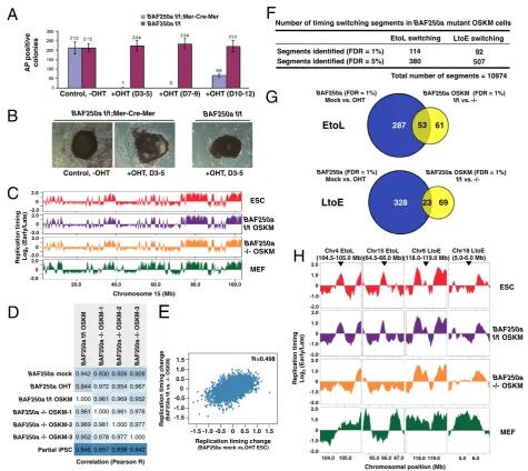
MEFs were treated with OHT at Days 3 to 5 after virus-mediated transduction of reprogramming factors (Oct4, Sox2, Klf4, and c-Myc; OSKM). We observed a significant decrease in the number of alkaline phos-phatase (AP)-positive colonies derived from cells expressing Mer-Cre-Mer compared to control cells (Figure 2A and B), demonstrating that BAF250a is
required for efficient somatic cell reprogramming, or for growth and viability of the latter. Also, colonies from OHT-treated BAF250a flox/flox Mer-Cre-Mer cells have an irregular unsmoothed shape (Figure 2B), which is reminiscent of the morphology of BAF250a -deficient ESCs [30]. Several iPSC-like colonies from
BAF250a flox/floxandBAF250a flox/flox Mer-Cre-Mer
Partial iPSC
BAF250a OHT
BAF250a mock
BAF250a -/- OSKM-1
BAF250a f/f OSKM
BAF250a -/- OSKM-2
BAF250a -/- OSKM-3
Correlation (Pearson R)
BAF250a f/f;Mer-Cre-Mer BAF250a f/f
Control, -OHT +OHT, D3-5 +OHT, D3-5
A
B
Control, -OHT +OHT (D3-5) +OHT (D7-9) +OHT (D10-12)
AP
positive
colonies
C
D
BAF250a f/f;Mer-Cre-Mer
BAF250a f/f
53
BAF250a (FDR = 1%)
Mock vs. OHT
BAF250a OSKM (FDR = 1%)
f/f vs.
-/-287 61
E
F
G
H
2.0
-2.0 0
2.0
-2.0 0
2.0
-2.0 0
2.0
-2.0 0
Chromosome 15 (Mb) Replication timing Log
2
(Early/Late)
20.0 40.0 60.0 80.0 100.0
0.942 0.930 0.944 0.972 1.000 0.961 0.961 1.000 0.969 0.981 0.952 0.978 0.845 0.857
0.928 0.928 0.954 0.967 0.969 0.952 0.981 0.978 1.000 0.977 0.977 1.000 0.838 0.842
BAF250a
OSKM-1
BAF250a f/f
OSKM
BAF250a
OSKM-2
BAF250a
OSKM-3
Number of timing switching segments in BAF250a mutant OSKM cells
LtoE switching EtoL switching
92 114
507 380
Total number of segments = 10974
23
328 69
Replication timing change
(
BAF250a f/f
vs.
OSKM)
-1.0 0 1.0 Replication timing change
(BAF250a mock vs.OHT ESC)
-0.5 0.5 R=0.498
1.5 -1.5
1.0 0.5 0 -0.5 -1.0 -1.5 1.5
EtoL
BAF250a (FDR = 1%)
Mock vs. OHT
BAF250a OSKM (FDR = 1%)
f/f vs.
-/-LtoE
Chr4 EtoL (104.5-105.0 Mb)
Chr15 EtoL (64.5-66.0 Mb)
Chr6 LtoE (118.0-119.0 Mb)
Chr18 LtoE (5.0-6.0 Mb)
ESC
BAF250a f/f OSKM
BAF250a -/- OSKM
MEF ESC
BAF250a f/f OSKM
BAF250a -/- OSKM
MEF
Chromosomal position (Mb) Replication timing Log
2
(Early/Late)
0
-1.0 1.0 2.0
104.0 105.0 65.0 66.0 67.0 118.0 119.0 5.0 6.0 -2.0
0
-1.0 1.0 2.0
-2.0 0
-1.0 1.0 2.0
-2.0 0
-1.0 1.0 2.0
-2.0
Figure 2Role of BAF250a in the regulation of replication domains in cells undergoing somatic cell reprogramming.
(A)Number of alkaline phosphatase (AP)-positive colonies derived from 4-hydroxytamoxifen (OHT)-treatedBAF250a flox/floxandBAF250a
cells were genotyped and confirmed to be BAF250a
flox/flox and BAF250a −/−, respectively. These col-onies, which we refer to as BAF250a flox/flox OSKM and BAF250a −/− OSKM, were also confirmed to ex-press high levels of ESC pluripotency markers (data not shown).
Next we performed replication-timing analysis of
BAF250a flox/flox OSKM and BAF250a −/− OSKM
cells. Despite the fact that loss of BAF250a significantly reduced the efficiency of AP-positive colony production, the genome-wide replication timing profiles of three in-dependent AP-positiveBAF250a−/−OSKM clones were almost identical to that of control BAF250a flox/flox OSKM or other ESC lines and were clearly more similar to pluripotent cells than to partially reprogrammed iPSCs (piPSCs; Figure 2C and D). This result suggests thatBAF250a−/−OSKM have passed the common epi-genetic block experienced by piPSCs [8]. Nonetheless,
BAF250a −/− OSKM cells display distinct replication timing differences from ESCs or control OSKM cells (Figure 2E-H). When replication timing differences in OSKM cells are compared to those in ESCs, we observed a conservation of BAF250a-affected domains between ESCs and OSKM cells (Figure 2E). Indeed, we identified a set of chromosomal domains that undergo replication timing switching in BAF250a-deficient OSKM cells (Figure 2F) and found that significant fraction of these switching domains overlap with those identified in
BAF250a-deficient ESCs (Figure 2G and H). These results confirm a role for BAF250a in replication timing regulation of specific chromosomal domains in the pluripotent state.
Loss of Brg1, but not BAF53a, affects an overlapping set of replication domains
Since BAF250a is a subunit of the esBAF complex in ESCs [32], we next examined the role of two other esBAF subunits, Brg1 (catalytic ATPase subunit) and BAF53a (another ESC-specific subunit), in the regulation of replication domain structures. To this end, we per-formed genome-wide replication timing analysis in Brg1 and BAF53a conditional knockout ESC lines [33,34]. Similar to BAF250a knockouts, the overall replication timing profiles of OHT-treated Brg1 flox/flox and
BAF53a flox/- ESCs are almost identical to that of parental (mock-treated) ESCs, suggesting an overall maintenance of stem-cell identity in these cells (Figure 3A and B). This was further confirmed by the finding that expression of Oct4 did not change in these mutant cells [see Additional file 4]. However, we found that loss of Brg1 induced altered replication timing profiles in a subset of chromosomal domains with a bias for EtoL switching (Figure 3C and D) and that these Brg1-sensitive domains significantly overlap with BAF250a-sensitive domains (Figure 3C and [see
Additional file 5]). On the other hand, loss of BAF53a did not induce any significant changes in replication timing (Figure 3C and D). This latter result, coupled with our previously published results with other Cre-inducible deletions [15], confirms that the changes in replication timing are not due to the activation of Cre recombinase by OHT treatment.
The fraction of chromosome domains that displayed EtoL switching in response to BAF250a loss (commonly misregulated in BAF250a-deficient ESCs and OSKM cells), showed a very similar tendency of replication timing switching in Brg1but not BAF53a mutant ESCs (Figure 3E-F). For example, at chromosome 4 (104.5-105.0 Mb) and chromosome 7 (82.5-83.0 Mb) domains where the BAF250a is required for early replication in both ESCs and OSKM cells, these domains are late repli-cating after Brg1 loss, while they remain early replirepli-cating in the absence of BAF53a (Figure 3F). Together, these results demonstrate a BAF53a-independent function of the esBAF complex is required for proper regulation of replication timing at specific replication domains. How-ever, the partial overlap in affected regions between Brg1 and BAF250a suggests the potential for independent roles of each subunit or gain of function effects of each subunit in the absence of the other.
BAF250a-dependent binding of esBAF complexes to affected domains independent of transcriptional regulation
Finally, we examined the effect of esBAF disruption on transcription of genes within the domains affected for replication timing [35]. None of the esBAF target genes in switching regions (0/19) was more than
two-fold up- or downregulated upon Brg1 knockdown (Brg1 KD). Genes in switching regions, regardless of whether they were targets of esBAF binding, have no coordination between EtoL/LtoE changes and
D
BAF250a mock BAF250a f/f OSKM BAF250a -/- OSKMF
Brg 1 mock Brg1 OHT BAF53a mock BAF53a OHTA
B
E
BAF250a OHT Chr4 EtoL (104.5-105.0 Mb) Chr7 EtoL (82.5-83.0 Mb)Chromosomal position (Mb) Replication timing Log
2 (Early/Late) Brg 1 mock Brg1 OHT BAF53a mock BAF53a OHT
Number of timing switching segments in Brg1 and BAF53a mutant ESCs
LtoE switching EtoL switching 4 54 19 116
Total number of segments = 10974 LtoE switching EtoL switching 0 0 0 0
Brg1 mutant BAF53a mutant
BAF250a ESC Replication timing Log
2 (Early/Late) 0.951 0.909 0.932 0.928 1.000 0.951 0.951 1.000 0.984 0.952 0.975 0.951 0.960 0.943 0.945 0.932 0.984 0.975 0.952 0.951 1.000 0.981 0.981 1.000 0.835 0.794 0.839 0.834
Brg1 OHT Brg1 mock BAF53a mock BAF53a OHT Partial iPSC
BAF250a OHT
BAF250a mock
Brg1 OHT
Brg1 mock
BAF53a mock
BAF53a OHT
Correlation (Pearson R)
EtoL switching LtoE switching
BAF250a OSKM Brg1 ESC BAF53a ESC BAF250a ESC BAF250a OSKM Brg1 ESC BAF53a ESC p=0.45 p=0.06 p=7.3e-8 p=3.9e-10 p=0.33 p=1.5e-7 p=1.6e-11 p=3.2e-13 2.0 -2.0 0 2.0 -2.0 0 2.0 -2.0 0 2.0 -2.0 0
Chromosome 15 (Mb)
20.0 40.0 60.0 80.0 100.0
C
0 -1.0 1.0 2.0 104.0 105.0 -2.082.0 82.5 83.0 0 -1.0 1.0 2.0 -2.0 0 -1.0 1.0 2.0 -2.0 0 -1.0 1.0 2.0 -2.0 0 -1.0 1.0 2.0 -2.0 0 -1.0 1.0 2.0 -2.0 0 -1.0 1.0 2.0 -2.0 0 -1.0 1.0 2.0 -2.0
1.000 0.498 0.377 0.062 0.498 1.000 0.393 0.135 0.377 0.393 1.000 0.137 0.062 0.135 0.137 1.000
Correlation (Pearson R)
BAF250a OSKM dRT
BAF250a ESC dRT
BAF53a ESC dRT
Brg1 ESC dRT
BAF250a OSKM dRT BAF250a ESC dRT BAF53a ESC dRT Brg1 ESC dRT 0 -1.0 1.0 2.0 -1.5 1.5 0.5 -0.5 0 -1.0 1.0 2.0 -1.5 1.5 0.5 -0.5 Replication timing Log
2
(Early/Late)
A
B
BAF250a mock
Brg1 sites per 200 Kb segment
4 6 8 10
0 2
-1.0 -0.5 0 0.5 1.0
Replication timing
Log2 (Early/Late)
Later Earlier
0
Brg1 sites per 200 Kb segment
0.5 1.0 1.5 2.0
EtoE EtoL (1%)
LtoE (1%)
LtoE (5%) EtoL
(5%) LtoL
Non-switching
0 0.1 0.2 0.3 0.4 0.5 0.6 0.7 0.8
Oct-4
Nkx2.5 1 2 3 4 1 2 3 4
Chr4 EtoL (104.5-105.0 Mb)
Chr7 EtoL (82.5-83.0 Mb)
Brg1 enrichment (% input)
C
BAF250a OHT
105.0
104.2 104.6 82.2 82.6 83.0 1.5
1.0 0.5
-0.5 -1.0 -1.5 0
1.5 1.0 0.5
-0.5 -1.0 -1.5 0
Replication timing Log
2
(Early/Late)
Chromosomal position (Mb)
Chr4 EtoL Chr7 EtoL
BAF250a mock
BAF250a f/f OSKM
Brg1 mock
BAF250a OHT
BAF250a -/- OSKM
Brg1 OHT
ns
* ns
* *
* *
*
transcriptional downregulation/upregulation, respect-ively (overall R = 0.02) (Figure 5A and B). This is con-sistent with the hypothesis that replication timing is associated more with transcriptional competence than transcription per se [36] and suggests that the role of esBAF in regulating replication timing is not a direct transcriptional role for this complex. Taken together, these results demonstrate that BAF250a-dependent Brg1-containing esBAF complexes are recruited to re-gions that require BAF250a and Brg1 for early replica-tion in ESCs, but most of these regions do not contain esBAF-regulated genes.
Conclusions
In summary, our data presented here reveal an unantici-pated effect of esBAF complex disruption on replication timing and, by proxy, higher-order chromatin folding [10,37,38]. Yeast transcription factors Fkh1 and Fkh2 are thought to modulate replication timing by bringing early replication origins in close proximity in the nuclear space independent of their transcriptional activity [16]. It is possible that the BAF complexes play a similar role in mammalian cells, thereby promoting the formation of an early replication domain. Indeed it has been shown that Brg1 is involved in cell type-specific chromatin loop formation at the beta-globin locus [39]. Interestingly, esBAF complexes are known to interact with the nuclear matrix protein Rif1 which has recently been identified as global replication timing regulators [18,19,32]. Currently, it is unclear why only a small subset of esBAF-enriched replication domains is sensitive to esBAF complex defi-ciency. For example, other early replicating domains har-boring genes such as Oct4 have multiple Brg1 binding
sites but maintain their early replication in the absence of BAF250a or Brg1 [see Additional file 6]. This suggests that there are additional mechanisms maintaining early replication of these domains, whereas we have identified a subset of domains at which esBAF presence has a major effect on replication timing. This may be related to whether or not the affected domains are capable of switching replication timing, as none of the affected do-mains were constitutively early replicating (Figure 1E). Future studies are warranted to uncover the mechanism by which BAF complexes influence replication timing during stem cell self-renewal and differentiation.
Methods
Embryonic stem cell culture
BAF250a flox/flox; Mer-Cre-MerESC lines were established from day 3.5 blastocysts obtained by crossing BAF250a
flox/+; Mer-Cre-Mer with BAF250a flox/flox and main-tained on feeder MEFs in the presence of leukemia inhibi-tory factor (LIF) as described previously [30].Mer-Cre-Mer mice were purchased from the Jackson Laboratory; Bar Harbor, ME USA (stock number: 008463). Brg1 flox/flox;
Actin-CreER and BAF53a flox/-; Actin-CreER ESC lines were maintained as described previously [33,34]. To gener-ate mutant ESCs, these ESC lines were tregener-ated with 1 μM 4-hydroxytamoxifen (OHT) for 24 h and harvested 48 h later, unless otherwise indicated. As a control, cells were treated with ethanol.
Somatic cell reprogramming
MEF cells derived fromBAF250a flox/flox; Mer-Cre-Mer and BAF250a +/+; Mer-Cre-Mer were infected with four reprogramming factors (Oct4, Sox2, Klf4, and c-Myc,
A
B
-1.0 0 0.5 1.0
Replication timing change Log2(Brg1 mock/OHT)
-0.5
T
ranscription change
Log
2
(Brg1 KD/control GFP
KD)
-2.0 -1.0 0 1.0 2.0
R=0.02
<2-fold down
>2-fold up
T
ranscription change
Log
2
(Brg1 KD/control GFP
KD)
-2.0 -1.0 0 1.0 2.0
<2-fold down
>2-fold up
LtoE (1%)
EtoL (1%)
LtoL EtoE Non-switching
1.5
-1.5 LtoE
(5%)
EtoL (5%)
LtoE switching EtoL switching
OSKM) [40]. Early passage fibroblasts (less than pas-sage 5) were cultured in 6-well dishes and about 4 × 10⁴cells in each well were infected overnight with viral supernatants freshly prepared by transfection of the retroviral packaging Plat-E cell line (Lipofectaine 2000, Invitrogen, Life Technologies, Carlsbad, CA, USA) con-taining the cDNAs of the mouse reprogramming factors. Three days after infection, cells were passaged into new wells and tamoxifen was added for three days (Days 3 to 5) or other time windows to ablate BAF250a. Control iPSC-like colonies (BAF250a +/+, OHT treatment or BAF250a
flox/flox; Mer-Cre-Mer, no OHT treatment) were typically picked 21 days after infection and iPSC-like colonies from
BAF250a flox/flox; Mer-Cre-Mer, OHT treated fibroblast culture were typically picked 30 days post infection. Genotyping ofBAF250awas performed by PCR. We used the primer sequences 5′-GTAATGGGAAAGCGACTAC TGGAG-3′and 5′-TGTTCATTTTTGTGGCGGGAG-3′, which amplify a 632-bp fragment from the WT locus, an 812-bp fragment from the floxed locus and a 298-bp frag-ment from the knockout locus, respectively. PCR reactions were carried out with 40 cycles (30 sec at 94°C, 30 sec at 59°C, 1 min at 72°C). For alkaline phosphatase (AP) staining, culture wells containing iPSC-like colonies were washed with PBS and cells were fixed with 4% paraformal-dehyde in PBS for 2 min at 20°C. Fixed cells were then rinsed twice with 0.5 ml of TBST (TBS plus 0.05% Tween-20) and incubated with fresh AP staining solution (4.5 μl 50 mg/ml nitro blue tetrazolium, 3.5 μl 50 mg/ml 5-bromo-4-chloro-3-indolyl phosphate in 100 mM Tris–HCl, pH 9.5, 100 mM NaCl, 50 mM MgCl2) in the dark room at
25°C for about 15 min. Stained cells were rinsed with PBS and kept at 4°C.
Chromatin Immunoprecipitation
ChIP was performed as previously described [41]. Two million cells were harvested and fixed in 1% formalde-hyde for 10 min at 25°C, then stop fixation in 0.125 M glycine. Fixed cells were sonicated to produce chromatin fragments 300 to 700 bp in length. Chromatin fragments were then immunoprecipitated with anti-Brg1 antibody [42]. The precipitated DNAs were then purified by ethanol precipitation after phenol-chloroform extraction. Quantitative PCR reactions were performed to detect the occupancy of Brg1 at multiple sites within the chromosome 4 and 7 EtoL domains. Quantitative PCR reactions included the following: 4 μl of ChIP product (200 μl per ChIP assay), 10 μl of 2X SYBR green PCR master mix (Applied Biosystem, Carlsbad, CA, USA, 4309155) and 25 nM of each primer. QPCR reactions were tripled and performed in ABI StepOnePlus system through 50 cycles (15 sec at 95°C, 45 sec at 60°C). Ct values were generated by ABI software. Standard errors in Figure 4C were generated from six individual
ChIP-qPCR experiments. Concentration of the ChIP samples was calculated as percent of input. QPCR was performed using primers for Oct4 promoter (forward, 5′-AGTGA GAAGGGCAGGAGGAT-3′; reverse, 5′-CCTACTTGC TCACACCACCA-3′), Nkx2.5 promoter (forward, 5′ -CCACCCCCAACCCTGCGTTT-3′; reverse, 5′-AGG GGCCGCGACACATTTGG-3′), Chr4 site-1: 104,654, 835-104,654,965 (forward, 5′- CAACAACCAACCTA GCTTTCCT-3′; reverse, 5′-GAGAGGATCGGTGG GAGGTC-3′), Chr4 site-2: 104,668,986-104,669,071 (forward, 5′- TCTGAGGGGGTTGGCATAGA-3′; re-verse, 5′-GATGTGTGCAAATGGGACCG-3′), Chr4 site-3: 104,693,231-104,693,309 (forward, 5′-TCCCT TACGTCACCGTCTGA-3′; reverse, 5′-AAACACCT TGACCAGAGGGC-3′), Chr 4 site-4: 104,713,676-104,713,776 (forward, 5′-GTTGGCGCTTGTGAACT GAG-3′; reverse, 5′-GTTAGGCAATGGCAGGAGG T-3′), Chr7 site-1: 82,610,306-82,610,419 (forward, 5′ -TCCTCGGGAACCTACTCCAG-3′; reverse, 5′-TACA GACACCGACTGAGGCT-3′), Chr7 site-2: 82,647, 473-82,647,844 (forward, 5′-GCTCGGGTCTCTGTG TCTGTC-3′; reverse, 5′-CGGGTGGGAGAAAGTG GAAGA-3′), Chr7 site-3: 82,660,145-82,660,243 (for-ward, 5′-CTCTGCAGCCTGTAAGTGGT-3′; reverse, 5′-ATGTACCACCAGCACACCAG-3′), and Chr7 site-4: 82,662,755-82,662,863 (forward, 5′-CTGATGCCCTGTA GTGCCTT-3′; reverse, 5′-TACAGGGTGGAGGTGGC TTT-3′).
Immunostaining
(200 ng/ml), washed with 4X SSC, and then mounted in 90% glycerol containing antifade reagent.
RNA FISH
RNA FISH was performed as described previously [43]. To generate RNA FISH probes, Rex1 genomic DNA fragments were amplified, cloned into pBluscript, and labeled by nick translation. Cells were treated with 0.5% Triton X-100 in CSK buffer (100 mM NaCl, 300 mM sucrose, 10 mM Pipes, pH 6.8, 3 mM MgCl2, 1 mM
EGTA) for 30 sec at 4°C, fixed with 4% paraformal-dehyde, and then immersed in 70% ethanol for 5 min at −20°C, dehydrated through a 90% and 100% ethanol series, and the denatured FISH probe mixture was hybridized to slides at 37°C for 16 h in a moist chamber. Slides were washed three times with 50% formamide in 2X SSC at 43°C and three times with 0.8X SSC at 60°C. Slides were then incubated for 30 min in a blocking solution (3% BSA, 0.1% Tween 20 in 2X SSC) at 37°C and incubated in a detection solution (in 1% BSA, 0.1% Tween 20 in 2X SSC) containing anti-digoxigenin-conjugated rhodamine (Roche, Nutley, New Jersey, USA, 11207750910) for 30 min at 37°C. Then slides were washed three times with 4X SSC, 0.1% Tween 20 for 5 min at 43°C. Before imaging, the slides were counterstained with DAPI (200 ng/ml), washed with 4X SSC, and then mounted in 90% glycerol containing antifade reagent.
Replication timing profiling by microarray
Replication timing analysis was performed as described previously [6,7]. In brief, cells were labeled with 50 μM BrdU for 2 h, washed twice with ice-cold PBS, trypsi-nized, and then were fixed in 75% ethanol. These cells were resuspended in PBS containing 1% FBS, stained with propidium iodide (50 μg/ml) for 30 min in the presence of RNaseA (0.5 mg/ml), and then were sorted into early and late S phase fractions by flow cytometry. After phenol-chloroform extraction of DNA, immuno-precipitation with BrdU mouse monoclonal anti-body (BD Biosciences, San Jose, CA, USA, 555627) was performed in each fraction to enrich BrdU-substituted replicating DNA. Isolated early and late replicating DNA were amplified by whole-genome amplification (WGA) (Sigma-Aldrich, St Louis, MO, USA, GenomePlex), labeled with Cy3 and Cy5, and hybridized to a mouse whole-genome microarray (NimbleGen Symtems, Madison WIS, USA, 2006-07-26_MM8_WG_CGH or 100718_MM9_ WG_CGH_HX3). Sample labeling, hybridization and data extraction were performed according to stand-ard NimbleGen Systems procedures. Data analyses were performed using R/Bioconductor (http://www.r-project.org; http://www.bioconductor.org). Obtained raw datasets were normalized using the limma pack-age in R/Bioconductor and loess-smoothed over a
300-Kb window size. These smoothed datasets were used to generate replication-timing plots in figures. For some analyses, datasets were averaged into 200-Kb win-dows (fixed position) and replication timing differential (that is, OHT ratio - mock ratio) was determined for each 200-Kb segment. In order to determine the significant repli-cation timing switching domains that are independent of changes between replicates, we determined Euclidian dis-tance at 10,974 200-Kb segments between groups (that is, mock versus OHT) and within groups (that is, mock replicate-1 versus mock replicate-2), which was used to calculate P values at each 200-Kb genomic segment. Statistical significance was then calculated using the qvalue package in R/Bioconductor, which yields a q-value for each segment that reflects the proportion of false-positives (False Discovery Rate; FDR) among segments deemed to have sig-nificant replication timing (RT) changes. High confidence replication timing switching domains were selected with a q-value cutoff of 0.01, corresponding to an overall FDR of 1%. A q-value cutoff of 0.05 was also used to identify a set of lower confidence domains. To examine alignment of timing switching domains to developmental domains, repli-cation timing data from 9 cell types (ESC/iPSC, EBM3/ EPL, EBM6/EpiSC, NPC, Mesoderm, Endoderm, partial iPSC, MEF, and Myoblast) were assembled from the Repli-cationDomain.org database [44] and plotted together with the data fromBAF250amock and OHT. Timing switching domains from chromosome 1 (largest-sized) and chromo-some 10 (middle-sized) were selected and their alignment to developmental domains was judged by visual inspection in Figure 1E. Indeed, when we examined statistical signifi-cance of replication timing changes ofBAF250aOHT com-pared to other cell lines, most domains examined in Figure 1E were not significantly different from at least one of nine cell types, even with a q-value cutoff of 0.2 (42/52 EtoL domains and 42/44 LtoE domains). The size of switching domains was determined using a segmentation algorithm in the DNAcopy package in R/Bioconductor as described previously [6]. Unsmoothed datasets consisting of replication timing (BAF250a OHT ratio - mock ratio) for all probes were processed for switching domain segmentation and the resultant EtoL and LtoE segment sizes were shown in Figure 1F. Replication timing datasets are downloadable from ReplicationDomain (http://www. replicationdomain.org).
Imaging system and measurement
Additional files
Additional file 1:Replication timing profile at chr15 (65.4-66.0 Mb)
domain. (A)TOP: The BAF250a protein level was monitored by
immunofluorescence staining at 0 (control), 24, and 72 h after 4-hydroxytamoxifen (OHT)-mediated induction of Cre recombinase. Circled are the nuclei of feeder mouse embryonic fibroblasts (MEFs), in which BAF250a protein level is not affected by the drug treatment, which serves as an internal immunostaining control. Bars, 10μm. BOTTOM: Western blot showing protein levels of BAF250a with (OHT) and without (Mock) Cre recombinase induction.(B)Replication timing profile of the chr15 domain shown in Figure 1D from untreated, 24 h mock-treated and 24 h OHT-treated embryonic stem cells (ESCs). Replication timing change at this domain was not observed during the 24 h experimental period. (C) Box plots show the replication timing of domains that are sensitive to BAF250a loss (false discovery rate (FDR) = 1% from Figure 1C) after 24 h and 72 h of OHT treatment.(D)Responses to BAF250a loss are reproducible. Top panels show average replication timing profile at the Chr15 domain. Replication timing profiles from two independent experiments are shown below.(E)Pvalue calculation based on the global Euclidian distances between groups and within replicates. Top plot is an examplary region showing replication timing ofBAF250ESC OHT in dark and light blue, and BAF250ESC mock in dark and light grey. Bottom plot shows global Probability Density Function (PDF) of Euclidian distance between groups (red) and within replicates (grey) calculated from replication timing in individual probes. The individual probes with significant replication timing differences are shown as red lines in the top plot.
Additional file 2:BAF250a-deficient embryonic stem cells (ESCs) have
pluripotency-specific replication profiles.Replication timing profile of
Dppa2/4andRex1domains derived from genome-wide analysis of various cell types. These domains are known to show early replication in pluripotent cells, but switch to late replication after differentiation [6].
Additional file 3:Pluripotency-associated marker expressions in
BAF250a-deficient embryonic stem cells (ESCs). (A)Immunofluorescence
analysis of Oct4 and Nanog proteins (left two panels) and RNA-FISH analysis of Rex1mRNA in mock- and 4-hydroxytamoxifen (OHT)-treated ESCs. Bars, 10μm.(B)Western blot showing protein levels of Oct4. The results for the loading control, tubulin, were the same as those in Additional file 1A.(C) RT-PCR expression level validation for pluripotency-associated genes. *P<0.05; nsP>0.1 (no significant difference). Statistical analysis was performed by a two-tailed Student’s t-test.
Additional file 4:Characterization of conditionalBrg1andBAF53a
knockout.Western blot showing protein levels of Brg1, BAF53a, and Oct4
after 4-hydroxytamoxifen (OHT)-mediated Cre recombinase induction. Additional file 5:Replication domains that are commonly
dysregulated both inBAF250aandBrg1mutants.Venn diagrams
show the overlap between domains that undergoes early to late (EtoL) switching (left) and late to early (LtoE) switching (right) upon BAF250a loss in embryonic stem cells (ESCs) versus Oct4, Sox2, Klf4, and c-Myc (OSKM) cells (false discovery rate (FDR) = 1% from Figure 1C and Figure 2F), as compared to Brg1-affected EtoL and LtoE segments (FDR = 1% from Figure 3D). Since the total number of affected segments is small, the overlap between BAF250a-affected EtoL segments and Brg1-affected EtoL segments is highly significant relative to what would be expected for a random distribution (P<0.001). However, limited overlaps between these segments may also suggest the existence of subunit-specific roles in replication timing regulation.
Additional file 6:Replication timing of the Oct4 domain.Replication timing profile of theOct4domain derived from genome-wide analysis of various cell types. The domain is early replicating before and after esBAF complex deficiency. Thirty seven Brg1 binding sites were identified by chromatin immunoprecipitation (ChIP)-seq [35] within the domain.
Abbreviations
BAF:Brg1/Brm associated factors; Brg1: Brahma-related gene 1; ChIP: Chromatin immunoprecipitation; ES cell: Embryonic stem cell; EtoL: Early to late; dRT: Replication timing differences; FDR: False discovery rate; GFP: Green fluorescent protein; iPSC: Induced pluripotent stem cells;
KD: Knockdown; LtoE: Late to early; OHT: 4-hydroxytamoxifen; MEFs: Mouse embryonic fibroblasts; OSKM: Oct4, Sox2, Klf4, and c-Myc; RT: Replication timing.
Competing interests
The authors declare that they have no competing interests.
Authors’contributions
ST and DMG designed the research; ST, TS, DB carried out the replication timing experiments; ST, TR, and VD analyzed the replication timing data; IL carried out the Brg1 ChIP experiment; XG, PF, YF, MAE, JT, GRC, and ZW generated mutant cell lines; and ST and DMG wrote the paper. All authors read and approved the final manuscript.
Acknowledgements
We thank Ruth Didier for sorting BrdU-labeled cells and Ichiro Hiratani for advice on the data analyses. This work was supported by NIH grant GM085354 (D.M.G.), American Heart Association Predoctoral Fellowship award 13PRE17060020 (V.D.), a Seed Grant from Harvard Stem Cell Institute (Z.W.), NIH Grant HL109054 (Z.W.), and a post-doctoral fellowship from the Uehara Memorial Foundation (S.T.).
Author details
1Department of Biological Science, Florida State University, 319 Stadium
Drive, Tallahassee, FL 32306, USA.2Cardiovascular Research Center, Massachusetts General Hospital, Harvard Medical School, Richard Simches Research Center, 185 Cambridge Street, Boston, MA 02114, USA.3Harvard Stem Cell Institute, 1350 Massachusetts Avenue, Cambridge, MA 02138, USA. 4Department of Cardiac Surgery, Cardiovascular Research Center, University
of Michigan Medical School, North Campus Research Complex, Ann Arbor, MI 48109, USA.5Stem Cell and Cancer Biology Group, Key Laboratory of Regenerative Biology, South China Institute for Stem Cell Biology and Regenerative Medicine, Guangzhou Institutes of Biomedicine and Health, Chinese Academy of Sciences, Guangzhou 510530, China.6Howard Hughes Medical Institute, Stanford University School of Medicine, Stanford, CA 94305, USA.
Received: 9 September 2013 Accepted: 2 December 2013 Published: 13 December 2013
References
1. Bantignies F, Roure V, Comet I, Leblanc B, Schuettengruber B, Bonnet J, Tixier V, Mas A, Cavalli G:Polycomb-dependent regulatory contacts
between distant Hox loci in Drosophila.Cell2011,144:214–226.
2. Hakim O, Sung MH, Voss TC, Splinter E, John S, Sabo PJ, Thurman RE, Stamatoyannopoulos JA, de Laat W, Hager GL:Diverse gene reprogramming events occur in the same spatial clusters of distal
regulatory elements.Genome Res2011,21:697–706.
3. Montavon T, Soshnikova N, Mascrez B, Joye E, Thevenet L, Splinter E, de Laat W, Spitz F, Duboule D:A regulatory archipelago controls Hox genes
transcription in digits.Cell2011,147:1132–1145.
4. Sexton T, Yaffe E, Kenigsberg E, Bantignies F, Leblanc B, Hoichman M, Parrinello H, Tanay A, Cavalli G:Three-dimensional folding and functional organization principles of the Drosophila genome. Cell2012,148:458–472.
5. Takebayashi S, Dileep V, Ryba T, Dennis JH, Gilbert DM: Chromatin-interaction compartment switch at developmentally regulated chromosomal domains reveals an unusual principle of chromatin
folding.Proc Natl Acad Sci USA2012,109:12574–12579.
6. Hiratani I, Ryba T, Itoh M, Yokochi T, Schwaiger M, Chang CW, Lyou Y, Townes TM, Schubeler D, Gilbert DM:Global reorganization of replication domains during embryonic stem cell differentiation. PLoS Biol2008,6:e245.
7. Ryba T, Battaglia D, Pope BD, Hiratani I, Gilbert DM:Genome-scale analysis
of replication timing: from bench to bioinformatics.Nat Protoc2011,
6:870–895.
9. Ryba T, Battaglia D, Chang BH, Shirley JW, Buckley Q, Pope BD, Devidas M, Druker BJ, Gilbert DM:Abnormal developmental control of replication
timing domains in pediatric acute lymphoblastic leukemia.Genome Res
2012,22:1833–1844.
10. Ryba T, Hiratani I, Lu J, Itoh M, Kulik M, Zhang J, Schulz TC, Robins AJ, Dalton S, Gilbert DM:Evolutionarily conserved replication timing profiles predict long-range chromatin interactions and distinguish closely related
cell types.Genome Res2010,20:761–770.
11. Yaffe E, Farkash-Amar S, Polten A, Yakhini Z, Tanay A, Simon I:Comparative analysis of DNA replication timing reveals conserved large-scale
chromosomal architecture.PLoS Genet2010,6:e1001011.
12. Pope BD, Chandra T, Buckley Q, Hoare M, Ryba T, Wiseman FK, Kuta A, Wilson MD, Odom DT, Gilbert DM:Replication-timing boundaries facilitate cell-type and species-specific regulation of a rearranged human
chromosome in mouse.Hum Mol Genet2012,21:4162–4170.
13. Wu R, Singh PB, Gilbert DM:Uncoupling global and fine-tuning replication
timing determinants for mouse pericentric heterochromatin.J Cell Biol
2006,174:185–194.
14. Jorgensen HF, Azuara V, Amoils S, Spivakov M, Terry A, Nesterova T, Cobb BS, Ramsahoye B, Merkenschlager M, Fisher AG:The impact of chromatin modifiers on the timing of locus replication in mouse embryonic stem
cells.Genome Biol2007,8:R169.
15. Yokochi T, Poduch K, Ryba T, Lu J, Hiratani I, Tachibana M, Shinkai Y, Gilbert DM: G9a selectively represses a class of late-replicating genes at the nuclear
periphery.Proc Natl Acad Sci USA2009,106:19363–19368.
16. Knott SR, Peace JM, Ostrow AZ, Gan Y, Rex AE, Viggiani CJ, Tavare S, Aparicio OM:Forkhead transcription factors establish origin timing and
long-range clustering in S. cerevisiae.Cell2012,148:99–111.
17. Hayano M, Kanoh Y, Matsumoto S, Renard-Guillet C, Shirahige K, Masai H: Rif1 is a global regulator of timing of replication origin firing in fission
yeast.Genes Dev2012,26:137–150.
18. Cornacchia D, Dileep V, Quivy JP, Foti R, Tili F, Santarella-Mellwig R, Antony C, Almouzni G, Gilbert DM, Buonomo SB:Mouse Rif1 is a key regulator of
the replication-timing programme in mammalian cells.EMBO J2012,
31:3678–3690.
19. Yamazaki S, Ishii A, Kanoh Y, Oda M, Nishito Y, Masai H:Rif1 regulates the
replication timing domains on the human genome.EMBO J2012,31:3667–3677.
20. Li J, Santoro R, Koberna K, Grummt I:The chromatin remodeling complex
NoRC controls replication timing of rRNA genes.EMBO J2005,24:120–127.
21. Yan Z, Wang Z, Sharova L, Sharov AA, Ling C, Piao Y, Aiba K, Matoba R, Wang W, Ko MS:BAF250B-associated SWI/SNF chromatin-remodeling complex is required to maintain undifferentiated mouse embryonic stem
cells.Stem Cells2008,26:1155–1165.
22. Wu JI, Lessard J, Crabtree GR:Understanding the words of chromatin
regulation.Cell2009,136:200–206.
23. Yoo AS, Staahl BT, Chen L, Crabtree GR:MicroRNA-mediated switching of
chromatin-remodelling complexes in neural development.Nature2009,
460:642–646.
24. Ho L, Crabtree GR:Chromatin remodelling during development.Nature 2010,463:474–484.
25. Ying QL, Wray J, Nichols J, Batlle-Morera L, Doble B, Woodgett J, Cohen P, Smith A:The ground state of embryonic stem cell self-renewal.Nature 2008,453:519–523.
26. Singhal N, Graumann J, Wu G, Arauzo-Bravo MJ, Han DW, Greber B, Gentile L, Mann M, Scholer HR:Chromatin-Remodeling Components of the BAF
Complex Facilitate Reprogramming.Cell2010,141:943–955.
27. Lessard J, Wu JI, Ranish JA, Wan M, Winslow MM, Staahl BT, Wu H, Aebersold R, Graef IA, Crabtree GR:An essential switch in subunit composition of a chromatin remodeling complex during neural
development.Neuron2007,55:201–215.
28. Yoo AS, Sun AX, Li L, Shcheglovitov A, Portmann T, Li Y, Lee-Messer C, Dolmetsch RE, Tsien RW, Crabtree GR:MicroRNA-mediated conversion of human
fibroblasts to neurons.Nature2011,476:228–231.
29. Takeuchi JK, Bruneau BG:Directed transdifferentiation of mouse
mesoderm to heart tissue by defined factors.Nature2009,459:708–711.
30. Gao X, Tate P, Hu P, Tjian R, Skarnes WC, Wang Z:ES cell pluripotency and germ-layer formation require the SWI/SNF chromatin remodeling
component BAF250a.Proc Natl Acad Sci USA2008,105:6656–6661.
31. Chandler RL, Brennan J, Schisler JC, Serber D, Patterson C, Magnuson T: ARID1a-DNA interactions are required for promoter occupancy by SWI/ SNF.Mol Cell Biol2013,33:265–280.
32. Ho L, Ronan JL, Wu J, Staahl BT, Chen L, Kuo A, Lessard J, Nesvizhskii AI, Ranish J, Crabtree GR:An embryonic stem cell chromatin remodeling complex, esBAF, is essential for embryonic stem cell self-renewal and
pluripotency.Proc Natl Acad Sci USA2009,106:5181–5186.
33. Ho L, Miller EL, Ronan JL, Ho WQ, Jothi R, Crabtree GR:esBAF facilitates pluripotency by conditioning the genome for LIF/STAT3 signalling and
by regulating polycomb function.Nat Cell Biol2011,13:903–913.
34. Krasteva V, Buscarlet M, Diaz-Tellez A, Bernard MA, Crabtree GR, Lessard JA: The BAF53a subunit of SWI/SNF-like BAF complexes is essential for
hemopoietic stem cell function.Blood2012,120:4720–4732.
35. Ho L, Jothi R, Ronan JL, Cui K, Zhao K, Crabtree GR:An embryonic stem cell chromatin remodeling complex, esBAF, is an essential component of
the core pluripotency transcriptional network.Proc Natl Acad Sci USA
2009,106:5187–5191.
36. Hiratani I, Takebayashi S, Lu J, Gilbert DM:Replication timing and
transcriptional control: beyond cause and effect–part II.Curr Opin Genet
Dev2009,19:142–149.
37. Lieberman-Aiden E, van Berkum NL, Williams L, Imakaev M, Ragoczy T, Telling A, Amit I, Lajoie BR, Sabo PJ, Dorschner MO, Sandstrom R, Bernstein B, Bender MA, Groudine M, Gnirke A, Stamatoyannopoulos J, Mirny LA, Lander ES, Dekker J: Comprehensive mapping of long-range interactions reveals folding principles
of the human genome.Science2009,326:289–293.
38. Dixon JR, Selvaraj S, Yue F, Kim A, Li Y, Shen Y, Hu M, Liu JS, Ren B: Topological domains in mammalian genomes identified by analysis of
chromatin interactions.Nature2012,485:376–380.
39. Kim SI, Bultman SJ, Kiefer CM, Dean A, Bresnick EH:BRG1 requirement for long-range interaction of a locus control region with a downstream
promoter.Proc Natl Acad Sci USA2009,106:2259–2264.
40. Takahashi K, Yamanaka S:Induction of pluripotent stem cells from mouse
embryonic and adult fibroblast cultures by defined factors.Cell2006,
126:663–676.
41. Lei I, Gao X, Sham MH, Wang Z:SWI/SNF component BAF250a regulates cardiac progenitor cell differentiation by modulating chromatin
accessibility during second heart field development.J Biol Chem2012,
287:24255–24262.
42. Wang W, Cote J, Xue Y, Zhou S, Khavari PA, Biggar SR, Muchardt C, Kalpana GV, Goff SP, Yaniv M, Workman JL, Crabtree GR:Purification and
biochemical heterogeneity of the mammalian SWI-SNF complex.EMBO J
1996,15:5370–5382.
43. Kagotani K, Nabeshima H, Kohda A, Nakao M, Taguchi H, Okumura K: Visualization of transcription-dependent association of imprinted genes
with the nuclear matrix.Exp Cell Res2002,274:189–196.
44. Weddington N, Stuy A, Hiratani I, Ryba T, Yokochi T, Gilbert DM: ReplicationDomain: a visualization tool and comparative database for
genome-wide replication timing data.BMC Bioinforma2008,9:530.
doi:10.1186/1756-8935-6-42
Cite this article as:Takebayashiet al.:Murine esBAF chromatin remodeling complex subunits BAF250a and Brg1 are necessary to maintain and reprogram pluripotency-specific replication timing of
select replication domains.Epigenetics & Chromatin20136:42.
Submit your next manuscript to BioMed Central and take full advantage of:
• Convenient online submission
• Thorough peer review
• No space constraints or color figure charges
• Immediate publication on acceptance
• Inclusion in PubMed, CAS, Scopus and Google Scholar
• Research which is freely available for redistribution