R E S E A R C H
Open Access
DHEA-induced ovarian hyperfibrosis is
mediated by TGF-
β
signaling pathway
Daojuan Wang
1, Wenqing Wang
1, Qiao Liang
1, Xuan He
1, Yanjie Xia
2, Shanmei Shen
3, Hongwei Wang
1,
Qian Gao
1and Yong Wang
1*Abstract
Background:The polycystic ovary syndrome (PCOS) is a common metabolic and endocrine disorder with pathological mechanisms remain unclear. The following study investigates the ovarian hyperfibrosis forming via transforming growth factor-β(TGF-β) signaling pathway in Dehydroepiandrosterone (DHEA)- induced polycystic ovary syndrome (PCOS) rat model. We furthermore explored whether TGF-βRI inhibitor (SB431542) decreases ovarian fibrosis by counterbalancing the expression of fibrotic biomarkers.
Methods:Thirty female Sprague-Dawley rats were randomly divided into Blank group (n= 6), Oil group (n = 6), and Oil + DHEA-induced model group (n = 6 + 12). The model groups were established by subcutaneous injection of DHEA for 35 consecutive days. The 12 successful model rats were additionally divided in vehicle group (n= 6) and SB431542-treated group (n = 6). Vehicle group and SB431542-treated group, served as administration group and were intraperitoneally injected with DMSO and SB431542 for additional 14 consecutive days. Ovarian morphology, fibrin and collagen localization and expression in ovaries were detected using H&E staining, immunohistochemistry and Sirius red staining. The ovarian protein and RNA were examined using Western blot and RT-PCR.
Results:In DHEA-induced ovary in rat, fibrin and collagen had significantly higher levels, while the main fibrosis markers (TGF-β, CTGF, fibronectin, a-SMA) were obviously upregulated. SB431542 significantly reduced the expression of pro-fibrotic molecules (TGF-β, Smad3, Smad2, a-SMA) and increased anti-fibrotic factor MMP2. Conclusion:TGF-βRI inhibitor (SB431542) inhibits the downstream signaling molecules of TGF-βand upregulates MMP2, which in turn prevent collagen deposition. Moreover, ovarian hyperfibrosis in DHEA-induced PCOS rat model could be improved by TGF-βRI inhibitor (SB431542) restraining the transcription of accelerating fibrosis genes and modulating EMT mediator.
Keywords:Polycystic ovary syndrome, Ovarian hyperfibrosis, TGF-β, SB431542
Background
The polycystic ovary syndrome (PCOS) is a common metabolic and endocrine disorder with pathological mech-anisms that are poorly understood [1, 2]. Generally, PCOS affects women of childbearing age, and is accompanied by ovarian dysfunction, infertility, hyperinsulinemia, hyperan-drogenism, and insulin resistance (IR) [3–5]. According to recent studies, metabolic disturbances are key factors of PCOS pathophysiology and different fasting regimens can have beneficial effects on ovarian function, androgen
excess and infertility in women in PCOS [6–8]. In recent years, advances in modern medicine have positively reflected on the study of organ fibrosis in lung, liver, kid-ney and pancreas. However, internationally or nationally, ovarian fibrosis has not attracted much attention. The key characteristic of PCOS is hyperandrogenism, which has been associated with ovarian hyperfibrosis [9]. Hughesdon was first to identify shared fibrosis features common in PCOS patients, such as increased collagen deposition, cortex and subcortical matrix thickening [10].
Fibrotic diseases are characterized by excessive scaring due to excessive production, deposition, and contraction of extracellular matrix [11]. Transforming growth factor (TGF-β) has been found to have an important role in * Correspondence:[email protected]
1State Key Laboratory of Analytacal Chemistry for Life Science & Jiangsu Key
Laboratory of Molecular Medicine, Medical school, Nanjing University, Nanjing 210093, China
Full list of author information is available at the end of the article
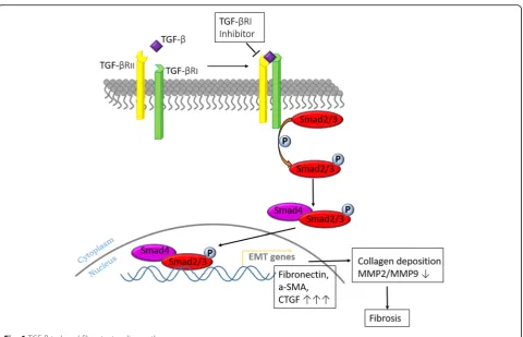
various tissues and organs fibrosis [12, 13]. There are mul-tiple mechanisms through which TGF-β can be involved in the fibrosis process. For instance, TGF-β can activate the TGF-β/Smads signaling pathway [14], promote fibro-blasts transformation into myofibrofibro-blasts [15] and destroy the balance between matrix metalloproteinases (MMPs) and tissue inhibitor of metalloproteinases (TIMPs).
TGF-β/Smads signaling has been shown to have a pivotal role in tissue fibrosis [16, 17]. Upon TGF-β activation, TGF-βreceptors propagate signal to the nucleus by phos-phorylating smad2 and smad3, which form a complex with smad4. The complexes accumulate in nucleus where they regulate the expression of fibrosis related genes [14, 18].
In addition, the overexpression of TGF-βcould induce activation and epithelial-mesenchymal transition (EMT) [19, 20], which facilitate fibronectin synthesis, thus, lead-ing to extracellular matrix (ECM) production [21, 22]. Furthermore, TGF-βcould cause the deposition of ECM and inhibit the degradation of ECM, by stimulating the secretion of growth factors that promote fibrosis, such as mesenchymal marker vimentin, and a-smooth muscle actin (a-SMA) [23]. These in turn could increase the dif-ferentiation of myofibroblast from fibroblasts, subepithe-lial myofibroblasts and smooth muscle cells [24, 25]. The maintenance of a stable internal environment in ECM depends on the coordination between MMPs and TIMPs. Once the balance is off, prolonged collagen deposition, eventually causes fibrosis. Although TGF-β occupies the central position in the development of fibrosis, the mo-lecular mechanisms have not been well characterized [26]. SB431542 is potent small molecular inhibitor that selectively inhibits transforming growth factor (TGF-β) type I receptor activin receptor-like kinase (ALK5). Ovarian fibrosis is characterized by abnormal prolifera-tion of fibroblasts and excessive deposiprolifera-tion of extracellu-lar matrix (ECM) [27]. The transcription of fibrotic genes is dependent on ALK5/Smad3 signaling [28]. It is still not fully understood if the inhibited expression of ALK5 could lead to decrease in fibrosis-related factors.
The main purpose of the present study was to evaluate the role of TGF-βin ovarian fibrosis in dehydroepiandros-terone (DHEA) -induced PCOS rat model. Moreover, we investigated whether ovarian fibrosis was induced via TGF-β/Smad signaling pathway, and whether it amelio-rated EMT. In addition, we wanted to test whether the fibrotic signaling pathway can be inhibited, and if inhibited TGF-βRIexpression would improve the symptoms of poly-cystic ovaries.
Methods Animals
Thirty female Sprague-Dawley (SD) rats, 21 days old, weighing ~50 g, were obtained from Qinglongshan, inc., Nanjing, China. The animals were housed in
specific-pathogen-free (SPF) environment (Jiangsu Key Laboratory of Molecular Medicine) with temperature of 22 ± 1 °C, relative humidity of 50 ± 1%, and a light/dark cycle of 12/ 12 h. Free access to food and water were provided.
All rats were randomly divided in three groups: Blank group (n= 6), Oil groups (n = 6), and Oil + DHEA-induced groups (n= 18). The treatment was given once a day, using hypodermic injectioncontaining: equivalent solvent (inject-able soybean oil) for the Oil group; 6 mg/100 (g·d) dehydro-epiandrosterone (DHEA, diluted in 0.2 ml injectable soybean oil) for the Oil + DHEA-induced groups; and sa-line solution for the blank group. The treatment lasted for 35 days [13, 29]. Eighteen days post- treatment, vaginal smears were collected from all the rats on daily basis at 9:00 am by judging cell types for 14 days, in order to deter-mine their estrous cycles daily. On day 36, all the rats in Blank and Oil-treated groups and six rats of DHEA-induced model groups were euthanized (using intraperito-neal injection of excess 5% chloral hydrate), blood was collected (from the superior vena cava), bilateral ovaries and uteri were dissected. Ovaries were fixed in 4% parafor-maldehyde for 24 h at 4 °C, and then embedded in paraffin. The rest of the tissues were frozen in −80 °C for further western blotting and real time- polymerase chain reaction (RT -PCR) analysis. The rest 12 rats from the DHEA-induced model groups were randomized in additional two groups: SB431542 (TGF-βRI Inhibitor) group and vehicle treatment group (control group), six rats per group. During this treatment, DHEA was no longer given to rats. The treatment was done by intraperitoneal injection contaning: 0.2 ml 0.1% DMSO for the vehicle treatment group; 0.2 ml 1uM TGF-βRI Inhibitor (SB431542, S1067, Selleckchem) for SB431542 group (during this treatment, DHEA was no longer given to rats). The treatment lasted for 2 weeks, after which all the animals were euthanized, and blood was collected, and bilateral ovaries and uteri were dissected following the instructions mentioned above.
Estrous cycle
On day 18 after DHEA treatment, vaginal smears were collected form all rats. Samples were then treated with toluidine blue for 30 min, and consequently cell morph-ology and estrous cycles were examined under the op-tical microscope (Leica Microsystems, Germany).
Serum hormones levels
Blood samples were collected from the superior vena cava. The serum was separated immediately and stored
at −20 °C for further hormones determination by
Haematoxylin- eosin (H&E) and Sirius red tissue staining
Ovaries were embedded in paraffin, and consequently sliced into 4 μm sections. Slices were then fixed in 4% paraformaldehyde for 24 h at 4 °C. Paraffin slices were stained with hematoxylin and eosin in order to examine the tissue morphology under the optical microscope (Leica Microsystems, Germany). Additionally, collagen from ovaries slices was stained by Sirius red to observe the effects of DHEA on rat ovarian fibrosis.
Immunohistochemistry (IHC)
Four μm tissue sections (ovaries), were first incubated with specific antibodies against TGF-β (3711S, Cell Sig-naling Technology, CST), P-Smad3 (#9520S, Cell Signal-ing Technology, CST), a-SMA (ab32575, Abcam), and Collagen IV (Abcam), overnight at 4 °C. All used anti-bodies were diluted to 1:100 before use. The sections were subsequently incubated with a secondary goat anti-rabbit IgG (H + L) HRP (BS13278, Bioworld Technology) at 37 °C for 30 min. Sections were consequently stained with DAB for 5 s, and counterstained with heamatoxylin (Beyotime Biotechnology), and then covered with cover-slips, and observed under optical microscope.
Real time- PCR (RT- PCR)
Ovarian RNA was extracted using TRIZOL (Beyotime Biotechnology) and the cDNA was synthesized using Reverse Transcription Kit (Takara, China) according to manufacturer’s instructions. RT-PCR reactions were per-formed with ABI Viia 7 system (USA) using SYBR® Green PCR Master Mix (Takara, China). The primer sequences of target genes are shown in Table 1. The crit-ical threshold cycle (CT) value was determined for each reaction, and the relative mRNA contents were calcu-lated as E = 2-ΔΔCt. The housekeeping gene, β- actin, was used as an internal control.
Western blotting
Ovarian proteins were extracted using RIPA lysis buffer (Beyotime) containing 1 mM Pierce™phosphatase inhibi-tor (#88667,Thermo Scientific) and 0.1% Halt™ Protease Inhibitor Cocktail (#1862209,Thermo Scientific). The extractive was mixed with 5× SDS-PAGE sample buffer (22.5% 1 M Tris-Cl, pH 6.8, 50% Glycerol, 5% SDS, 0.05% Bromophenol blue, 3.856% DTT). Equal amount of proteins was separated by 10% SDS-PAGE and trans-ferred to PVDF membrane (Merck Millipore), and con-sequently blocked with 5% BSA blocking buffer for 1.5 h. Target bands were incubated with corresponding primary
antibodies, anti-TGF-β (CST, USA), anti-Smad3 (CST,
USA), anti-P-Smad3 (CST, USA), anti-Smad2 (CST, USA), P-Smad2 (CST, USA), CTGF (CST, USA), anti-Fibronectin (CST, USA), anti-a-SMA (abcam), anti-MMP2 (Bioworld, USA), MMP9 (CST, USA) and anti-GAPDH (Bioworld, USA) at 4 °C overnight. After washing with TBST three times for 5 min each time, target bands were incubated with secondary antibody (Goat anti rabbit IgG (H + L) HRP) (Bioworld, USA) for 1.5 h at room temperature. At the end, protein strips were stained with Immobilon western Chemiluminescent HRP Substrate (A liquid and B liquid at a ratio of 1:1) (MA01821, MILLI-PORE, USA) for rational time. GAPDH was used as internal control.
Statistical analysis
Data were expressed using mean ± standard deviation (S.D.) or standard error of the mean (s.e.m.) from at least three independent experiments. Differences were analyzed using GraphPad Pism 6.07 software. The multiple com-parison was done by one-way ANOVA software using Tukey’s post-hoc test. Binary variables were compared with t-test.P≤0.05 was considered statistically significant.
Results
DHEA-induced PCOS-like rat and ovarian fibrosis
Increased serum hormones levels of T, E2 and LH/FSH were observed in Oil + DHEA-treated rats; however, the change in E2 showed no statistical differences. Interest-ingly, the LH, FSH and E2/T levels were markedly decreased in Oil + DHEA- group (Table 2 and Additional file 1: Fig. S1). LH and FSH are essential for estrogen production [30]. Under high level LH, the luteinization of theca cells increases [31]. In this study, both LH and FSH levels were significantly reduced. These data suggested that exogenous excessive androgen was not converted to estrogen. The estrous cycle dis-order, mainly in the estrus, was observed in the model group (Additional file 1: Fig. S2 A). The mirror images of each period are shown in (Additional file 1: Fig. S2 B). Additionally, ovarian cystic expansion, increased the number of follicles, granular cell layer thinning and the
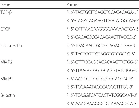
Table 1Primer sequences for real-time RT-PCR
Gene Primer
TGF-β F: 5’-TACTGCTTCAGCTCCACAGAGA-3′
R: 5’-CAGACAGAAGTTGGCATGGTAG-3′
CTGF F: 5’-CATTAAGAAGGGCAAAAAGTGA-3′
R: 5’-CACACCCCACAGAACTTAGCC-3′
Fibronectin F: 5’-TGACAACTGCCGTAGACCTGG-3’
R: 5’-TACTGGTTGTAGGTGTGGCCG-3’
MMP2 F: 5’-CTTTGCAGGAGACAAGTTCTGG-3’
R: 5’-TTAAGGTGGTGCAGGTATCTGG-3’
MMP9 F: 5’-AAGCCTTGGTGTGGCACGAC-3’
R: 5’-TGGAAATACGCAGGGTTTGC-3’
β- actin F: 5’-TCAGGTCATCACTATCGGCAAT-3’
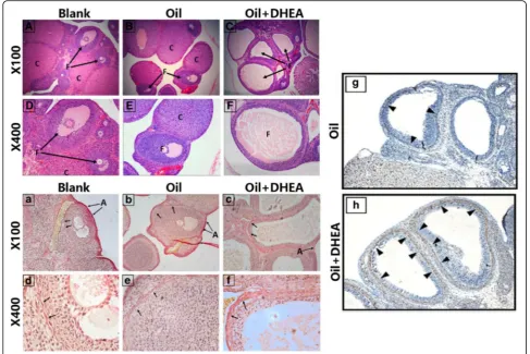
thickening of theca cell layer, while the vast majority of no corpus luteum formation was observed in DHEA-induced rats. Furthermore, cystic follicles were detected in the DHEA group compared to the control (Fig. 1A-F). Sirius red staining of ovarian tissues revealed a mark-edly higher level of fibrin and collagen in Oil +
DHEA-induced rat ovaries compared to no-treated and Oil-treated rat ovaries; high level of fibrin and collagen were detected in the follicular basic membrane follicles and interstitial areas (Fig. 1 a-f ). Due to the significantly posi-tive expression of fibrosis in DHEA-induced group, we further investigated the collagen IV in ovaries by immunohistochemi-cal analysis (IHC). Our results showed extremely profuse expression in DHEA-treated rat follicular membrane relative to vehicle-treated rats (Fig. 1g and h).
TGF-βand Smads/p-Smads are up-regulated in oil + DHEA-induced rats
Based on our previous findings, which revealed higher expression of fibrosis in Oil + DHEA-induced rats,
we further investigated whether the TGF-β/Smad3
signaling pathway might be associated with ovarian
fibers. TGF-β and p-Smad3 were mainly expressed in
theca-interstitial cells and granulosa cells. Briefly,
higher expression of TGF-β and p-Smad3 was found
Table 2The serum hormone levels in Control and DHEA
treated rats
Blank Oil Oil + DHEA
T(ng/ml) 0.488 ± 0.034 0.497 ± 0.051 1.356 ± 0.176 *
E2(pg/ml) 120.137 ± 8.756 119.553 ± 3.485 131.592 ± 7.210
LH(IU/L) 224.737 ± 37.577 217.783 ± 56.983 81.353 ± 15.160*
FSH(ng/ml) 154.58 ± 2.07 73.33 ± 14.8 31.64 ± 11.967**
LH/FSH 1.492 ± 0.313 2.743 ± 0.316 2.958 ± 0.272**
E2/T 0.302 ± 0.030 0.274 ± 0.019 0.128 ± 0.019***
*p≤0.05, **p≤0.01, ***p≤0.02, significantly different from Blank; n = 6 in each group; Data are mean ± SEM
in the theca cells, while almost no expression was detected within theca extema cells. Importantly, in
Oil + DHEA-induced rats, the levels of TGF-β were
significantly more supernal compared to bank control and the oil control group, especially in the granulosa cells. However, according to quantification, we can see that p-Smad3 protein levels have no difference among three groups (Fig. 2a and b). Consequently, we
examined TGF-β and p-Smad3 at protein and mRNA
level, and detected its expression. Briefly, the
expression of TGF-β protein, but not the mRNA,
evi-dently increased in Oil + DHEA-induced rat ovaries (Fig. 2c). Moreover, the expression levels of Smad3 and Smad2 protein in Oil + DHEA-induced rat ovar-ies were significantly higher compared to Oil-treated group. Similarly, DHEA stimulated Smad2/3 phos-phorylation in rat ovaries via TGF-β signaling path-way (Fig. 2d and e).
Expression of TGF-βdownstream signaling molecules and collagen deposition-related protein in DHEA-induced PCOS-like rats
Connective tissue growth factor (CTGF), which is the downstream signaling molecule of TGF-β, interacts with
TGF-β to promote ovarian fibrosis [32]. In the DHEA
up-regulated rats, both ovarian CTGF and fibronectin protein and mRNA levels were notably enhanced (Fig. 3a and b). In addition, TGF-βcan also facilitate extracellu-lar matrix (ECM)-producing cells express a-smooth muscle actin (a-SMA), thus promoting the transformation of fibroblasts into muscle fibroblasts, and therefore lead-ing to ECM over-synthesis and dysregulation of MMPs-TIMPs balance [26]. In Oil + DHEA-induced rats, a-SMA protein showed increasing tendency (Fig. 3d-f). Based on previous data, we detected MMP2 and MMP9 genes expression via RT-PCR analysis; and we observed an obvi-ous down-regulation of MMP2 (Fig. 3c).
SB431542 improve DHEA-induced ovarian fibrosis
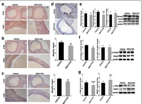
TGF-βRI is the main receptor of TGF-β, which forms a complex to stimulate smad3 phosphorylation, and con-sequently promotes target genes transcription. After treating rats with SB431542, no difference in the serum total T was observed compared to the vehicle group; however, significant difference in E2/T levels was ob-served in the modeling group and the treatment group (Additional file 2: Table S1). These data suggested that body has a strong self-regulating ability. After inhibition of ALK5, the corpus luteum was enhanced and the fol-licular number was reduced. However, no visible thick-ening of the granular cell layer was observed (Fig. 4a-d). Moreover, it seemed that the inhibition of TGF-βdidn’t have much effect on hormone regulation. The relative hormone levels did not significantly improve after SB431542 treatment. Furthermore, after suspending DHEA treatment, prominent hormone improvement was observed in the vehicle and SB431542 treated group compared to DHEA-induced group (Additional file 2: Table S1 and Additional file 1: Fig. S3), which may be due to innate response ability to directly or indirect effect and adjusted the hormonal levels in the body.
After cutting off the DHEA supply, and consequently treating rats with SB431542, no significant decrease in fibrin and collagen protein expression was observed
(Fig. 5a). Interestingly, in model and in blank group (Fig. 1A-F), as well as in the administration group (Fig. 5a), the levels of expression of fibroin in the tu-nica albuginea ovarii were extremely high. These data suggested that the ovarian albuginea has a high degree of fibrosis and may not be susceptible to medicine. To fur-ther examine the correlation between TGF-β/Smad3 sig-naling pathway and ovarian fibers, we treated rats with SB431542 (TGF-βRI Inhibitor). Briefly, lower expression
of TGF-β and Smad3 expressed in granulosa cells was
observed in the SB431542 group compared to vehicle-control rats; however, in the theca cells, the level of
TGF-β and p-Smad3 did not change prominently (Fig. 5b-c). Moreover, the expression of TGF-β and Smad3 protein were dramatically down-regulated. Nevertheless, it ap-peared that inhibiting TGF-βto combine with its recep-tors could not restrain Smad3 phosphorylation (Fig. 5e). Moreover, the TGF-βRI inhibitor led to downregulation of Smad2, but not of p-Smad2 (Fig. 5f).
Additionally, correlation analyses revealed that elevated ovary TGF-βand smad2/3 expression could be associated with ovarian fibrosis, and that blocking of TGF-β/Smad signaling pathway can ease ovarian fibrosis. Furthermore, TGF-βRI Inhibitor can mitigate DHEA exposed ovarian fibrosis by downregulating TGF-β downstream molecular
one of ECM-independent factors - MMP2, thus attenuating the accretion of collagen in ovary tissue of vehicle-treated PCOS-like rats (Fig. 5d and g). To sum up, DHEA-induced ovarian fibrosis was mediated by TGF-βsignaling pathway. However, despite striking improvements in ovarian function and fibrosis related factors, fibrotic phenotype did not reach effective modification. To conclude, these results suggested that drug could inhibit the excessive production of fibrosis, nevertheless it could not reverse the existing fibrosis.
Discussion
In this study, we reported that ovarian fibrosis in
DHEA-induced PCOS rats was mediated by TGF-β
sig-naling, while TGF-βRI inhibitors attenuated fibrogenic
response by suppressing the elevation of TGF-β and
Smad3 (Fig. 6). This caused accumulation of ECM, which promoted ovarian interstitial fibrosis by disrupting the balance between MMPs and TIMPs [33]. A few cys-tic follicles and a marginal accretion of ovarian granular cell layer were observed in ovaries of SB431542-exposed rat. Accordingly, we showed that ovarian fibrosis in
PCOS rat model was formed via TGF-β signaling.
Des-pite the fact that treating PCOS rats with TGF-βRI in-hibitors for 2 weeks did not reveal any improvements in Sirius red stained tissue sections following SB431542 ex-posure, the key factor in fibrosis, TGF-β signaling and its downstream molecular effects all showed a tendency to down-regulate.
Tissue fibrosis is generally considered to arise due to the failure of the normal wound healing or the
accumulation of senescence cells [34, 35]. Our results showed that the DHEA-induced PCOS model in rats was a resource of profibrotic signaling observed in ovar-ian fibrosis (Fig. 1a-f ). Ovarovar-ian follicular granule cell layer in PCOS rats was thinner compared to normal rats (Fig. 1A-F). It could be that ovarian fibrosis results from the degeneration of granulosa cells, which causes inter-stitial fibroblast hyperplasia, fibrinogen and collagen de-position. These proteins include profibrotic proteins
TGF-β, CTGF and ECM. We believe that TGF-β
recep-tor is an especially important target since TGF-βmight activate Smad3 and Smad2. Subsequently, TGF-β/Smads complex might promote CTGF, fibronectin and a-SMA transcription in nucleus.
to have any significant role on improving hormonal levels. One of the limitations of the present study relates to the levels of hormones that widely vary among indi-viduals, due to their inconsistency in the estrous cycle. Being an androgen hormone, we hypothesized that DHEA might cause ovarian dysfunction by binding to several nuclear receptors [36], which were possibly inde-pendent of TGF-βregulation.
Many recent studies have showed that tissue fibrosis is very hard to reverse, even if targeting certain mecha-nisms could promote tissue remodeling and tissue func-tion improvement [35, 37], including pulmonary fibrosis and kidney fibrosis [38]. Furthermore, ovaries from SB431542-treated rats didn’t show a reversible appear-ance compared to vehicle-control rats (Fig. 5a). Never-theless, even though the fibrotic phenotype did not
improve, all the fibrosis biomarkers, such as TGF-β, CTGF and a-SMA were noticeably down-regulated.
Ovarian fibrosis appears challenging but not impossible to reverse. Resveratrol and metformin have shown to be very effective in reversing fibrosis by upregulating phos-phorylated AMPK (p-AMPK) expression which attenuates ROS-induced oxidative stress [39–41]. Consequently, in our future studies we plan to address the effect of resvera-trol and metformin on ovarian fibrosis upon p-AMPK/ ROS signaling pathway in vivo or in vitro.
MMP9 did not show any significant variations, which implies that MMP2 could have an important role in the development of fibrosis, and that SB431542 has a crucial regulatory effect on MMP2.
Although there are numerous studies on fibrosis, the ovarian fibrosis pathogenesis is still poorly understood. The aim of the present study was to explore whether ovar-ian fibrosis occurs through TGF-βsignaling pathway, and
whether the intervention on the TGF-β inhibitor can
reverse this phenomenon. Another study limitation is the nature of fibrosis in relation to PCOS, i.e. whether the fibrosis was the cause of PCOS or the result.
Conclusion
TGF-βRI inhibitor (SB431542) inhibits the downstream
signaling molecules of TGF-β and upregulates MMP2,
which consequently prevents collagen deposition. More-over, ovarian hyperfibrosis in DHEA-induced PCOS rat
model could be improved by TGF-βRI inhibitor
(SB431542) restraining the transcription of accelerating fibrosis genes and modulating EMT mediator. In short, we have demonstrated that DHEA could induce rat
ovarian over-fibrosis, which is mediated by TGF-β
signaling pathway. Although this hyperfibrosis was hard to reverse, all the fibrotic factors were down-regulated.
Additional files
Additional file 1: Figure S1.The serum testosterone (T) and(Estrogen)E2/ T levels in control, DHEA and SB431542-treated rats. *p≤0.05, ***p≤0.002. Data are shown as mean ± SEM.Figure S2.The estrous cycle of DHEA-induced PCOS rats disordered.(A)The estrous cycle of the Blank group, Oil group and Oil + DHEA group rats. The abscissa indicates the age of rats. In theproestrus, many nucleated epithelial cells (NEC) were observed. In theestrus, high number of corneous cells (CC) were detected. In themetestrus, visible nucleated epithelial cells, corneous cells and leucocyte (L) were discovered. In thediestrus, a mass of leucocyte can be seen in the field of view.(B)Microscopic examination of vaginal smears stained by toluidine blue. × 200.. D = diestrus; P = proestrus; E = estrus; M = metestrus.Figure S3.The hormone levels after SB431542 treatment.(A)Serum total T levels;(B)E2/T of serum. (DOCX 467 kb)
Additional file 2: Table S1.The serum hormonal levels in Vehicle and SB431542 treated rats.Δp≤0.05,ΔΔp≤0.01, significantly different from Oil + DHEA;#p≤0.05, significantly different from Vehicle. Dates are mean ± SEM. (XLSX 11 kb)
Abbreviations
ALK5:activin receptor-like kinase; a-SMA: a-smooth muscle actin;
CTGF: Connective tissue growth factor; E2: Estradiol; ECM: Extracellular matrix; EMT: Pithelial-mesenchymal transition; IHC: Immunohistochemistry; LH: Luteinizing hormone; MMPs: Matrix metalloproteinases; RT- PCR: Real Time- PCR; T: Testosterone; TGF-β: Transforming growth factor; TIMPs: Tissue inhibitor of metalloproteinases
Acknowledgments
We are grateful to Lei Wang, Shiyu Song and Mengyuan Niu for their technical support and helpful suggestions.
Funding
This work was supported by the National Natural Science Foundation of China (81471422 and 81170541) and the Foundation of State Key Laboratory of Analytical Chemistry for Life Science (5431ZZXM1603).
Availability of data and materials
All the data were collected and analyzed by the authors. The additional information regarding data and manuscript are available within this article (Additional file 1 and Additional file 2).
Author’s contributions
DJW, WQW, HWW, QG and YW were the principal investigators and they contributed to the study design, method investigation, experiment performance and the manuscript writing. DJW, QL and XH participated in the animal experiments. DJW, YJX and SMS carried out the analysis of Western blot and RT-PCR. The final version was approved by all the authors.
Ethics approval
All animal studies (including the mice euthanasia procedure) were done in compliance with the regulations and guidelines of Nanjing University Institutional Animal Care and conducted according to the AAALAC and the IACUC guidelines.
Consent for publication Not applicable.
Competing interests
The authors declare that they have no competing interests.
Publisher’s Note
Springer Nature remains neutral with regard to jurisdictional claims in published maps and institutional affiliations.
Author details
1State Key Laboratory of Analytacal Chemistry for Life Science & Jiangsu Key
Laboratory of Molecular Medicine, Medical school, Nanjing University, Nanjing 210093, China.2Prenatal Diagnosis Center, the First Affiliated Hospital of Zhengzhou University, Zhengzhou, Henan 450052, China. 3Divisions of Endocrinology, the Affiliated Drum Tower Hospital, Medical
School, Nanjing University, Nanjing 210093, China.
Received: 14 July 2017 Accepted: 22 December 2017
References
1. Chiofalo B, Lagana AS, Palmara V, Granese R, Corrado G, Mancini E, et al. Fasting as possible complementary approach for polycystic ovary syndrome: hope or hype? Med Hypotheses. 2017;105:1–3.
2. Asemi Z, Samimi M, Taghizadeh M, Esmaillzadeh A. Effects of Ramadan fasting on glucose homeostasis, lipid profiles, inflammation and oxidative stress in women with polycystic ovary syndrome in Kashan. Iran ARCH IRAN MED. 2015;18:806–10.
3. Franks S. Controversy in clinical endocrinology: diagnosis of polycystic ovarian syndrome: in defense of the Rotterdam criteria. J Clin Endocrinol Metab. 2006;91:786–9.
4. Qu J, Wang Y, Wu X, Gao L, Hou L, Erkkola R. Insulin resistance directly contributes to androgenic potential within ovarian theca cells. Fertil Steril. 2009;91:1990–7.
5. Lagana AS, Pizzo A. Authors' reply to: "empiric" inositol supplementation in normal-weight non-insulin resistant women with polycystic ovarian disease: from the absence of benefit to the potential adverse effects. Arch Gynecol Obstet. 2015;291:959–60.
6. Lagana AS, Sapia F, La Rosa VL, Vitale SG. Comment on "Inositols: from physiology to rational therapy in gynecological clinical practice". Expert Opin Drug Metab Toxicol. 2016;12:1527.
7. Vitale SG, Rossetti P, Corrado F, Rapisarda AM, La Vignera S, Condorelli RA, et al. How to achieve high-quality Oocytes? The key role of Myo-Inositol and melatonin. Int J Endocrinol. 2016;2016:4987436.
8. Schubel R, Graf ME, Nattenmuller J, Nabers D, Sookthai D, Gruner LF, et al. The effects of intermittent calorie restriction on metabolic health: rationale and study design of the HELENA trial. CONTEMP CLIN TRIALS. 2016;51:28–33.
9. Zhang X, Zhang C, Shen S, Xia Y, Yi L, Gao Q, et al. Dehydroepiandrosterone induces ovarian and uterine hyperfibrosis in female rats. Hum Reprod. 2013; 28:3074–85.
10. Hughesdon PE. Morphology and morphogenesis of the stein-Leventhal ovary and of so-called "hyperthecosis". OBSTET GYNECOL SURV. 1982;37:59–77. 11. Leask A, Abraham DJ. TGF-beta signaling and the fibrotic response. FASEB J.
2004;18:816–27.
12. Xu L, Cui WH, Zhou WC, Li DL, Li LC, Zhao P, et al. Activation of Wnt/beta-catenin signalling is required for TGF-beta/Smad2/3 signalling during myofibroblast proliferation. J Cell Mol Med. 2017;21:1545–54.
13. Miao ZL, Guo L, Wang YX, Cui R, Yang N, Huang MQ, et al. The intervention effect of Rosiglitozone in ovarian fibrosis of PCOS rats. Biomed Environ Sci. 2012;25:46–52.
14. DiRenzo DM, Chaudhary MA, Shi X, Franco SR, Zent J, Wang K, et al. A crosstalk between TGF-beta/Smad3 and Wnt/beta-catenin pathways promotes vascular smooth muscle cell proliferation. Cell Signal. 2016;28:498–505.
15. Sime PJ, Xing Z, Graham FL, Csaky KG, Gauldie J. Adenovector-mediated gene transfer of active transforming growth factor-beta1 induces prolonged severe fibrosis in rat lung. J Clin Invest. 1997;100:768–76.
16. Chan EC, Dusting GJ, Guo N, Peshavariya HM, Taylor CJ, Dilley R, et al. Prostacyclin receptor suppresses cardiac fibrosis: role of CREB phosphorylation. J Mol Cell Cardiol. 2010;49:176–85.
17. Chen Y, Yang S, Yao W, Zhu H, Xu X, Meng G, et al. Prostacyclin analogue beraprost inhibits cardiac fibroblast proliferation depending on prostacyclin receptor activation through a TGF beta-Smad signal pathway. PLoS One. 2014;9:e98483.
18. Steinway SN, Zanudo JGT, Ding W, Rountree CB, Feith DJ, Loughran TP, et al. Network modeling of TGF signaling in Hepatocellular carcinoma epithelial-to-Mesenchymal transition reveals joint sonic hedgehog and Wnt pathway activation. Cancer Res. 2014;74:5963–77.
19. Zheng L, Zhang C, Li L, Hu C, Hu M, Sidikejiang N, et al. Baicalin ameliorates renal fibrosis via inhibition of transforming growth factor beta1 production and downstream signal transduction. Mol Med Rep. 2017;15:1702–12. 20. Song Y, Peng C, Lv S, Cheng J, Liu S, Wen Q, et al. Adipose-derived stem
cells ameliorate renal interstitial fibrosis through inhibition of EMT and inflammatory response via TGF-beta1 signaling pathway. Int Immunopharmacol. 2017;44:115–22.
21. Varga J, Pasche B. Transforming growth factor beta as a therapeutic target in systemic sclerosis. Nat Rev Rheumatol. 2009;5:200–6.
22. Lu J, Liu Q, Wang L, Tu W, Chu H, Ding W, et al. Increased expression of latent TGF-beta-binding protein 4 affects the fibrotic process in scleroderma by TGF-beta/SMAD signaling. Lab Investig. 2017;97:591–601.
23. Yang J, Tian B, Brasier AR. Targeting chromatin remodeling in inflammation and fibrosis. Adv Protein Chem Struct Biol. 2017;107:1–36.
24. Speca S, Giusti I, Rieder F, Latella G. Cellular and molecular mechanisms of intestinal fibrosis. World J Gastroenterol. 2012;18:3635–61.
25. Di Gregorio J, Sferra R, Speca S, Vetuschi A, Dubuquoy C, Desreumaux P, et al. Role of glycogen synthase kinase-3beta and PPAR-gamma on epithelial-to-mesenchymal transition in DSS-induced colorectal fibrosis. PLoS One. 2017;12:e171093.
26. Xu M, Wang G, Zhou H, Cai J, Li P, Zhou M, et al. TGF-beta1-miR-200a-PTEN induces epithelial-mesenchymal transition and fibrosis of pancreatic stellate cells. Mol Cell Biochem. 2017;431:161–8.
27. Chen YH, Wang Q, Li CY, Hou JW, Chen XM, Zhou Q, et al. Haplodeficiency of activin receptor-like kinase 4 alleviates myocardial infarction-induced cardiac fibrosis and preserves cardiac function. J Mol Cell Cardiol. 2017;105:1–11. 28. Ding Q, Subramanian I, Luckhardt TR, Che P, Waghray M, Zhao XK, et al.
Focal adhesion kinase signaling determines the fate of lung epithelial cells in response to TGF-beta. Am J Physiol Lung Cell Mol Physiol. 2017;312:L926–35. 29. Anderson E, Lee GY, O'Brien K. Polycystic ovarian condition in the
dehydroepiandrosterone-treated rat model: hyperandrogenism and the resumption of meiosis are major initial events associated with cystogenesis of antral follicles. Anat Rec. 1997;249:44–53.
30. Whirledge S, Cidlowski JA. Glucocorticoids, stress, and fertility. Minerva Endocrinol. 2010;35:109–25.
31. Gleicher N, Weghofer A, Barad DH. The role of androgens in follicle maturation and ovulation induction: friend or foe of infertility treatment? Reprod Biol Endocrinol. 2011;9:116.
33. Zhou F, Shi LB, Zhang SY. Ovarian fibrosis: a phenomenon of concern. Chin Med J. 2017;130:365–71.
34. Eckes B, Zigrino P, Kessler D, Holtkotter O, Shephard P, Mauch C, et al. Fibroblast-matrix interactions in wound healing and fibrosis. Matrix Biol. 2000;19:325–32.
35. Schafer MJ, White TA, Iijima K, Haak AJ, Ligresti G, Atkinson EJ, et al. Cellular senescence mediates fibrotic pulmonary disease. Nat Commun. 2017;8:14532. 36. Webb SJ, Geoghegan TE, Prough RA, Michael MK. The biological actions of
dehydroepiandrosterone involves multiple receptors. Drug Metab Rev. 2006;38:89–116.
37. Bickelhaupt S, Erbel C, Timke C, Wirkner U, Dadrich M, Flechsig P, et al. Effects of CTGF blockade on attenuation and reversal of radiation-induced pulmonary fibrosis. J Natl Cancer Inst. 2017;109
38. Sutariya B, Saraf M. Betanin, isolated from fruits of Opuntia Elatior mill attenuates renal fibrosis in diabetic rats through regulating oxidative stress and TGF-beta pathway. J Ethnopharmacol. 2017;198:432–43.
39. Yu W, Fu YC, Zhou XH, Chen CJ, Wang X, Lin RB, et al. Effects of resveratrol on H(2)O(2)-induced apoptosis and expression of SIRTs in H9c2 cells. J Cell Biochem. 2009;107:741–7.
40. Lee MS, Kim SH, Kim DS, Min KS, Yoon JT. Metformin enhances the action of insulin on porcine granulosa-lutein cells in vitro. Anim Reprod Sci. 2012;136:100–7. 41. Kumar S, Kim YR, Vikram A, Naqvi A, Li Q, Kassan M, et al. Sirtuin1-regulated
lysine acetylation of p66Shc governs diabetes-induced vascular oxidative stress and endothelial dysfunction. Proc Natl Acad Sci U S A. 2017;114:1714–9. 42. Theodore LN, Hagedorn EJ, Cortes M, Natsuhara K, Liu SY, Perlin JR, et al.
Distinct roles for matrix Metalloproteinases 2 and 9 in embryonic hematopoietic stem cell emergence, migration, and niche colonization. Stem Cell Reports. 2017;8:1226–41.
• We accept pre-submission inquiries
• Our selector tool helps you to find the most relevant journal
• We provide round the clock customer support
• Convenient online submission
• Thorough peer review
• Inclusion in PubMed and all major indexing services
• Maximum visibility for your research
Submit your manuscript at www.biomedcentral.com/submit