H E A L T H A N D M E D I C I N E
Focused ultrasound delivery of a selective TrkA agonist
rescues cholinergic function in a mouse model
of Alzheimer’s disease
K. Xhima1,2, K. Markham-Coultes1, H. Nedev3,4, S. Heinen1, H. U. Saragovi3,4,
K. Hynynen5,6, I. Aubert1,2*
The degeneration of cholinergic neurons is a prominent feature of Alzheimer’s disease (AD). In animal models of injury and aging, nerve growth factor (NGF) enhances cholinergic cell survival and function, contributing to im-proved memory. In the presence of AD pathology, however, NGF-related therapeutics have yet to fulfill their regen-erative potential. We propose that stimulating the TrkA receptor, without p75NTR activation, is key for therapeutic
efficacy. Supporting this hypothesis, the selective TrkA agonist D3 rescued neurotrophin signaling in TgCRND8 mice, whereas NGF, interacting with both TrkA and p75NTR, did not. D3, delivered intravenously and noninvasively
to the basal forebrain using MRI-guided focused ultrasound (MRIgFUS)–mediated blood-brain barrier (BBB) permeability activated TrkA-related signaling cascades and enhanced cholinergic neurotransmission. Recent clinical trials support the safety and feasibility of MRIgFUS BBB modulation in AD patients. Neuroprotective agents targeting TrkA, combined with MRIgFUS BBB modulation, represent a promising strategy to counter neuro-degeneration in AD.
INTRODUCTION
Alzheimer’s disease (AD) is a neurodegenerative disorder characterized by progressive cognitive decline. Among the widespread neuronal and synaptic deficits in AD, degeneration of basal forebrain cholinergic neurons (BFCNs) and loss of cholinergic innervation to the cortex (CTX) and hippocampal formation (HF) substantially contribute to cognitive decline in AD (1). During the prodromal and earliest stages of AD, cholinergic neurons undergo trophic changes, including alterations in nerve growth factor (NGF) processing or transport, expression of NGF receptors, and activation of downstream NGF signaling components (2). The loss of neurotrophic support to BFCNs, beginning in early- stage AD, contributes to impaired cho-linergic neurotransmission, followed by neuronal degeneration that is observed in late-stage AD (2).
In the brain, NGF promotes neuronal survival, growth, and syn-aptic plasticity by binding to tropomyosin receptor kinase A (TrkA) and p75 neurotrophin receptor (p75NTR) (3). NGF is produced in the medial septum (MS), diagonal band of Broca (DBB), and nucleus basalis of Meynert (NBM), which together compose the basal fore-brain, HF, and CTX. In response to NGF binding, TrkA receptors expressed by BFCNs trigger activation of the tyrosine kinase do-main, receptor phosphorylation, and intracellular signaling via the mitogen-activated protein kinase (MAPK) and phosphoinositide 3-kinase (PI3K)/Akt cascades (3). NGF also binds p75NTR and can, in the absence of TrkA, induce neuronal apoptosis and glial inflam-mation through Jun N-terminal kinase (JNK) signaling (3, 4). In addition, BFCNs respond to NGF-induced activation of TrkA by
enhancing their cholinergic phenotype. TrkA activation engages the cholinergic gene locus to increase choline acetyltransferase (ChAT) activity, the enzyme that synthesizes the neurotransmitter acetylcholine (ACh), which, in turn, promotes ACh release at cho-linergic nerve terminals (3).
The importance of NGF in neuronal health and disease has been long recognized, yet translating a treatment strategy for AD remains a major challenge. The therapeutic efficacy of NGF is limited by its short half-life, detrimental effects related to p75NTR activation, and its inability to cross the blood-brain barrier (BBB) (5). Furthermore, assessment of the preclinical efficacy of NGF-based therapeutics has been limited to injury models, namely, the fimbria-fornix lesion- induced degeneration of BFCNs or aging models (5), in the absence of toxic amyloid- (A) peptides and tau pathology. Native NGF can bind to both TrkA and p75NTR, and in the presence of defective TrkA signaling (as observed in human AD), p75NTR activation facil-itates A- and tau-induced signaling and neurodegeneration (6–8). Recent clinical trials for NGF therapy in AD showed some growth of cholinergic afferents but ultimately failed to slow cognitive decline (9, 10). Further elucidation of the molecular interplay between NGF and pathological features of AD is necessary to facilitate the devel-opment of successful NGF-based therapies.
In comparison to native NGF, ligands with desirable pharmaco-logical properties, designed to specifically activate TrkA, and not p75NTR, may be a preferable therapeutic approach to up regulate the cholinergic phenotype of BFCNs (11–14). Saragovi and colleagues (11–13) have previously demonstrated that D3, a peptidomimetic agonist of TrkA, promotes cell survival, outgrowth, cholinergic activ-ity, and rescued learning and memory deficits in aged rats and a transgenic (Tg) mouse model of AD. However, like NGF, D3 does not cross the BBB.
In this study, we achieved noninvasive delivery of D3 to the basal forebrain in a mouse model of AD using magnetic resonance imaging– guided focused ultrasound (MRIgFUS). MRIgFUS, combined with intravenous microbubbles, allows a localized and transient increase
1Hurvitz Brain Sciences Research Program, Biological Sciences, Sunnybrook Research Institute, Toronto, ON, Canada. 2Department of Laboratory Medicine and Pathobiology, Faculty of Medicine, University of Toronto, Toronto, ON, Canada. 3Lady Davis Institute, Jewish General Hospital, Montreal, QC, Canada. 4Department of Pharmacology and Therapeutics, McGill University, Montreal, QC, Canada. 5Physical Sciences, Sunnybrook Research Institute, Toronto, ON, Canada. 6Department of Medical Biophysics, Univer-sity of Toronto, Toronto, ON, Canada.
*Corresponding author. Email: [email protected]
Copyright © 2020 The Authors, some rights reserved; exclusive licensee American Association for the Advancement of Science. No claim to original U.S. Government Works. Distributed under a Creative Commons Attribution NonCommercial License 4.0 (CC BY-NC).
on September 17, 2020
http://advances.sciencemag.org/
mice (21, 22). Cholinergic atrophy and cell loss have been reported, coinciding with a reduction in the levels of ACh release in the pre-frontal cortex of 7-month-old TgCRND8 compared to non-Tg mice (22). Consistent with TrkA deficits observed in AD postmor-tem tissue (23, 24), single-cell RNA sequencing also revealed lower mRNA levels of TrkA in TgCRND8 mice compared to non-Tg lit-termates (25).
The status of NGF signaling in relation to cholinergic function remains poorly understood in animal models of AD. Here, we re-port deficits in NGF levels, TrkA receptor levels, and TrkA-mediated signaling cascades in parallel with cholinergic dysfunction in the TgCRND8 model of AD, thus providing a disease-relevant pheno-type to test NGF-based therapeutics. To this end, we delivered D3 to BFCNs using MRIgFUS in TgCRND8 mice. D3 was physiologi-cally active in vivo after MRIgFUS delivery, significantly increasing TrkA phosphorylation and downstream signaling cascades in both TgCRND8 mice and non-Tg littermates. Moreover, we report that D3—and not NGF—successfully activated TrkA signaling pathways in TgCRND8 mice, representing an additional advantage of our therapeutic approach favoring TrkA activation without engaging p75NTR. MRIgFUS-mediated D3 delivery in the basal forebrain in-creased ChAT activity locally and enhanced ACh release from hip-pocampal and cortical slices. These results suggest that noninvasive, selective TrkA activation in BFCNs is sufficient to increase cho-linergic function in the presence of A pathology and that it can be achieved via MRIgFUS delivery of D3. TrkA receptor ligands, com-bined with MRIgFUS targeted delivery, merit consideration as a treatment strategy for AD.
RESULTS
NGF/TrkA signaling declines with age in TgCRND8 mice
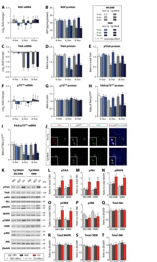
We comprehensively evaluated age-dependent changes in NGF sig-naling in an AD animal model to identify a relevant, early-stage, AD phenotype for testing NGF-related therapies. We measured regional differences in the levels of NGF, TrkA, and p75NTR mRNA [quanti-tative real-time polymerase chain reaction (qRT-PCR) and in situ hybridization] and protein [enzyme-linked immunosorbent assay (ELISA) and Western blot] in TgCRND8 and non-Tg mice at 4, 6, and 8 months of age (Fig. 1 and fig. S1).
Relative to age-matched non-Tg mice, NGF mRNA levels in the MS/DBB and NBM were unaltered in TgCRND8 mice (Fig. 1A). In contrast, NGF protein levels were reduced in 6- and 8-month-old Tg mice (Fig. 1B). Similar analysis in the HF and CTX revealed no change in NGF mRNA and protein levels between groups at all time points examined (fig. S1, A and B).
(Fig. 1, I and J) in 6- and 8-month-old Tg mice, as a result of a shift in the balance of NGF and TrkA expression. Our results demon-strate that deficits in the NGF/TrkA system observed in TgCRND8 mice resemble those observed in human AD and provide a relevant model to evaluate NGF-based therapeutic strategies for AD.
D3, not NGF, stimulates TrkA-dependent signaling in TgCRND8 mice
Next, we directly compared the in vivo efficacy of native NGF and compound D3, a selective TrkA agonist, to induce TrkA-dependent signaling cascades in 6-month-old TgCRND8 mice. Following direct intraparenchymal injection to the MS/DBB and NBM in TgCRND8 mice, D3, unlike native NGF, stimulated neurotrophin signaling (Fig. 1, K to T). Specifically, we found elevated levels of pTrkA, pAkt (Thr308), pMAPK (Thr202/Tyr204), and pCREB (phosphorylated cyclic adenosine monophosphate response element-binding protein; Ser133) within 90 min after D3 treatment relative to NGF-treated and phosphate-buffered saline (PBS)–treated TgCRND8 mice (Fig. 1, K to O). Downstream of p75NTR, we found reduced JNK activity (Thr183/Tyr185) in D3- and NGF-treated mice relative to PBS con-trols (Fig. 1, K and P). No differences were detected in total protein level normalized to -tubulin-III (tubIII) among all conditions (Fig. 1, Q to T). As a positive control, we performed intraparenchymal injection of D3 to the basal forebrain in age-matched, non-Tg mice to confirm signal transduction pathways activated by D3 (fig. S2, A to F) and NGF (fig. S3, A to F). Total protein levels normalized to tubIII remained unaltered across all treatment groups (figs. S2, G to J, and S3, G to J). These findings provide experimental evidence that A pathology interferes with NGF bioactivity in vivo and demonstrate that selective TrkA stimulation, in the absence of p75NTR interaction, is required to promote TrkA-dependent survival and plasticity in the presence of A pathology.
Localized MRIgFUS-mediated BBB disruption in the basal forebrain
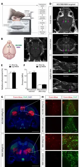
To rescue NGF/TrkA signaling deficits in the TgCRND8 model, we delivered the selective TrkA ligand, D3, to BFCNs using MRIgFUS. The MRIgFUS experimental setup is illustrated in Fig. 2A. The MS/ DBB region of the basal forebrain was targeted bilaterally with one spot (Fig. 2B, green dot) and the NBM with two foci (Fig. 2B, purple dots) on T2-weighted (T2w) MR images (Fig. 2C). BBB opening was confirmed on T1w contrast-enhanced MR images, visualized by the influx of Gadovist (MRI contrast agent) from the blood-stream into sonicated areas (Fig. 2D). We found no difference in the level of contrast enhancement between TgCRND8 and non-Tg mice (Fig. 2E). The mean increase in signal intensity in the sonicated areas
on September 17, 2020
http://advances.sciencemag.org/
Fig. 1. Intraparenchymal injection of D3, a selective TrkA agonist, but not NGF, increased TrkA-dependent signaling in 6-month-old TgCRND8 mice with NGF and TrkA deficits. (A) No change in NGF mRNA levels was found in the MS/DBB and NBM from 4 to 8 months of age in TgCRND8 and non-Tg mice. (B) NGF protein,
(C) TrkA mRNA, (D) TrkA protein, and (E) pTrkA levels were reduced in 6- and 8-month-old TgCRND8 relative to non-Tg mice. (F) p75NTR mRNA and (G) p75NTR protein levels remained stable from 4 to 8 months of age in both genotypes. The relative levels of TrkA to p75NTR (H) protein and (I) mRNA in TgCRND8 mice were decreased by 6 months of age compared to non-Tg mice. (J) Fluorescence in situ hybridization revealed down-regulation of TrkA (red) but not p75NTR (white) mRNA transcripts in ChAT+ cholinergic somata (green) of TgCRND8 mice. Scale bars, 20 m. (K) Representative Western blots from the MS/DBB and NBM after injection of PBS, NGF, and D3 in TgCRND8 mice. D3-treated mice demonstrated increased levels of (L) pTrkA, (M) pAkt, (N) pMAPK, and (O) pCREB relative to NGF- and PBS-injected TgCRND8 mice. (P) pJNK levels were decreased following NGF and D3 treatment compared to PBS-treated mice. There were no changes in total levels of (Q) Akt, (R) MAPK, (S) CREB, and (T) JNK normalized to tubIII. All mRNA levels were quantified by qRT-PCR. NGF protein was analyzed by ELISA. TrkA and p75NTR protein levels were measured by Western blot. All phosphorylation signal intensity values were normalized to their respective total protein levels. Statistics: Repeated-measures two-way analysis of variance (ANOVA) (A to I) and one-way ANOVA (L to T). Significance: *,^,†P < 0.05; **,††P < 0.01; ***,†††P < 0.001; * indicates comparison to age-matched non-Tg mice; ^ and † indicate comparison with PBS-treated mice. Data represent means ± SEM; n = 8 per group (A to I) and n = 4 per group (L to T).
on September 17, 2020
http://advances.sciencemag.org/
Fig. 2. Noninvasive FUS-induced BBB permeability localized to the basal forebrain under MRI guidance. (A) Schematic of the experimental setup. Mice were
posi-tioned supine on an MRI-compatible sled with the head coupled to a polyimide membrane with ultrasound gel and placed in a tank filled with degassed, deionized water, housing the transducer and hydrophone. (B) Ultrasound foci in the MS/DBB and NBM regions of the basal forebrain were targeted on (C) axial T2w MR images. (D) BBB permeability was visualized by the influx of an MRI contrast agent (Gadovist) on contrast-enhanced T1w images in the MS/DBB (green arrows) and NBM (purple arrows). (E and F) There were no significant differences in (E) contrast enhancement or (F) pressure required to induce BBB modulation in 6-month-old TgCRND8 mice compared to non-Tg littermates. (G) Histologically, Evans blue dye (red) extravasation was detected in MRIgFUS-targeted regions of the basal forebrain (MS/DBB, top; NBM, bottom) 90 min after sonication. (H) Higher-magnification confocal micrographs in the MS/DBB (top) and NBM (bottom) revealed the entry of Evans blue in the parenchyma of animals treated with MRIgFUS compared to untreated animals, injected with Evans blue without MRIgFUS. ChAT immunoreactivity (green) was used to identify cholinergic cell bodies. Scale bars, 5 mm (C and D), 1 mm (G), and 100 m (H). Statistics: Student’s t test (E and F). Data represent means + SEM; n = 12 per genotype.
on September 17, 2020
http://advances.sciencemag.org/
was 20 ± 8% and 25 ± 9% in non-Tg and TgCRND8 mice, respec-tively. Concomitantly with relative enhancement, no difference in the average peak pressure required to induce subharmonic emis-sions was observed between genotypes. The mean peak pressure reached during sonication was 1.29 ± 0.15 MPa in the TgCRND8 mice and 1.21 ± 0.11 MPa in the non-Tg controls (Fig. 2F). Relative contrast enhancement and mean acoustic pressure were comparable between treatment groups as well as subregionally in the basal forebrain (fig. S4). These data suggest that the increase in BBB permeability was consistent between genotypes, treatment groups, and distinct nuclei within the basal forebrain.
Evans blue dye, which binds to serum albumin, and endogenous antibodies, immunoglobulin G (IgG) and IgM, proteins that normally do not cross the BBB, were used to histologically confirm the sites targeted by MRIgFUS. Immunohistochemical analyses revealed a 1-mm-wide focal spot of Evans blue dye at the level of the MS/DBB and two 1-mm-wide focal spots in the NBM (Fig. 2G). In contrast, Evans blue was not detected in brain regions nontargeted by FUS (Fig. 2G) and in control, non-FUS–treated, mice (Fig. 2H). Extrav-asation of blood-borne IgG and IgM into the basal forebrain was observed 90 min after MRIgFUS (fig. S5). Immunohistochemical (fig. S5A) and Western blot (fig. S5, B to E) analyses revealed greater levels of IgG and IgM in MRIgFUS-treated regions relative to un-sonicated areas, in both TgCRND8 and non-Tg mice. Furthermore, there was a positive correlation between IgM levels and Gadovist contrast enhancement (fig. S5F), as previously reported in the same experimental model (26).
We found no evidence of erythrocyte extravasation or neuronal cell death in sonicated areas. Ter119-positive erythrocytes were confined to vascular structures identified by tomato lectin in MRIgFUS- targeted regions, without red blood cell infiltration into the brain parenchyma (fig. S6). We did not detect either cleaved caspase-3 or terminal deoxynucleotidyl transferase–mediated deoxyuridine tri-phosphate nick end labeling (TUNEL) immunoreactivity in neuronal nuclei (NeuN)–positive cells (fig. S7). The olfactory bulb was used as a positive control for caspase-3 and TUNEL due to the relatively high rate of granule cell turnover in this region (18). Together, these findings suggest that MRIgFUS allows BBB opening in the basal forebrain without overt tissue damage.
MRIgFUS effectively delivers D3 to the basal forebrain
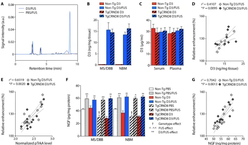
Using quantitative high-performance liquid chromatography (HPLC), we measured D3 concentrations in MRIgFUS-targeted and non-sonicated brain regions of TgCRND8 mice and non-Tg controls after intravenous D3 injection. D3 was detected in D3/FUS-treated tissue 30 min after treatment (Fig. 3, A and B). The mean D3 levels in the MS/DBB were 15 ± 2 and 13 ± 3 ng/mg tissue in TgCRND8 and non-Tg mice, respectively. Similarly, D3 levels in the NBM were 16 ± 2 and 12 ± 2 ng/mg tissue in TgCRND8 and non-Tg mice, respectively. The drug concentration achieved after MRIgFUS was not significantly different between genotypes in both neuroanatomical locations. By contrast, D3 levels were below the limit of detection by HPLC in D3-treated, non-FUS controls (Fig. 3B). The mean D3 con-centration in the serum and plasma was comparable between D3- and D3/FUS-treated mice for both genotypes (Fig. 3C).
The mean D3 concentration and relative contrast enhancement were positively correlated, with no apparent differences in correla-tion between non-Tg and TgCRND8 mice (Fig. 3D). In addicorrela-tion, there was a positive correlation between TrkA activation and
rela-tive contrast enhancement, consistent with enhanced D3 bioavail-ability in the MRIgFUS-targeted regions (Fig. 3E). Overall, these data support the feasibility of using MRIgFUS to deliver D3 locally to the basal forebrain in a noninvasive manner.
Given that the sonication scheme used to achieve MRIgFUS BBB opening in this study would potentially allow the entry of serum NGF (26 kDa), or factors stabilizing NGF and/or its release, we evaluated NGF protein levels by ELISA. Elevated levels of NGF were found in PBS/FUS relative to PBS-treated mice and D3/FUS relative to D3-treated animals in the MS/DBB and NBM for both TgCRND8 and non-Tg mice (Fig. 3F). There was a positive correla-tion between NGF levels and contrast-enhanced T1w voxel intensity in MRIgFUS-treated non-Tg and TgCRND8 mice, indicating that Gadovist enhancement may provide a means of estimating NGF alter-ations in the targeted tissue volume (Fig. 3G). The elevated local NGF levels resulting from MRIgFUS lend support to the hypothesis that our therapeutic paradigm may be used to promote endogenous brain plasticity and repair for the treatment of AD.
MRIgFUS-mediated delivery of D3 activates TrkA-dependent signaling pathways
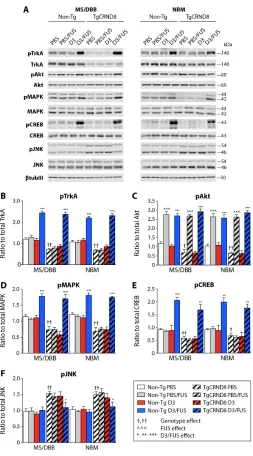
To further elucidate TrkA signaling deficits in the TgCRND8 mouse model of AD, we compared signal transduction targets modulated downstream of TrkA phosphorylation in the basal forebrain (Fig. 4). In an independent cohort of 6-month-old mice, we confirmed the reduction in pTrkA levels in TgCRND8 relative to non-Tg mice in the MS/DBB and NBM (Fig. 4B). Parallel to reduced levels of pTrkA, pAkt, pMAPK, and pCREB were also decreased in TgCRND8 mice (Fig. 4, C to E). Downstream of p75NTR, we found increased JNK activity in TgCRND8 compared to non-Tg mice (Fig. 4F).
Thus, we assessed the functional effects of MRIgFUS delivery of D3 on TrkA-dependent signaling pathways in 6-month-old TgCRND8 and non-Tg littermates (Fig. 4). We found elevated pTrkA levels in the MS/DBB and NBM within 90 min after D3/FUS treatment rel-ative to PBS/FUS-, PBS-, and D3-treated mice in both genotypes (Fig. 4, A and B). TrkA activation was achieved in 6-month-old TgCRND8 mice in the presence of decreased TrkA expression, and the level of pTrkA was not significantly different from D3/FUS- treated non-Tg mice (Fig. 4B).
We observed elevated pAkt in PBS/FUS- and D3/FUS-treated mice compared to PBS- and D3-treated animals, respectively (Fig. 4C). These data are in line with recent studies, which demon-strate that MRIgFUS BBB opening leads to elevated pAkt levels, 90 min after sonication (27, 28). Similarly, we found an increase in MAPK activation in D3/FUS-treated compared to PBS/FUS-, D3-, and PBS-treated mice (Fig. 4D). CREB function was acutely in-creased after D3/FUS treatment (Fig. 4E), likely to be due to phos-phorylation at Ser133 by multiple kinases, including MAPK and Akt (3). JNK activity was decreased after D3/FUS relative to control conditions in TgCRND8 mice (Fig. 4F). For each protein in the NGF/TrkA signaling pathway, no difference was detected in total pro-tein level normalized to tubIII among all treatment groups (fig. S8).
MRIgFUS-mediated delivery of D3 enhances cholinergic function
We established whether deficits in TrkA signaling correspond to diminished cholinergic function in TgCRND8 mice. ChAT activity in the basal forebrain (enriched in cholinergic cell bodies) and ACh release in the HF and CTX (enriched in cholinergic axon terminals)
on September 17, 2020
http://advances.sciencemag.org/
were measured (Fig. 5). In 6-month-old TgCRND8 mice, ChAT activity was reduced by 30 to 40% relative to non-Tg littermates (Fig. 5, A and B). Furthermore, basal and potassium- induced ACh release was lower in TgCRND8 compared to non-Tg slices (Fig. 5, C and D), consistent with the loss of enzymatic activity related to ACh synthesis in TgCRND8 mice. These results indicate that 6-month-old TgCRND8 and non-Tg mice represent valid experi-mental models of impaired and unimpaired cholinergic function, respectively.
Thus, we evaluated the potential of MRIgFUS delivery of D3 to increase cholinergic function in TgCRND8 and non-Tg mice. Three days after sonication, elevated ChAT activity was found in the D3/ FUS-treated MS/DBB (Fig. 5A) and NBM (Fig. 5B) relative to ani-mals that received intravenous D3 administration only, for both genotypes. In addition, we examined the impact of D3/FUS treat-ment on basal and depolarization-evoked ACh release in hippocam-pal and cortical slices, also at 3 days after FUS. Potassium-evoked ACh release was elevated in D3/FUS compared to intravenous D3 treated animals of both genotypes in hippocampal (Fig. 5C) and cortical preparations (Fig. 5D). Together, our data demonstrate
that a single D3/FUS treatment can stimulate cholinergic function in TgCRND8 mice up to 3 days after sonication.
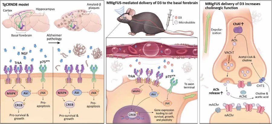
Overall, these findings highlight the in vivo efficacy of our treat-ment paradigm—that is, to selectively engage TrkA receptors and regulate critical intracellular signaling cascades that are sufficient to promote cholinergic function in the presence of A pathology (Fig. 6). Thus, enhancing cholinergic neurotransmission by MRIgFUS- mediated delivery of D3 represents a possible therapeutic approach to limit cognitive decline in AD.
DISCUSSION
There is an urgent need for therapeutics and delivery methods that slow or reverse the degeneration of neurons in AD, including cho-linergic neurons. Acetylcholinesterase inhibitors, which represent the most common pharmacological compounds used for the treat-ment of AD, do not restore dying or degenerating neurons. In con-trast, NGF-related therapeutics bode well for the maintenance of cholinergic morphology, survival, and function. In particular, strat-egies in which NGF-mediated activation of BFCN function can Fig. 3. Enhanced D3 delivery and increased NGF protein levels in the basal forebrain following MRIgFUS-induced BBB opening. (A) Representative HPLC elution
profile corresponding to D3 detection in the basal forebrain following D3/FUS treatment (blue line) relative to PBS/FUS control (black line). a.u., arbitrary units. (B) D3 levels were negligible in the brain after intravenous D3 injection without MRIgFUS (red bars). With MRIgFUS, D3 significantly entered the targeted MS/DBB and NBM in non-Tg and TgCRND8 mice (blue bars). (C) Plasma and serum D3 levels between unsonicated and sonicated animals for both genotypes were comparable. (D) Increases in D3 concentration, and (E) TrkA phosphorylation, were proportional to the contrast enhancement in MRIgFUS-treated regions for both genotypes. (F) NGF protein levels were increased in PBS/FUS- and D3/FUS-treated relative to PBS- and D3-treated animals, respectively. NGF protein levels were decreased in 6-month-old TgCRND8 mice com-pared to non-Tg littermates, but FUS-induced NGF protein levels were comparable between genotypes. (G) There was a positive correlation between the relative contrast enhancement and NGF levels measured. Statistics: Two-way ANOVA (B, C, and F). Significance: ^,†P < 0.05, **,^^P < 0.01; † indicates comparison with PBS-treated non-Tg mice (genotype effect); ^ indicates comparison with PBS-treated mice of the same genotype (FUS effect); * indicates comparison with D3-treated mice (i.e., intravenous D3, no MRIgFUS) of the same genotype (D3/FUS effect). (D, E, and G) Linear regression analysis. Dashed lines indicate a 95% confidence interval. Data represent means + SEM;
n = 4 per group (B to D) and n = 6 per group (E to G).
on September 17, 2020
http://advances.sciencemag.org/
be safely and effectively accomplished hold therapeutic potential for AD.
In this study, we show that MRIgFUS delivery of D3 is a novel therapeutic approach to enhance cholinergic function, addressing fundamental limitations of current NGF-based treatment modalities for AD. MRIgFUS-mediated delivery of D3 led to selective TrkA signaling in BFCNs while reducing p75NTR activation, and elevated ChAT activity and ACh release in a mouse model of amyloidosis with deficits in NGF, TrkA and cholinergic function—pathologies that are present in human AD. Our findings are consistent with the suggestion that NGF administration directly to cholinergic cell bodies, rather than in axon terminals, bypasses transport defects and allows NGF/TrkA signaling at the soma (29–31). Even in the presence of reduced TrkA levels, D3/FUS treatment increased TrkA signaling in TgCNRD8 mice, as assessed by activation of TrkA and downstream effectors including Akt, MAPK, and CREB (Fig. 4). Al-though p75NTR expression levels remain stable, downstream p75NTR activity decreased in response to D3/FUS treatment as measured by JNK activation. TrkA signaling has been shown to silence p75NTR- mediated pathways such as JNK via PI3K/Akt (3). Because D3 does not interact with p75NTR directly, Akt activation may represent a potential mechanism by which D3/FUS treatment decreases JNK phosphorylation. Moreover, MRIgFUS delivery of D3 rescued cho-linergic function, i.e., depolarization-induced ACh release in amyloid- burdened hippocampal and cortical regions, and ChAT activity in the basal forebrain. The levels of evoked ACh release and ChAT activity for up to 3 days after D3/FUS treatment were similar to those observed in untreated, non-Tg controls, which have levels of cholinergic signaling sufficient to support normal cognitive function (Fig. 5). In this way, localized D3 delivery to the basal forebrain by MRIgFUS led to widespread restoration of cholinergic transmission in the associated projection regions of the HF and CTX.
To date, NGF-based therapies have been validated in preclinical models of aging and injury. Although these studies lend further support to the rationale of NGF as a therapeutic agent for AD, the predictive validity of these models was limited by the absence of A and tau pathology. Multiple lines of evidence suggest that A and tau are inextricably linked to NGF-related signaling cascades in vitro and in vivo (6–8). Here, we discovered alterations in the status of NGF bioactivity related to NGF mRNA and protein levels, NGF re-ceptor levels, and downstream signaling components in a preclinical model of amyloidosis, which, taken together, appear to recapitulate the signaling alterations reported in early-stage AD patients (2). Ev-idence from preclinical and human studies indicate that the shift in TrkA/p75NTR levels may represent a switch favoring the binding of NGF to p75NTR, possibly activating apoptosis over survival path-ways in BFCNs and contributing to cholinergic dysfunction before cell loss in AD (2, 3). Our signaling data also suggest a TrkA/p75NTR signaling imbalance, which can contribute to cholinergic dysfunc-tion in AD. Previous studies suggest that increased p75NTR activity coupled with increased A load exacerbates A-induced neuro-degeneration (6–8). Moreover, our data in the TgCRND8 model indicate that the amount of ACh synthesized (Fig. 5, A and B) and released (Fig. 5, C and D) is impaired with disease progression and may, in part, underlie the cognitive deficits observed in this model of AD (20). Likewise, Bellucci et al. (22) reported decreased ChAT immunoreactivity in the NBM and release of cortical ACh in 7-month-old TgCRND8 mice relative to non-Tg controls. To this end, the TgCRND8 model is suitable for testing NGF-related therapeutic Fig. 4. MRIgFUS delivery of D3 increased TrkA-dependent signaling cascades
in the basal forebrain 90 min after treatment. (A) Representative Western blots
from the MS/DBB and NBM after PBS, PBS/FUS, D3, or D3/FUS treatment. Blots were probed for pTrkA, pAkt, pMAPK, pCREB, pJNK, the corresponding total proteins, and tubIII to control for loading. Levels of (B) pTrkA, (C) pAkt, (D) pMAPK, and (E) pCREB were reduced in PBS-treated TgCRND8 mice relative to non-Tg mice and stimulated in D3/FUS-treated mice compared to PBS- and D3-treated (no MRIgFUS) controls in the MS/DBB and NBM for both genotypes. (F) Elevated levels of pJNK were found in control TgCRND8 mice relative to non-Tg mice. pJNK was decreased in TgCRND8 mice after D3/FUS treatment compared to control conditions in Tg mice, but levels remained unaltered in non-Tg mice. Statistics: Two-way ANOVA. Significance: *,†P < 0.05; **,††P < 0.01, ***,^^^P < 0.001; † indicates comparison with PBS-treated non-Tg mice (genotype effect); ^ indicates comparison with PBS-treated mice of the same genotype (FUS effect); * indicates comparison with D3-treated group (i.e., intravenous D3, no MRIgFUS) of the same genotype (D3/FUS effect). Data represent means + SEM; n = 6 per group.
on September 17, 2020
http://advances.sciencemag.org/
agents as a means to improve cholinergic function in the presence of A pathology.
Within this framework, selective activation of TrkA, avoiding p75NTR, was necessary to induce TrkA-related signaling pathways and sufficient to up-regulate cholinergic function in the context of amyloid pathology. Our results demonstrate that D3, in contrast to native NGF, stimulates TrkA signaling in the TgCRND8 mice (Fig. 1, K to T). Our findings draw attention to a previously
un-known role of amyloid on NGF bioactivity in vivo, including the relative imbalance in TrkA/p75NTR signaling, amyloid pathology, and potentially other mechanisms affecting NGF stability, and could partly explain the limited efficacy of NGF-directed treatments for AD to date (9, 10, 32).
A major concern for NGF-based therapy is that undesirable pleiotropic effects can reduce the therapeutic benefit of NGF. For example, p75NTR activation can negate the benefit of engaging TrkA Fig. 6. MRIgFUS delivery of D3, a selective TrkA agonist, to the basal forebrain rescued neurotrophin signaling and cholinergic function in the TgCRND8 model of AD. (Left) By 6 months of age, TgCRND8 mice demonstrated deficits in the NGF/TrkA signaling system, similar to what is observed in human AD. TgCRND8 mice had
reduced NGF protein levels, TrkA expression, and activation of downstream signaling cascades, coupled with increased JNK activity in the basal forebrain. (Middle) MRIgFUS was used to noninvasively and locally increase BBB permeability in the basal forebrain, enhancing the bioavailability of intravenous D3 to cholinergic cell bodies in the MS/DBB and NBM. Using this therapeutic approach in TgCNRD8 mice, we stimulated TrkA and downstream signaling effectors—pAkt, pMAPK, and pCREB—while simul-taneously decreasing pJNK known to be triggered by p75NTR activation. (Right) MRIgFUS delivery of D3 to the basal forebrain led to enhanced cholinergic neurotransmis-sion in widespread axon terminal regions of the CTX and HF, as evidenced by enhanced ChAT activity, the enzyme that synthesizes ACh, and ACh release.
Fig. 5. MRIgFUS delivery of D3 enhances cholinergic function 3 days after treatment. (A and B) ChAT enzymatic activity was significantly reduced in TgCRND8 relative to
non-Tg littermates in the (A) MS/DBB and (B) NBM. ChAT activity was significantly increased in D3/FUS-treated compared to D3-treated (no MRIgFUS) controls in the MS/DBB and NBM for both TgCRND8 and non-Tg mice. (C) Basal and potassium depolarization–induced ACh release from hippocampal slices was significantly reduced in TgCRND8 mice compared to non-Tg controls. Evoked hippocampal ACh release was increased after D3/FUS treatment compared to D3-treated controls in both genotypes. Basal release of ACh was not significantly different in D3/FUS-treated compared to D3-treated animals for both genotypes. (D) TgCRND8 mice had decreased levels of basal and potassium- evoked cortical ACh release relative to non-Tg littermates. Evoked ACh release was also elevated in cortical slices from both TgCRND8 and non-Tg mice following D3/FUS treatment, but basal levels of ACh release remained unaltered in both genotypes. Statistics: Two-way ANOVA. Significance: *,†,#P < 0.05; **,††, ##P < 0.01, ***,###P < 0.001; † indi-cates comparison with D3-treated non-Tg mice (genotype effect); # indiindi-cates comparison with basal levels of ACh release of the same treatment and genotype (depolarization effect); * indicates comparison with D3-treated group (i.e., intravenous D3, no MRIgFUS) of the same genotype (D3 /FUS effect). Data represent means + SEM; n = 5 per group.
on September 17, 2020
http://advances.sciencemag.org/
(14). Delivery of D3, a highly selective and potent partial TrkA ago-nist, is a promising approach to promote TrkA signaling (11), and studies in animal models of aging and AD have shown improvements in memory performance (12, 13). Clinically, D3 has been shown to be a well-tolerable and effective treatment for dry eye, without in-ducing potential treatment-related side effects such as aberrant growth of sensory fibers (33).
In addition, the delivery of NGF-related therapeutic agents to the brain, to date, required invasive surgery due to their limited ability to cross the BBB (5). We demonstrate that MRIgFUS is an effective method for targeted drug delivery to the basal forebrain in a noninvasive manner with real-time feedback monitoring. In hu-man AD, clinical trials for NGF and vector-based NGF delivery all require complex intracranial surgical procedures that produce BBB breakage and tissue damage and carry substantial risk for severe hemorrhages and infection. Moreover, these surgeries pose a higher risk of adverse events among elderly individuals. These detrimental effects could rapidly outweigh the potential benefit of NGF therapy (9, 10, 32). The MRIgFUS parameters used here did not induce erythrocyte extravasation (fig. S6) or neuronal apoptosis (fig. S7). These data are in line with previous studies, which demonstrated that these MRIgFUS settings are below the threshold of damage (34, 35). Focused ultrasound combined with MRI as an imaging method for targeting the brain allows ultrasound to be applied throughout the entire basal forebrain, without significant diffusion of the drug to distal brain regions, especially in the ventricles and meninges containing cerebrospinal fluid. Here, an intravenous dose of 500 g of D3 was sufficient to activate TrkA in the basal forebrain targeted by MRIgFUS, without drug diffusion to other brain re-gions (D3 levels below the detection threshold by HPLC).
Further work to optimize experimental design parameters should be conducted with the goal of achieving consistent BBB modulation of permeability and simultaneously minimizing po-tential damage. Factors such as sonication scheme, microbubble composition, and brain subregion can influence the extent of BBB permeability and associated biological effects following FUS treatment. Microbubble dose, size, shell properties, and half-life circulation can affect the degree of BBB permeability (36). In this study, we used the recommended clinical imaging dose of Defini-ty microbubbles (20 l/kg) to approximate an equivalent micro-bubble dose between animals. Recently, McMahon and Hynynen (34) reported that Definity at 20 l/kg compared to 100 l/kg (10 times higher than the clinical imaging dose) can be used to increase BBB permeability without activation of inflammatory markers (i.e., nuclear factor B signaling pathway genes), tissue damage, or MRI evidence of edema and hemorrhage. The probability of FUS-induced BBB permeability using a clinical imaging dose of Definity and Optison is similar across a range of pressure ampli-tudes, as the ones achieved in our study and established previously by McDannold et al. (37).
We monitored and controlled FUS-mediated BBB modulation in real time using an acoustic controller. The applied acoustic pressure was incrementally increased until subharmonic emissions (characteristic of stable cavitation) were detected, thereby increas-ing BBB permeability below the threshold for tissue damage (35). This method has been shown to improve sonication consistency and safety. A similar approach was used in the first clinical trial for BBB opening in AD using MRIgFUS with no adverse events after sonication (19). Immunohistochemical analysis in our study
revealed no neuronal loss or erythrocyte extravasation in the basal forebrain 90 min after sonication (figs. S6 and S7). These findings are in accordance with other preclinical studies that demonstrate increased BBB permeability in the absence of edema, neuronal damage, and minimal to no erythrocyte extravasation using real- time feedback monitoring (34, 35, 38, 39).
Previous evidence suggests that local differences in vasculature and brain region can lead to variable effects of FUS-induced BBB permeability (40). Here, we show that similar pressure amplitudes were required to induce subharmonic emissions in the subregions of the basal forebrain (fig. S4), and acoustic pressures were compa-rable to those achieved following FUS-mediated BBB opening in the hippocampus of TgCRND8 mice and non-Tg littermates using an identical transducer/hydrophone configuration and feedback controller (38). We observed no differences in BBB opening be-tween TgCRND8 and non-Tg mice (Fig. 2F) despite vascular dys-function in TgCRND8 mice by 6 months of age (41). Future work is necessary to evaluate whether BBB recovery varies temporally in different basal forebrain regions and due to the presence of AD pathology.
In recent years, the potential use of MRIgFUS for AD has evolved beyond a method for drug delivery. In several mouse models of AD, FUS-mediated BBB opening reduced amyloid and tau pathology, even in the absence of drug delivery (26, 38, 39, 42, 43). Here, we combined FUS, as a delivery tool with disease-modulatory effects, with a pharmacological agent that is designed to enhance plasticity and resilience of BFCNs vulnerable to degeneration in AD. In this way, our therapeutic approach targets multiple pathologies impli-cated in AD and thus may prove more efficacious than a single tar-get paradigm to slow or reverse disease progression. We found elevated levels of NGF in the brain parenchyma following MRIgFUS (Fig. 3, F and G). The source of increased NGF levels at sites of FUS-BBB modulation remains to be identified. Candidates include cir-culating NGF present in the bloodstream, cells capable of rapid NGF release residing on the parenchymal side of the BBB and known to be first responders [e.g., mast cells; reviewed in (44)], and microglia and astrocytes known to be activated by FUS (26, 28) that secrete NGF as early as 3 hours after stimulation in vitro (45). However, it remains to be tested whether glial cells can release sufficient amounts of NGF within 90 min in vivo, to account for the levels detected here.
In the adult mouse, basal NGF levels in the serum equivalent to 50 to 100 pg/ml may be physiologically meaningful in view of NGF binding affinity for the TrkA receptor [Kd (dissociation constant) of ~10−11 M] (3, 11, 46). Our results demonstrate that elevated levels of NGF are not sufficient to induce TrkA phosphorylation in non-Tg and TgCRND8 mice at 90 min following MRIgFUS alone. This re-sult may be explained by NGF instability in solution and sensitivity to proteolysis and supported by the fact that NGF bioactivity is op-timal at nanomolar concentrations (11). Nevertheless, this finding has important implications for the therapeutic action of D3 in MRIgFUS-treated areas. Because NGF and D3 can bind TrkA con-comitantly, D3 can potentiate TrkA signals activated by low pico-molar levels of NGF (11, 47). Therefore, MRIgFUS-induced increase in NGF and D3 may together further potentiate TrkA-dependent signaling cascades in the context of A pathology.
An important part of preclinical development toward clinical translation will be dosing optimization for D3/FUS treatment. Pre-vious work has shown that a single intraparenchymal injection of
on September 17, 2020
http://advances.sciencemag.org/
of BFCNs) and affect cognition. Preclinical studies demonstrated that administration of NGF and specific agonists of TrkA for at least 2 weeks in aged or lesioned animal models reversed BFCN atrophy and ameliorated behavioral impairments; however, the deficits re-appeared after cessation of treatment (5, 12, 50, 51).
The first AD BBB modulation clinical trial recently revealed that single and repeated FUS treatments, monitored in real time using passive cavitation detection, are well tolerated in AD patients (19), supporting the feasibility of FUS delivery of D3 in AD patients. Sub-sequent larger clinical trials will help determine the potential thera-peutic benefit of FUS-induced BBB permeability alone. An important question for future work will be to assess whether repeated admin-istration of a TrkA agonist using FUS can act as a combination ther-apy to restore cholinergic function and reduce A deposition and tau pathology in ways that promote cognitive function.
Evidence suggests that administration of NGF or TrkA ligands can, on one hand, improve performance in cognitively impaired ro-dents and, on the other hand, compromise intact function in healthy animals (12, 13, 51–53). This differential effect in diseased compared to healthy brains has to be taken into consideration when treatment strategies are planned. NGF modulation of cholinergic function is tightly regulated such that either excess or lack of the neurotrophin can be detrimental. Excessive and adverse neuronal plasticity as a result of TrkA activation is unlikely to occur in the presence of toxic A species that are negatively influencing TrkA signaling contin-uously; however, this remains to be tested.
Pharmacological treatment of AD with D3 will require personalized strategies to address the disease process of each individual. Promising advances in neuroimaging may be eventually used to identify patients who might be most responsive to a therapeutic regimen incorporat-ing agonists to TrkA. For instance, Trk-targeted radiotracers for positron emission tomography imaging may reveal spatiotemporal changes in TrkA receptor expression level—an early molecular al-teration in mild cognitive impairment (MCI) and AD correlated with cognitive impairment (54). Alternatively, radioligands designed to quantify and map in vivo degeneration of cholinergic terminals in AD patients could be used for clinical identification, as well as monitoring the efficacy of TrkA-based interventions (55).
In conclusion, our study demonstrates that, in contrast to native NGF, only a selective TrkA agonist, without engaging p75NTR, can stimulate prosurvival and regenerative signaling in the context of A pathology. The compound D3, and not NGF, activated TrkA- dependent signaling cascades in the TgCRND8 model of AD. Our results reveal a relative imbalance of TrkA/p75NTR activity in TgCRND8 mice, as observed in human AD, which may partly ex-plain the failure of NGF reported here and, more broadly, why NGF
MATERIALS AND METHODS
Study design
Our study aimed to (i) investigate how the NGF/TrkA signaling system is altered with disease progression in a mouse model of AD, (ii) compare the efficacy of native NGF and selective TrkA agonist D3 in a disease-relevant phenotype, and (iii) establish the feasibility of noninvasive and localized D3 delivery to the basal forebrain with MRIgFUS. Accordingly, we treated and analyzed four cohorts of TgCRND8 and non-Tg littermates. A first cohort of sex-balanced TgCRND8 and non-Tg mice at 4, 6, and 8 months of age (n = 8 per group) was used to characterize the genotype- and age-associated alterations in the NGF/TrkA signaling system using qRT-PCR, in situ hybridization, ELISA, Western blotting, and confocal microscopy.
In a second cohort, sex-balanced, 6-month-old TgCRND8 and non-Tg mice were randomly assigned to receive PBS, 2 g of NGF, or 2 g of D3 injected to the basal forebrain via intraparenchymal injection. Mice were sacrificed 90 min after injection (n = 4 per group). Using immunoprecipitation and Western blotting, we mea-sured TrkA-dependent signaling pathways activated in the basal forebrain following PBS, NGF, and D3 administration.
A third cohort of sex-balanced, 6-month-old TgCRND8 and non-Tg littermates was tested. Mice were randomly assigned to treatment groups as follows: (i) D3/FUS-treated (500 g, intravenous dose of D3 with MRIgFUS), (ii) PBS/FUS-treated (intravenous PBS with MRIgFUS), (iii) D3-treated (500 g, intravenous dose of D3, no MRIgFUS), or (iv) PBS-treated (intravenous PBS, no MRIgFUS). Using histological methods, Western blotting, ELISA, and confocal microscopy, we evaluated the extent of BBB permeability in the basal
forebrain 90 min following MRIgFUS (n = 6 PBS/FUS- and
PBS-treated non-Tg and Tg mice). HPLC was used to measure the concentration of D3 delivered to the basal forebrain targeted with MRIgFUS, and mice were sacrificed 30 min after treatment to max-imize therapeutic detection (n = 4 per group). Using immuno-precipitation and Western blotting, we evaluated the effect of D3/ FUS treatment on TrkA signaling cascades 90 min after sonication (n = 6 per group). Treatment allocation was kept blinded until anal-ysis. All animals were included in the analanal-ysis.
In the fourth cohort, sex-balanced, 6-month-old TgCRND8 and non-Tg mice were randomly assigned to either a D3/FUS-treated (500 g, intravenous dose of D3 with MRIgFUS) or D3-treated (500 g, intravenous dose of D3, no MRIgFUS) experimental group. Mice were sacrificed 3 days following treatment (n = 5 per group) for the assessment of ChAT activity in the basal forebrain, in addition to basal and depolarization-evoked ACh release in cholinergic termi-nal regions following D3 or D3/FUS treatment.
on September 17, 2020
http://advances.sciencemag.org/
Animals
TgCRND8 mice and their age-matched non-Tg littermates were used in this study (20). All procedures were conducted in accordance with guidelines established by the Canadian Council on Animal Care and protocols approved by the Sunnybrook Research Institute Animal Care Committee.
Intracranial injections
Anesthesia was induced and maintained with isoflurane in oxygen. Mice were placed in a stereotaxic apparatus (David Kopf Instruments). The skull was exposed, and burr holes were made with a Stoelting drill to the following coordinates: MS/DBB: anteroposterior, +0.8 mm; mediolateral, +0.3 mm; dorsoventral, −4.4 mm (measured from bregma with a 4° vertical angle alignment); NBM: anteroposterior, −0.3 mm; mediolateral, +1.5 mm; dorsoventral, −5.6 mm (measured from bregma). A 33-gauge needle attached to a 5-l Hamilton syringe containing the solution to be injected was lowered to the injection site, and 1-l injection was made over 10 min. Following each injec-tion, the needle was held in place at the injection site for 5 min and then removed from the brain.
MRI-guided focused ultrasound
Mice were anesthetized with 5% isoflurane and maintained at 2% isoflurane in medical air. Head fur was removed with depilatory cream, and a 26-gauge angiocatheter was inserted into the tail vein. The MRIgFUS experimental setup is illustrated in Fig. 2A. MRIgFUS was performed using the in-house build prototype system (similar to LP100, FUS Instruments Inc.), and animals were imaged using a 7.0-T MRI (Bruker). The acoustic beam was generated using a spherically focused transducer (1.68-MHz frequency, 75 mm diameter, 60 mm radius of curvature), with standard BBB disruption parameters (10 ms bursts, 1 Hz burst repetition frequency, 120-s duration). Axial T2w MR images were used to target three focal spots, covering the basal forebrain bilaterally.
At the time of sonication, mice received an intravenous injection of D3 (500 g) diluted in PBS, or PBS only, Definity microbubbles (0.02 ml/kg; Lantheus Medical Imaging), and Gadovist (0.2 ml/kg, Schering AG) via the tail vein. For animals that received Evans blue, the dye (2% solution in PBS; 4 ml/kg; Sigma-Aldrich) was injected immediately after the sonication. Each injectable was administered individually, separated by a 150 l saline flush to ensure transfer into the bloodstream.
Acoustic emissions were recorded using a polyvinylidene fluoride (PVDF) hydrophone coupled to an acoustic controller (34). The applied acoustic pressure was increased incrementally after each pulse until subharmonic emissions were detected, then reduced to 25%, and maintained for the remainder of the sonication. BBB permeability was visualized by an increase in Gadovist signal intensity on T1w MR images acquired 5 min after sonication. Contrast enhancement was assessed by measuring pixel intensity in a 2 mm × 2 mm target region normalized to a reference region of the brain. A custom image analysis program was used to calculate relative contrast en-hancement (MATLAB, MathWorks).
Biodistribution and pharmacokinetic analysis of D3
D3 was quantified using HPLC versus standard curves as previously described (11). Mice were intracardially perfused with saline 30 min after treatment, and brains were removed and flash-frozen in liquid nitrogen. Tissue was homogenized in methanol using a mini-pestle
fitted in a pellet pestle motor (Kontes) for 1 min at 4°C and centri-fuged at 12,000 rpm for 15 min at 4°C for phase separation. The organic phase was collected and evaporated using nitrogen gas and resuspended in 40 l of methanol for further analysis.
RNA isolation, reverse transcription, and qRT-PCR
Mice were intracardially perfused with ice-cold saline, and brains were removed, flash-frozen in liquid nitrogen, and stored at −80°C until analysis. Samples were homogenized (Minilys homogenizer, Bertin Technologies) in 1 ml of TRIzol (Invitrogen), and RNA was isolated with the PureLink RNA Mini Kit (Invitrogen). RNA concentration and purity were confirmed by spectrophotometry (NanoDrop Technologies). Synthesis of complementary DNA (cDNA) was performed using the SuperScript III First-Strand Synthesis SuperMix Kit (Invitrogen), followed by qRT-PCR using the ViiA 7 Real-Time PCR System (Applied Biosystems) with SYBR Green qPCR Master Mix (Applied Biosystems). Relative gene expression of NGF, TrkA, and p75NTR was normalized to the mean C
t value of three housekeeping genes (-actin, GAPDH, and HPRT1), using the Ct method. All samples were run in triplicate. Primers used in this study are listed in table S1.
In situ hybridization
Mice were intracardially perfused with ice-cold saline, followed by 4% paraformaldehyde (PFA). Brains were isolated, postfixed in 4% PFA for 24 hours, and equilibrated in 30% sucrose (0.1 M PO4 buffer). Brains were sectioned axially at 40 m and stored in cryoprotectant [50 mM phosphate buffer, 30% ethylene glycol, and 25% glycerin (pH 7.4)] at −20°C. Fluorescence in situ hybridization was performed using RNAscope (ACD Newark, CA, USA) according to the manu-facturer’s instructions with slight modifications. Briefly, brain sec-tions were washed in PBS, and endogenous peroxidase activity was quenched with 3% (v/v) H2O2 in PBS for 45 min at room tempera-ture (RT). Antigen retrieval was performed at 95°C for 20 min using ACD antigen retrieval buffer. Sections were washed with PBS, mounted on Superfrost Plus slides (Fisher Scientific), and dried overnight at 37°C. Sections were then permeabilized with 0.3% Triton X-100 (Sigma-Aldrich) in PBS for 30 min at RT and then washed in double- distilled H2O. Probes for TrkA (ACD ref.: 435791-C1), ChAT (ACD ref.: 408731-C2), and p75NTR (ACD ref.: 494261-C3) were added in a 50:1:1 ratio. Sections were covered with parafilm and incubated overnight at 40°C in a humidified chamber (HybEZ oven, ACD). Subsequent washes were performed with 0.025% Tween 20 in 0.1× SSC (SSCT). Incubation times for additional steps were as follows, each succeeded by three washes in SSCT for 2 min at RT: Amp1-3 for 60 min at 40°C, horseradish peroxidase (HRP)–C1-C3 for 25 min at 40°C, TSA Plus fluorophore (TSA dyes, PerkinElmer) for 30 min at 40°C, and HRP blocker (ACD) for 20 min at 40°C; nuclei were labeled with 4′,6-diamidino-2-phenylindole (DAPI; ACD).
Protein extraction
Mice were intracardially perfused with ice-cold saline, and brains were removed, flash-frozen in liquid nitrogen, and stored at −80°C until analysis. Tissue lysates were prepared by homogenizing tissue in ice-cold lysis buffer containing 20 mM tris-HCl (pH 7.5), 137 mM NaCl, 2 mM EDTA, 4% NP-40 detergent, and protease and phospha-tase inhibitors (Sigma-Aldrich), using the Minilys homogenizer (Bertin Technologies), and then centrifuged at 12,000 rpm for 15 min
on September 17, 2020
http://advances.sciencemag.org/
(1:2000; Cell Signaling, 9102), pJNK (1:1000; Cell Signaling, 9251), JNK (1:1000; Cell Signaling, 9252), tubIII (1:5000; Covance, MMS-435P), and -actin (1:5000; Sigma- Aldrich, A5316). Membranes were washed in TBST, incubated with corresponding IRDye sec-ondary antibodies at 1:15,000 for 1 hour at RT (LI-COR Biosciences), and imaged with an Odyssey infrared imaging system (LI-COR Biosciences). Densitometry analysis was performed using ImageStudio software.
NGF ELISA
NGF levels were quantified using a commercially available ELISA kit from RayBiotech (ELM-bNGF-1) according to the manufacturer’s protocol. Samples (1:5 dilution in tissue lysis buffer) and controls were analyzed in triplicates.
Immunoprecipitation
Tissue lysates containing 500 g of protein were left shaking overnight at 4°C with 5 g of TrkA antibody (Santa Cruz Biotechnology, sc-118). Protein A agarose beads (50 l; Santa Cruz Biotechnology, sc-2001) were added and left shaking at 4°C for 4 hours. The beads were washed with lysis buffer. SDS-PAGE and Western blotting were per-formed as described above. TrkA phosphorylation was measured by blotting for phosphotyrosine (1:1000; Upstate, 16-103) and total TrkA (1:1000; Cell Signaling, 2505).
Immunohistochemistry
Mice were intracardially perfused with saline, followed by 4% PFA. Brains were isolated, postfixed in 4% PFA for 24 hours, and equili-brated in 30% sucrose. Brains were sectioned axially at 40 m and stored in cryoprotectant at −20°C. Sections were rinsed with 0.1 M PBS and then incubated for 1 hour at RT in 10% donkey serum and 0.3% Triton X-100 in PBS. Sections were incubated with mouse anti-IgM (1:400; Jackson ImmunoResearch, C839P24), mouse anti-IgG (1:400; Chemicon, AP192C), goat anti-ChAT (1:500; EMD Millipore, AB144P), tomato lectin (1:400; Vector Laboratories, DL1174), or Ter119 (1:400; BioLegend, 116207) for 72 hours at 4°C. Sections were incubated with fluorescently labeled secondary antibodies appro-priate for the primary antibodies used (1:200; Jackson ImmunoResearch) for 2 hours at RT and then counterstained with DAPI. For TUNEL, we used the DeadEnd Fluorometric TUNEL System (Promega) as per the manufacturer’s instructions. For double immunostaining with cleaved caspase-3 and NeuN, TUNEL-stained sections were incubated in blocking solution (10% donkey serum, 0.3% Triton X-100 in PBS) for 2 hours at RT and then with anti-cleaved caspase-3 (1:300; Cell Signaling, 9661) and anti-NeuN (1:500; EMD Millipore, ABN90) for 24 hours at 4°C. Sections were rinsed with PBS and then
treatment, mice were deeply anesthetized, decapitated, and selected brain regions (i.e., MS/DBB, NBM, HF, and CTX) were dissected rapidly on ice. The MS/DBB and NBM were flash-frozen in liquid nitrogen and kept at −80°C until ChAT activity quantification. Tissue was homogenized in 5% (w/v) buffer containing 50 mM phosphate buffer, 10 mM EDTA, 1× protease inhibitor, and 5% Triton X-100 (pH 7.4). ChAT activity was measured by incorporation of [14C]acetyl– coenzyme A into [14C]ACh as described previously (56). ChAT activity is expressed as nanomoles of radioactive ACh formed per milligram of protein per hour. To measure [14C]ACh release, the freshly dissected CTX and HF were cut into 500-m-thick traverse slices. The release of labeled ACh was determined following a preincubation with labeled choline as described previously (56). [14C]ACh release is expressed as a percentage of total radioactivity. The total radioactivity was mea-sured using liquid scintillation counting (Beckman Coulter). The BCA assay was used to determine the protein content of the samples (Pierce Biotechnology).
Statistical analysis
Prism 7 software (GraphPad) was used for statistical analysis and graphical presentation. All values are presented as the means ± SEM. Differences between two groups were assessed using unpaired Student’s
t tests. Three groups were compared using a one-way ANOVA.
Correlation analyses were performed using linear regression analysis with 95% confidence intervals. All other analyses were performed using a two-way ANOVA (with or without repeated measures, as indicated). Multiple comparisons between groups were performed with Bonferroni’s post hoc analysis for significant ANOVAs. In all cases, P < 0.05 was considered statistically significant.
SUPPLEMENTARY MATERIALS
Supplementary material for this article is available at http://advances.sciencemag.org/cgi/ content/full/6/4/eaax6646/DC1
Fig. S1. Age-dependent NGF and TrkA deficits in the HF and CTX of TgCRND8 mice. Fig. S2. Intraparenchymal injection of D3-induced TrkA-dependent signaling in the basal forebrain of 6-month-old non-Tg mice.
Fig. S3. Intraparenchymal injection of NGF-stimulated TrkA signaling pathways in the basal forebrain of 6-month-old non-Tg mice.
Fig. S4. MRIgFUS-mediated BBB disruption in the basal forebrain after intravenous administration of PBS or D3.
Fig. S5. Increased extravasation of endogenous immunoglobulins into the basal forebrain following MRIgFUS.
Fig. S6. MRIgFUS BBB disruption in the basal forebrain did not cause erythrocyte extravasation. Fig. S7. MRIgFUS-BBB disruption in the basal forebrain did not induce neuronal apoptosis. Fig. S8. Western blot analysis of total Akt, MAPK, CREB, and JNK protein levels after MRIgFUS-mediated delivery of D3.
Table S1. Primer sequences for qRT-PCR.
View/request a protocol for this paper from Bio-protocol.
on September 17, 2020
http://advances.sciencemag.org/
REFERENCES AND NOTES
1. R. T. Bartus, R. L. Dean III, B. Beer, A. S. Lippa, The cholinergic hypothesis of geriatric memory dysfunction. Science 217, 408–414 (1982).
2. B. J. Williams, M. Eriksdotter-Jonhagen, A.-C. Granholm, Nerve growth factor in treatment and pathogenesis of Alzheimer’s disease. Prog. Neurobiol. 80, 114–128 (2006). 3. K. Deinhardt, M. V. Chao, Trk receptors. Handb. Exp. Pharmacol. 220, 103–119 (2014). 4. Y. Bai, P. Dergham, H. Nedev, J. Xu, A. Galan, J. C. Rivera, S. ZhiHua, H. M. Mehta, S. B. Woo,
M. V. Sarunic, K. E. Neet, H. U. Saragovi, Chronic and acute models of retinal neurodegeneration TrkA activity are neuroprotective whereas p75NTR activity is
neurotoxic through a paracrine mechanism. J. Biol. Chem. 285, 39392–39400 (2010). 5. M. H. Tuszynski, Nerve growth factor gene delivery: Animal models to clinical trials.
Dev. Neurobiol. 67, 1204–1215 (2007).
6. C. Matrone, M. T. Ciotti, D. Mercanti, R. Marolda, P. Calissano, NGF and BDNF signaling control amyloidogenic route and A production in hippocampal neurons. Proc. Natl.
Acad. Sci. U.S.A. 105, 13139–13144 (2008).
7. J. K. Knowles, J. Rajadas, T.-V. Nguyen, T. Yang, M. C. LeMieux, L. Vander Griend, C. Ishikawa, S. M. Massa, T. Wyss-Coray, F. M. Longo, The p75 neurotrophin receptor promotes amyloid-(1-42)-induced neuritic dystrophy in vitro and in vivo. J. Neurosci. 29, 10627–10637 (2009).
8. L.-L. Shen, N. B. Mañucat-Tan, S.-H. Gao, W.-W. Li, F. Zeng, C. Zhu, J. Wang, X.-L. Bu, Y.-H. Liu, C. Y. Gao, Z. Q. Xu, L. Bobrovskaya, P. Lei, J.-T. Yu, W. Song, H.-D. Zhou, X.-Q. Yao, X.-F. Zhou, Y.-J. Wang, The ProNGF/p75NTR pathway induces tau pathology and is a therapeutic target for FTLD-tau. Mol. Psychiatry 23, 1813–1824 (2018).
9. M. S. Rafii, M. H. Tuszynski, R. G. Thomas, D. Barba, J. B. Brewer, R. A. Rissman, J. Siffert, P. S. Aisen; AAV2-NGF Study Team, Adeno-associated viral vector (serotype 2)-nerve growth factor for patients with Alzheimer disease: A randomized clinical trial. JAMA
Neurol. 75, 834–841 (2018).
10. M. H. Tuszynski, J. H. Yang, D. Barba, H.-S. U, R. A. E. Bakay, M. M. Pay, E. Masliah, J. M. Conner, P. Kobalka, S. Roy, A. H. Nagahara, Nerve growth factor gene therapy: Activation of neuronal responses in Alzheimer disease. JAMA Neurol. 72, 1139–1147 (2015).
11. S. Maliartchouk, Y. Feng, L. Ivanisevic, T. Debeir, A. C. Cuello, K. Burgess, H. U. Saragovi, A designed peptidomimetic agonistic ligand of TrkA nerve growth factor receptors.
Mol. Pharmacol. 57, 385–391 (2000).
12. M. A. Bruno, P. B. S. Clarke, A. Seltzer, R. Quirion, K. Burgess, A. C. Cuello, H. U. Saragovi, Long-lasting rescue of age-associated deficits in cognition and the CNS cholinergic phenotype by a partial agonist peptidomimetic ligand of TrkA. J. Neurosci. 24, 8009–8018 (2004).
13. T. Aboulkassim, X.-K. Tong, Y. C. Tse, T.-P. Wong, S. B. Woo, K. E. Neet, F. Brahimi, E. Hamel, H. U. Saragovi, Ligand-dependent TrkA activity in brain differentially affects spatial learning and long-term memory. Mol. Pharmacol. 80, 498–508 (2011).
14. S. Josephy-Hernandez, S. Jmaeff, I. Pirvulescu, T. Aboulkassim, H. U. Saragovi, Neurotrophin receptor agonists and antagonists as therapeutic agents: An evolving paradigm. Neurobiol. Dis. 97, 139–155 (2017).
15. K. Hynynen, N. McDannold, N. Vykhodtseva, F. A. Jolesz, Noninvasive MR imaging–guided focal opening of the blood-brain barrier in rabbits. Radiology 220, 640–646 (2001). 16. J. F. Jordão, C. A. Ayala-Grosso, K. Markham, Y. Huang, R. Chopra, J. McLaurin, K. Hynynen,
I. Aubert, Antibodies targeted to the brain with image-guided focused ultrasound reduces amyloid- plaque load in the TgCRND8 mouse model of Alzheimer’s disease.
PLOS ONE 5, e10549 (2010).
17. A. Burgess, C. A. Ayala-Grosso, M. Ganguly, J. F. Jordão, I. Aubert, K. Hynynen, Targeted delivery of neural stem cells to the brain using MRI-guided focused ultrasound to disrupt the blood-brain barrier. PLOS ONE 6, e27877 (2011).
18. K. Xhima, F. Nabbouh, K. Hynynen, I. Aubert, A. Tandon, Noninvasive delivery of an -synuclein gene silencing vector with magnetic resonance–guided focused ultrasound. Mov. Disord. 33, 1567–1579 (2018).
19. N. Lipsman, Y. Meng, A. J. Bethune, Y. Huang, B. Lam, M. Masellis, N. Herrmann, C. Heyn, I. Aubert, A. Boutet, G. S. Smith, K. Hynynen, S. E. Black, Blood–brain barrier opening in Alzheimer’s disease using MR-guided focused ultrasound. Nat. Commun. 9, 2336 (2018). 20. M. A. Chishti, D.-S. Yang, C. Janus, A. L. Phinney, P. Horne, J. Pearson, R. Strome, N. Zuker,
J. Loukides, J. French, S. Turner, G. Lozza, M. Grilli, S. Kunicki, C. Morissette, J. Paquette, F. Gervais, C. Bergeron, P. E. Fraser, G. A. Carlson, P. S. George-Hyslop, D. Westaway, Early-onset amyloid deposition and cognitive deficits in transgenic mice expressing a double mutant form of amyloid precursor protein 695. J. Biol. Chem. 276, 21562–21570 (2001).
21. S. Krantic, N. Isorce, N. Mechawar, M. A. Davoli, E. Vignault, M. Albuquerque, J.-G. Chabot, E. Moyse, J.-P. Chauvin, I. Aubert, J. McLaurin, R. Quirion, Hippocampal GABAergic neurons are susceptible to amyloid- toxicity in vitro and are decreased in number in the Alzheimer’s disease TgCRND8 mouse model. J. Alzheimers Dis. 29, 293–308 (2012). 22. A. Bellucci, I. Luccarini, C. Scali, C. Prosperi, M. G. Giovannini, G. Pepeu, F. Casamenti,
Cholinergic dysfunction, neuronal damage and axonal loss in TgCRND8 mice. Neurobiol. Dis. 23, 260–272 (2006).
23. E. J. Mufson, S. E. Counts, S. D. Ginsberg, Gene expression profiles of cholinergic nucleus basalis neurons in Alzheimer’s disease. Neurochem. Res. 27, 1035–1048 (2002). 24. S. D. Ginsberg, S. Che, J. Wuu, S. E. Counts, E. J. Mufson, Down regulation of trk but not
p75NTR gene expression in single cholinergic basal forebrain neurons mark
the progression of Alzheimer’s disease. J. Neurochem. 97, 475–487 (2006). 25. P. M. McKeever, T. Kim, A. R. Hesketh, L. MacNair, D. Miletic, G. Favrin, S. G. Oliver,
Z. Zhang, P. St George-Hyslop, J. Robertson, Cholinergic neuron gene expression differences captured by translational profiling in a mouse model of Alzheimer's disease.
Neurobiol. Aging 57, 104–119 (2017).
26. J. F. Jordão, E. Thévenot, K. Markham-Coultes, T. Scarcelli, Y.-Q. Weng, K. Xhima, M. O’Reilly, Y. Huang, J. McLaurin, K. Hynynen, I. Aubert, Amyloid- plaque reduction, endogenous antibody delivery and glial activation by brain-targeted, transcranial focused ultrasound. Exp. Neurol. 248, 16–29 (2013).
27. S. Jalali, Y. Huang, D. J. Dumont, K. Hynynen, Focused ultrasound-mediated BBB disruption is associated with an increase in activation of AKT: Experimental study in rats.
BMC Neurol. 10, 114 (2010).
28. Z. I. Kovacs, S. Kim, N. Jikaria, F. Qureshi, B. Milo, B. K. Lewis, M. Bresler, S. R. Burks, J. A. Frank, Disrupting the blood–brain barrier by focused ultrasound induces sterile inflammation. Proc. Natl. Acad. Sci. U.S.A. 114, E75–E84 (2017).
29. J. H. Kordower, S. R. Winn, Y. T. Liu, E. J. Mufson, J. R. Sladek Jr., J. P. Hammang, E. E. Baetge, D. F. Emerich, The aged monkey basal forebrain: Rescue and sprouting of axotomized basal forebrain neurons after grafts of encapsulated cells secreting human nerve growth factor. Proc. Natl. Acad. Sci. U.S.A. 91, 10898–10902 (1994).
30. J. D. Cooper, A. Salehi, J.-D. Delcroix, C. L. Howe, P. V. Belichenko, J. Chua-Couzens, J. F. Kilbridge, E. J. Carlson, C. J. Epstein, W. C. Mobley, Failed retrograde transport of NGF in a mouse model of Down’s syndrome: Reversal of cholinergic
neurodegenerative phenotypes following NGF infusion. Proc. Natl. Acad. Sci. U.S.A. 98, 10439–10444 (2001).
31. J. M. Conner, M. A. Darracq, J. Roberts, M. H. Tuszynski, Nontropic actions of neurotrophins: Subcortical nerve growth factor gene delivery reverses age-related degeneration of primate cortical cholinergic innervation. Proc. Natl. Acad. Sci. U.S.A. 98, 1941–1946 (2001).
32. L. S. Honig, Gene therapy in Alzheimer disease—It may be feasible, but will it be beneficial? JAMA Neurol. 75, 791–793 (2018).
33. K. Meerovitch, G. Torkildsen, J. Lonsdale, H. Goldfarb, T. Lama, G. Cumberlidge, G. W. Ousler 3rd, Safety and efficacy of MIM-D3 ophthalmic solutions in a randomized, placebo-controlled phase 2 clinical trial in patients with dry eye. Clin. Ophthalmol. 7, 1275–1285 (2013).
34. D. McMahon, K. Hynynen, Acute inflammatory response following increased blood-brain barrier permeability induced by focused ultrasound is dependent on microbubble dose.
Theranostics 7, 3989–4000 (2017).
35. M. A. O’Reilly, K. Hynynen, Blood-brain barrier: Real-time feedback-controlled focused ultrasound disruption by using an acoustic emissions–based controller. Radiology 263, 96–106 (2012).
36. D. McMahon, C. Poon, K. Hynynen, Evaluating the safety profile of focused ultrasound and microbubble-mediated treatments to increase blood-brain barrier permeability.
Expert Opin. Drug Deliv. 16, 129–142 (2019).
37. N. McDannold, N. Vykhodtseva, K. Hynynen, Use of ultrasound pulses combined with Definity for targeted blood-brain barrier disruption: A feasibility study. Ultrasound
Med. Biol. 33, 584–590 (2007).
38. A. Burgess, S. Dubey, S. Yeung, O. Hough, N. Eterman, I. Aubert, K. Hynynen, Alzheimer disease in a mouse model: MR imaging–guided focused ultrasound targeted to the hippocampus opens the blood-brain barrier and improves pathologic abnormalities and behavior. Radiology 273, 736–745 (2014).
39. C. T. Poon, K. Shah, C. Lin, R. Tse, K. K. Kim, S. Mooney, I. Aubert, B. Stefanovic, K. Hynynen, Time course of focused ultrasound effects on -amyloid plaque pathology in the TgCRND8 mouse model of Alzheimer’s disease. Sci. Rep. 8, 14061 (2018).
40. J. J. Choi, J. A. Feshitan, B. Baseri, S. Wang, Y.-S. Tung, M. A. Borden, E. E. Konofagou, Microbubble-size dependence of focused ultrasound-induced blood–brain barrier opening in mice in vivo. IEEE Trans. Biomed. Eng. 57, 145–154 (2010).
41. A. Y. Lai, A. Dorr, L. A. Thomason, M. M. Koletar, J. G. Sled, B. Stefanovic, J. McLaurin, Venular degeneration leads to vascular dysfunction in a transgenic model of Alzheimer’s disease. Brain 138, 1046–1058 (2015).
42. G. Leinenga, J. Götz, Scanning ultrasound removes amyloid- and restores memory in an Alzheimer’s disease mouse model. Sci. Transl. Med. 7, 278ra33 (2015). 43. R. Pandit, G. Leinenga, J. Götz, Repeated ultrasound treatment of tau transgenic mice
clears neuronal tau by autophagy and improves behavioral functions. Theranostics 9, 3754–3767 (2019).
44. R. Silver, J. P. Curley, Mast cells on the mind: New insights and opportunities.
Trends Neurosci. 36, 513–521 (2013).
on September 17, 2020
http://advances.sciencemag.org/