R E S E A R C H A R T I C L E
Open Access
Identification, method development,
validation, and characterization of Aza
sugars by an ion-chromatography,
high-resolution mass spectrometer, and LC-MS/
MS
Nagaraju Rajana
1*, P. Madhavan
1, J. Moses Babu
1, Balasaheb b Deore
1, K. Basavaiah
2and Dharamasoth Rama Devi
3Abstract
Background:Aza sugars are organi c sugars having nitrogen containing polyhydroxyl sugar molecules. These molecules are active pharmaceutical ingredients; these are not well separated and eluted early in the HPLC and UPLC columns due to high polar nature. Aza sugars are having high conductivity hence the ion chromatography validated method has been established for the castanospermin and celgosivir along with its degradation studies (impurities).
Methods:An ion chromatography with conductivity detector-cation column was used to determine the assay of castanospermin and celgosivir as in the form of bulk active pharmaceutical ingredients. The degradation impurities were identified and characterized by using the UPLC-TOF and the LCMS/MS techniques.
Results:An ion chromatography method was developed and determined the assay for castanospermin and celgosivir as in the form of bulk active pharmaceutical ingredients with the specificity of the miglitol and 1-deoxynojirimycin. Validation was performed for assay of the castanospermin and celgosivir. The method precision %RSD results at 0.25 mg/mL concentration of castanospermin and celgosivir were 1.1 and 0.7 respectively. The linearity was performed from 25 to 200% (w.r.t 0.25 mg/mL); the results were 1.000 and 0.999 coefficient for the castanospermin and celgosivir respectively. The recovery studies, robustness, ruggedness, and solution stability results were within the acceptance limits of the ICHQ2 (R1) guidelines. The stress study for the castanospermin and celgosivir active pharmaceutical ingredients was performed by using 0.5N HCl solution, 0.5N NaOH solution, 3.0% H202 solution, UV-visible and the thermal conditions. The castanospermin was degraded as 20.8% of n-oxide impurity, and celgosivir was degraded as 10.0% n-n-oxide impurity under 3.0% H202 solution. In base degradation, the celgosivir was back converted completely to castanospermin. These n-oxide impurities were identified and characterized by using UPLC-TOF and LCMS/MS techniques after collection from the ion chromatography. Castanospermin and celgosivir are stable in remaining stress conditions.
Conclusions:From the present study, it was found that robust analytical ion chromatography technique is used for the determination of assay in Aza sugar, especially assay for the castanospermin and celgosivir with minimum usage of test sample 0.25 mg/mL and used green chemistry solvents. The study also explains that the unique degradation of castanospermin and celgosivir under oxidative and base hydrolysis, Oxidative degradation impurities were identified and characterized as n-oxides of its respective castanospermin and celgosivir active pharmaceutical ingredients by using HRMS and LC-MS/MS.
* Correspondence:[email protected]
1Analytical Research and Development, Dr. Reddy’s Laboratories, Hyderabad, Telangana 500049, India
Full list of author information is available at the end of the article
Background
1-Deoxynojirimycin, miglitol, castanospermin, and celgosi-vir are some of the selected Aza sugars, which are now used for the treatment of diabetes, dengue, and hepatitis C
virus (HCV) diseases respectively. Celgosivir is in
development by Migenix for the treatment of HCV infec-tion; it is an oral prodrug of the natural product. Castanos-permin inhibits alpha-glucosidase, an enzyme that plays a critical role in viral maturation by initiating the processing of the N-linked oligosaccharides of
viral envelope glycoproteins. Celgosivir is well
Fig. 1UPLC method chromatogram of Aza sugars
Fig. 2Development of Aza sugars (1-deoxynojirimycin, miglitol, castanospermin, and celgosivir) with universal cation column in ion chromatography
Fig. 3Blend chromatograms of selected Aza sugars with Metrosep C4 250/4.0 column
absorbed in vitro and in vivo and rapidly gets con-verted to castanospermin. Celgosivir has a novel mechanism of action and demonstrates broad antiviral activity in vitro. Celgosivir is not efficient as a mono-therapy for the treatment of HCV but has a synergis-tic effect in combination with pegylated interferon alfa-2b plus ribavirin, both in vitro and in phase II clinical trials that last up to 1 year in patients with chronic HCV infection. Celgosivir is a 6-0-butanoyl ester derivative of castanospermin, a compound de-rived from the Australian chestnut with activity against hepatitis C virus. Celgosivir rapidly converts to castanospermin in the body, where it is a potent
inhibitor of alpha-glucosidase (Belley et al. 2013), a host enzyme required for viral assembly, release, and infectivity. Castanospermin is an indolizidine alkaloid
first isolated from the seeds of Castanospermum
aus-trale. It is a potent inhibitor of some glucosidase en-zymes and has antiviral activity in vitro and in mouse models. 1-Deoxynoijirimycin is also called as duvoglu-stat or moranolin. It is an inhibitor; it is mostly present in the mulberry leaves and also present by brewing little quantities from the (herbal tea) of the mulberry leaves. Miglitol is an oral anti-diabetic drug; it is acting as an inhibitor, the ability to break down complex carbohydrates into glucose. It is used in
diabetes mellitus type 2 for establishing greater gly-cemic control which prevents the digestion of disac-charides, oligosacdisac-charides, and polysaccharides into monosaccharides which can be absorbed by the body.
As per the knowledge of the author, there was no analytical method proposed for Aza sugars with selec-tion. Many HPLC methods were there for the deter-mination of miglitol in drug products and drug
substances (Balakumaran et al. 2016; Dhole et
al. 2012; Chittora et al. 2009). Some of the HPLC methods were available for the determination of miglitol content in human plasma. The liquid chro-matography is an available method for the identifica-tion and quantificaidentifica-tion of miglitol in drug substance, drug product, and human plasma (Li et al. 2007; Nirogi et al. 2006; Wang et al. 2005). The unique
separation techniques like electrophoresis, kinetic
study instruments are used for the determination of miglitol in bulk drug substance (Cahours et al. 2002;
Ibrahim et al. 2007). Similarly, many HPLC, UPLC, and ion chromatographic methods are available for the determination of 1-deoxynojirimycin as bulk and in mulberry plants (Japan Intl. Research Center for Agricultural Sciences et al. 2010; Rudraprasad Reddy et al. 2014; Kimura et al. 2004; Rajana et al. 2016).
The castanospermin and celgosivir are well biological active drugs for curing the different diseases (Budavari et al. 1989; Hohenschutz et al. 1981; Saul et al. 1985; Whitby et al. 2005; Durantel et al. 2009; Whitby et al. 2004); these are highly polar polyhydroxyl Aza com-pounds. As per the author, no best analytical method can be determined for castanospermin and celgosivir as single method. The present study is novel and also cap-able of characterizing the process impurities and degrad-ation impurities for castanospermin and celgosivir as active pharmaceutical ingredients (International Confer-ence on Harmonization (ICH) 2005; 1996; USP39 2016; Council of Europe 2015). The other polyhydroxyl Aza
sugars are characterized by high-resolution mass spec-trometry and LC-MS/MS.
Methods
Chemicals and reagents
1-Deoxynojirimycin, miglitol, castanospermin, and celgosivir are synthesized and purified from the process research and development of Custom Pharmaceutical Services of Dr. Reddy’s Laboratories Limited. Analytical reagent grade tartaric acid, pyridine-2,6-dicarboxylic acid, sodium hydrox-ide, hydrochloric acid, and hydrogen peroxide were purchased from Rankem (Mumbai, India). Millipore Milli Q purification system purchased from Bangalore, India. Photo stability chamber is with UV meter and master digital lux meter purchased from Mack PharmaTech, India. Vacuum oven was purchased from Cintex, India.
Ion chromatography—optimization of chromatographic conditions
Metrohm ion chromatography instrument with MagIC Net 3.2 software was used for the total study. The
ion chromatography system was bought from
Metrohm, Herisau, Switzerland. The ion chromatog-raphy system is equipped with 858 professional sam-ple processor, 818 IC pump, sampling injector with a
20-μL loop, 882 compact IC plus with a cation
sup-pressor, and a conductivity detector. Analysis of Aza sugars have been performed from the output signal, monitored, and processed using the MagIC Net 3.2 version software on a Compaq computer (Digital Equipment Corp.). Dilutions were performed with Hamilton Precision Pipettes (Bondaiz, Switzerland).
Chromatographic separation was performed on
Metrosep C4 150/4.0 column at ambient temperature
with 4 mM tartaric acid and 1.5 mM
pyridine-2,6-di-carboxylic acid mixed buffer and acetone (%v/v
90:10). The analysis was performed at 0.7 mL flow
and 20-injection volume loop with conductivity
detector.
High-resolution mass spectrometry—optimization of mass spectrometry conditions
Water AQUATY UPLC-TOF instrument with LCT Premier XE Mass Lynx TM software was used for the identification, characterization of process impurities, and degradation impurities of castanospermin and celgosivir. The mobile phase used for this study was 10 mM ammonium acetate and acetonitrile (%v/v 50:50). The
injection volume was 2.0μL on Acquity TM binary solv-ent manager with waters TOF with LCT Premier XE.
LC-MS/MS—optimization of mass spectrometry conditions
LC-MS/MS study of castanospermin and celgosivir and their degradation impurities were recorded with LC-MS G6410 QqQ instrument (Agilent Technolo-gies). The used condition was source temperatures at 325 °C, gas flow was 10 L/min, capillary voltage was
4000 V, and the mass range was 100–800 a.m.u. The
heater temperatures were 100 °C for the both MS1 and MS2 heaters. The EMV was 10 for mass spec-trometer. The collision gas was nitrogen for MS/MS of APIs and degradation impurities. The different
collision energies were used for the complete study of unknown degradation impurities. The LC condition used were (% v/v) 25:75: 0.1% formic acid in water and 0.1% formic acid in acetonitrile, the injection
vol-ume for the present study of LC-MS/MS was 5.0 μL,
and diluent was (%v/v: 10:90): acetonitrile and water. The MS/MS study was performed by using the flow 0.5 mL/min, run time 1.0 min, 0.25 mg/mL sample concentration and union.
Standard and sample solution preparation
The Aza sugars’ standard solution was prepared by
weighing 25 mg of each catanospermin and celgosivir and transferring separately in to 100 mL volumetric flask containing 25 mL of water. Make up to the
mark with diluent and cyclomix standards with vortex cyclomixer. The solution was used for the ion chro-matography injection.
Specificity solution
The specificity of the ion chromatography method was demonstrated with 1-deoxynojirimycin and miglitol along with castanospermin and celgosivir. The specificity standard solution used for this present study was weighed and transferred about each 25 mg of 1-deoxynojirimycin, miglitol, castanospermin, and celgosi-vir in a 100-mL volumetric flask contain 25 mL of water and make up to the mark with diluent, cyclomix stan-dards with vortex cyclomixer.
Method validation of castanospermin and celgosivir by ion chromatography
The optimized ion chromatography method was vali-dated as per the ICH guidelines and validation of com-pendial procedures as per USP general chapter 1225. The validation parameters for the ion chromatographic method were specificity, precision, accuracy, linearity, robustness, and ruggedness.
Specificity
Specificity is the ability to assess unequivocally the ana-lyte in the presence of components which may be
expected to be present. Typically, these might include impurities, degradants, matrix, etc. The specificity was done for the no interference with other matrix and degradants along with studied active pharmaceutical in-gredients. The degradation study was done by acid and base hydrolysis and oxidation; the degradation impurities were identified by using high-resolution mass spectrom-etry and LC-MS/MS.
Precision
The precision of an analytical procedure expresses the closeness of agreement between a series of measurements
obtained from multiple sampling of the same homoge-neous sample under the prescribed conditions. Precision may be considered at three levels: repeatability, intermedi-ate precision, and reproducibility. The precision was stud-ied for the system suitability test and method precision, 50% level and 150% level with respect to 100% level. Average, standard deviation, and variance for the six prep-arations were calculated for each parameter.
Accuracy
It is also called trueness. The accuracy of an analytical procedure expresses the closeness of agreement between the value which is accepted either as a conventional true value or an accepted reference value and the value found. The accuracy at 50, 100, and 150% levels were calculated, and average recovery was calculated at all levels.
Linearity
The linearity of an analytical procedure is its ability to obtain test results which are directly proportional to the concentration of analyte in the sample. The linearity of the ion chromatography method study for the 25 to 200% (w.r.t test concentration 0.25 mg/mL) has been studied, and correlation coefficient was calculated for both castanospermin and celgosivir.
Robustness
The robustness of an analytical procedure is a measure of its capacity to remain unaffected by small, but deliber-ate variations in method parameters, and provides an indication of its reliability during normal usage. To test the robustness parameter, the precision and accuracy were done by changing flow and buffer concentration, and the %RSD were calculated.
Ruggedness
The ruggedness parameter is consisting of intermediate precision and repeatability. The intermediate precision was performed by changing the different column and different instrument in the same lab, the study was extended in different laboratories, and the results were calculated and reported the %RSD for two labs.
Solution and mobile phase stability
Solution stability is the stability for the same solution of drug substance at different intervals of time with opti-mized conditions. Mobile phase is stable at fresh prepar-ation in different intervals of time with the same mobile phase along with other method parameters.
Results and discussion
Ion chromatography—method development and optimization
The objective of the ion chromatographic method is to de-velop the novel isocratic method for assay determination in Aza sugars having polyhydroxyl compounds such as 1-deoxynojirimycin, miglitol, castanospermin, and celgosivir. The degradation study of castanospermin and celgosivir has been carried out by ion chromatography and characterization by high-resolution mass spectrometry and LC-MS/MS. In initial stage, analytical method development
has been started with ultra-performance liquid
chromatography. The peaks of castanospermin and celgosi-vir were eluted early, retained in C18 column with high-concentration potassium buffer with sample high-concentration of 10 mg/mL, and they are less UV active in Fig. 1. Hence-forth, the analytical method development has been started by using ion chromatography technique with cation col-umn, conductivity detector, and ion chromatography
com-patible buffers. In ion chromatography, the initial
development was started by using the universal column with 3 mM nitric acid buffer and conductivity detector; the Aza sugars were eluted as broad peaks with co-elution (Fig. 2). The universal column has been replaced with Metrosep C4 150/4.0 column with 3.0 mM nitric acid buf-fer: Acetone: 90:10 (%v/v), the peaks of 1-deoxynojirimycin, miglitol, and castanospermin were separated but not re-solved properly. Instead of Metrosep C4 150/4.0, the long column (Metrosep C4 250/4.0) with the same conditions like the previous trials were used for resolution of 1-deoxynojirimycin, miglitol, and castanospermin but the profile of all peaks came late and peaks become broad and no good resolution was observed between the 1-deoxynojirimycin, miglitol, and castanospermin (Fig. 3). The weak organic buffer such as 4 mM tartaric acid, 0.75 mM pyridine-2,6-dicarboxylic acid mixed buffer and acetonitrile used (%v/v90:10), but the resolution between 1-deoxynojirimycin and miglitol was less. The study was continued with acetone instead of the acetonitrile; the
Table 1m/z values of castanospermin, celgosivir, and its oxidative impurities
Name of product Molecular ion (m/z) Collison energy (eV) Fragment ions (m/z)
Celgosivir 260.10 0 ND
260.10 10 242,172,154,136,116,72
260.10 20 242,172,154,136,126,116,98,80,72,56
260.10 30 172,154,136,126,116,108,98,80,72,56
260.10 50 136,126,116,108,98,80,72,56
Castanospermin 190.10 0 ND
190.10 10 172,154,136,86,72
190.10 20 172,154,136,126,112,98,86,80,72,58
190.10 30 172,154,136,118,112,100,86,69,56
260.10 50 136,112,93,82,68,56
Celgosivir 276.10 0 ND
(Oxidative impurity) 276.10 10 206,158
276.10 20 206,171,158,124,102,71
276.10 30 206,171,158,145,114,86,68
276.10 50 158,112,98,86,68
Castanospermin 206.10 0 ND
(Oxidative impurity) 206.10 10 172
206.10 20 188,172,154,145,116,98,82
206.10 30 188,172,154,145,128,116,98,86,68,57
206.10 50 106,98,86,68,57
resolution increased more than acetonitrile trial, then the concentration of tartaric acid increased from 4 to 5 mM the all peaks came early with same conditions of above tri-als, all the peaks eluted early than previous profile and ob-served the resolution also less. Finally, 4 mM tartaric acid and 1.5 mM pyridine-2,6-dicarboxylic acid mixed buffer and acetone (%v/v90:10) was used with 0.7 mL flow and 20-injection volume loop with conductivity detector and Metrosep C4 150/4.0 column, and analysis was done at am-bient temperature (Fig. 4). The profile of all selected Aza sugars and degradation impurities was well resolved. The final method was validated against the ICHQ2 (R1).
High-resolution mass spectrometry—method development, optimization, identification, and characterization of unknown degradation impurities
The development of the ESI method was done with DIP mass and LC-MS; the mass of 1-deoxynojirimycin, migli-tol, castanospermin, and celgosivir were observed with
proton adduct, i.e., [M + H] + and the unknown degrad-ation peaks of castanospermin and celgosivir were identi-fied as n-oxides of castanospermin and celgosivir. The mobile phase used for this study was 10 mM ammonium acetate and acetonitrile (%v/v50:50). The injection volume
was 2.0 μL on Acquity ™ binary solvent manager with
waters TOF with LCT Premier XE. Instrument parameters were polarity, ES+, Analyser, W-Mode, Capillary (V) 1500.0, sample cone (V): 20.0, desolvation temp (°C):250.0, source temp (°C):120.0, cone gas flow: 25.0, desolvation flow: 250.0, ion guide one: 5.0, aperture 1 voltage: 5.0. ion energy(V): 29.0, aperture 2 voltage: 1.5, Hex pole DC volt-age:5.0, aperture 3 voltage: 10.0, acceleration (V): 90.0, Y focus (V): 68.0, steering (V): 2.1, tube lens (V): 30.0, attenu-ated: Z, focus (V): 85.9, TOF fight tube(V): 7200.0, reflec-tron (V): 1800.0, pusher (V): 900.0, pusher offset voltage: 0.9, puller voltage: 752.0, puller offset (V): 0.0, MCP detector (V): 2600, PusherCycleTimeAuto(60), PusherFre-quency:16666.67, pusherwidthe: 400, Centroidthreshold:
1.0, Minpoints: 2.0, Npmultiplier: 0.70, resolution: 9533.0, Lteff: 2225.0000, Veff: 7193.7285, trigger threshold (Mv): 600.0000. Signal Threshold (mV) 60.0000, Data Threshold: 0.0000, DXC Temperature: 25.0, Scans in function: 277, Cycle time (secs): 0.410, Scan duration (secs):0.40, Inter scan delay (s):0.01, retention window (mins): 0.000 to 10.000, Ionization mode: ES+, function type: TOF MS, Mass range: 105 to 1000 (Figs. 6 and 7).
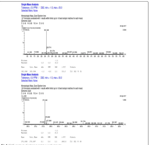
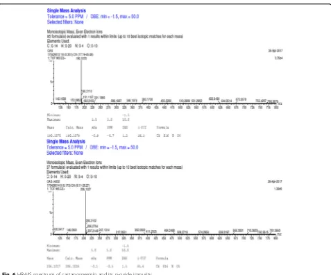
The formulae of all selected Aza sugars and its degrad-ation impurities were matching with theoretical mass num-ber with less than 5.0 ppm error value. The celgosivir molecular formula (C12H22NO5: [M + H]) accuracy error ppm was −4.6 ppm, and the celgosivir peroxide degrad-ation impurity molecular formula (C12H22NO6: [M + H]) was 0.4 ppm with 2.5 DBE. Hence, the celgosivir peroxide degradation impurity was proposed as n-oxide impurity of celgosivir (Fig. 5). Similarly, the castanospermin molecular formula (C18H16NO4: [M + H]) accuracy error ppm was
−4.7 ppm and the castanospermin peroxide degradation impurity molecular formula (C18H16NO5: [M + H]) was
−0.5 ppm with 1.5 DBE. Hence, the castanospermin perox-ide degradation impurity was proposed as n-oxperox-ide impurity of castanospermin (Fig. 6).
LC-MS/MS—method development, optimization, identification, and characterization of unknown degradation impurities
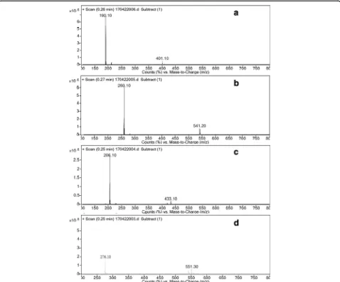
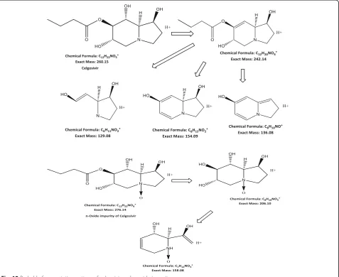
The MS/MS spectra of celgosivir and castanospermin and their peroxide degradation impurities are shown in Figs. 7 and 8. At lower collision energies, i.e., 10 and 20 eV, the molecular ion peak at 260 and 190 m/z was observed for celgosivir and castanospermin respectively (Figs. 8, 9, and 10). At higher collision energies, i.e., 30 and 50 eV, the buta-none group in celgosivir and –OH group in castanosper-min were elicastanosper-minated and observed 206 m/z for celgosivir and 172 m/z for castanospermin, further fragmented to 158 m/z for the celgosivir (Fig. 8) and 154 m/z for casta-nospermin (Fig. 9). HR-MS data supported the fragmenta-tion profile of celgosivir and castanospermin (Figs. 6 and 7). At the lower collision energies, the stable molecular ion
peaks were at 276 and 206 m/z for n-oxides of celgosivir and castanospermin respectively. At higher collision ener-gies, the stable molecular ion peaks were at 206 and 188 m/z for n-oxides of celgosivir and castanospermin re-spectively. The hydroxyl group loss stated that 206 and 188 m/z for celgosivir and castanospermin, respectively, was observed. Further fragmentation gave continuous loss of hydroxyl group by using higher collision energies (Figs. 8, 9, 10, and 11). The fragmentation pattern difference ob-served was 16 mass number such as APIs and its n-oxide
impurities (Table 1, Additional file 1). HR-MS data sup-ported the fragmentation profile of n-oxides of celgosivir and castanospermin and characterized as n-oxides of celgo-sivir and castanospermin (Figs. 12, 13, and 14; Table 2).
Method validation for castanospermin and celgosivir by ion chromatography
Specificity
Chromatography obtained with mixer of castanosper-min and celgosivir shows no interference with other
Aza sugars such as 1-deocynojirimycine, miglitol, and their degradants. During the degradation study, the n-oxide impurities of castanospermin and celgosivir were formed with 3% hydrogen peroxide (Table 3) and no interference with other cations. The n-oxide impurities of castanospermin and celgosivir were identified by isolation at the column end and charac-terized by high-resolution mass spectrometry and LC-MS/MS. Degradation study of castanospermin and celgosivir were performed by acid hydrolysis (0.5N HCl), base hydrolysis (0.5% Tri ethyl amine and 0.5N NaOH), oxidation (3% H202), photo degradation at
1.2 million lux hours, visible light, 200 W/m2 UV
light, and thermal degradation at 105 °C. The degrad-ation peaks have been identified and characterized as n-oxides of castanospermin and celgosivir by high-resolution mass spectrometry and LC-MS/MS. The peak purity was evaluated by high-resolution mass spectrometry and LC-MS/MS. The mass balance is demonstrated in Table 3.
Precision
The precision data obtained for the evaluated method is demonstrated in Table 4. The precision at 50, 100, and 150% were evaluated for castanospermin and cel-gosivir in a mixer solution. The %RSD at (n= 6) all levels were less than the 2.0% assuming acceptable precision.
Accuracy
Accuracy was performed by means of recovery studies using the developed method. The percentage of recoveries after spiking with additional mixer of casta-nospermin and celgosivir was at all levels such as 50,
100, and 150% in the range of 98–102%, and the
results are listed in Table 4.
Linearity
A linear relationship was presented between the concen-tration and peak area. The linearity concenconcen-tration for the mixer of castanospermin and celgosivir was taken to seven levels such as 25, 50, 75, 100, 125, 150, and 200%. The correlation coefficient value (r) and regression coef-ficient value (r) obtained for both was more than 0.999, which explains the linearity of the method, and the re-sults are listed in Table 4, (Plot 1 and Plot 2).
Table 2HR-MS pseudo fragmentation profile for formulae confirm of the n-oxide impurity of castanospermin and celgosivir
Name of product Fragmentation Formulae
Castanospermin 190.1070 [M + H]+ C8H16NO4
172.0965 C8H14NO3
154.0872 C8H12NO2
136.0753 C8H10NO
112.0743 C6H10NO
n-Oxide impurity of CAS (unknown impurity)
206.1027 [M + H]+ C8H16NO5
172.0968 C8H14NO3
154.0857 C8H12NO2
Celgosivir 260.1486[M + H]+ C12H22NO5
172.0972 C8H14NO3
154.0834 C8H12NO2
242.1272 C12H20NO4
Oxide impurity of CEL (unknown impurity)
276.1448 [M + H]+ C12H22NO6
206.1008 C8H16NO5
158.0816 C7H12NO3
Table 3Summary of forced degradation results
Stress condition Duration Assay of after forced degradation castanospermin (%w/w)
Assay of after forced degradation of celgosivir (%w/w)
Content of major degradant (%w/w)
Remarks
Acid hydrolysis 10 days 100 100 – No degradation
products formed for both
Base hydrolysis 0 h .0 100 100 of CAS formed
from CIL after 0 h
No degradation products formed for CAS
Oxidation 6 h 79.2 90.0 20.8% n-oxide of CAS,
10% of n-oxide CIL
n-oxides both sugars
Thermal (105 °C) 7 days 100 100 – No degradation
products formed for both
UV light 200 W/m2 100 100 – No degradation
products formed for both
Visible light 1.2 million lux hours
100 100 – No degradation
Robustness
Robustness was performed by changing flow, i.e., from 10% to lower to actual flow and 10% higher flow from actual flow, and buffer concentration flow, i.e., the study has been performed from 10% to lower concentration and 10% higher concentration from actual concentra-tion. The evaluated parameters were performed for the precision, %RSD were calculated and the results are listed in Table 4.
Ruggedness
The %RSD values for intermediate precision and repeat-ability reported were found to be less than 2% which shows ruggedness of the ion chromatography method. The results of ruggedness parameters are listed in Table 4.
Solution and mobile phase stability
Solution stability was performed with same mixer of cas-tanospermin and celgosivir in active pharmaceutical in-gredient solution at 24 and 48 h. The mobile phase was observed as stable in fresh mixer (for the assay of casta-nospermin and celgosivir in active pharmaceutical ingre-dients) at 24 and 48 h. The % variations of assay for both are less than−8.5%.
Conclusions
The present research provides the ion chromato-graphic method for the assay determination of the castanospermin and celgosivir as active pharmaceut-ical ingredients. Identification of the other Aza sugars such as 1-deoxynojirimycin and miglitol in the same
method with less amount of the sample quantity was determined. The method was validated as per ICH Q2 (R1); the proposed ion chromatography method is found to be simple, sensitive, accurate, precise, spe-cific and green chemistry type of analysis for diluent and mobile phase preparation; it can be used for intended purposes in drug substance and drug prod-uct. The n-oxide degradation impurities of castanos-permin and celgosivir under peroxide degradation were identified and characterized by using the high-resolution mass spectrometry and the LC-MS/MS. These novel techniques will help to improve the qual-ity of the drugs because the most of Aza sugars are using in diabetic research and dengue research.
Additional file
Additional file 1:Fragmentation pattern differences. (DOCX 761 kb)
Acknowledgements
The authors would like to thank Dr. Reddy’s Laboratories Ltd., Hyderabad, India, for providing the facilities to carry out this study. Cooperation from colleagues of Analytical Research and Development and the process research and development of Dr. Reddy’s Laboratories Ltd. is thankfully acknowledged.
Authors’contributions
PM, JMB, KB, and DRD conceived and designed the experiments. NR and BbD performed the experiments and wrote the manuscript. All authors read and approved the final manuscript.
Competing interests
The authors declare no that they have no competing interests.
Publisher’s Note
Springer Nature remains neutral with regard to jurisdictional claims in published maps and institutional affiliations.
Author details
1Analytical Research and Development, Dr. Reddy’s Laboratories, Hyderabad, Telangana 500049, India.2Department of Inorganic and Analytical Chemistry, Andhra University, Visakhapatnam, Andhra pradesh 530003, India.3AU College of Pharmaceutical Sciences, Andhra University, Visakhapatnam, Andhra pradesh 530003, India.
Received: 26 September 2017 Accepted: 10 January 2018
References
Balakumaran K, Janagili M, Rajana N, Papureddy S, Anireddy J. Development and Validation of Miglitol and Its Impurities by RP-HPLC and Characterization Using Mass Spectrometry Techniques. Sci Pharm. 2016;84(4):654-70. https:// doi.org/10.3390/scipharm84040654.
Belley M, Leger S, Roy P, Xiang YB, Labelle M, Guay D, Miglitol D. MedlinePlus Drug Information. MedlinePlus. National Institutes of Health. 2013. Retrieved 13 April 2013. European Patent 0480717B1, April 15 1998.
Budavari S, O'Neil MJ, Smith A, PE Heckelman, editors. The Merck Index: An Encyclopedia of Chemicals, Drugs and Biologicals. Eleventh Edition. Rahway: Merck & Co. Inc.; 1989.
Cahours X, Daali Y, Cherkaoui S, Veuthey JL. Simultaneous analysis of poly hydroxylated alkaloids by capillary electrophoresis using borate complexation and evaluation of sweeping technique for sensitivity improvement. Chromatographia. 2002;55:211–6.
Table 4Summary of validation results
Parameter Castanospermin Celgosivir
Correlation coefficient (r) 1.000 0.999
Intercept (c) −0.01 −0.01
Slope (m) 0.1 0.03
Method precision (%RSD) 1.1 0.7
Intermediate precision (%RSD) 3.2 3.1
Precision at 50% 1.2 1.7
Precision at 150% 0.4 1.9
Accuracy at 50% (%recovery) 100.1 95.4
Accuracy at 100% (%recovery) 99.7 99.7
Accuracy at 150% (%recovery) 97.0 82.3
Precision at flow 0.6 mL/min (%RSD) 0.5 0.4
Precision at Flow 0.8 mL/min (%RSD) 1.1 0.7
low buffer strength (%RSD) 0.5 1.5
High buffer strength (%RSD) 2.4 0.5
Solution stability and mobile phase
difference −
1.0 −8.5
Chittora NC, Shrivastava A, Jain A. New RP-HPLC method of miglitol in tablet dosage form including forced degradation studies and estimation in spiked rabbit plasma. J Young Pharm. 2009;1:364–70.
Council of Europe. European Pharmacopoeia (Ph. Eur.), 8th ed. Strasbourg: Council of Europe; 2015.
Dhole SM, Khedekar PB, Amnerkar ND. Validated high performance liquid chromatography method for determination of miglitol in tablet dosage form. Pharm Methods. 2012;3(2):68–72. https://doi.org/10.4103/2229-4708.103875. Durantel D. Celgosivir, an alpha-glucosidase I inhibitor for the potential treatment
of HCV infection. Curr Opin Investig Drugs. 2009;10(8):860-70.
Hohenschutz LD, Bell EA, Jewess PJ, Leworthy DP, Pryce RJ, Arnold E, Clardy J. Castanospermin, a 1,6,7,8-tetrahydroxyoctahydroindolizine alkaloid, from seeds of castanospermin australe. Photochemistry. 1981;20(4):811–4. https:// doi.org/10.1016/0031-9422(81)85181-3.
Ibrahim FA, Ali FA. Kinetic determination of acarbose and miglitol in bulk and pharmaceutical formulations using alkaline potassium permanganate. Int J Biomed Sci. 2007;3:20–30.
International Conference on Harmonization. Q2 (R1), Validation of Analytical Procedures: Text and Methodology. ICH Guidelines; 2005.
International Conference on Harmonization. Q1B, Photo stability Testing of New Drug Substances and Products. ICH Guidelines; 1996.
Japan Intl. Research Center for Agricultural Sciences. A simple, selective, and rapid method of high-performance anion-exchange chromatography with pulsed amperometric detection (HPAEC-PAD) to quantify DNJ in mulberry-based food products, Postharvest Science and Technology Div. Tsukuba: Japan Intl. Research Center for Agricultural Sciences; 2010. https://doi.org/10. 1111/j.1750-3841.2010.01528.
Kimura T, Nakagawa K, Saito Y, Yamagishi K, Suzuki M, Yamaki K, Shinmoto H, Miyazawa T. Simple and rapid determination of 1-deoxynojirimycin in mulberry leaves. Biofactors. 2004;22(1-4):341-5.
Li X, Wang Y, Wang J, Fawcett JP, Zhao L, Gu J. Determination of miglitol in human plasma by liquid chromatography/tandem mass spectrometry. Rapid Commun Mass Spectrum. 2007;21:247–51. https://doi.org/10.1016/j.jchromb. 2005.09.021.
Nirogi RV, Kandikere VN, Shukla M, Mudigonda K, Maurya S, Boosi RK, Yerramilli A. Liquid chromatographic tandem mass spectrometry method for the quantification of miglitol in human plasma. Arzneimittelforschung. 2006;56: 328–36.
Rajana N, Lasker R, Venkatesh P, Yarbagi K, Balakumaran K, Babu JM. A novel assay method of 1-deoxy nojirimycine (DNJ) by ion chromatography. Int J Pharm Sci Res. 2016;7(4):1580–9. https://doi.org/10.13040/IJPSR.0975-8232. 7(4).1580-89.
Rudraprasad Reddy J, Pramod Kumar R, Appa rao KMCh, Ramakrishna K. A sensitive UPLC method development and validation with LC-MS compatible for the determination of 1-Deoxynojirimycin in mulberry leaves using fluorescence detection. Am J Pharm Tech Res. 2014;4(3). ISSN: 2249-3387. Saul R, Ghidoni JJ, Molyneux RJ, Elbein AD. Castanospermine inhibits
alpha-glucosidase activities and alters glycogen distribution in animals. PNAS. 1985; 82(1):93–7. https://doi.org/10.1073/pnas.82.1.93. PMC 396977. PMID 3881759. The United States Pharmacopoeia 39 and National Formulary 34, Asian ed.
United States Pharmacopoeia. Rockville: Convection; 2016.
Wang F, Jin XZ, Xiu-mei D. Determination of miglitol in human plasma by HPLC-MS/ESI. Chin Pharm J. 2005;40:51–3.
Whitby K, Pierson TC, Geiss B, Lane K, Engle M, Zhou Y, Doms RW, Diamond MS. Castanospermin, a potent inhibitor of dengue virus infection in vitro and in vivo. J Virol. 2005;79(14):8698–706. https://doi.org/10.1128/JVI.79.14.8698-8706. PMC 1168722. PMID. 15994763