Introduction
Hematopoietic stem cell transplantation (HSCT) is a treatment modality for patients with hematological malignancy such as acute myeloid leukemia, chronic myeloid leukemia, acute lymphoblastic leukemia, and Hodgkin’s lymphoma. Earlier, it was called bone marrow transplantation; however, hematopoietic stem cells can be obtained from a variety of sources such as bone marrow, peripheral blood, and umbilical cord blood. Hence, the term bone marrow transplant been replaced by hematopoietic stem cell transplantation.[1]
Hematopoietic stem cell can be obtained from the patient him/herself which is called autologous transplantation. When it is obtained from
ABSTRACT
Hematopoietic stem cell transplantation (HSCT) is a treatment modality for patients with hematological malignancy. Oral mucositis is the most serious complication in these group of patients. Among various risk factors for oral mucositis, oral microbial colonization is considered the most important factor which greatly affects the quality of life in these patients. A systematic review was carried out to assess the colonization of microbes in individuals who developed oral mucositis following to HSCT. The literature study was done using PubMed, Medline, Google Scholar, Cochrane databases with keywords of oral mucositis, bacteria, fungi, viruses, hematologic malignancy, and HSCT, from the January 1989 to June 2017. Furthermore, hand searches were taken from the back references for assessing microbial colonization in oral mucositis following HSCT. Data extraction done from the included studies. The study quality was assessed using Ottawa-Newcastle scale. Four studies investigating microbial colonization in oral mucositis following HSCT were included. The available literature shows no clear pattern or association between oral mucositis and oral microflora in patients undergoing HSCT as a treatment modality for various hematological malignancies. Future research is needed to determine the relationship between the nature of oral microflora and its role in the development of mucositis subsequent to HSCT therapy. Data from such work would direct toward the development and testing of selective antimicrobial therapies for the prevention and management of mucositis subsequent to HSCT.
Keywords: Oral mucositis, stomatitis, mucositis, hematopoietic stem cell transplantation, stem cell transplantation, hematopoietic stem cell transplant, oral microbes
Microbial colonization in hematopoietic stem cell
transplantation-induced oral mucositis: A systematic review
Nandhini Subramaniam, Arvind Muthukrishnan
Department of Oral Medicine and Radiology, Saveetha Dental College, Saveetha University, Chennai, Tamil Nadu, India
Correspondence: Dr. Arvind Muthukrishnan, Department of Oral Medicine and Radiology, Saveetha Dental College, Saveetha University, 162, Poonamallee High Road, Chennai - 600 077, Tamil Nadu, India. E-mail: [email protected]
an identical twin, it is referred as synergic transplant, and transplant other than identical twin/from patient himself is called allogeneic transplantation.
In patients receiving HSCT, most serious complication is oral
mucositis[2] which is almost seen in 70–90% of patients who
receive HSCT[3,4] and 30–50% of patients with HSCT complain
that mucositis is their most significant toxicity. There is 4 phase pathophysiological models in the development of oral mucositis: Phase 1 - vascular phase (1–3rd day), Phase 2 - epithelial phase (4–5th day), Phase 3 - ulcerative phase (6–12th day), and Phase 4 - healing phase (12th day onward)[5] which has been replaced by a 5 phase pathobiological model[6,7] beginning with (i) initiation, (ii) upregulation and message generation, (iii) signal amplification, (iv) ulceration, and (v) healing.
Oral mucositis has severe physical and mental disability during the course of treatment. Among various risk factors for oral mucositis, development of bacterial, viral, and fungal infections is considered the most important factor[2] which greatly affects the quality of life in these patients. It has been suggested that the microbes on the ulcerated surface are active contributors of mucositis process.[8] The endotoxins released by the microorganisms penetrate the submucosa which exerts a chemotactic activity on polymorphonuclear
How to cite this article: Subramaniam N, Muthukrishnan A. Microbial colonization in hematopoietic stem cell transplantation-induced oral mucositis: A systematic review. J Adv Pharm Edu Res 2017;7(2):49-54.
Source of Support: Nil, Conflict of Interest: None declared..
leukocytes and macrophages and stimulates further pro-inflammatory cytokines that intensifies the pro-inflammatory process as well as exacerbates the ulcer.[9] Therefore, we need a better understanding of the role of oral microflora in the development of oral mucositis which begins with identifying changes in the oral flora during HSCT therapy.
Thus, the objective behind carrying out this systematic review was to assess the microbial colonization in patients developing oral mucositis following HSCT.
Materials and Methods
The literature search was done using the keywords of “oral mucositis” with “HSCT” and “oral microbes” in databases of PubMed, Medline, Cochrane library, and Google Scholar from January 1989 to 20th June 2017 for English language publications. After database search reference lists were hand-searched to identify additional studies.
Research methodology
Inclusion and exclusion criteria of this review were set a priori
A studies published in English between January 1st 1989 and June 20th 2017. Studies discussing microbial colonization from samples taken from oral cavity in patients developing oral mucositis following HSCT were included. Experimental studies, observational studies, and systematic reviews involving human subjects only were included in the study. While case reports, case series, animal studies, studies in which patients developed oral mucositis following radiotherapy, and studies that assessed microbial colonization from blood samples were excluded from the study.
Data retrieval
After the initial literature search, the authors reviewed all titles and abstracts to identify suitable articles for inclusion. All articles identified as suitable were read in their entirety and considered for inclusion and were summarized based on study design, sample population, methodology, results, and conclusion.
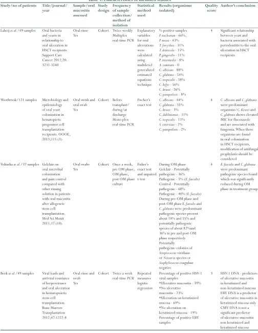
Table 1: Characteristics of included studies
Study/no of patients Title/journal/
year Sample/oral mucositis assessed
Study
design Frequency of sample collection/ method of isolation Statistical method used Results (organisms
isolated) Quality score Author’s conclusion
Laheij et al./49 samples Oral bacteria and yeasts in relationship to oral ulceration in HSCT recipients. Support Care Cancer 2012;20: 3231-3240
Oral rinse
Yes Cohort Twice weeklyMultiplex
real-time PCR Explanatory variables for oral ulcerations were calculated using multilevel generalized estimated equations technique
% positive samples
F. nucleatum - 66%,
P. micra - 43%
T. forcythia - 31%
T. denticola - 15%
P. gingivalis - 11%
P. intermedia - 8%
A. comitans - 0
C. albicans - 88%
C. glabrata - 54%
C. tropicalis - 58%
C. kefyr - 56%
C. krusei - 26%
C. parapsilosis - 8%
4 Significant relationship between yeast and bacteria associated with periodontitis to the oral ulceration in HSCT recipients
Westbrook/121 samples Microbiology and
epidemiology of oral yeast colonization in hematopietic progenitor cell transplantation recipients. OOOE, 2013;115 (3).
Oral swish and oral swab Yes Cohort Before transplant/ during/at discharge Mono plex real-time PCR Fischer’s exact test
C. albicans - 44%
C. glabrata - 35%
C. krusei - 3%
C. dubliniensis - 11%
C. tropicalis - 11%
S. cerevisiae - 2%
C. parapsilosis - 2%
4 C. albicans and C. glabrata
were predominant organisms/C. Krusei and
C. glabrata shows elevated MIC for fluconazole and are associated with fungemia. When these organisms are found in oral colonization in HSCT recipients, modification of antifungal prophylaxis should be considered Vokurka et al./37 samples Gelclair on
oral microbial colonization and pain control compared with other rinsing solution in patients with oral mucositis after allogeneic stem cell transplantation. Med Sci Monit 2011;17 (10).
Oral swabs
Yes Cohort Once a week, pre OM phase, OM phase, post OM phase culture
Fisher’s exact test and unpaired t-test
During OM phase Gelclair - Potentially pathogenic - 36% Pathogenic - 5% (E. faecalis)
Control - Potentially pathogenic - 60% Pathogenic - 40% (E. faecalis)
During pre-OM phase and post-OM phase E. faecalis and
C. glabrata were predominant pathogenic species present about 18% and 55% and potentially pathogenic species of about 82%and 36% in pre and post-OM phase respectively Potentially
pathogenic-colonies of
Streptococcus viridians or Neisseria species or
Staphylococcus coagulase negative
4 E. faecalis and C. glabrata
were predominant pathogenic species found which was significantly reduced during OM phase in treatment group
Beek et al./49 samples Viral loads and antiviral resistance of herpesviruses and oral ulceration in hematopoietic stem cell transplantation. Bone Marrow Transplantation 2012;47:1222-8
Oral rinse and swab Yes
Cohort Twice a week
real-time PCR Repeated measures logistic regression
Percentage of positive HSV-1 viral samples
*Ulcerative mucositis - 39% *No ulcerative
mucositis - 23% *Ulceration on keratinized mucosa - 69%
*No ulceration on keratinized mucosa - 19% Percentage of positive EBV samples
3 HSV-1 DNA - predictors
of ulcerative mucositis in keratinized and non-keratinized mucosa EBV DNA is a predictor of ulcerative mucositis in keratinized mucosa only CMV DNA is not a significant predictor of ulcerative mucositis non keratinized and keratinized mucosa
Quality assessment
The authors evaluated the quality of the included studies using a published scoring system. Newcastle–Ottawa quality assessment scale, a 9-item checklist, was used to score cohort studies. Their scores were summarized in Table 1.
Results
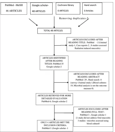
The database search and the hand search identified 48 studies. After initial title screening, 11 were excluded, and after abstract screening, 29 abstracts were excluded. The remaining 8 articles were retrieved, and the full text was reviewed. Of these, 4 articles were excluded. Of the 4 studies excluded at this stage, 3 were excluded because they assessed microbes in blood samples while 1 study was excluded because it did not specifically investigate oral mucositis. The entire data have been tabulated I according to PRISMA flowchart [Figure 1]. The remaining 4 articles that assessed microbial colonization in oral mucositis following HSCT were included in the present study, and their characteristics are described in Table 1.
Levels of evidence
The included observational studies showed the level of evidence - II-B.
Grading of oral mucositis - All 4 included studies used standardized scoring system – the WHO scoring system to assess mucositis in all 4 studies additionally OMAS and visual analog scale were used in 2 studies.
The 4 studies retrieved from the literature assessed bacterial, fungal, and viral colonization using oral samples. However, it is difficult to compare studies as considerable heterogeneity was observed
between the studies. The available literature showed no clear pattern or association between oral mucositis and oral microflora in patients undergoing HSCT as a treatment modality for various hematological malignancies.
Discussion
One of the most important complications of HSCT is oral mucositis occurring in 75–99% of patients.[10-13] It usually develops 6–10 days after transplantation that greatly affects the quality of life in these patients. The commonly involved sites are buccal mucosa, labial mucosa, floor of the mouth, ventral surface of the tongue, and esophageal mucosa.[7,14] The phase initiates with basal cell damage and release of reactive oxygen species followed by which there is activation of transcription factors and upregulation of genes and production of destructive proteins and pro-inflammatory cytokines leading to apoptosis and cell injury, also there is positive feedback loop (signal amplification) which leads to destructive process, and there is break in oral epithelium and ulcerates where infective process comes into play complicates. It is the ecological shift in the oral microbes that complicate the oral mucositis.[15,16] Thus, in this review, we have analyzed the studies assessing the oral microbial colonization in oral mucositis among hematopoietic stem cell transplant recipients.
The primary outcome measure in the 4 included studies was assessing the microbial colonization. In a study involving 49 HSCT recipients by Laheij et al.,[17] the authors found that Fusobacterium nucleatum was the most frequent bacteria isolated and Porphyromonas gingivalis
was consistently isolated from the samples. Candida albicans was the most frequent fungi isolated, and the study concluded that there was a significant relationship between yeast and bacteria associated with periodontitis to the oral mucositis in hematopoietic stem cell
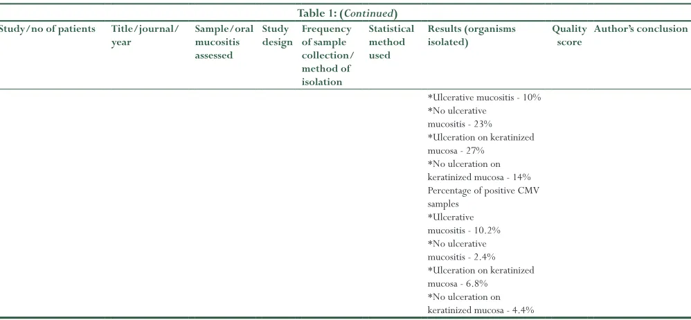
HSCT: Hematopoietic stem cell transplantation, PCR: Polymerase chain reaction, F. nucleatum: Fusobacterium nucleatum, P. micra: Prevotella micra, T. forcythia: Tannerella forsythia, T. denticola: Treponema denticola, P. gingivalis: Porphyromonas gingivalis, Candida albicans, P. intermedia: Prevotella intermedia, A. comitans: Aggregatibacter actinomycetemcomitans, C. albicans: Candida albicans, C. glabrata: Candida glabrata, C. tropicalis: Candida tropicalis, C. kefyr: Candida kefyr, C. krusei: Candida krusei, C. parapsilosis: Candida parapsilosis, MIC: Minimum inhibitory concentration, S. cerevisiae: Saccharomyces cerevisiae, C. dubliniensis: Candida dubliniensis, E. faecalis: Enterococcus faecalis, HSV: Herpes simplex virus, EBV: Epstein‑Barr virus, CMV: Cytomegalovirus
*Ulcerative mucositis - 10% *No ulcerative
mucositis - 23% *Ulceration on keratinized mucosa - 27%
*No ulceration on keratinized mucosa - 14% Percentage of positive CMV samples
*Ulcerative mucositis - 10.2% *No ulcerative mucositis - 2.4% *Ulceration on keratinized mucosa - 6.8%
*No ulceration on keratinized mucosa - 4.4%
Table 1: (Continued)
Study/no of patients Title/journal/
year Sample/oral mucositis assessed
Study
design Frequency of sample collection/ method of isolation
Statistical method used
Results (organisms
transplant recipients. Moreover, Candidakefyr, a rare species but very high colonization rate (94% of patients), is found among the study population.
Furthermore, the authors stated that there is the absence of
Actinobacillus actinomycetemcomitans in the study samples which is normally present in subgingival biofilm of 15–30% of healthy population. It is important to note that this study had the lower quality score because follow-up was not adequate and explanation for the absence of A. actinomycetemcomitans was not given; however, this could be due to the use of antibiotics.
In a prospective cohort study, Westbrook et al.[18] evaluated oral yeast colonization in patients undergoing stem cell transplantation who received fluconazole prophylaxis. C. albicans and Candida glabrata were predominant organisms. Both Candida krusei and C. glabrata showed elevated MIC for fluconazole and are associated with fungemia. This finding concluded that when these organisms are found in oral colonization in HSCT recipients, modification of antifungal prophylaxis should be considered.
In a prospective observational study involving 37 HSCT recipients, Vokura et al.[19] found that Enterococcus faecalis and C. glabrata were the predominant pathogenic organisms in 15 patients and these organisms were significantly reduced during oral mucositis phase in 22 patients who were given Gelclair. However, this study assessed microbial colonization by culture-based method.
van der beek et al.[20] found that the presence of HSV-1 DNA was found to be a predictor of ulcerative mucositis in keratinized and non-keratinized. Furthermore, antiviral-resistant strains were isolated from the samples. EBV was found to be a predictor of ulcerations in the keratinized mucosa only. CMV was not a significant predictor of ulcerative mucositis on the non-keratinized and keratinized mucosa. It is important to note that this study had lowest quality score of included studies because follow-up of cohorts not adequate and no information regarding loss of follow-up given in the study.
Method of collecting samples is not uniform in all 4 studies where 2 studies used both oral swish and oral swab as collection method while other two involved only oral rinse and oral swab for collecting sample. Furthermore, the methodology for detection and quantification of microflora in the included studies shows the molecular biologic technique in 3 studies while one employed culture-based isolation of microbes which is less sensitive than real-time PCR, so low loads could be missed.
All patients who underwent HSCT in the included studies developed oral mucositis, and they were all given different conditioning regimen as prophylaxis for graft versus host disease.
Although oral prophylaxis was given in all 4 included studies, consistent increase in microbes found during mucositis phase. This may be due to the selection of inappropriate antimicrobials and the way it is administered. When antimicrobials are given orally, patients might have difficulty in sucking due to dry mouth which could be the reason for low compliance of prophylactic antimicrobials.
Not all included studies evaluated the relationship between WBC counts and quantitative oral flora changes. Combination of oral mucositis and granulocytopenia increases the risk of systemic infection.
The studies included in this review assessed bacterial, fungal, and viral colonization using oral samples. However, it is difficult to compare studies since they differ on the population that were studied, conditioning regimen administered, sample collection methods and intervals, sample analysis methods, microorganisms studied. The available literature shows no clear pattern or association between oral mucositis and oral microflora in patients undergoing HSCT as a treatment modality for various hematological malignancies.
Conclusion
Future research is needed to determine the relationship between the nature of oral microflora and its role in the development of mucositis subsequent to HSCT therapy. This would give a better understanding of the potential role oral microflora in the development and exacerbation of oral mucositis. Data from such work would direct toward the development and testing of selective antimicrobial therapies for the prevention and management of mucositis subsequent to HSCT.
References
1. Hatzimichael E, Tuthill M. Hematopoietic stem cell transplantation. Stem Cells Cloning 2010;3:103-17.
2. Scully C, Sonis S, Diz PD. Oral mucositis. Oral Dis 2006;12:229-41. 3. Napeñas JJ, Brennan MT, Bahrani-Mougeot FK, Fox PC, Lockhart PB. Relationship
between mucositis and changes in oral microflora during cancer chemotherapy. Oral Surg Oral Med Oral Pathol Oral Radiol Endod 2007;103:48-59. 4. Botti S, De Cecco V, Galgano L, Gargiulo G, Magarò A, Orlando L. Oral
Mucositis in Hematopoietic Stem Cell(HSCT): Position Statement by Gruppo Italiano Trapianto di Midollo Osseo (GITMO) Nurses Group, DCTH; 2014. p. 205-23.
5. Sonis ST. The pathobiology of mucositis. Nat Rev Cancer 2004;4:277-84. 6. Cawley MM, Benson LM. Current trends in managing oral mucositis. Clin J
Oncol Nurs 2005;9:584-92.
7. Sonis ST. Mucositis: The impact, biology and therapeutic opportunities of oral mucositis. Oral Oncol 2009;45:1015-20.
8. Vanhoecke B, De Ryck T, Stringer A, Van de Wiele T, Keefe D. Microbiota and their role in the pathogenesis of oral mucositis. Oral Dis 2015;21:17-30. 9. Sonis ST. Mucositis as a biological process: A new hypothesis for the development
of chemotherapy-induced stomatotoxicity. Oral Oncol 1998;34:39-43. 10. Sonis ST, Elting LS, Keefe D, Peterson DE, Schubert M,
Hauser-Jensen M, et al. Perspectives on cancer therapy induced mucosal injury: Pathogenesis,measurement, epidemiology and consequences for patients. Cancer 2004;100 9 Suppl:1995-2025.
11. Stiff P. Mucositis associated with stem cells transplantation: Current status and innovative approaches to management. Bone Marrow Transplant 2001;27 Suppl 2:S3-11.
12. Gabriel DA, Shea T, Olajida O, Serody JS, Comeau T. The effect of oral mucositis on morbidity and mortality in bone marrow transplant. Semin Oncol 2003;30 6 Suppl 18:76-83.
13. Rubenstein EB, Peterson DE, Schubert M, Keefe D, McGuire D, Epstein J,
Suppl:2026-46.
14. Molinterni A, Zambetti M. Danni da farmaci. In: Bonadonna G, Cuna GR, Valgusa P, editors. Medicina Oncologica. 7th ed. Milano: Masson; 2004. p. 1647-61.
15. Zollner-Schwetz I, Auner HW, Paulitsch-Fuchs AH, Krause R. Oral and intestinal candida colonization in patients undergoing hematopoietic stem-cll transplantation. J Infect Dis 2008;198:150-3.
16. Balletto E, Mikulska M. Bacterial infections in hematopoietic stem cell transplant recipients. Mediterr J Hematol Infect Dis 2015;7:e2015045. 17. Laheij AM, de Soet JJ, von dem Borne PA, Kuijper ED, Kraneveld EA, van
Loveren C, et al. Oral bacteria and yeasts in relationship to oral ulcerations in hematopoietic stem cell transplant recipients. Support Care Cancer 2012;20(12):3231-40.
18. Westbrook SD, Kirkpatrick WR, Wiederhold N, Redding SW. Microbiology and epidemiology of oral yeast colonization in hemopoietic progenitor cell transplant recipients. Oral Surg Oral Med Oral Pathol Endod 2013;115:354-8.
19. Vokurka S, Skardova J, Hruskova R, Kabatova-Maxova K, Svoboda T, Bystricka E, et al. The effect of polyvinylpyrrolidone-sodium hyaluronate gel (Gelclair) on oral microbial colonization and pain control compared with other rinsing solutions in patients with oral mucositis after allogeneic stem cells transplantation. Med Sci Monit 2011;17:CR572-6.