Int.J.Adv.Microbiol.Health.Res.2018; 2(2):40-55
40
3 International Journal of Advanced Microbiology and Health Research ISSN: 2457-077X. Volume 2, Issue 2 (June 2018), PP. 40-55
Available online atwww.ijamhr.com
Review Article
Nipah: A killer virus
Gurjeet Singh1, Raksha2, Anant D. Urhekar3 Assistant Professor1&2, Professor and Head3.
1&2
Department of Microbiology, N.C. Medical College and Hospital, Israna, Panipat-132107, Haryana, India. 3
Department of Microbiology, MGM Medical College and Hospital, Kamothe, Navi Mumbai-410209, Maharashtra, India.
ABSTRACT
Received 30th May, 2018 Revised 2nd June, 2018 Accepted 5th June, 2018 Published online 5th June, 2018
Nipah virus
Zoonotic
Diagnosis
Prevention
ELISA
PCR
Article Info
Nipah virus (NiV) is member of the genus Henipavirus in the family Paramyxoviridae. NiV disease is a zoonotic disease characterized by fever, constitutional symptoms, and encephalitis, sometimes accompanied by respiratory illness. The name „Nipah virus‟ originated from Sungai Nipah (Nipah River Village). A Nipah virus disease outbreak was reported from Kozhikode district of Kerala, India on 19 May 2018. This is the first NiV outbreak in South India. As of 28 May, there are 14 deaths, 16 confirmed cases and 12 suspected cases. Transmission of the NiV disease occurred from direct contact with sick pigs or their contaminated tissues also via respiratory droplets, contact with throat or nasal secretions from the pigs, or contact with the tissue of a sick animal. Consumption of fruits or fruit products (e.g. raw date palm juice) contaminated with urine or saliva from infected fruit bats was the most likely source of infection. Limited human to human transmission of NiV has also been reported among family and care givers of infected NiV patients. In Siliguri, India, transmission of the virus was also reported within a health-care setting (nosocomial), where 75% of cases occurred among hospital staff or visitors. This infection can occur in humans without showing any symptoms. However, it is essential for people to look out for influenza-like symptoms. Fever, sore throat, headaches, vomiting and muscle pain (myalgia) are some of the common signs. The infection progresses to acute respiratory infection (mild to severe) causing interference in breathing. During this phase, people experience atypical pneumonia and acute respiratory distress, which further leads to severe problems (fatal encephalitis). Initially develop influenza-like symptoms of fever, headaches, myalgia (muscle pain), vomiting and sore throat, followed by dizziness, drowsiness, altered consciousness, and neurological signs that indicate acute encephalitis. Some people can also experience atypical pneumonia and severe respiratory problems, including acute respiratory distress. Encephalitis and seizures occur in severe cases, progressing to coma within 24 to 48 hours. The incubation period (interval from infection to the onset of symptoms) is believed to range from 4 to 18 days. However, an incubation period as long as 45 days has been reported. BSL 2 facilities are sufficient if the virus can be first inactivated during specimen collection. Laboratory diagnosis of NiV includes serology, histopathology, Polymerase Chain Reaction (PCR) and virus isolation. Serum Neutralization Test, Enzyme Linked Immunosorbent Assay (ELISA), Reverse Transcriptase Polymerase Chain Reaction (RT-PCR) are used for laboratory confirmation. The 2018 review of the WHO list of Blueprint priority diseases indicates that there is an urgent need for accelerated research and development for the Nipah virus.
Int.J.Adv.Microbiol.Health.Res.2018; 2(2):40-55
41
INTRODUCTION
Nipah virus (NiV) is member of the genus Henipavirus in the family Paramyxoviridae. It is classified as a Biosafety Level-4 (BSL-4) agent due to its highly pathogenicity and relative new findings. The Centers for Disease Control and Prevention (CDC) and the National Institute of Allergy and Infectious Diseases (NIAID) have classified NiV as a Category C priority pathogen. [1] NiV disease is a zoonotic disease characterized by fever, constitutional symptoms, and encephalitis, sometimes accompanied by respiratory illness. NiV has an envelope with filamentous nucleocapsids, the genome consists of a single-stranded negative-sense RNA of approximately 18.2 kb. The genome encodes for six major structural proteins: nucleocapsid (N), phosphoprotein (P), matrix protein (M), fusion protein (F), glycoprotein (G), and large protein or RNA polymerase (L). [2]
The name „Nipah virus‟ originated from Sungai Nipah (Nipah River Village), where the first isolates were obtained [3–5]. Bats of the genus Pteropus appear to be the natural reservoir of the virus. Nipah virus swept through numerous piggeries in Malaysia and killed 1100 people during the period from 1998 through 1999. [6-7] NiV was identified as the etiological agent responsible of an outbreak, in pigs and humans, in Malaysia and Singapore. Transmission may be from consumption of contaminated food by bats secretion, or contact with infected pigs. Another way can be human-to-human spread. Since 1998 there have been several cases of infections in Bangladesh and India, collectively causing hundreds of cases with lethality rates sometimes in excess of 70%. [8–22]
The natural reservoir for NiV is pteropid fruit bats [2], and direct bat to human transmission can occur, frequently as a result of consumption of date palm sap contaminated with saliva or urine from infected bats [22]. Alternatively, transmission to the human population can proceed via an amplifying host, as during the initial NiV outbreak in Malaysia, in which transmission was from close contact with infected domesticated swine [14]. Transmission is primarily via the oronasopharyngeal route, with initial infection in the respiratory mucosa, followed by viral dissemination and high levels of viral replication in the endothelial cells of the central nervous system vasculature, causing the often-fatal
encephalitis [23,24]. No intermediate host was implicated in the Nipah virus outbreaks in India and Bangladesh. Rather, epidemiological data suggest transmission of Nipah virus from bats to humans through the consumption of fruit or date palm sap contaminated by infected fruit bats. [17,25] In most outbreaks, limited chains of direct human-to-human transmission have been documented, usually from an infected patient with respiratory symptoms to a direct care-giver, both in community and hospital settings [22,26–28].
For the Nipah virus outbreaks in Bangladesh between 2001 and 2007, it was estimated that,50% of Nipah virus cases were the result of human-to-human transmission events. Nipah virus has been isolated from human urine, saliva, nasal and oropharyngeal secretions and epidemiological data suggest that direct contact with these secretions of Nipah virus spreaders resulted in greater risk of Nipah virus infection. Three potential modes of human-to-human transmission of Nipah virus could be transmission via fomites, direct contact or aerosols. [29] Currently there are no vaccines or therapeutic medicine specifically recorded to either prevent or treat patients infected with Nipah virus. [30] The World Health Organization has included Nipah in its priority list of emerging diseases that could cause a global pandemic, along with Crimean Congo fever, Ebola, Middle East respiratory syndrome coronavirus and Zika. [31]
Int.J.Adv.Microbiol.Health.Res.2018; 2(2):40-55
42
EPIDEMIOLOGY
Paramyxoviruses are characterized by broad host range and for this reason they show an important zoonotic potential, like Nipah virus originating from bats. Bats represent the most successful mammals on earth including about 1200 chiropteran species distributed worldwide. In the last decades Hendra virus, Nipah virus and other zoonotic viruses like Ebola, Marburg, and SARS virus, have been identified in various Pteropus species fruit bats [33-36, 56].
Fruit-eating bats (Pteropus species) are the natural reservoir for the henipaviruses. Humans are usually infected via the intermediate hosts. In case of Nipah virus, pigs are the usual intermediate hosts. But exposures to infected fruit bats or materials contaminated by infected bats or direct human-to-human transmission is also possible. Bats are classified in the order Chiroptera (from the Greek 'cheiros,' meaning hand; and 'pteros,' meaning wing) and it is within the genus Pteropus in the family Pteropodidae or old world fruit bats, that we find the natural hosts of HV and NV. Pteropid bats are commonly referred to as 'flying foxes.' Sixty-five Pteropus species are distributed from Madagascar through the Indian subcontinent to southeastern Asia and Australia and as Far East as the Cook Islands. Some Pteropus species are among the largest of all bats, weighing as much as 1.2 kg and displaying a wingspan of up to 1.7 m.
Pteropus species are unique because they lack the complex neural and behavioral mechanisms required for echolocation that characterize the vast majority of bat species. Instead, they have large eyes and they navigate visually, feeding mainly on fruits and flowers, which they locate by smell.[37-39]
Although brought into much attention by the epidemic of NV encephalitis in Malaysia in 1998-99 (vide infra), isolated cases of Hendra virus causing encephalitic illness amongst animal handlers were being reported since as early as 1994. NV and HV having close genomic similarity were difficult to differentiate serologically earlier. In fact, even during the Malaysian epidemic of 1998-99, initial results indicated the causative organism to be Hendra virus. Later of course, with viral isolation and development of a specific serological marker, the identity of the NV was
made known. The word 'Nipah' originated from the name of a village 'Sungai Nipah' in the Malaysian peninsula, one of the first villages where pig farmers developed an encephalitic disease. Studies have been reported Nipah encephalitic illnesses among human and animal [40-62].
The first known human infection with NV was detected during an outbreak of severe febrile encephalitis in peninsular Malaysia and Singapore [61-62] in 1998-1999. Direct contact with pigs was the primary source of human infection. A total of 276 patients with viral encephalitis were reported in that epidemic. Most of the victims were adult males involved in pig farming or pork production. The spread of virus within the pig farms and between states of Malaysia was due to movement of pigs. NiV disease transmission among pigs in the same farm was attributed to direct contact with excretions and secretions i.e. urine, saliva, laryngeal and pharyngeal secretions. Iatrogenic transmission by use of same needles was also implicated. [39,62]
But what exactly led to the spillage of the virus from its natural reservoir into the pigs remains a subject of speculation. Species jumping of viruses can be due to evolutionary or ecological reasons. But NV is an old virus and has not undergone any evolutionary change. Most authorities believe that ecological factors led to their emergence. [39] This can be due to a change in the number density and management of pigs. But more importantly, the curse of unplanned deforestation of pulpwood has taken its toll on the natural habitat of the fruit bats in the last two decades. This coupled with the EL Nino Southern Oscillation - related drought prompted migration of bats from their natural habitat in the costal forest on to the villages where the pigsties (piggeries) were located. [39]
Int.J.Adv.Microbiol.Health.Res.2018; 2(2):40-55
43 in several ways. With destruction of trees, there had been shortage of food in the forest, especially for the fruit-eating bats. Second, the dry weather resulted in forest fires, causing further loss of trees and lack of food supply for the fruit-eating bats. Thirdly, the forest fire caused a severe haze, resulting in poor visibility for the bats, which preferred to migrate to the cleaner 'air' of the villages closer to human and pig habitation. These EL Nino related factors coupled with 'intentional' deforestation played a major role in upsetting the ecological balance, perpetuating transmission of NV from bats to pigs and pigs to humans in Malaysia.[39]
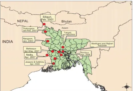
The route of introduction of virus into the pigs was also facilitated by the practice of growing fruit trees adjacent to the piggeries. In April and May 2001, a cluster of febrile neurological illnesses with nine deaths was reported in a village in Meherpur district, Bangladesh. Preliminary investigations by the Bangladesh Ministry of Health and the World Health Organization (WHO) excluded a diagnosis of Japanese encephalitis, dengue fever or malaria, but 2 of 42 serum specimens obtained from village residents in May 2001 showed reactive antibodies to Nipah virus antigen in tests performed at the US Center for Disease Control and Prevention (CDC). However, a comprehensive investigation of this outbreak was not conducted. [57]
In January 2003, a further cluster of febrile illnesses with neurological features and eight reported deaths occurred in adjoining villages in Naogaon district, 150 km from the village in Meherpur district. Similarities in the clinical manifestations observed among patients in Naogaon and Meherpur raised the question of whether the outbreaks were caused by the same agent. But unlike the Malaysia epidemic, no intermediate amplifying host could be identified. This led to the conjecture that the virus was transmitted directly or indirectly from bats to the humans. [58] Two outbreaks consisting of 48 cases of NV were detected in 2004 in two adjacent districts (30 km apart) of central Bangladesh (Rajbari and Faridpur) with a case-fatality rate of nearly 75%. Because of heightened surveillance, other small clusters and isolated cases (n=19) were identified during the same period in seven other districts in central and northwest Bangladesh. Although antibodies to NV were detected in fruit bats from the affected areas in 2004, an
intermediate animal host was not identified, which suggests that the virus was transmitted from bats to humans. In fact, human-to-human transmission of NV was documented during the Faridpur outbreak of 2004. [59]
A study showed four NV isolates from Bangladesh share 99.1% homology but exhibit more inter strain nucleotide heterogeneity than the sequences of the human isolates in Malaysia, which were nearly identical. These varying amounts of genetic variability may reflect differences in the mode of transmission of NV in the two countries. A further outbreak in Tangail district of Bangladesh occurred in end 2004 early 2005 and has also been reported.[60]
Unlike the Malaysia outbreak, 'bat to pig to human' transmission was unlikely to have occurred in Bangladesh. For religious reasons, pig farming is not practiced in Bangladesh and pig population is low. Hence direct bat-to-human transmission and then human-to-human transmission seemed most likely. Tan, in a talk delivered at the World Congress of Neurology, November 2005, at Sydney,[63] proposed that this route might be through contaminated date palm juice, which humans consume. Collecting date palm juice in earthenware pots hung atop date palm trees (after making an incision in the bark of the trees) is a common practice in rural areas of eastern India and Bangladesh during winter months. The bats feed on the juice, thus contaminating the juice with their saliva, which is subsequently drunk by humans.
Int.J.Adv.Microbiol.Health.Res.2018; 2(2):40-55
44
Fig. 1: Structure of Nipah Virus Infection (NiV)
Source: https://en.wikipedia.org/wiki/Henipavirus#Nipah_virus
The boundaries and name shown on this map do not imply any expression or any opinion what so ever on the part of World Health Organization concerning the legal status of any country, territory, city or area of its authorities or concerning the delimitation of its frontiers or boundaries.
Int.J.Adv.Microbiol.Health.Res.2018; 2(2):40-55
45
Table 1: Morbidity and mortality due to Nipah or Nipah-like virus encephalitis in WHO South-East Asia Region, 2001-2018
Country: Bangladesh
Month/Year Location No. of
cases
No. of deaths
Fatality Rate
April, May 2001 Meherpur 13 9 69%
January 2003 Naogaon 12 8 67%
Jan 2004 Apr 2004
Rajbari Faridpur
31 36
23 27
74% 75%
Jan- Mar 2005 Tangail 12 11 92%
Jan-Feb 2007 Mar 2007 Apr 2007
Thakurgaon Kushtia
Pabna, Natore and Naogaon
7 8 3
3 5 1
43% 63% 33%
Feb 2008 Apr 2008
Manikgonj Rajbari
4 7
4 5
100% 71%
Jan 2009 Gaibandha, Rangpur and Nilphamari Rajbari
3 1
0 1
0% 100%
Feb-Mar 2010 Faridpur
Faridpur, Rajbari, Gopalgonj, Kurigram,
8 8 1
7 7 1
87.50% 87.50% 100%
Jan-Feb 2011 Lalmohirhat, Dinajpur, Comilla Nilphamari, Faridpur, Rajbari
44 40 91%
Jan 2012 Joypurhat 12 10 83%
Jan- Apr 2013 Pabna, Natore, Naogaon, Gaibandha, Manikganj
24 21 88%
Jan-Feb 2014 13 districts 18 9 50%
Jan-Feb 2015 Nilphamari, Ponchoghor, Faridpur, Magura, Naugaon, Rajbari
9 6 67%
Country: India
Month/Year Location No. of cases No. of death Case Fatality
Rate
Feb 2001 Siliguri 66 45 68%
Apr 2007 Nadia 5 5 100%
May* 2018 Kerala 14 12 86%
*As of 24 May 2018
Int.J.Adv.Microbiol.Health.Res.2018; 2(2):40-55
46
MODE OF TRANSMISSION
During the initial outbreaks in Malaysia and Singapore, most human infections resulted from direct contact with sick pigs or their contaminated tissues. Transmission is thought to have occurred via respiratory droplets, contact with throat or nasal secretions from the pigs, or contact with the tissue of a sick animal.
In the Bangladesh and India outbreaks, consumption of fruits or fruit products (e.g. raw date palm juice) contaminated with urine or saliva from infected fruit bats was the most likely source of infection.
Limited human to human transmission of NiV has also been reported among family and care givers of infected NiV patients. During the later outbreaks in Bangladesh and India, Nipah virus spread directly from human-to-human through close contact with people's secretions and excretions.
In Siliguri, India, transmission of the virus was also reported within a health-care setting (nosocomial), where 75% of cases occurred among hospital staff or visitors. From 2001 to 2008, around half of reported cases in Bangladesh were due to human-to-human transmission through providing care to infected patients. [68]
Transmission also occurs from direct exposure to infected bats. A common example is consumption of raw date palm sap contaminated with infectious bat excretions. [69]
The possible mechanical transmission by repetitive use of same needles or equipment without further sterilization after each use for health intervention and artificial insemination and sharing of boar semen within a farm were also implicated. The possible role of transmission by infected dogs and cats found in the affected farm could not be excluded [39].
SIGNS AND SYMPTOMS
This infection can occur in humans without showing any symptoms. However, it is essential for people to look out for influenza-like symptoms. Fever, sore throat, headaches, vomiting and muscle pain (myalgia) are some of the common signs.
The infection progresses to acute respiratory infection (mild to severe) causing interference in breathing. During this phase, people experience atypical pneumonia and acute respiratory distress, which further leads to severe problems (fatal encephalitis).
Infected people initially develop influenza-like symptoms of fever, headaches, myalgia (muscle pain), vomiting and sore throat. This can be followed by dizziness, drowsiness, altered consciousness, and neurological signs that indicate acute encephalitis. Some people can also experience atypical pneumonia and severe respiratory problems, including acute respiratory distress. Encephalitis and seizures occur in severe cases, progressing to coma within 24 to 48 hours.
The incubation period (interval from infection to the onset of symptoms) is believed to range from 4 to 18 days. However, an incubation period as long as 45 days has been reported. Most people who survive acute encephalitis make a full recovery, but long term neurologic conditions have been reported in survivors. Approximately 20% of patients are left with residual neurological consequences such as seizure disorder and personality changes.
A small number of people who recover subsequently relapse or develop delayed onset encephalitis. The case fatality rate is estimated at 40% to 75%. This rate can vary by outbreak depending on local capabilities for epidemiological surveillance and clinical management. [70-71]
DIFFERENTIAL DIAGNOSIS
• It is important to note that this disease has human health implications and all field investigations should take necessary precautions to prevent infection
• Any respiratory or neurological conditions of swine in an area known to have Pteropid bats, should consider Nipah as a rule out
Int.J.Adv.Microbiol.Health.Res.2018; 2(2):40-55
47
LABORATORY DIAGNOSIS
Procedures for the laboratory diagnosis of NiV include serology, histopathology, PCR and virus isolation. Serum Neutralization Test, ELISA, RT-PCR are used for laboratory confirmation.
Most countries in the South-East Asia Region do not have adequate facilities for diagnosing the virus or on ways of controlling it. Bangladesh, India and Thailand have developed laboratory capacity for diagnostic and research purposes.
Nipah virus is classified internationally as a biosecurity level (BSL) 4 agent. BSL 2 facilities are sufficient if the virus can be first inactivated during specimen collection. There are a few laboratories in which the virus can be studied safely without a risk of it “escaping” and infecting more people. [72]
Virus Isolation
Virus isolation is an important primary diagnostic approach for NiV infections.
NiV grow well in Vero cells, and the range of specimens yielding isolates in either natural or experimental cases. Brain, lung, kidney and spleen should always be submitted.
Tissues are handled under sterile conditions for preparation of 10% suspensions in cell culture media.
These are clarified by centrifugation and the supernatant used for inoculation of cell cultures.
A CPE usually develops within 3 days, but two 5-day passages are recommended before judging the attempt unsuccessful.
Initially after low multiplicity infection of cell monolayers, the CPE is manifested by the formation of syncytia that may contain up to 20 or more nuclei.
Subsequently syncytia lift from the substrate, leaving punctate holes in the cell monolayer.
The syncytia formed by NiV in Vero cell monolayers are significantly larger than those created by HeV in the same time period.
Interestingly, the distribution of nuclei differs between NiV-induced syncytia and can be used to differentiate the two viruses (see Hyatt et al. in this Current focus for more details).
Identification methodologies for virus isolates include immunostaining of fixed, infected cells, neutralization with specific antisera, PCR of culture supernatants, electron microscopy and immunoelectron microscopy.
The later techniques are useful for preliminary characterization of the isolate since HeV and NiV have distinct ultrastructural characteristics. [74]
Immunohistochemistry
Immunohistochemistry has proven one of the most useful tests in NiV detection.
Performed on formalin-fixed tissues, it is safe and has allowed retrospective investigations on archival material.
In NiV infections there is a wide range of tissues in which viral antigen can be detected, since the primary pathology occurs in the vascular endothelium.
It has been suspected that viral antigens may be cleared from lung tissue somewhat early in the course of infection and so the diagnostic submission should include a range of tissues, not just lung.
Ideally a submission for immunohistochemistry would include samples of the brain at various levels, lung, mediastinal lymph nodes, spleen and kidney.
In pregnant animals the uterus, placenta and foetal tissues should be included.
Antiserum was used in the initial NiV investigations, but was subsequently replaced by convalescent pig and cat anti-NiV antisera.
The preferred reagent now is a rabbit antiserum raised to plaque purified NiV.
Int.J.Adv.Microbiol.Health.Res.2018; 2(2):40-55
48
Electron Microscopy
Nipah virus grows in cultured cells to titres as high as 108 TCID50 or PFU/mL.
Visualization of viruses in the medium of infected cells by negative contrast electron microscopy and detection of virus-antibody interactions by immunoelectron microscopy rapidly provide valuable information on virus structure and antigenic reactivity, even during primary isolation of the virus.
Other ultrastructural techniques such as grid cell culture, in which cells are grown, infected and visualized on electron microscope grids, and identification of replicating viruses and inclusion bodies in thin sections of fixed, embedded cell cultures and infected tissues complement the diagnostic effort. [74]
Serum Neutralization Test
For serology the SNT is accepted as the reference standard.
In the test, performed under PC4 conditions, sera are incubated with virus in the wells of 96-cell microtitre plates prior to the addition of Vero cells.
Sera are screened at a 1:2 dilution, although this occasionally leads to problems with serum-induced cytotoxicity.
Where sample quality is poor or sample volumes small, as may be the case with flying fox (Pteropus species) or microbat sera, an initial dilution of 1:5 may be used.
Cultures are read at 3 days, and those sera that completely block development of CPE are designated as positive.
NiV have been quantified using a plaque assay procedure and the procedure modified to create a second neutralization procedure.
The viruses are titrated on Vero cell monolayers in 96-well plates and after 18–24 h, foci of infection are detected immunologically in methanol-fixed cells using an antiserum to a bacterial expressed portion of the HeV P protein. Rabbit anti-P antibodies are detected using immuno-peroxidaseconjugated secondary antibody.
This procedure has been modified in the traditional manner by incubating a specific number of plaque-forming units with dilutions of test serum prior to adsorption to the cell
monolayers. Unadsorbed virus is removed and virus-induced syncytia detected 24 hours later.
Such a plaque-reduction neutralization test has merit if cytotoxicity is a problem, because virus-serum mixtures are removed after an adsorption period, and if the volumes of sera available are low. [74]
ELISA
In initial investigations of outbreaks caused by NiV, in subsequent epidemiological studies and for ongoing surveillance there is a need for serological tests that can be conducted safely and quickly without access to PC4 facilities.
ELISA meets these requirements.
The first development was an indirect ELISA for detection of IgG antibodies to HeV in horses.
Lysates of NiV-infected cells have been prepared using non-ionic detergents and virus-specific material removed from the cytoskeleton by shearing in a Dounce homogenizer.
Nuclei are removed by centrifugation and for safety purposes the antigens are irradiated with 6 kilo-Grays prior to use.
This treatment has negligible effect on antigen titre.
At CDC the approach has been to not only have an indirect ELISA for detection of IgG but to also employ a capture ELISA for detection of IgM.
The CDC ELISAs for detection of both anti-HeV IgG and IgM antibodies were the initial tests transferred to Malaysia in response to the NiV outbreak.
They were used to confirm the diagnosis in both human and porcine populations, the identification of cases and to establish the serological profile of infected pig herds as a prelude to designing a national surveillance program.
A panel of sera from farms involved in the outbreak was tested in this ELISA, and the neutralization results on the panel were used to calculate a relative sensitivity and specificity of > 70 and > 95%, respectively. [74]
Reverse Transcriptase Polymerase Chain
Reaction (RT-PCR)
Int.J.Adv.Microbiol.Health.Res.2018; 2(2):40-55
49
Sets of nested primers are employed for amplification of segments of the M genes, coding for the relatively conserved matrix protein.
RT-PCRs can be used for detection of viral sequences in fixed or fresh tissue or cerebrospinal fluid diagnostic specimens or as an adjunct to the rapid characterization of viral isolates from cell culture.
An alternative approach at CDC has been to develop a diagnostic PCR based on the N gene coding for the nucleoprotein, although the initial work was done with a PCR for the P gene.
In the NiV outbreak in Malaysia isolates from human cases and from pigs were shown to be identical, as was an isolate from a human case in Singapore.
A phylogenetic analysis of members of the subfamily Paramyxovirinae has been conducted on the basis of N gene sequences. NiV was shown to group and to be more closely related to viruses in the genus Morbillivirus than to viruses in the genus Rubulavirus. [74]
RISK OF EXPOSURE
The risk of exposure of Nipah virus is high for hospital workers and caretakers of those infected with the virus. In Malaysia and Singapore, Nipah virus infection occurred in those with close contact to infected pigs. In Bangladesh and India, the disease has been linked to consumption of raw date palm sap (toddy) and contact with bats. [18]
PREVENTION
Human-to-human transmission of NiV has been reported in recent outbreaks demonstrating a risk of transmission of the virus from infected patients to healthcare workers through contact with infected secretions, excretions, blood or tissues. The Healthcare workers who working for patients with suspected or confirmed NiV should implement Standard Precautions when caring for patients and handling specimens from them.
The following general measures can be effective from prevention of Nipah virus infection:
Avoid close (unprotected) physical contact with infected people
Wear NH95-grade and higher masks
Wash hands regularly with soap
Avoid consuming partly eaten fruits or unpasteurised fruit juices
Avoid being around anima pens
Boil freshly collected date palm juice before consuming
Thoroughly wash and peel fruits before consuming
Maintain your and children's personal hygiene
Cover your household properly.
Sanitary prophylaxis
Strict biosecurity of swine installations with the aim of avoiding contact with fruit bats and their secretions is essential, including: fruit tree set-back, using screens at open-air access and appropriate disposal of roof run-off
An active surveillance program with rapid detection and immediate culling of seropositive swine is critical in preventing spread of disease and infection of humans
Effective quarantines and control of animal movements must also be implemented early in an outbreak
All materials and equipment from affected farms should be cleaned and disinfected before transport
Control of any access to swine by wild or domestic animals must be enacted. [75]
TREATMENT
Int.J.Adv.Microbiol.Health.Res.2018; 2(2):40-55
50
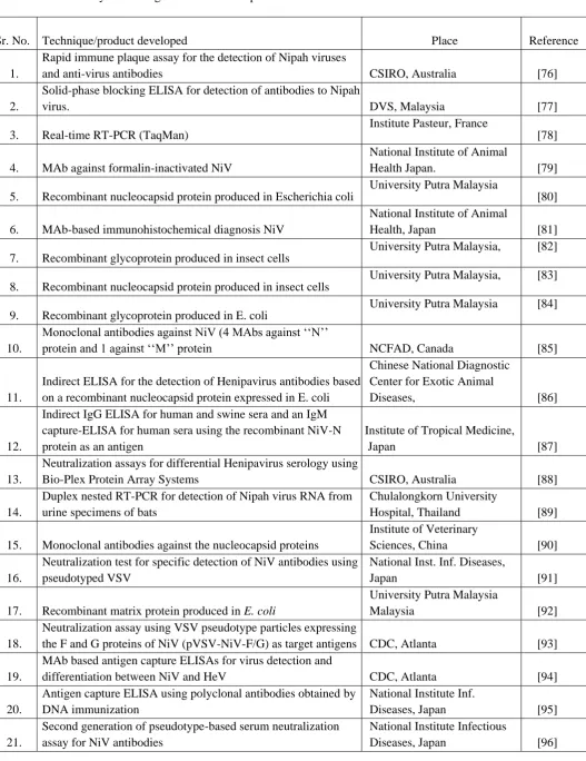
Table 2 Brief summary of lab diagnostic tests developed in various laboratories
Sr. No. Technique/product developed Place Reference
1.
Rapid immune plaque assay for the detection of Nipah viruses
and anti-virus antibodies CSIRO, Australia [76]
2.
Solid-phase blocking ELISA for detection of antibodies to Nipah
virus. DVS, Malaysia [77]
3. Real-time RT-PCR (TaqMan)
Institute Pasteur, France
[78]
4. MAb against formalin-inactivated NiV
National Institute of Animal
Health Japan. [79]
5. Recombinant nucleocapsid protein produced in Escherichia coli
University Putra Malaysia
[80]
6. MAb-based immunohistochemical diagnosis NiV
National Institute of Animal
Health, Japan [81]
7. Recombinant glycoprotein produced in insect cells
University Putra Malaysia, [82]
8. Recombinant nucleocapsid protein produced in insect cells
University Putra Malaysia, [83]
9. Recombinant glycoprotein produced in E. coli
University Putra Malaysia [84]
10.
Monoclonal antibodies against NiV (4 MAbs against „„N‟‟
protein and 1 against „„M‟‟ protein NCFAD, Canada [85]
11.
Indirect ELISA for the detection of Henipavirus antibodies based on a recombinant nucleocapsid protein expressed in E. coli
Chinese National Diagnostic Center for Exotic Animal
Diseases, [86]
12.
Indirect IgG ELISA for human and swine sera and an IgM capture-ELISA for human sera using the recombinant NiV-N protein as an antigen
Institute of Tropical Medicine,
Japan [87]
13.
Neutralization assays for differential Henipavirus serology using
Bio-Plex Protein Array Systems CSIRO, Australia [88]
14.
Duplex nested RT-PCR for detection of Nipah virus RNA from urine specimens of bats
Chulalongkorn University
Hospital, Thailand [89]
15. Monoclonal antibodies against the nucleocapsid proteins
Institute of Veterinary
Sciences, China [90]
16.
Neutralization test for specific detection of NiV antibodies using pseudotyped VSV
National Inst. Inf. Diseases,
Japan [91]
17. Recombinant matrix protein produced in E. coli
University Putra Malaysia
Malaysia [92]
18.
Neutralization assay using VSV pseudotype particles expressing
the F and G proteins of NiV (pVSV-NiV-F/G) as target antigens CDC, Atlanta [93]
19.
MAb based antigen capture ELISAs for virus detection and
differentiation between NiV and HeV CDC, Atlanta [94]
20.
Antigen capture ELISA using polyclonal antibodies obtained by DNA immunization
National Institute Inf.
Diseases, Japan [95]
21.
Second generation of pseudotype-based serum neutralization assay for NiV antibodies
National Institute Infectious
Int.J.Adv.Microbiol.Health.Res.2018; 2(2):40-55
51
CONCLUSION
Nipah virus (NiV) causes a recently discovered zoonotic disease endemic in South Asia, where sporadic outbreaks have been reported in Malaysia, Singapore, India, and Bangladesh. NiV infection in humans causes a range of clinical presentations, from asymptomatic infection (subclinical) to acute respiratory infection and fatal encephalitis. The case fatality rate is estimated at 40% to 75%. This rate can vary by outbreak depending on local capabilities for epidemiological surveillance and clinical management. NiV can be transmitted to humans from animals (i.e. bats or pigs), or contaminated foods and can also be transmitted directly from human-to-human. Fruit bats of the Pteropodidae family are the natural host of NiV. At present there is no antiviral drug available for NiV disease and the treatment is just supportive. There is no vaccine available for either people or animals. The primary treatment for humans is supportive care. The 2018 review of the WHO list of Blueprint priority diseases indicates that there is an urgent need for accelerated research and development for the Nipah virus.
REFERENCES
1. Angeletti S, Presti AL, Cella E, Ciccozzi M. Molecular epidemiology and phylogeny of Nipah virus infection: A mini review. Asian Pacific Journal of Tropical Medicine 2016; 9(7): 630–634.
2. Halpin K, Hyatt AD, Fogarty R, Middleton D, Bingham J, Epstein JH, et al. Pteropid bats are confirmed as the reservoir hosts of henipaviruses: a comprehensive experimental study of virus transmission. Am J Trop Med Hyg 2011; 85: 946-951.
3. Wang L, Harcourt BH, Yu M, Tamin A, Rota PA, Bellini WJ, et al. Molecular biology of Hendra and Nipah viruses. Microbes Infect 2001; 3: 279-287.
4. Lee KE, Umapathi T, Tan CB, Tjia HT, Chua TS, Oh HM, et al. The neurological manifestations of Nipah virus encephalitis, a novel paramyxovirus. Ann Neurol 1999; 46: 428-432.
5. Centers for disease control and prevention. Update: outbreak of Nipah virus-Malaysia and Singapore, 1999. MMWR Morb Mortal Wkly Rep 1999; 48: 335-337.
6. Centers for disease control and prevention. Outbreak of Hendralike virus-Malaysia and Singapore, 1998–1999. MMWR Morb Mortal Wkly Rep 1999; 48: 265-269.
7. Broder CC, Xu Kai, Nikolov DB, Zhu Z, Dimitrov DS, Middleton D, Pallister J, Geisbert TW, Bossart KN, Wang LF. A treatment for and vaccine against the deadly Hendra and Nipah viruses. Antiviral Research, 2013; 100(1): 8-13.
8. Hsu VP, Hossain MJ, Parashar UD, Ali MM, Ksiazek TG, Kuzmin I, et al. Nipah virus encephalitis reemergence, Bangladesh. Emerg Infect Dis 2004; 10: 2082-2087.
9. Nipah encephalitis outbreak over wide area of western Bangladesh, 2004. Health Sci Bull 2004; 2: 7-11.
10.Nipah virus outbreak from date palm juice. Health Sci Bull 2005; 3: 1-5.
11.Anonymous. Nipah virus, fatal-India (West Bengal). Pro-med Int Soc Infect Dis 2007. 12.Anonymous. Nipah virus, fatal-Bangladesh.
Pro-med Int Soc InfectDis 2008. ProMED archive number: 20130128.1518442 18.
13.Harit AK, Ichhpujani RL, Gupta S, Gill KS, Lal S, Ganguly NK, et al. Nipah/Hendra virus outbreak in Siliguri, West Bengal, India in 2001. Indian J Med Res 2006; 123: 553-560. 14.Chua KB, Bellini WJ, Rota PA, Harcourt BH,
Tamin A, Lam SK, et al. Nipah virus: a recently emergent deadly paramyxovirus. Science 2000; 288: 1432-1435.
15.Epstein JH, Rahman S, Zambriski J, Halpin K, Meehan G, Jamaluddin AA, et al. Feral cats and risk for Nipah virus transmission. Emerg Infect Dis 2006; 12: 1178-1179.
16.Epstein JH, Field HE, Luby S, Pulliam JR, Daszak P. Nipah virus: impact, origins, and causes of emergence. Curr Infect Dis Rep 2006; 8(1): 59-65.
17.Luby SP, Rahman M, Hossain MJ, Blum LS, Husain MM, Gurley E, et al. Food borne transmission of Nipah virus, Bangladesh. Emerg Infect Dis 2006; 12: 1888-1894.
18.Luby SP, Gurley ES, Hossain MJ. Transmission of human infection with Nipah virus. Clin Infect Dis 2009; 49: 1743-1748. 19.Chadha MS, Comer JA, Lowe L, Rota PA,
Rollin PE, Bellini WJ, et al. Nipah virus-associated encephalitis outbreak, Siliguri, India. Emerg Infect Dis 2006; 12: 235-240. 20.Gurley ES, Montgomery JM, Hossain MJ, Bell
Int.J.Adv.Microbiol.Health.Res.2018; 2(2):40-55
52 person transmission of Nipah virus in a Bangladeshi community. Emerg Infect Dis 2007; 13: 1031-1037.
21.Eaton BT, Broder CC, Middleton D and Wang LF (2006) Hendra and Nipah viruses: different and dangerous. Nat Rev Microbiol 4, 23–35. 22.Islam MS, Sazzad HMS, Satter SM, Sultana S,
Hossain MJ, Hasan M, Rahman M, Campbell S, Cannon DL, Str€oher U et al. (2016) Nipah virus transmission from bats to humans associated with drinking traditional liquor made from date palm sap, Bangladesh, 2011– 2014. Emerg Infect Dis 22, 664–670.
23.Wong KT, Shieh W-J, Kumar S, Norain K, Abdullah W, Guarner J, Goldsmith CS, Chua KB, Lam SK, Tan CT et al. (2002) Nipah virus infection. Am J Pathol 161, 2153–2167.
24.De Wit E and Munster VJ (2015) Animal models of disease shed light on Nipah virus pathogenesis and transmission. J Pathol 235, 196–205.
25.Rahman MA, Hossain MJ, Sultana S, Homaira N, Khan SU, et al. (2011) Date Palm Sap Linked to Nipah Virus Outbreak in Bangladesh, 2008. Vector Borne Zoonotic Dis. 26.Luby SP (2013) The pandemic potential of
Nipah virus. Antiviral Res 100, 38–43.
27.Homaira N, Rahman M, Hossain MJ, Epstein JH, Sultana R, Khan MSU, Podder G, Nahar K, Ahmed B, Gurley ES et al. (2010) Nipah virus outbreak with person-to-person transmission in a district of Bangladesh, 2007. Epidemiol Infect 138, 1630–1636.
28.Sazzad HMS, Hossain MJ, Gurley ES, Ameen KMH, Parveen S, Islam MS, Faruque LI, Podder G, Banu SS, Lo MK et al. (2013) Nipah virus infection outbreak with nosocomial and corpse-to-human transmission, Bangladesh. Emerg Infect Dis 19, 210–217. 29.Luby SP, Hossain MJ, Gurley ES, Ahmed BN,
Banu S, et al. (2009) Recurrent zoonotic transmission of Nipah virus into humans, Bangladesh, 2001–2007. Emerg Infect Dis 15: 1229–1235.
30.Wolf M C, Negrete OA, Lee B. (2007). Pathobiology of henipavirus entry: insights into therapeutic strategies.
31.Nine days after detecting Nipah, Kerala is yet to nail the carrier of the deadly virus [Accessed
on 29 May 2018],
https://scroll.in/article/880626/nine-days-after- detecting-nipah-kerala-is-yet-to-nail-the-carrier-of-the-deadly-virus.
32.Nipah Virus: Outbreak of Nipah virus encephalitis in Kerala state of India. [Accessed
on 29 May 2018],
http://www.searo.who.int/entity/emerging_dise ases/links/nipah_virus/en/.
33.Halpin K, Young PL, Field HE, Mackenzie JS. Isolation of Hendra virus from pteropid bats: a natural reservoir of Hendra virus. J GenVirol 2000; 81: 1927-1932.
34.Leroy EM, Kumulungui B, Pourrut X, Rouquet P, Hassanin A, Yaba P, et al. Fruit bats as reservoirs of Ebola virus. Nature 2005; 438: 575-576.
35.Li W, Shi Z, Yu M, Ren W, Smith C, Epstein JH, et al. Bats are natural reservoirs of SARS-like coronaviruses. Science 2005; 310:676-679.
36.Towner JS, Amman BR, Sealy TK, Carroll SA, Comer JA, Kemp A, et al. Isolation of genetically diverse Marburg viruses from Egyptian fruit bats. PLoS Pathog 2009; 5: e1000536.
37.Eason BT, Broder CC, Middleton D, Wang LF. Hendra and Nipah viruses: Different and dangerous. Nat Rev Microbiol 2006;4:23-35. 38.Olivae KJ, Daszak P. The ecology of emerging
neurotropic viruses. J Neurovirol 2005;11:441-6.
39.Chua KB. Nipah virus outbreak in Malaysia. J Clin Virol 2003;26:265-75.
40.Rogers RJ, Douglas IC, Baldock FC, Glanville RJ, Seppanen KT, Gleeson LJ, et al . Investigation of a second focus of equine morbillivirus infection in coastal Queensland. Aust Vet J 1996;74:243-4.
41.Murray K, Selleck P, Hooper P, Hyatt A, Gould A, Gleeson L, et al . A morbillivirus that caused fatal disease in horses and humans. Science 1995;268:94-7.
42.Selvey LA, Wells RM, McCormack JG, Ansford AJ, Murray K, Rogers RJ, et al . Infection of humans and horses by a newly described morbillivirus. Med J Aus 1995;162:642-5.
43.O'Sullivan JD, Allworth AM, Paterson DL, Snow TM, Boots R, Gleeson LJ, et al . Fatal encephalitis due to novel paramyxovirus transmitted from horses. Lancet 1997;349:93-5.
Int.J.Adv.Microbiol.Health.Res.2018; 2(2):40-55
53 morbillivirus infection. Aus Vet J 1996;74:244-5.
45.Young PL, Halpin K, Selleck PW, Field HE, Gravel JL, Kelly MA, et al . Serologic evidence for the presence in Pteropus bats of a paramyxovirus related to equine morbillivirus. Emerg Infect Dis 1996;2:239-40.
46.Mohd Nor MN, Gan CH, Ong BL. Nipah virus infection of pigs in peninsular Malaysia. Rev Sci Tech 2000;19:160-5.
47.Chua KB, Goh KJ, Wong KT, Kamarulzaman A, Tan PS, Ksiazek TG, et al . Fatal encephalitis due to Nipah virus among pig-farmers in Malaysia. Lancet 1999;354:1257-9. 48.Halpin K, Bankamp B, Harcourt BH, Bellini
WJ, Rota PA. Nipah virus conforms to the rule of six in a minigenome repliction assay. J Gen Virol 2004;85:701-7.
49.Chua KB, Bellini WJ, Rota PA, Harcourt BH, Tamin A, Lam SK, et al . Nipah virus: A recently emergent deadly paramyxovirus. Science 2000;288:1432-5.
50.Field HE, Barratt PC, Hughes RJ, Shield J, Sulivan ND. A fatal case of Hendra virus infection in a horse in north Queensland. Clinical and epidemiological features. Aust Vet J 2000;78:279-80.
51.Field H, Young P, Yob JM, Mills J, Hall L, Mackenzie J. The natural history of Hendra and Nipah viruses. Microbes Infect 2001;3:307-14.
52.Yob JM, Field H, Rashdi AM, Morrissy C, van der Heide B, Rota P, et al . Nipah virus infection in bats (order Chiroptera) in Peninsular Malaysia. Emerg Infect Dis 2001;7:439-41.
53.Wong KT, Shieh WJ, Kumar S, Norain K, Abdullah W, Guarner J, et al . Nipah virus infection: Pathology and pathogenesis of an emerging paramyxoviral zoonosis. Am J Pathol 2002;161:2153-67.
54.Anonymous. Outbreak of viral encephalitis due to Nipah/Hendra like viruses. Western Bangladesh. Health Sci Bult 2003;1:1-6. 55.Hsu VP, Hossain MJ, Parashar UD, Ali MM,
et al . Nipah virus encephalitis reemergence. Bangladesh. Emerg Infect Dis 2004;12:2082-7. 56.Chua KB, Koh CL, Hooi PS, Wee KF, et al . Isolation of Nipah virus from Malaysian Island flying foxes. Mocrobes Infect 2002;4:145-51. 57.Anonymous. Hendra virus - Australia
(Queensland). ProMED Archive Number 2004;1214:3307.
58.Anonymous. Nipah virus encephalitis outbreak over wide area of western Bangladesh. Health Sci Bult 2004;2:7-11.
59.Anonymous. Person to person tramsmission of Nipah virus during outbreak in Faridpur district. Health Sci Bult 2004;2:5-9.
60.Anonymous. Nipah virus - Bangladesh (Tangail) ProMED Archive Number 2005;0211:0468.
61.Paton NI, Leo YS, Zaki SR, Auchus AP, Lee KE, Ling AE, et al . Outbreak of Nipah virus infection among abattoir workers in Singapore. Lancet 1999;354:1253-6.
62.Goh KJ, Tan CT, Chew NK, Tan PS, Kamarulzaman A, Sarji SA, et al . Clinical features of Nipah virus encephalitis among pig farmers in Malaysia. N Eng J Med 2000;342:1229-35.
63.Tan CT, Viral encephalitis. Lecture delivered at world Congress of Neurology. Sydney, Australia: 2005.
64.Chadha MS, Comer JA, Lowe L, Rota PA, Rollin PE, Bellini WJ, et al . Nipah virus - Associated encephalitis outbreak, Siliguri, India. Emerg Infect Dis 2006;12:235-40. 65.Halder NR, Dasgupta K, Khandelwal AK,
Halder N, Banerjee S, et al . First epidemic of viral encephalitis in 2001 of highly infections nature with feeding mortality at Siliguri (abstract). J Neurol Sci 2005;S121.
66.Halder A, Chakravarty A. Nipah virus encephalitis: A cause for concern for Indian neurologists?.Ann Indian Acad Neurol 2006;9:137-144.
67.Morbidity and mortality due to Nipah or Nipah-like virus encephalitis in WHO South-East Asia Region, 2001-2018 [Accessed on 29
May 2018],
http://www.searo.who.int/entity/emerging_dise ases/links/morbidity-and-mortality-nipah-sear-2001-2018.pdf?ua=1.
68.Nipah virus: transmission, treatment, World Health Organization (WHO) [Accessed on 29 May 2018], http://www.who.int/news-room/fact-sheets/detail/nipah-virus
69.Nipah Virus (NiV): Transmission, Centers for Disease Control and Prevention [Accessed on
29 May 2018],
https://www.cdc.gov/vhf/nipah/transmission/in dex.html
Int.J.Adv.Microbiol.Health.Res.2018; 2(2):40-55
54 es/panache/throbbing-headache-nausea-facts-
symptoms-prevention-of-nipah-virus/articleshow/64285621.cms
71.Nipah virus: signs and symptoms, World Health Organization (WHO) [Accessed on 29 May 2018], http://www.who.int/news-room/fact-sheets/detail/nipah-virus
72.Daniels P, Ksiazek TG, Eaton BT. Laboratory diagnosis of Nipah virus and Hendra infections. Microbes Infect., 3. 2001:289-295. 73.Watkinson RE and Lee B. Nipah virus matrix
protein: expert hacker of cellular machines. Federation of European Biochemical Societies, Letters 2016; 590:2494–2511.
74.Danielsa P, Ksiazekb T, Eatona BT. Laboratory diagnosis of Nipah and Hendra virus infections. Microbes and Infection, 2001; 3:289−295.
75.Nipah virus: OIE - World Organisation for Animal Health. [Accessed on 29 May 2018], http://www.oie.int/en/animal-health-in-the-world/animal-diseases/Nipah-Virus/
76.Crameri G, Wang LF, Morrissy C, White J, Eaton BT. A rapid immune plaque assay for the detection of Hendra and Nipah viruses and anti-virus antibodies. J Virol Methods. 2002;99(1–2):41–51.
77.Kashiwazaki Y, Na YN, Tanimura N, Imada T. A solid-phase blocking ELISA for detection of antibodies to Nipah virus. J Virol Methods. 2004;121(2):259–61.
78.Guillaume V, Lefeuvre A, Faure C, Marianneau P, Buckland R, Lam SK, Wild TF, Deubel V. Specific detection of Nipah virus using real-time RT-PCR (TaqMan). J Virol Methods. 2004;120(2):229–37.
79.Imada T, Abdul Rahman MA, Kashiwazaki Y, Tanimura N, Syed Hassan S, Jamaluddin A. Production and characterization of monoclonal antibodies against formalin-inactivated Nipah virus isolated from the lungs of a pig. J Vet Med Sci. 2004;66(1):81–3.
80.Tan WS, Ong ST, Eshaghi M, Foo SS, Yusoff K. Solubility, immunogenicity and physical properties of the nucleocapsid protein of Nipah virus produced in Escherichia coli. J Med Virol. 2004;73(1):105–12.
81.Tanimura N, Imada T, Kashiwazaki Y, Shahirudin S, Sharifah SH, Aziz AJ.
Monoclonal antibody-based
immunohistochemical diagnosis of Malaysian Nipah virus infection in pigs. J Comp Pathol. 2004;131(2–3):199–206.
82.Eshaghi M, Tan WS, Mohidin TB, Yusoff K. Nipah virus gly-coprotein: production in baculovirus and application in diag-nosis. Virus Res. 2004;106(1):71–6.
83.Eshaghi M, Tan WS, Ong ST, Yusoff K. Purification and characterization of Nipah virus nucleocapsid protein produced in insect cells. J Clin Microbiol. 2005;43(7):3172–7. 84.Eshaghi M, Tan WS, Chin WK, Yusoff K.
Purification of the extra-cellular domain of Nipah virus glycoprotein produced in Escherichia coli and possible application in diagnosis. J Bio-technol. 2005;116(3):221–6. 85.Berhane Y, Berry JD, Ranadheera C, Marszal
P, Nicolas B, Yuan X, Czub M, Weingartl H. Production and characterization of monoclonal antibodies against binary ethylenimine inacti-vated Nipah virus. J Virol Methods. 2006;132(1–2):59–68.
86.Chen JM, Yu M, Morrissy C, Zhao YG, Meehan G, Sun YX, Wang QH, Zhang W, Wang LF, Wang ZL. A comparative indirect ELISA for the detection of Henipavirus antibodies based on a recombinant nucleocapsid protein expressed in Escherichia coli. J Virol Methods. 2006;136(1–2):273–6. 87.Yu F, Khairullah NS, Inoue S,
Balasubramaniam V, Berendam SJ, Teh LK, Ibrahim NS, Abdul Rahman S, Hassan SS, Hasebe F, Sinniah M, Morita K. Serodiagnosis using recombinant Nipah virus nucleocapsid protein expressed in Escherichia coli. J Clin Microbiol. 2006;44:3134–8.
88.Bossart KN, McEachern JA, Hickey AC, Choudhry V, Dimitrov DS, Eaton BT, Wang LF. Neutralization assays for differential Henipavirus serology using Bio-Plex protein array systems. J Virol Methods. 2007;142(1– 2):29–40.
89.Wacharapluesadee S, Hemachudha T. Duplex nested RT-PCR for detection of Nipah virus RNA from urine specimens of bats. J Virol Methods. 2007;141(1):97–101.
90.Xiao C, Liu Y, Jiang Y, Magoffin DE, Guo H, Xuan H, Wang G, Wang LF, Tu C. Monoclonal antibodies against the nucleo-capsid proteins of Henipaviruses: production, epitope mapping and application in immunohistochemistry. Arch Virol. 2008;153(2):273–81.
Int.J.Adv.Microbiol.Health.Res.2018; 2(2):40-55
55 Wang LF. A neutralization test for specific detection of Nipah virus antibodies using pseudotyped vesicular stomatitis virus expressing green fluorescent protein. J Virol Methods. 2009;160(1–2):7–13.
92.Subramanian SK, Tey BT, Hamid M, Tan WS. Production of the matrix protein of Nipah virus in Escherichia coli: virus-like particles and possible application for diagnosis. J Virol Methods. 2009;162(1–2):179–83.
93.Tamin A, Harcourt BH, Lo MK, Roth JA, Wolf MC, Lee B, Weingartl H, Audonnet JC, Bellini WJ, Rota PA. Development of a neutralization assay for Nipah virus using pseudotype par-ticles. J Virol Methods. 2009;160(1–2):1–6.
94.Chiang CF, Lo MK, Rota PA, Spiropoulou CF, Rollin PE. Use of monoclonal antibodies
against Hendra and Nipah viruses in an antigen capture-ELISA. Virol J. 2010;7:115.
95.Kaku Y, Noguchi A, Marsh GA, Barr JA, Okutani A, Hotta K, Bazartseren B, Broder CC, Yamada A, Inoue S, Wang LF. Antigens capture ELISA system for Henipaviruses using poly-clonal antibodies obtained by DNA immunization. Arch Virol. 2012;157(8):1605– 9.
96.Kaku Y, Noguchi A, Marsh GA, Barr JA, Okutani A, Hotta K, Bazartseren B, Fukushi S, Broder CC, Yamada A, Inoue S, Wang LF. Second generation of pseudotype-based serum neu-tralization assay for Nipah virus antibodies: sensitive and high-throughput analysis utilizing secreted alkaline phosphatase. J Virol Methods. 2012;179(1):226–32.
Corresponding Author: Dr. Gurjeet Singh Assistant Professor, Department of Microbiology, N.C. Medical College and Hospital, Israna, Panipat-132107, Haryana, Indian
E-mail: [email protected]
How to cite this article:
Singh G, Raksha, Urhekar AD. Nipah: A killer virus. Int. J. Adv.Microbiol.Health.Res., 2018; 2(2):40-55.