University of Pennsylvania
ScholarlyCommons
Publicly Accessible Penn Dissertations
Fall 12-22-2009
Cellular and Molecular Analyses of Neural and
Synaptic Development in Zebrafish
Yuanquan Song
University of Pennsylvania, [email protected]
Follow this and additional works at:http://repository.upenn.edu/edissertations Part of theDevelopmental Neuroscience Commons, and theMolecular and Cellular Neuroscience Commons
This paper is posted at ScholarlyCommons.http://repository.upenn.edu/edissertations/297 For more information, please [email protected].
Recommended Citation
Song, Yuanquan, "Cellular and Molecular Analyses of Neural and Synaptic Development in Zebrafish" (2009).Publicly Accessible Penn Dissertations. 297.
Cellular and Molecular Analyses of Neural and Synaptic Development in
Zebrafish
Abstract
Proper function of the nervous system requires the precise wiring of neuronal circuitry, which is established during development via mechanisms that guide cells to establish a correct identity, direct axons to navigate to and make synaptic connections with appropriate targets, and establish and maintain the function of synaptic circuits. Dysfunction of genes implicated in one or more of these processes has been linked to human neurological disorders with behavioral and cognitive manifestations. However, our understanding of how specific gene defects affect circuitry formation, function and in turn behavior remains fragmentary. I have used zebrafish as a genetic model system to begin to address some aspects of this central question. I performed in vivo imaging and cellular analyses of the formation of a peripheral neural circuit, synapses between motor neurons and muscle fibers, called neuromuscular junctions. I then characterized two zebrafish mutants, identified from a small-scale genetic screen for neuromuscular synaptic defects, slytherin (srn) and xavier (xav). Analyses of srn uncovered a previously underappreciated role for protein fucosylation in several aspects of neural development. Analyses of xav suggest critical roles for mitochondria during neural
development. Given that the corresponding mutations in humans result in disorders with poorly explored neural defects, the molecular and cellular characterization of these mutants may shed light on our
understanding of the neural and synaptic phenotypes in human patients.
Degree Type Dissertation
Degree Name
Doctor of Philosophy (PhD)
Graduate Group Neuroscience
First Advisor
Rita J. Balice-Gordon
Keywords
zebrafish, synaptogenesis, fucosylation, notch, etfdh, madd
Subject Categories
Developmental Neuroscience | Molecular and Cellular Neuroscience
CELLULAR AND MOLECULAR ANALYSES OF NEURAL AND SYNAPTIC
DEVELOPMENT IN ZEBRAFISH
Yuanquan Song
A DISSERTATION
in
Neuroscience
Presented to the Faculties of the University of Pennsylvania
in
Partial Fulfillment of the Requirements for the
Degree of Doctor of Philosophy
2009
Supervisor (or co-Supervisors) of Dissertation
________________________
Rita J. Balice-Gordon, Ph.D.
Graduate Group Chairperson
________________________
Rita J. Balice-Gordon, Ph.D.
Dissertation Committee (typed names and title; no signatures)
Steven S. Scherer, M.D., Ph.D.
Greg Bashaw, Ph.D.
Jonathan A. Raper, Ph.D.
ii
ABSTRACT
CELLULAR AND MOLECULAR ANALYSES OF NEURAL AND
SYNAPTIC DEVELOPMENT IN ZEBRAFISH
Yuanquan Song
Thesis advisor: Rita J. Balice-Gordon
Proper function of the nervous system requires the precise wiring of neuronal
circuitry, which is established during development via mechanisms that guide cells to
establish a correct identity, direct axons to navigate to and make synaptic connections
with appropriate targets, and establish and maintain the function of synaptic circuits.
Dysfunction of genes implicated in one or more of these processes has been linked to
human neurological disorders with behavioral and cognitive manifestations. However,
our understanding of how specific gene defects affect circuitry formation, function and in
turn behavior remains fragmentary.
I have used zebrafish as a genetic model system to begin to address some aspects
of this central question. I performed in vivo imaging and cellular analyses of the
formation of a peripheral neural circuit, synapses between motor neurons and muscle
fibers, called neuromuscular junctions. I then characterized two zebrafish mutants,
identified from a small-scale genetic screen for neuromuscular synaptic defects, slytherin
(srn) and xavier (xav). Analyses of srn uncovered a previously underappreciated role for
iii
critical roles for mitochondria during neural development. Given that the corresponding
mutations in humans result in disorders with poorly explored neural defects, the
molecular and cellular characterization of these mutants may shed light on our
iv
TABLE OF CONTENTS
Abstract ii
Table of Contents iv
List of Figures vi
List of Tables ix
Chapter 1: Introduction 1
References 14
Chapter 2: In vivo imaging of preferential motor axon outgrowth to and synaptogenesis at prepatterned acetylcholine receptor clusters in embryonic zebrafish skeletal muscle
Abstract 22
Introduction 23
Materials and Methods 26
Results 34
Discussion 45
References 53
Figures and Legends 61
Chapter 3: Notch-dependent and -independent mechanisms underlie neural and synaptic defects in slytherin, a zebrafish model for human congenital disorders of glycosylation
Abstract 89
Introduction 90
Materials and Methods 93
Results 97
Discussion 108
v
Figures and Legends 119
Supplemental Methods, Results and Figures 140
Chapter 4: Mutations in electron transfer flavoprotein (ETF) and electron transfer flavoprotein dehydrogenase (ETFDH), cause fatty acid metabolism and mitochondrial dysfunction, unbalanced oxidative phosphorylation and glycolysis, and lead to severe neural defects in zebrafish and humans
Abstract 158
Introduction 159
Materials and Methods 162
Results 170
Discussion 183
References 186
Figures and Legentds 193
Supplemental Results and Figures 208
Chapter 5: General Conclusions and Future Directions 236
vi
LIST OF FIGURES
Chapter 2:
Fig. 1 Location of prepatterned AChR clusters in myotomal muscle. 61
Fig. 2 Dynamics of prepatterned AChR clusters. 63
Fig. 3 Motor axon growth cones preferentially extend toward prepatterned AChR
clusters. 67
Fig. 4 Motor axon filopodia preferentially extend toward and contact prepatterned AChR
clusters. 70
Fig. 5 Filopodia are preferentially extended from synapses. 73
Fig. 6 Postsynaptic AChR clusters precede presynaptic vesicle clusters during initial
neuromuscular synaptogenesis. 75
Fig. 7 Insertion of new AChRs and redistribution of prepatterned AChRs during initial
neuromuscular synaptogenesis. 78
Fig. 8. AChR activity or AChR clusters are not required for motor axon outgrowth or
neuromuscular synaptogenesis. 80
Supplemental Fig. 1 VAMP-GFP marks sites of presynaptic vesicle clusters and does not
alter motor axon outgrowth. 83
Supplemental Fig. 2 Blockade of sodium channels with tricaine does not affect dispersal
of prepatterned AChR clusters or initial motor axon outgrowth. 85
Chapter 3:
Figure 1. Slytherin external phenotype, genotype, cloning and mRNA rescue of srn
mutants. 119
Figure 2. slytherin mutants exhibit reduced protein fucosylation as measured by AAL
staining. 122
Figure 3. Supplementation with exogenous GDP-fucose rescues srn external phenotypes
and restores AAL staining. 124
Figure 4. Reduction in Notch-Delta signaling accounts for some srn phenotypes. 126
Figure 5. mib and DAPT treatment exclude srn phenotypes. 129
Figure 6. NICD rescues srn neuro- and gliogenesis phenotypes. 131
Figure 7. srn mutants showed aberrant expression of Notch responsive genes similar to
vii
Figure 8. Slytherin mutants exhibit defects in neuromuscular synaptogenesis due in part
to reduction in Notch-Delta signaling. 135
Figure 9. Slytherin mutants exhibit defects in axon branching and CNS synaptic
connectivity that are independent of Notch-Delta signaling. 137
Supplemental Figure 1. Gmds mRNA localization by in situ hybridization in wild type
zebrafish embryos from 12 to 72 hpf. 143
Supplemental Figure 2. Modeling of zebrafish GMDS protein structure. 145
Supplemental Figure 3. GDP-fucose rescue of srn and morpholino knockdown of gmds. 147 Supplemental Figure 4. Secondary motor neuron number is reduced in mib but not srn,
des or dla compared to wild type embryos. 150
Supplemental Figure 5. Reduction in Notch-Delta signaling accounts for some srn
phenotypes in the retina. 152
Supplemental Figure 6. Muscle patterning is grossly normal in srn mutants. 154
Chapter 4:
Figure 1. xavier external phenotype, genotype, cloning and morpholino phenocopy of xav
mutants 193
Figure 2. xavier mutants display abnormal acylcarnitine and organic acid profile 195
Figure 3. xav mutants exhibit mitochondrial dysfunction 197
Figure 4. Human MADD fibroblast cells display similar mitochondrial defects as xav 200
Figure 5. Increased aerobic glycolysis in MADD fibroblasts and xav 202
Figure 6. xav exhibits increased neural cell proliferation as a result of increased
glycolysis, due to perturbation of the PPARG-ERK pathway 205
Supplemental Figure 1. Genetic map of the xav locus 215
Supplemental Figure 2. Nonsense mediated decay and nonsense mediated alternative
splicing of etfdh transcript in xav, and morpholino knock down of etfdh 217
Supplemental Figure 3. xav mutants display polycystic kidney like phenotypes. 219
Supplemental Figure 4. xav mutants exhibit respiratory deficiency 221
Supplemental Figure 5. xav mutants exhibit neural and glial defects and cell death 223 Supplemental Figure 6. xav mutants exhibit reduced motor axon branching and
neuromuscular synaptogenesis that are not caused by change of motor neuron number or
viii
Supplemental Figure 7. xav mutants display electrophysiological properties in the
muscle, comparable to WT 227
Supplemental Figure 8. xav mutants exhibit aberrant mitochondria distribution in motor
neurons 229
Supplemental Figure 9. xav mutants exhibit cell death throughout the nervous system that is rescued by p53 morpholino knockdown and does not account for the motor axon
ix
LIST OF TABLES
Chapter 3:
Supplemental Table 1. Primers used for qRT-PCR. 142
Chapter 4:
Supplemental Table 1. Primer sequences for new zebrafish simple sequence repeat
(SSR) markers 233
Supplemental Table 2. Primers for qRT-PCR analyses of gene expression in xav and
1 Chapter 1
Introduction
This chapter is published in
International Anesthesiology Clinics:
Spring 2006 - Volume 44 - Issue 2 - pp 145-178
Formation and Plasticity of Neuromuscular Synaptic Connections
Song, Yuanquan BS; Panzer, Jessica A. MD, PhD; Wyatt, Ryan M. BS; Balice-Gordon,
Rita J. PhD
The nervous system becomes wired into circuits that are fine tuned to subserve
particular functions and behaviors through a series of events including neurogenesis and
differentiation; cell migration, axon guidance and synapse formation; synaptic pruning
and circuit maturation and maintenance. Perturbation of any of these steps results in
neurodevelopmental, cognitive or behavioral abnormalities, as well as neurological and
psychiatric disorders. My thesis project utilized zebrafish as a model organism to
characterize some of the cellular and molecular mechanisms underlying synapse
formation, and also revealed several mechanisms that impact other aspects of neural
development. Here I review our current understanding of mechanisms underlying
2
The specificity of synaptic connections that is essential for nervous system
function arises during development through a series of events, including axon outgrowth
and guidance, target selection, synaptogenesis and synapse elimination. Neuromuscular
synapses between spinal motor neurons and skeletal muscle fibers have become one of
the most widely used model systems to study these events due to its relatively large size,
accessibility and the wealth of molecular and functional information about their
formation, maintenance and plasticity (Goda and Davis, 2003; Sanes and Lichtman,
1999).
Neuromuscular synapses are established through complex multi-directional
signaling among presynaptic motor neurons, postsynaptic muscle fibers (Burden, 2002;
Luo et al., 2003; Sanes and Lichtman, 2001) and perisynaptic glia (Koenig et al., 1998).
These synapses consists of specialized regions of the presynaptic membrane, termed
active zones, at which clustered synaptic vesicles fuse and release the neurotransmitter
acetylcholine (ACh), as well as peptides such as calcitonin gene related peptide (CGRP)
and other signaling molecules. Directly apposed to the presynaptic active zones are
acetylcholine receptors (AChRs) which cluster at the crests of junctional folds in the
muscle fiber membrane. These AChRs bind ACh that has diffused across the synaptic
cleft, leading to subsequent depolarization, which, if above threshold, leads to contraction
of the muscle fiber (Sanes and Lichtman, 1999). The temporal and spatial extent of ACh
signaling is regulated by acetycholinesterase (AChE), which is located within the basal
lamina that invaginates the synaptic cleft, and cleaves ACh, thereby terminating the
3
Schwann cells, specialized glia that have been found to modulate synaptic structure,
activity, and response to injury (Koenig et al., 1998; Son et al., 1996).
The role of the Agrin – MuSK – Lrp4 –Rapsyn pathway in neuromuscular synaptogenesis
Neuromuscular synaptic function depends critically on the precise spatial
apposition of presynaptic motor neuron acetylcholine release sites with high-density
clusters of AChRs in the postsynaptic muscle fiber membrane. During neuromuscular
synaptogenesis, AChRs are clustered before innervation, prepatterning a central muscle
region where synapses will later be established. Motor neuron signals refine the muscle
prepattern by clustering AChRs beneath terminals and dispersing uninnervated clusters so
that AChRs become localized to, and are stably maintained, at nascent synapses. Over
the last 15 years, work from a number of groups has uncovered the basic signaling
mechanisms that underlie these events (reviewed in Sanes and Licthman 2001). Muscle
specific kinase (MuSK), a receptor tyrosine kinase expressed by postsynaptic muscle
fibers, is essential for the formation of aneural, prepatterned AChR clusters as well as for
the formation and maintenance of later, innervated AChR clusters (Lin et al., 2001; Yang
et al., 2001). The presynaptically released proteoglycan Agrin, that activates MuSK
signaling, is now more fully understood to be important as an anti-declustering, AChR
cluster maintenance factor. A role for the neurotransmitter ACh as a cluster dispersion
factor for non-innervated AChR clusters has also recently come to be appreciated
4
A third protein shown to be crucial for the clustering of AChRs is the cytoskeletal
linker protein rapsyn. Following activation by MuSK, Rapsyn, which binds both AChRs
and β-dystroglycan, clusters at synapses, resulting in the synaptic clustering of AChRs
(Gautam et al., 1995). Although AChR clustering is completely absent in Rapsyn mutant
mice (Gautam et al., 1995), many aspects of synaptic differentiation, including
concentration of MuSK at synapses and selective transcription of AChR genes by
synaptic nuclei, are unaffected (Apel et al., 1997; Gautam et al., 1995). Thus, although
necessary for neuromuscular synaptogenesis, it may be that Rapsyn plays a role in a
relatively late step in the Agrin-MuSK signaling cascade. The understanding of Rapsyn’s
role in directing synaptic AChRs clustering is further complicated by recent findings that
AChRs themselves are necessary to direct Rapsyn clusters to synapses (Grow and
Gordon, 2000; Huh and Fuhrer, 2002; Missias et al., 1997; Ono et al., 2001).
While a wealth of genetic evidence supports the Agrin-MuSK hypothesis,
evidence for a protein-protein interaction between Agrin and MuSK has been lacking.
Earlier work had proposed that an additional protein complex that directly bind Agrin and
was expressed specifically in muscle cells, called myotube-associated specificity
component (MASC), was required to constitute a fully functional receptor complex that
both binds and responds to Agrin (Glass et al., 1996). Recently mice lacking Lrp4
expression was reported to display neuromuscular synaptic defects strikingly similar to
those present in mice lacking MuSK expression (DeChiara et al., 1996), namely the
absence of postsynaptic AChR clusters, extensive aberrant presynaptic branching and
reduced formation of presynaptic terminals (Weatherbee et al., 2006). Prompted by this
5
long sought MASC, and that it binds, clusters and works in concert with Agrin to activate
MuSK (Kim et al., 2008; Zhang et al., 2008). These observations raise the question of
whether Lrp4 clustering precedes and in turn leads to MuSK clustering, priming
transcriptional mechanisms in sub-synaptic nuclei and protein-protein interactions that
lead to the formation of aneural AChR clusters and ultimately the formation of functional
neuromuscular synapses.
Intracellular signaling mechanisms downstream of MuSK activation
Although the roles of Agrin, MuSK, and Rapsyn in neuromuscular synaptogenesis
have been relatively well established, much less is known about the identity and role of
effectors downstream of MuSK activation. Recent work, however, has begun to fill in
some of these gaps (Luo et al., 2003). The activity of several muscle enzymes, including
Rho-family GTPases, NO synthetases (NOS), and geranylgeranyl-transferase I, has been
shown to increase in response to Agrin (Luo et al., 2002), and both NO and cGMP have
been implicated in the regulation of AChR clustering (Jones and Werle, 2000; Luck et al.,
2000). AChRs are tightly associated with cytoskeletal proteins, and since Rho GTPases
are well-known regulators of the cytoskeleton, they may play a role in translating MuSK
activation into the cytoskeletal reorganization required to cluster AChRs synaptically
(Luo et al., 2003). One Rho-family GTPase, Rac1 has been implicated in initial
induction of small AChR clusters by Agrin, and Rho itself appears to be required to then
6
Given MuSK’s importance in neuromuscular synapse formation, much attention
has focused on identifying its downstream interactors. Recent work has identified
Disheveled (Dvl) as a MuSK binding protein (Luo et al., 2002). Dvl was originally
identified in Drosophila as a molecule that is activated by the Wingless (Wnt) receptor
Frizzled (Dierick and Bejsovec, 1999). Interestingly, MuSK shares a conserved
extracellular domain with Wnt receptors (Saldanha et al., 1998). MuSK activation of Dvl
signaling may result in AChR clustering through the function of the Rho GTPases
discussed above, as it is known that Dvl activation results in activation of Rho GTPases
(Habas et al., 2001). In addition, Dvl is also known to inhibit phosphorylation of β
-catenin (Cadigan and Nusse, 1997), and β-catenin itself has been shown to bind Rapsyn
(Luo et al., 2003) and inhibit the clustering activity of Agrin (Zhang et al., 2001). These
data are consistent with a model in which Dvl signaling results in reduced binding of β
-catenin to Rapsyn, allowing Rapsyn to cluster AChRs (Luo et al., 2003), although this
model remains to be tested. Interestingly, Wnt signaling has been implicated in the
development of both CNS synapses (Hall et al., 2000) and of the glutamatergic
neuromuscular synapses in Drosophila. Thus, it may be that this evolutionarily
conserved pathway is involved in the formation of a diverse array of structurally and
functionally distinct synapses.
The role of Agrin – MuSK – Lrp4 signaling in presynaptic differentiation
In addition to lacking postsynaptic specializations, Agrin, MuSK and Lrp4 mutant
7
discrete endplate band, but instead branch extensively throughout the muscle. Thus,
Agrin, MuSK and Lrp4 signaling must induce a retrograde signal that instructs axons to
stop and undergo presynaptic differentiation (DeChiara et al., 1996; Gautam et al., 1996).
In vitro experiments have demonstrated that Agrin inhibits neurite outgrowth (Bixby et
al., 2002; Campagna et al., 1995; Chang et al., 1997; Halfter et al., 1997; Mantych and
Ferreira, 2001), and can initiate some aspects of presynaptic differentiation at sites of
neurite contact with Agrin expressing cells (Campagna et al., 1995). The current
understanding of the cellular and molecular events underlying presynaptic development,
however, is much less advanced than that of postsynaptic differentiation.
The role of prepatterned AChR clusters in neuromuscular synaptogenesis
The presence of prepatterned (aneural) AChR clusters was not well appreciated
until recently. That muscle might be patterned independent of motor innervation was
first strongly suggested in studies of the mouse topisomerase IIβ knockout (Yang et al.,
2000). In these mutants, although motor axons fail to branch within muscles, a band of
AChR clusters is observed within the central region of the muscle (Yang et al., 2000).
These AChR clusters are present in an area ca. two-fold wider than the normal endplate
zone (Yang et al., 2000). This finding has been confirmed in additional mouse mutants in
which motor nerves are disrupted (Lin et al., 2000) or fail to form (Lin et al., 2001; Yang
8
In light of these findings, researchers have re-examined the early embryonic
stages of neuromuscular synaptogenesis and found that AChR clusters are present in wild
type muscle along the endplate band prior to the formation of neuromuscular synapses in
a distribution similar to that seen in the genetically manipulated animals discussed above
(Feng et al., 2000; Lin et al., 2001; Lupa and Hall, 1989; Morris et al., 1999). These
phenomena have been extensively examined in the mouse, where AChR clusters appear
1-2 days after the first motor axons reach the muscle, at approximately the same time that
motor axon branches first contact myotubes (Lin et al., 2001; Misgeld et al., 2002).
Moreover, recent studies in zebrafish have demonstrated that AChR clusters are also
present in myotomal muscles, but well before motor axons extend from the spinal cord
(Flanagan-Steet et al., 2005; Panzer et al., 2005; Panzer et al., 2006). Thus postsynaptic
specializations are prepatterned in vertebrate muscles well in advance of innervation.
Less well studied then the mechanisms underlying the formation of prepatterned
AChR clusters is their actual role in synapse formation. In vitro studies of nerve-muscle
co-cultures demonstrated that motor axon growth cones did not preferentially contact
“hot spots,” but rather appeared to contact myotubes at random, inducing new AChR
clusters at sites of contact (Anderson and Cohen, 1977; Frank and Fischbach, 1979).
These and related studies (Kuromi and Kidokoro, 1984; Role et al., 1985;
Ziskind-Conhaim et al., 1984) further showed that “hot spots” are stable in the absence of nerves,
but rapidly disperse following synaptogenesis. It may be that, in vivo, motor axons
9
In contrast, during axon regeneration in adult muscle, pre-existing AChR clusters
are selectively reinnervated (Bennett and Pettigrew, 1976). Similarly, during
development, motor axons might contact prepatterned AChR clusters, either via an as-yet
unidentified attractive cue or via random exploration, and incorporate them into newly
formed synapses. In mutant mice in which motor axons branch extensively and grow
beyond the confines of the endplate zone, synaptic terminals are still restricted to the
central region of the muscle, indicating that the prepattern may play a key role in defining
the location of future synapses (Yang et al., 2001).
Further supporting the idea that motor axons might selectively innervate
prepatterned AChR clusters is the fact that no uninnervated AChR clusters are observed
in ChAT knockout mice in which prepattern dispersal is theoretically absent (Brandon et
al., 2003; Misgeld et al., 2002). If prepattern dispersal is indeed disrupted in ChAT
mutants, the observed lack of uninnervated AChR clusters must indicate that random
exploration by growth cones is sufficient to result in contact with and innervation of all
prepatterned AChR clusters, or that there is a cue attracting motor axon growth cones to
AChR clusters. In support of the latter hypothesis, it is thought that muscle-intrinsic cues
are present at future postsynaptic sites on drosophila muscle fibers, and that these cues
trigger axon termination and synaptogenesis (Broadie and Bate, 1993).
Recent work in zebrafish, however, has provided direct evidence revealing a
critical role of postsynaptic muscle prepatterning during neuromuscular synaptogenesis.
In vivo time lapse studies have shown that some prepatterned AChR clusters are directly
10
selective outgrowth of growth cones and filopodia towards prepatterned AChR clusters
have been observed, suggesting that one function of the muscle prepattern is to determine
the sites of future synapses (Panzer et al., 2006). Although the underlying mechanism
remains unclear, pharmacological and genetic analysis have demonstrated that AChR
activity, overall excitability and AChR protein itself are dispensable for these events to
occur, suggesting that other cues that are co-patterned with AChR clusters may play this
instructive role (Panzer et al., 2006). One candidate is MuSK or a downstream signaling
component. In mouse, MuSK has been shown to be colocalized with prepatterned AChR
clusters (Burden, 2002), and defects in motor axon outgrowth, branching and initial
neuromuscular synaptogenesis are observed in MuSK mutant mice (Sanes and Lichtman,
1999) and in unplugged mutant zebrafish in which the zebrafish MuSK homolog is
absent (Zhang et al., 2004).
Taken together, two distinct modes of synaptogenesis can be proposed. In the
first mode, initial synapses are formed at sites that contain preexisting neurotransmitter
receptor clusters. In a second and possibly contemporaneous mode, additional synapses
are formed by the clustering of receptors beneath presynaptic nerve terminals
(Flanagan-Steet et al., 2005; Panzer et al., 2006). These latter events are mediated by Agrin-MuSK
and other signaling (Sanes and Lichtman, 1999).
Mutagenesis in zebrafish to study neuromuscular synapse formation
As discussed above, several of the molecular events underlying the differentiation
11
these events into a coherent signaling pathway remain to be elucidated. In addition, the
cell-cell signaling mechanisms mediating the differentiation of presynaptic nerve
terminals and regulating synapse number, size and location are poorly understood. The
technical limitations inherent in studying early embryonic development in a mammalian
system and the difficulty of performing forward genetic screens in mammals has limited
our understanding of the cellular events that take place during neuromuscular
synaptogenesis and hampered the discovery of the molecules which underlie them.
Zebrafish is a useful model system in which to address these issues. The
development of spinal motor neurons and myotomal muscle targets has been described in
detail (Eisen, 1998; Westerfield and Eisen, 1988). The optical transparency, external
fertilization and rapid development of embryos facilitate the cellular analysis of early
stages in neuromuscular synaptogenesis. These advantages, combined with the large
number of embryos produced per clutch, has allowed performing mutagenesis screens
which have revealed genetic, molecular and cellular mechanisms underlying many
aspects of neural development.
Towards this goal, our lab previously performed a small-scale forward
mutagenesis screen to identify genes important for neuromuscular synaptogenesis
(Panzer et al., 2005). Form this screen, six novel mutants were identified that display
defects including axon outgrowth, branching, pathfinding, AChR clustering and
synaptogenesis. Uncovering the genetic mutations underlying these mutants and
12
a more complete understanding of the establishment and function of neuromuscular as
well as central synapses.
Summary of Dissertation Chapters
Although the mechanisms underlying the formation of neuromuscular synapses
remain amongst the most well-understood of any synapse, it is clear that many questions
still remain. The identity of the repertoire of molecules that mediate and modulate the
clustering of AChRs at synapses, as well as the molecular mediators and modulators of
presynaptic differentiation, remain unknown. In addition, the events initiating the
dynamic clustering and dispersal of prepatterned AChRs, as well as their role in
neuromuscular synaptogenesis, remains poorly understood.
To begin to address these questions, I examined neuromuscular synaptogenesis in
live zebrafish embryos, and established a potential role for prepatterned AChRs in the
formation of mature neuromuscular connectivity (see Chapter 2, published as Panzer,
Song et al., 2006). I then characterized two mutants identified in a small-scale genetic
screen (Panzer et al., 2005), that provided insight into several of the mechanisms
underlying synapse formation, in muscle as well as in the central nervous system.
Analyses of one of these mutants, slytherin (srn), whose mutation we found resides in
GDP-mannose 4,6 dehydratase (GMDS), the rate-limiting enzyme in protein
fucosylation, including that of Notch, demonstrated that defects in protein fucosylation
leads to defects in neuronal differentiation, maintenance, axon branching, and synapse
13
Chapter 3, Song et al., in revision in Development). Analyses of a second mutant, xavier
(xav), that we found has a nonsense mutation in electron transfer flavoprotein
dehydrogenase (etfdh) critical for fatty acid metabolism and electron transport in
mitochondria, demonstrated that defects in electron transfer flavoprotein genes cause
fatty acid metabolism and mitochondrial dysfunction, unbalanced oxidative
phosphorylation leading to an increase in glycolysis, in turn leading to severe neural
defects in zebrafish and humans, at least in part, due to perturbation of the pparγ-ERK
14 References
Anderson, M. J. and Cohen, M. W. (1977). Nerve-induced and spontaneous
redistribution of acetylcholine receptors on cultured muscle cells. J Physiol 268, 757-73.
Apel, E. D., Glass, D. J., Moscoso, L. M., Yancopoulos, G. D. and Sanes, J. R. (1997).
Rapsyn is required for MuSK signaling and recruits synaptic components to a
MuSK-containing scaffold. Neuron 18, 623-35.
Bennett, M. R. and Pettigrew, A. G. (1976). The formation of neuromuscular synapses.
Cold Spring Harb Symp Quant Biol 40, 409-24.
Bixby, J. L., Baerwald-De la Torre, K., Wang, C., Rathjen, F. G. and Ruegg, M. A.
(2002). A neuronal inhibitory domain in the N-terminal half of agrin. J Neurobiol 50,
164-79.
Brandon, E. P., Lin, W., D'Amour, K. A., Pizzo, D. P., Dominguez, B., Sugiura, Y.,
Thode, S., Ko, C. P., Thal, L. J., Gage, F. H. et al. (2003). Aberrant patterning of
neuromuscular synapses in choline acetyltransferase-deficient mice. Journal of
Neuroscience 23, 539-49.
Broadie, K. and Bate, M. (1993). Innervation directs receptor synthesis and localization
in Drosophila embryo synaptogenesis. Nature 361, 350-3.
Burden, S. J. (2002). Building the vertebrate neuromuscular synapse. J Neurobiol 53,
501-11.
Cadigan, K. M. and Nusse, R. (1997). Wnt signaling: a common theme in animal
development. Genes Dev 11, 3286-305.
Campagna, J. A., Ruegg, M. A. and Bixby, J. L. (1995). Agrin is a
15
Chang, D., Woo, J. S., Campanelli, J., Scheller, R. H. and Ignatius, M. J. (1997).
Agrin inhibits neurite outgrowth but promotes attachment of embryonic motor and
sensory neurons. Dev Biol 181, 21-35.
DeChiara, T. M., Bowen, D. C., Valenzuela, D. M., Simmons, M. V., Poueymirou,
W. T., Thomas, S., Kinetz, E., Compton, D. L., Rojas, E., Park, J. S. et al. (1996).
The receptor tyrosine kinase MuSK is required for neuromuscular junction formation in
vivo. Cell 85, 501-12.
Dierick, H. and Bejsovec, A. (1999). Cellular mechanisms of wingless/Wnt signal
transduction. Curr Top Dev Biol 43, 153-90.
Eisen, J. S. (1998). Genetic and molecular analyses of motoneuron development. Current
Opinion in Neurobiology 8, 697-704.
Feng, G., Mellor, R. H., Bernstein, M., Keller-Peck, C., Nguyen, Q. T., Wallace, M.,
Nerbonne, J. M., Lichtman, J. W. and Sanes, J. R. (2000). Imaging neuronal subsets
in transgenic mice expressing multiple spectral variants of GFP. Neuron 28, 41-51.
Flanagan-Steet, H., Fox, M. A., Meyer, D. and Sanes, J. R. (2005). Neuromuscular
synapses can form in vivo by incorporation of initially aneural postsynaptic
specializations. Development 132, 4471-81.
Frank, E. and Fischbach, G. D. (1979). Early events in neuromuscular junction
formation in vitro: induction of acetylcholine receptor clusters in the postsynaptic
membrane and morphology of newly formed synapses. J Cell Biol 83, 143-58.
Gautam, M., Noakes, P. G., Moscoso, L., Rupp, F., Scheller, R. H., Merlie, J. P. and
Sanes, J. R. (1996). Defective neuromuscular synaptogenesis in agrin-deficient mutant
16
Gautam, M., Noakes, P. G., Mudd, J., Nichol, M., Chu, G. C., Sanes, J. R. and
Merlie, J. P. (1995). Failure of postsynaptic specialization to develop at neuromuscular
junctions of rapsyn-deficient mice. Nature 377, 232-6.
Glass, D. J., DeChiara, T. M., Stitt, T. N., DiStefano, P. S., Valenzuela, D. M. and
Yancopoulos, G. D. (1996). The receptor tyrosine kinase MuSK is required for
neuromuscular junction formation and is a functional receptor for agrin. Cold Spring
Harb Symp Quant Biol 61, 435-44.
Goda, Y. and Davis, G. W. (2003). Mechanisms of synapse assembly and disassembly.
Neuron 40, 243-64.
Grow, W. A. and Gordon, H. (2000). Acetylcholine receptors are required for
postsynaptic aggregation driven by the agrin signalling pathway. Eur J Neurosci 12,
467-72.
Habas, R., Kato, Y. and He, X. (2001). Wnt/Frizzled activation of Rho regulates
vertebrate gastrulation and requires a novel Formin homology protein Daam1. Cell 107,
843-54.
Halfter, W., Schurer, B., Yip, J., Yip, L., Tsen, G., Lee, J. A. and Cole, G. J. (1997).
Distribution and substrate properties of agrin, a heparan sulfate proteoglycan of
developing axonal pathways. J Comp Neurol 383, 1-17.
Hall, A. C., Lucas, F. R. and Salinas, P. C. (2000). Axonal remodeling and synaptic
differentiation in the cerebellum is regulated by WNT-7a signaling. Cell 100, 525-35.
Huh, K. H. and Fuhrer, C. (2002). Clustering of nicotinic acetylcholine receptors: from
17
Jones, M. A. and Werle, M. J. (2000). Nitric oxide is a downstream mediator of
agrin-induced acetylcholine receptor aggregation. Mol Cell Neurosci 16, 649-60.
Kim, N., Stiegler, A. L., Cameron, T. O., Hallock, P. T., Gomez, A. M., Huang, J. H.,
Hubbard, S. R., Dustin, M. L. and Burden, S. J. (2008). Lrp4 is a receptor for Agrin
and forms a complex with MuSK. Cell 135, 334-42.
Koenig, J., de La Porte, S. and Chapron, J. (1998). The Schwann cell at the
neuromuscular junction. J Physiol Paris 92, 153-5.
Kuromi, H. and Kidokoro, Y. (1984). Nerve disperses preexisting acetylcholine
receptor clusters prior to induction of receptor accumulation in Xenopus muscle cultures.
Dev Biol 103, 53-61.
Lin, W., Burgess, R. W., Dominguez, B., Pfaff, S. L., Sanes, J. R. and Lee, K. F.
(2001). Distinct roles of nerve and muscle in postsynaptic differentiation of the
neuromuscular synapse. Nature 410, 1057-64.
Lin, W., Sanchez, H. B., Deerinck, T., Morris, J. K., Ellisman, M. and Lee, K. F.
(2000). Aberrant development of motor axons and neuromuscular synapses in
erbB2-deficient mice. Proc Natl Acad Sci U S A 97, 1299-304.
Luck, G., Hoch, W., Hopf, C. and Blottner, D. (2000). Nitric oxide synthase (NOS-1)
coclustered with agrin-induced AChR-specializations on cultured skeletal myotubes. Mol
Cell Neurosci 16, 269-81.
Luo, Z., Wang, Q., Dobbins, G. C., Levy, S., Xiong, W. C. and Mei, L. (2003).
18
Luo, Z. G., Wang, Q., Zhou, J. Z., Wang, J., Luo, Z., Liu, M., He, X.,
Wynshaw-Boris, A., Xiong, W. C., Lu, B. et al. (2002). Regulation of AChR clustering by
Dishevelled interacting with MuSK and PAK1. Neuron 35, 489-505.
Lupa, M. T. and Hall, Z. W. (1989). Progressive restriction of synaptic vesicle protein
to the nerve terminal during development of the neuromuscular junction. Journal of
Neuroscience 9, 3937-45.
Mantych, K. B. and Ferreira, A. (2001). Agrin differentially regulates the rates of
axonal and dendritic elongation in cultured hippocampal neurons. J Neurosci 21, 6802-9.
Misgeld, T., Burgess, R. W., Lewis, R. M., Cunningham, J. M., Lichtman, J. W. and
Sanes, J. R. (2002). Roles of neurotransmitter in synapse formation: development of
neuromuscular junctions lacking choline acetyltransferase. Neuron 36, 635-48.
Missias, A. C., Mudd, J., Cunningham, J. M., Steinbach, J. H., Merlie, J. P. and
Sanes, J. R. (1997). Deficient development and maintenance of postsynaptic
specializations in mutant mice lacking an 'adult' acetylcholine receptor subunit.
Development 124, 5075-86.
Morris, J. K., Lin, W., Hauser, C., Marchuk, Y., Getman, D. and Lee, K. F. (1999).
Rescue of the cardiac defect in ErbB2 mutant mice reveals essential roles of ErbB2 in
peripheral nervous system development. Neuron 23, 273-83.
Ono, F., Higashijima, S., Shcherbatko, A., Fetcho, J. R. and Brehm, P. (2001).
Paralytic zebrafish lacking acetylcholine receptors fail to localize rapsyn clusters to the
19
Panzer, J. A., Gibbs, S. M., Dosch, R., Wagner, D., Mullins, M. C., Granato, M. and
Balice-Gordon, R. J. (2005). Neuromuscular synaptogenesis in wild-type and mutant
zebrafish. Dev Biol 285, 340-57.
Panzer, J. A., Song, Y. and Balice-Gordon, R. J. (2006). In vivo imaging of
preferential motor axon outgrowth to and synaptogenesis at prepatterned acetylcholine
receptor clusters in embryonic zebrafish skeletal muscle. J Neurosci 26, 934-47.
Role, L. W., Matossian, V. R., O'Brien, R. J. and Fischbach, G. D. (1985). On the
mechanism of acetylcholine receptor accumulation at newly formed synapses on chick
myotubes. J Neurosci 5, 2197-204.
Rotundo, R. L. (2003). Expression and localization of acetylcholinesterase at the
neuromuscular junction. J Neurocytol 32, 743-66.
Saldanha, J., Singh, J. and Mahadevan, D. (1998). Identification of a Frizzled-like
cysteine rich domain in the extracellular region of developmental receptor tyrosine
kinases. Protein Sci 7, 1632-5.
Sanes, J. R. and Lichtman, J. W. (1999). Development of the vertebrate neuromuscular
junction. Annu Rev Neurosci 22, 389-442.
Sanes, J. R. and Lichtman, J. W. (2001). Induction, assembly, maturation and
maintenance of a postsynaptic apparatus. Nat Rev Neurosci 2, 791-805.
Son, Y. J., Trachtenberg, J. T. and Thompson, W. J. (1996). Schwann cells induce
and guide sprouting and reinnervation of neuromuscular junctions. Trends Neurosci 19,
20
Weatherbee, S. D., Anderson, K. V. and Niswander, L. A. (2006).
LDL-receptor-related protein 4 is crucial for formation of the neuromuscular junction. Development
133, 4993-5000.
Westerfield, M. and Eisen, J. S. (1988). Neuromuscular specificity: pathfinding by
identified motor growth cones in a vertebrate embryo. Trends Neurosci 11, 18-22.
Weston, C., Gordon, C., Teressa, G., Hod, E., Ren, X. D. and Prives, J. (2003).
Cooperative regulation by Rac and Rho of agrin-induced acetylcholine receptor
clustering in muscle cells. J Biol Chem 278, 6450-5.
Yang, X., Arber, S., William, C., Li, L., Tanabe, Y., Jessell, T. M., Birchmeier, C.
and Burden, S. J. (2001). Patterning of muscle acetylcholine receptor gene expression in
the absence of motor innervation. Neuron 30, 399-410.
Yang, X., Li, W., Prescott, E. D., Burden, S. J. and Wang, J. C. (2000). DNA
topoisomerase IIbeta and neural development. Science 287, 131-4.
Zhang, B., Luo, S., Wang, Q., Suzuki, T., Xiong, W. C. and Mei, L. (2008). LRP4
serves as a coreceptor of agrin. Neuron 60, 285-97.
Zhang, J., Lefebvre, J. L., Zhao, S. and Granato, M. (2004). Zebrafish unplugged
reveals a role for muscle-specific kinase homologs in axonal pathway choice. Nat
Neurosci 7, 1303-9.
Zhang, J., Malayaman, S., Davis, C. and Granato, M. (2001). A dual role for the
zebrafish unplugged gene in motor axon pathfinding and pharyngeal development. Dev
Biol 240, 560-73.
Ziskind-Conhaim, L., Geffen, I. and Hall, Z. W. (1984). Redistribution of
21
Chapter 2
In vivo imaging of preferential motor axon outgrowth to and synaptogenesis at
prepatterned acetylcholine receptor clusters in embryonic zebrafish skeletal muscle
This chapter is published in
The Journal of Neuroscience, January 18, 2006, 26(3):934-947
In Vivo Imaging of Preferential Motor Axon Outgrowth to and Synaptogenesis at
Prepatterned Acetylcholine Receptor Clusters in Embryonic Zebrafish Skeletal
Muscle
22 Abstract
Little is known about the spatial and temporal dynamics of pre- and postsynaptic
specializations that culminate in synaptogenesis. Here we imaged presynaptic vesicle
clusters in motor axons and postsynaptic acetylcholine receptor (AChR) clusters in
embryonic zebrafish to study the earliest events in synaptogenesis in vivo. Prepatterned
AChR clusters are present on muscle fibers in advance of motor axon outgrowth from the
spinal cord. Motor axon growth cones and filopodia are selectively extended toward and
contact prepatterned AChR clusters, followed by the rapid clustering of presynaptic
vesicles and insertion of additional AChRs, hallmarks of synaptogenesis. All initially
formed neuromuscular synapses contain AChRs that were inserted into the membrane at
the time the prepattern is present. Examination of embryos in which AChRs were
blocked or clustering is absent showed that neither receptor activity or receptor protein is
required for these events to occur. Thus during initial synaptogenesis, postsynaptic
differentiation precedes presynaptic differentiation, and prepatterned neurotransmitter
clusters mark sites destined for synapse formation.
23 Introduction
Neuronal circuitry becomes wired during development via mechanisms that direct
axons to make synaptic connections with appropriate postsynaptic targets. However, the
spatial and temporal dynamics of these events are poorly understood. Some studies have
suggested that presynaptic differentiation precedes and initiates postsynaptic
differentiation (Rao et al., 1998; Friedman et al., 2000; Okabe et al., 2001; Washbourne
et al., 2002), whereas others have suggested that the opposite occurs (Cooper et al., 1992;
Saito et al., 1992). These questions remain unresolved in large part because, to date, few
studies have simultaneously imaged both presynaptic terminals and postsynaptic
specializations in vivo (Javaherian and Cline, 2005), and none have examined the
dynamism of both pre- and postsynaptic specializations simultaneously in living animals
over time.
Neuromuscular synapses between motor neurons and muscle fibers have been
used for studies of synaptogenesis in several invertebrate and vertebrate species (Sanes
and Lichtman, 1999; Jin, 2002; Goda and Davis, 2003). Over the last 2 decades, studies
of neuromuscular synaptogenesis in rodents, amphibians and fish have suggested that the
clustering of postsynaptic acetylcholine receptors (AChRs) is induced by motor axon
contact with muscle fibers by presynaptic release of the proteoglycan agrin and signaling
through the tyrosine kinase receptor, MuSK, in the muscle fiber membrane (Sanes and
Lichtman, 1999). In contrast, work in nerve-muscle cocultures has shown that small,
non-synaptic AChR clusters were present in the absence of neurite contact. These
24
clustered de novo at sites of neurite contact with muscle fiber membranes (Fischbach and
Cohen, 1973; Sytkowski et al., 1973; Anderson and Cohen, 1977; Frank and Fischbach,
1979; Bloch, 1988).
More recently, observations made in rodents during early stages of
synaptogenesis and in mutant mice lacking motor neurons showed that, prior to and in the
absence of innervation, AChR clusters are present on central region of muscle fibers
(endplate band) through which the ingrowing nerve normally extends (Harris et al., 1981;
Lupa and Hall, 1989; Morris et al., 1999; Feng et al., 2000; Yang et al., 2000; Lin et al.,
2001; reviewed in Arber et al., 2002). We recently showed that AChR clusters are
present in myotomal muscle of zebrafish, well before motor axons extend from the spinal
cord (Panzer et al., 2005; see also Flanagan-Steet et al., 2005). Thus postsynaptic
specializations are prepatterned in vertebrate muscles well in advance of innervation.
Contrary to the prevailing belief that AChR clustering was dependent on the activation of
a muscle-specific kinase (MuSK) via agrin released from motor axon growth cones
(Sanes and Lichtman, 1999), in mice, prepatterned AChR clusters are formed in an
agrin-independent, although MuSK-dependent fashion (Lin et al., 2001; Yang et al., 2001).
Similarly, non-synaptic neurotransmitter receptor clusters are present on the dendrites
and soma of CNS neurons prior to axon contact both in vivo and in vitro (Aoki et al.,
1994; Rao et al., 1998; Washbourne et al., 2002). However, the fate of prepatterned
neurotransmitter receptor clusters and their role in subsequent synaptogenesis remain
25
Here we report observations made using in vivo imaging of the spatial and
temporal dynamics of motor axon growth cones and nascent terminals, and of the fate of
prepatterned AChR clusters in zebrafish embryos. The optical transparency and rapid
development of zebrafish embryos facilitate studies of neuromuscular synaptogenesis at
developmental stages that are inaccessible in mammals. These observations show that
motor axon growth cones preferentially contact prepatterned AChR clusters and form
synapses at those sites. Thus, prepatterning of postsynaptic targets determines the spatial
location of initial synaptogenesis in muscle, and may play a similar role during
26 Materials and Methods
Zebrafish strains
Wild type, HuC:GFP (Park et al., 2000) and sofa potato (Ono et al., 2001)
embryos were obtained from crosses between adult zebrafish. The HuC promoter drives
GFP expression in all neurons, and was used here to visualize motor axons in some
experiments.
Generation of transient transgenic embryos expressing VAMP-GFP
A plasmid encoding UAS-VAMP2-GFP (Jontes et al., 2004) was co-injected with
a plasmid encoding a-tubulin-GAL4 (7.5-15 ng/ml) in Yamamoto Ringers (in mM: 17
NaCl, 0.4 KCl, 0.27 CaCl2, 0.5 Mg Cl2, 2.4 NaHCO3, pH 7.3 plus 0.05% Phenol red) into
embryos at the 1-4 cell stage. Thus GAL4 activation of UAS drove VAMP2-GFP
expression in all cells in a mosaic fashion. Embryos were then raised to 18-20 hours post
fertilization (hpf) at 28.5° C in E3 medium (in mM: 5 NaCl, 0.17 KCl, 0.33 CaCl2, 0.33
MgSO4) and evaluated for GFP expression in the spinal cord. Embryos in which primary
motor neurons were VAMP-GFP+ were selected for rhodamine a-bungarotoxin (αBTX)
staining and subsequent imaging as described below.
VAMP-GFP expression was observed in many types of neurons, including
primary motor neurons CaP, MiP, RoP, and VaP. Motor neuron identity was determined
by the location and size of the neuronal cell body and the territory innervated by the
27
immunostaining. Diffuse VAMP-GFP within axons and punctate VAMP-GFP clusters
were observed in motor neuron axons, growth cones, and fine filopodia during early
stages of axon outgrowth and synaptogenesis, as previously reported for VAMP-GFP and
other synaptic vesicle proteins during early neural development (Sabo and McAllister,
2003; Jontes et al., 2004). Colocalization of VAMP-GFP with the synaptic vesicle
protein SV2 after immunostaining (see immunostaining section below) confirms that
VAMP-GFP accurately marks the location of synaptic vesicle clusters in zebrafish
(Supplemental Fig. 1A; (Panzer et al., 2005)). In addition, VAMP-GFP expression did
not affect motor axon outgrowth or synaptogenesis (Supplemental Fig. 1B).
Fluorescent αBTX staining in live embryos
At ca. 18-24 hpf, wild type, HuC:GFP or transient transgenic embryos were
briefly anesthetized in 0.02% Tricaine (Sigma, St. Louis, MO) in Hank's solution (in
mM: 137 NaCl, 5.4 KCl, 0.25 mM Na2HPO4, 0.44 KH2PO4, 1.3 CaCl2, 1.0 MgSO4, 4.2
NaHCO3). The most caudal 1 to 2 segments of the tail were removed using a scalpel.
Embryos were rinsed in Hank's solution, and incubated in rhodamine αBTX (15 mg/ml;
Molecular Probes, Eugene, OR) for 1.5 hours at room temperature followed by extensive
washing in Hank’s solution. We empirically determined that this αBTX staining protocol
resulted in labeling of AChRs that was optically saturating but non-paralytic. Absence of
paralysis was determined by normal response of embryos to head or tail tap. Optical
saturation of AChRs was defined as failure of Cy5 αBTX, applied immediately after the
28
could be detected optically, even with the highest gain settings of the confocal
photomultiplier tube. Optically saturating but non-paralytic labeling of AChR receptors
did not significantly alter motor axon outgrowth or neuromuscular synapse formation.
Embryos were then rinsed with Hank's solution prior to imaging. Using this technique,
AChRs inserted into the muscle fiber membrane after αBTX application are unlabeled,
and therefore not detectable during in vivo imaging.
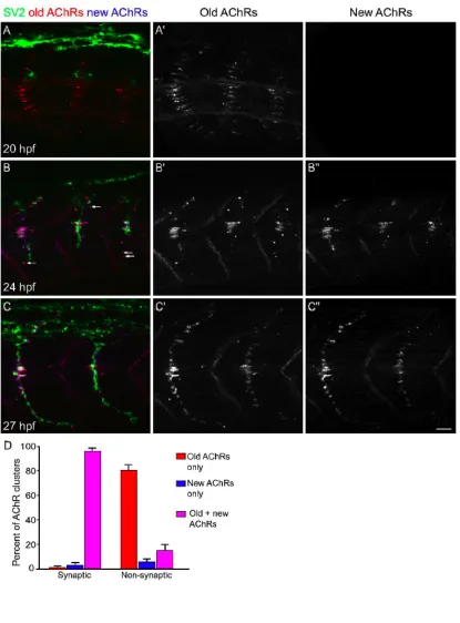
To examine the localization of newly inserted receptors, in one series of
experiments, the location of AChRs that were inserted in the muscle fiber membrane at
the time of initial rhodamine αBTX labeling (old AChRs) was compared to the location
of subsequently inserted AChRs (new AChRs) that were labeled with Cy5 αBTX after a
2, 4 or 7 hour delay. Embryos were then processed for immunostaining.
In vivo imaging
Embryos were placed in 1.2% low melting temperature agarose (SeaPlaque;
Cambrex) in Hank's solution in a modified imaging chamber (Warner Instrument Co.)
perfused with Hank’s solution at 28.5 °C. In transient transgenic VAMP-GFP embryos,
confocal z-stacks of images were obtained approximately every 10-20 minutes for 2-8
hours (Leica TCS 4D or SP2 system). In HuC:GFP embryos, confocal z-stacks were
obtained every 1 hour for 6 hours. Unless otherwise stated, each movie panel is a single
plane projection of a z-stack of 20-60 1 µm thick planes taken at the indicated time
interval. Z-stacks of up to 60 microns were necessary in some embryos because the
initial path of outgrowing motor axons is not flat and because, in older embryos, motor
29
Spontaneous movements and blood cell circulation, indicators of embryo viability, were
observed throughout the duration of movies that were subsequently analyzed. At the
conclusion of the imaging session, embryos were re-anesthetized, fixed and processed for
immunostaining.
In transient transgenic embryos expressing VAMP-GFP at ca. 24 hpf, relatively
few motor axon branches or neuromuscular synapses are present, whereas by 72 hpf
axons have branched into their appropriate, cell specific territory and many
neuromuscular synapses are present (Supplemental Fig. 1C). In vivo imaging of
VAMP-GFP+ primary motor neurons showed that motor axons were extended at a rate of 10.1 ±
0.8 µm / hour (mean ± s.e.m; N = 10 22-30 hpf embryos, 10 neurons), similar to that
observed in previous in vivo analyses using single cell fills (Eisen et al., 1986; Myers et
al., 1986; Westerfield et al., 1986). Thus the imaging procedures used in the present
study do not interfere with axon outgrowth or neuromuscular synaptogenesis.
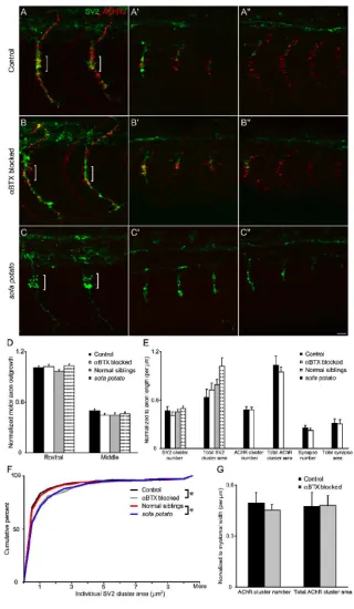
AChR blockade
Rhodamine αBTX was diluted to 0.25 mM in Yamamoto Ringers with 0.05%
Phenol red and injected directly into the yolk of 12-14 hpf embryos (Lefevre et al., 2004).
This approach was used to achieve sustained paralysis, since it likely achieves a higher
concentration of αBTX within embryonic muscle than bath application does, both by
bypassing the skin, a natural barrier to toxins, and by providing sustained exposure to
30
motility by evaluating responses to head and tail tap, and only those embryos that were
completely paralyzed were collected, fixed and immunostained.
Sodium channel blockade
Embryos were raised from 10 to 21 hpf in 0.01% tricaine in E3. At 21 hpf,
embryos were scored for motility by evaluating spontaneous tail movements, and only
those embryos that were completely immotile were collected, fixed and immunostained.
Whole mount immunostaining of zebrafish embryos
Embryos were anesthetized, fixed and immunostained as described previously
(Panzer et al., 2005) using antibodies against SV2 (Developmental Studies Hybridoma
Bank (DSHB)) and/or GFP (Chemicon, Inc.) and a fluorescently conjugated secondary
antibody (Jackson Labs, Inc.). Immunostained embryos were examined using confocal
microscopy. Unless otherwise stated, each figure panel showing immunostaining is a
single plane projection of a z-stack of 20-60 1 µm thick planes.
Analyses of prepatterned AChR clusters, presynaptic vesicle clusters and synapse
formation
AChR or presynaptic vesicle cluster number and area were measured from single
plane projections of confocal image stacks using interactive software (Metamorph,
31
longest axis of a cluster were measured to analyze changes in AChR cluster length and
intensity over time. Pre- and postsynaptic clusters were scored as synapses if there was at
least 30% pixel overlap between presynaptic and postsynaptic labeling.
To quantify the contribution of prepatterned AChR clusters to synapses, each
cluster present in the first frame of a time lapse movie was analyzed and placed into one
of three categories: Present but subsequently disappeared; present with a VAMP-GFP+
cluster overlying at least 30% of the AChR cluster, and thus innervated (synaptic); and
present throughout the movie but not innervated during the imaging interval
(non-synaptic). Preliminary analyses suggested that the 30% colocalization criteria is the
minimum for reliable identification of a synapse at the early embryonic ages examined
here (see also Panzer et al., 2005). Synapses were confirmed after subsequent
immunostaining of presynaptic vesicles with antibodies against SV2. The percentage of
events in which an AChR cluster was present in advance of a presynaptic vesicle cluster
and in which a presynaptic vesicle cluster was present followed by appearance of an
AChR cluster was also determined.
Quantification of growth cone and filopodia dynamics
To quantify axon and growth cone outgrowth with respect to prepatterned AChR
clusters, the position of a growth cone was determined from a point in its geometric
center. The closest AChR cluster in advance of the growth cone was defined as a
potential target AChR cluster. Three angles were then measured: angle 1, the angle
32
between the actual trajectory and the initial trajectory if the growth cone were to grow in
a straight line; and angle 3, the angle between the initial trajectory and the target AChR
cluster (see Fig. 3D). The distribution of angles 1 and 2 was evaluated, and the
relationship between angle 2 and angle 3 was analyzed by linear regression. In some
cases, more than one time interval of axon outgrowth was analyzed per neuron, and this
number is reported as an outgrowth event.
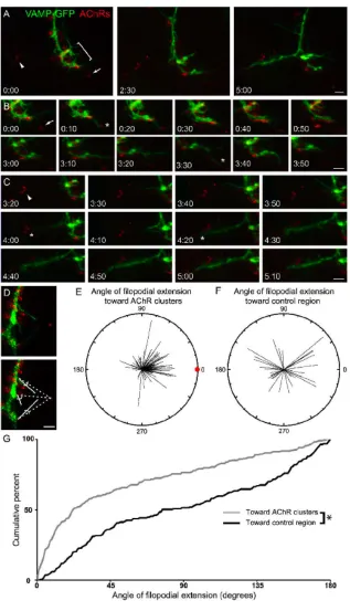
To quantify the direction of filopodial extension with respect to prepatterned
AChR clusters, only filopodia extended near AChR clusters that were not along the direct
path of axon extension were analyzed. This is because we wished to avoid a bias in the
quantification that would have arisen if filopodia tended to grow out in the direction of
axon extension, and AChR clusters happened to be along this pathway. All filopodia
within a 5 µm radius of an axon or growth cone region nearest the target AChR cluster
were analyzed. The mean distance from an axon or growth cone to a target AChR cluster
was 15 µm. The presence of multiple AChR clusters that were an equal distance from an
axon or filopodia was rare, and these cases were not analyzed.
The length of the filopodia, the actual trajectory of the filopodia and the
straightest, shortest trajectory between the base of the filopodia at the axon and an AChR
cluster were determined in each movie frame (see Fig. 4D). The length of the filopodia
and the angle between its actual trajectory and the straightest trajectory with respect to an
AChR cluster were plotted using polar coordinates where each line segment represents
filopodia length. Similarly, the length of filopodia, and the angles between the filopodia
33
AChR clusters in the vicinity were also measured and plotted. The cumulative percent of
filopodia extended at various angles with respect to an AChR cluster or control region
was plotted and differences assessed (Komolgorov-Smirnoff test).
Acknowledgements
We thank A. Kugath, M. Scott and H.-Y. Zhou for technical assistance, Dr. P.
Brehm for providing heterozygous carriers of the sofa potato mutation, Dr. S. Smith for
providing the UAS-VAMP-GFP construct, and Dr. S. Gibbs for helpful discussions.
Supported by grants from the NIH to R. B. G. (NS45919, NS50524) and a Howard
34 Results
Prepatterned AChR clusters are dynamic
Each of the three primary motor neurons per spinal cord hemisegment (CaP, MiP,
and RoP) sends an axon out of the spinal cord between 16-24 hours post-fertilization
(hpf). Primary motor axons then grow ventrally along the lateral surface of the notochord
to the so-called choice point at the horizontal myosepta (midline) of the somite. Axons
pause at the choice point for several hours, then grow out across the medial surface (CaP,
MiP), or branch near the midline (RoP, VaP) of the myotome, and subsequently all axons
branch extensively (Eisen et al., 1986). Neuromuscular synapses are formed en passant
along the axon shaft and branches (Eisen et al., 1986; Myers et al., 1986; Westerfield et
al., 1986; Panzer et al., 2005). Approximately 5 hours after initiation of primary motor
axon outgrowth, the axons of secondary motor axons (ca. 20-30 per hemisegment) begin
to extend into the periphery along the pathways pioneered by primary motor axons and
also form en passant synapses (Myers et al., 1986; Westerfield et al., 1986).
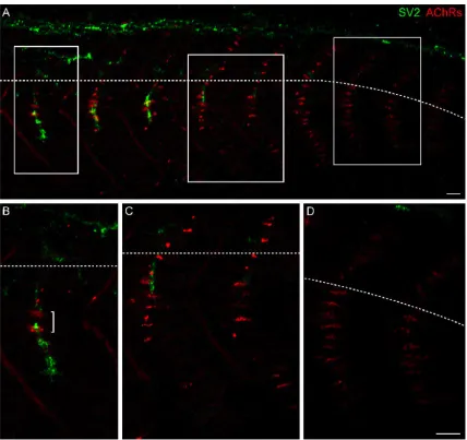
In fixed and immunostained embryos at 24 hpf, AChR clusters are present on
each muscle fiber along the medial surface of the myotome (Fig. 1A, caudal most
segments, Fig. 1D), on many muscle fibers in lateral muscle layers, and along the lateral
myosepta, well in advance of motor axon outgrowth from the spinal cord (Panzer et al.,
2005). These AChR clusters are thus prepatterned on muscle fibers in advance of
innervation (Yang et al., 2000; Lin et al., 2001; Yang et al., 2001). Prepatterned AChR
clusters are elongated and diffuse in caudal and thus younger myotomes, but are small,
35
largest prepatterned AChR clusters are present at the choice point; these clusters persist
as motor axons enter the myotome and the first neuromuscular synapses are made at these
prepatterned AChR clusters (Fig. 1B, bracket; Panzer et al., 2005). These observations
suggest that initially diffuse, elongated prepatterned AChR clusters coalesce, and some
AChR clusters disappear, as innervation occurs.
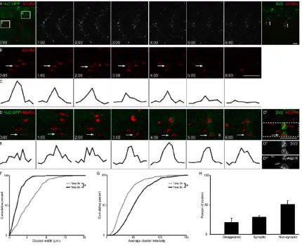
To directly determine the fate of prepatterned AChR clusters as motor axons grow
into the myotome, AChRs were labeled with a non-paralyzing dose of rhodamine αBTX
in HuC:GFP embryos, in which all neurons including primary motor neurons and axons
express GFP, and confocal z-stacks were obtained over time (N = 4 20-26 hpf embryos,
19 myotome segments). At the beginning of imaging, motor axons had not exited the
spinal cord (Fig. 2A, panel 0:00). Prepatterned AChR clusters are initially elongated and
diffuse, and over several hours, most if not all clusters coalesce (Fig. 2A; compare panel
1:00 with panel 3:00). During a 6 hour interval, AChR cluster width is reduced (Fig. 2F)
and fluorescence intensity increases (Fig. 2G), consistent with the coalescing of each
cluster. During this interval, 20% of all prepatterned AChR clusters disappear, as
demonstrated both by comparing subsequent images qualitatively (Fig. 2B, arrow) as
well as quantifying changes in individual cluster length and intensity (Fig. 2C, F-H).
This dynamic redistribution of AChRs usually begins in advance of motor axon
outgrowth into the myotome. Thirty percent of all prepatterned AChR clusters persist
and become innervated during a 6 hour interval (Fig. 2D, arrow), as demonstrated by the
presence of GFP+ axons apposed to AChR clusters (Fig. 2D, panel 5:00, asterisk) as well
as by post-imaging immunostaining for presynaptic vesicles (Fig. 2D, last panel). As
36
However, about half of the prepatterned AChR clusters are neither dispersed nor
innervated during a 6 hour imaging interval. Given the absence of uninnervated AChR
clusters in older embryos (Panzer et al., 2005), these clusters must either be innervated or
disappear over longer intervals. These observations show that prepatterned AChR
clusters are highly dynamic before and during motor axon outgrowth into the myotome,
and that some of these clusters are incorporated into synapses, whereas others are
dispersed or persist in an uninnervated state.
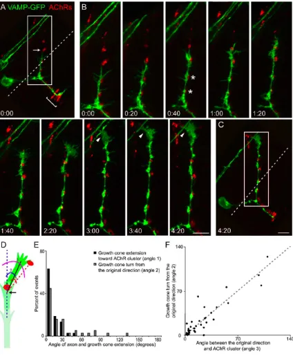
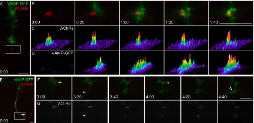
Motor axon outgrowth occurs along prepatterned AChR clusters
In vivo imaging of VAMP-GFP+ primary motor axon outgrowth in transiently
transgenic embryos was used to examine the spatial and temporal dynamics of these
events with respect to prepatterned AChR clusters over time (N = 20 20-30 hpf embryos,
29 growth cones, 44 outgrowth events). In vivo imaging showed that, at any given time,
AChR clusters are observed in advance, or within 2-3 muscle fibers, of ca. 75% (33 / 44)
of outgrowing motor axons (c.f. Fig. 1C; Fig. 3A; Fig. 4A; Fig. 6A, E). Motor axon
growth cones grow directly towards, then along and eventually innervate, prepatterned
AChR clusters (Fig. 3B). Continual growth towards, and innervation of, prepatterned
AChR clusters result in motor axon extension across the medial surface of the myotome
(Fig. 3B). Thus, motor axons appear to follow a pathway across the medial surface of the
myotome that is spatially coincident with prepatterned AChR clusters. These results
contrast with previous reports that AChR clustering and motor axon outgrowth are
37
least some cases, prepatterned AChR clusters themselves, or cues that are co-patterned
with such clusters, might direct motor axon outgrowth.
These possibilities are further supported by the observation that the location of
prepatterned AChR clusters predicts growth cone turning. For example, in Fig. 3B,
prepatterned, previously uncontacted, AChR clusters are located ca. 15 µm rostral (and
somewhat lateral) to the motor axon growth cone in panel 2:20, and in the subsequent 2
hours, the growth cone turns 65° toward and contacts these AChR clusters. Sixty-three
percent of growth cones are extended with an angle ≤ 10º, and 95% are extended at an
angle of ≤ 30º, with respect to a prepatterned, previously uncontacted, AChR cluster (Fig.
3E, black bars). Growth cones were observed to turn toward a prepatterned AChR cluster
over a wide range of angles, from 10º to 130º (Fig. 3E, grey bars). For the population of
growth cones that were observed over time, a significant correlation exists between the
location of a prepatterned AChR cluster with respect to the motor axon’s initial
trajectory, and the angle that the growth cone eventually turns toward that cluster (Fig.
3F). These observations show that the direction of motor axon outgrowth is dynamically
altered so that axons continually extend along a pathway that is spatially coincident with
prepatterned AChR clusters that they will eventually contact.
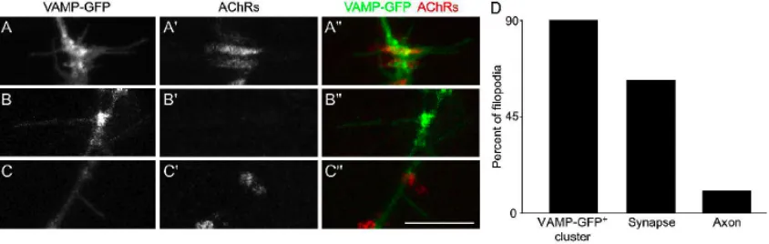
Motor axon filopodia are preferentially extended toward prepatterned AChR clusters
In the course of time lapse imaging of motor axon outgrowth to, and interactions