265 | P a g e
BRAIN TUMOR SEGMENTATION
Dr. D. Haritha
Computer Science and Engineering Department, Jawaharlal Nehru Technological University
Kakinada, Kakinada(India)
ABSTRACT
Brain tumor is an abnormal growth of the cells in the brain. The location, shape and region of the tumor is very
important for identifying the tumor. Even the doctors can identify these tasks with their knowledge, but, it is a
time consuming process. In order to save the time of the doctors, there is a need for the automation of the Brain
tumor segmentation. Here, we have made an attempt for developing the algorithm for Brain tumor
segmentation. Illumination normalization is an important criterion for the images, for reducing the lighting
effects on images. In order to avoid the wrong prediction of the tumor location in brain images, which was
caused due to illumination, there is a need to eliminate the illumination. So, here, illumination also considered,
while detecting the tumor. We used the preprocessing techniques Local Binary Pattern and for segmentation
used Morphological operations. The proposed algorithm is developed using MATLAB.
Keywords: Morphological operations, Local binary pattern, Threshold Segmentation, erosion,
dialation, Structure element.
I. INTRODUCTION
Brain is an important part in the human body. If Brain is not functioning, then there is no proper functioning of the remaining parts of the human body. Brain tumor is an abnormal growth of the cell in the brain, which become an obstacle for the functioning of the brain due to the occupancy of space within the brain. Further it leads to the damage of the blood vessels. Brain tumor is identified using several considerations like MRI scan, spinal tap biopsy, brain angiogram, etc... Image processing techniques like image segmentation, classification, Morphological operations..etc., are used for detecting the Brain tumor. Brain tumor is divided into two groups based on the growth of the tumor. They are Benign and Malignant. Non-cancerous tumors are called as Benign. Benign tumors are seldom grows back. Cancerous tumors are called as Malignant. Malignant tumors rapidly grows and effect the surrounding brain cells, which further damage the health of the patient in a short period, further leads to the death of the patient. Hence, identifying the brain tumor in early stage is also an important criteria in brain tumor detection [1].
and burden of the doctor there is a need for the automation of the brain tumor. Segmentation is a crucial method for extracting the interested regions in the images. For detecting the exact location of the tumor in brain images, segmentation methods are used. There are several segmentation techniques available for obtaining the interested region or segment of the image. Some of them are: water shed algorithm, region growing and splitting, threshold segmentation, etc. [2].
While developing the automation system for detecting the Brain tumor, we have to remember mainly two points.
The identification of the Brain tumor in early stage.
The location, size and shape of the brain tumor in brain MRI images. Hence, based on these criteria‟s, doctors can start the treatment for curing the disease.
This paper is divided into six sections. Section I deals with the introduction part. Literature survey was discussed in Section II. The problems of existing system and the proposed work are presented based on the study of the literature work. System overview is discussed in section III. LBP and Morphological operations and the importance of the erosion and dilation are mentioned in this section. Experimental Results are shown in section 4. Final conclusion was discussed in section 5.
II. LITERATURE WORK
Several Researchers worked for the Brain tumor detection using the different Image processing techniques. Atiq et al., proposed a stochastic method for Brain tumor. The proposed multifractal feature based brain tumor segmentation method is compared with the Gabor-like multi scale texton feature. Novel patient-independent tumor segmentation of tumors scheme proposed by extending the AdaBoost algorithm. Modified AdaBoost algorithm assigns weights for the component classifiers. Experimentation was done by considering 300 images of 14 patients[3]. A new approach fluid vector flow was proposed by Tao et. al., Fluid vector flow given better results compared to the boundary vector flow, gradient vector flow and magnetostatic active contour. Experiment was done on three sets of images: brain tumor MRI images, diatric head MRI images and synthetic images[4].
Andac Hamamciet. al, presents a fast and robust tool for the brain tumor segmentation.This cellular automata differentiate necrotic and enhancing tumor tissue content. CA based segmentation connection was established. Furthermore, this CA algorithm compares the necrotic and enhancing tumor tissue content. Experimentation was done on the clinical and synthetic brain tumor datasets and found 80% to 90% performance with the existing system. Performance is done based on the computational time[5]. Anahita et al., proposed a multi scale for the gradient vector flow algorithm, in order to solve the poor robustness against noise and lack of convergence. This method further extended with threshold-based edge detector. This method proved as an effective method. They found 30% improvement in the accuracy of the tumor segmentation compared to the traditional gradient vector segmentation algorithm.[6].
267 | P a g e Discrete Wavelet Transform using the soft-thresholding[8]. Kimmiet. al., proposed a hybrid method for brain tumor segmentation, which uses edge detection, watershed segmentation, morphological operations and threshold segmentation. With this method, they found the exact location and size of the tumor in brain images. Experimentation was done using the MATLAB.[9].
Xiaoyang et al., proposed the reliable face recognition system under uncontrolled lighting conditions. Here, they used LBP for eliminating the lighting effects and performed the illumination normalization[10]. Shufu et al., proposed local pattern method with Gabor magnitude and phase for face recognition. Dimensionality reduction is done by block based fisher‟s linear discriminant. The proposed model outperforms for most of the state-of the art approaches[11].
Based on the literature survey, we found that the existing methods developed by different researchers concentrate on the detection of the brain tumor using various segmentation techniques like water shed algorithm, threshold segmentation, etc. We need to consider one important criteria i.e., illumination, while detecting the tumor. In order to avoid the wrong prediction of the tumor location in brain images, which was caused due to illumination, Here, we used the preprocessing techniques Local Binary Pattern(LBP). Even though, several methods proposed, but, less no of literature work found in the preprocessing step. So, here, we made an attempt for eliminating the illumination on brain images. The effect of illumination gives an impact for tumor in brain tumor segmentation.
III. SYSTEM ARCHITECTURE
Fig.1 Shows flowchart for overview of the system, which deals with the step by step actions performed on the brain MRI images for obtaining the required segmented tumor image. Experimentation was done on MATLAB Platform
Fig.1: Overview of the system
A.
Local Binary Pattern
LBP is a simple classifier and it‟s computationally simple. Due to these tasks, it has gained more popularity. LBP operator is used for measuring the texture, which is the interest texture in a local neighborhood. LBP classifier was using in many of the application like medical image analysis, face recognition, video analysis, criminal identification etc.. Based on the performance of the LBP classifier, we have applied this LBP technique for the Brain MRI images[11, 12].
he basic concept of the LBP is: First we have to apply the LBP operator for the 3X3 block from the leaf image. If the pixel value at any position(except the center pixel) is greater than the center pixel then give the value as 1, otherwise give the value is 0 at that location. The obtained Pixel value is multiplied by the powers of two. Sum of these pixel values is placed at the center pixel position. Like this, we have to obtain the center pixel value for every 3X3 block in the leaf image until it reaches the size of the image. [13, 14].
After applying the LBP for the brain MRI image, the resultant image is called as LBP image. The obtained LBP image is given for segmenting the regions using the Morphological operations.
B.
Morphological operations
Morphological operations are derived from the set theory. These Morphological operations are used for separating the tumor from the brain image. Tumor part has high intensity. Separation of this tumor part from the brain images, shown to be awhite spot on the screen[15].
Here, Dilation and erosion was used for filling the gaps and for eliminating the irrelevant cells from the brain image. For binary images, dilation is best method and the relevant and broken segments are easily joined by this dilation.
With A and B are sets in Z2 ,The dilation of A by B is defined by: (1) Here, set B is called as structuring element in dilation.
For applying the erosion, we have to consider the structuring elements. Based on the size of these structuring elements, we can display the required portion of the interest, which means unwanted portion is removed from the image.
With A and B are sets in Z2 ,The erosion of A by B is defined by: (2)
For the obtained LBP image, apply the Morphological operations. By applying these threshold segmentation, dilation and erosion, we obtain the segmented image[16].
IV. EXPERIMENTAL RESULTS
A sample of three brain MRI images was considered for Experimentation. Experimentation is done on the platform of the MATLAB, which is a compatible environment for the analysis of images and for the image processing applications.
269 | P a g e segmenting the image using Threshold segmentation, erosion and dilation. Dilation and erosion was used for filling the gaps and for eliminating the irrelevant cells from the brain image.
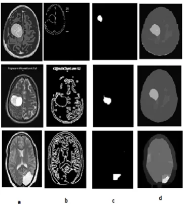
Fig. 2 deals with the resultant images after the segmentation. For experimentation, we have considered 3 sample Brain MRI images. The first column i.e., named as „a‟ shows the sample of Brain MRI images. The input brain MRI image was processed with the LBP operator and obtained LBP image is shown in the second column, i.e., „b‟. The third column, „c‟ refers to the segmented tumor in brain images. This resultant image in column c was obtained by performing LBP and Morphological operation. The last column, ‟d‟ refers to the brain image which is obtained by applying only the morphological operations. With these images, it clearly gives the comparison of the resultant image obtained by LBP and Morphological operations and with the resultant image, which is obtained by only applying the Morphological operations. Based on these results, we can find the exact location of the tumor in brain MRI images.
V. CONCLUSION
The Proposed method deals with the brain tumor segmentation based on the preprocessing using Local Binary Pattern and segmentation using Threshold segmentation and Morphological operations. Most of the existing methods mainly concentrate on the detection of the brain tumor detection. Here, we proposed a method for elimination of illumination effects by using local binary pattern in order to obtain the exact location for the tumor. Wrong detection of tumor may lead to collapsing of patient health. The location of the tumor has an effect on the normal functioning of human. Finally, Morphological operations applied for the occurred LBP image, for obtaining the segmented image. The proposed method has compared with the existing Threshold segmentation method based on Morphological operations. The proposed method was giving the exact location compared to the existing methods.
In this paper, presented the brain tumor detection based on the preprocessing technique LBP. There are several variations for the LBP techniques. Further, this system can be extended by using the extended LBP methods. Clustering methods are also used for segmenting the images. Hence, this system can be further extended by using different clustering methods.
REFERENCES
[1] S.U. Aswathy, G. Glan Deva Dhas, S.S. Kumar, “A Survey on Detection of Brain Tumor From MRI Brain Images”, 2014 international conference on control, Instrumentation, Communication and Computational Technologies(ICCICCT), 2014, 871-877.
[2] A. K. Chaudhari and J. V. Kulkarni, “Local Entropy based Brain MR image segmentation”, 3rd IEEE international Advance Computing Conference, 2013, 1229-1233.
[3] Atiqislam, Syed M. S. Reza and Khan M Iftekharuddin, “Multifractal Texture Estimation for Detection and Segmentation of Brain Tumors”, IEEE Transactions on Biomedical Engineering, 60(11), 2013, 3204-3215.
[4] Tao Wang, Irene Cheng and AnupBasu, “ Fluid Vector flow and applications in Brain Tumor Segmentation”, IEEE transactions on Biomedical Engineering, 56(3), 2009, 781-789.
[5] AndacHamamci, Nadir Kucuk, KutlayKaraman, KayihanEngin and GozdeUnal, “Tumor-cut: Segmentation of Brain Tunors on Contrast Enhanced MR images for Radio surgery Applications”, IEEE transactions on Medical Imaging, 31(3), 2012, 790-804.
[6] AnahitaFthiKazerooni and AlirezaAhmadian, “Segmentation of Brain Tumors in MRI Images using Multi-scale Gradient Vector Flow”, 33rd
Annual international conference of the IEEE EMBS, 2011, 7973-7976. [7] NailahAfshan, ShaimaQureshi, Syed MujtibaHussain, “Comparative study of Tumor Detection on
algorithms”, international conference on Medical imaging, m-Health and Emerging Communication Systems, 2014, 251-256.
[8] Tapas Si, Arunava De and Anup Kumar Bhattacharjee, “Brain MRI segmentation for Tumor Detection using grammatical swarm based clustering algorithm”, international conference on Circuit, Power and Computing Technologies, 2014, 1196-1201.
[9] KimmiVerma, ShabanaUrooj and Rituvijay, “Effective Evaluation of tumor region in brain MR images using Hybrid segmentation”, International conference on Comupting for sustainable Global Development, 570-574.
[10] Xiaoyang Tan and Bill Triggs, “Enhance Local Texuture Feature Sets for Face Reognition Under Difficult Lighting Conditions”, IEEE Transactions on Image Processing, 19(6), 2010, 1635-1650.
[11] Shufu Xie, Shiguang Shan, Xilin Chen and Jie Chen, “ Fusing Local patterns of Gabor Magnitude and Phase for face Recognition”, IEEE transactions on Image Processing, 19(5), 2010, 1349-1360.
[12] Ma Li, R.C. Staunton, “Optimum Gabor filter design and local binary patterns for texture segmentation”, Patter Recognition Letters, 29(5), 2008, 664-672.
[13] T. Ojala,M. Pietikäinen and D. Harwood, “A comparative study of texture measures with classification based on feature distributions”, Pattern Recoginition, 29(1), 1996, 51–59.
[14] D. Haritha, K. Srinivasa Rao, and Ch. Satyanarayana, ―Performance evaluation of the effect of combining the DCT and LBP on face recognition system”, International journal of Modern education andComputer Scienc , 2012, 4(11), 21-32.