Timing of Follow-up Voiding Cystourethrogram in Children
With Primary Vesicoureteral Reflux: Development and
Application of a Clinical Algorithm
Matthew Thompson, MD*; Stephen D. Simon, PhD‡; Vidya Sharma, MBBS, MPH‡; and Uri S. Alon, MD*
ABSTRACT. Background and Objectives. Of children diagnosed with urinary tract infection, 30% to 40% have primary vesicoureteral reflux (VUR). For the majority of these children, treatment involves long-term prophylac-tic antibioprophylac-tics (ABX) and a periodic voiding cystourethro-gram (VCUG) until resolution of VUR as detected by VCUG. Radiation exposure and considerable discomfort have been associated with VCUG. To date, no clear guidelines exist regarding the timing of follow-up VCUGs. The objective of this study was to develop a clinically applicable algorithm for the optimal timing of repeat VCUGs and validate this algorithm in a retrospec-tive cohort of children with VUR.
Methods. Based on previously published data regard-ing the probability of resolution of VUR over time, a decision-tree model (DTM) was developed. The DTM compared the differential impact of 3 timing schedules of VCUGs (yearly, every 2 years, and every 3 years) on the average numbers of VCUGs performed, years of ABX exposure, and overall costs. Based on the DTM, an algo-rithm optimizing the timing of VCUG was developed. The algorithm then was validated in a retrospective co-hort of patients at an urban pediatric referral center. Data were extracted from the medical records regarding num-ber of VCUGs, time of ABX prophylaxis, and complica-tions associated with either. VUR in patients in the co-hort was grouped into mild VUR (grades I and II and unilateral grade III for those<2 years old), and
moder-ate/severe VUR (other grade III and grade IV). Kaplan-Meier survival curves were created from the cohort data. From the survival curves, the median times to resolution of VUR were determined for the cohort, and these times were compared with the median times to VUR resolution of the data used for the DTM. The numbers of VCUGs performed, time of ABX exposure, and costs in the cohort were compared with those that would have occurred if the algorithm had been applied to both mild and mod-erate/severe VUR groups.
Results. Using an algorithm that results in a recom-mendation of VCUGs every 2 years in mild VUR would reduce the average number of VCUGs by 42% and costs by 33%, with an increase in ABX exposure of 16%, com-pared with a schedule of yearly VCUGs. For moderate/
severe VUR, a VCUG performed every 3 years would reduce the average number of VCUGs by 63% and costs by 51%, with an increase in ABX exposure of 10%. Ap-plying this algorithm to the retrospective cohort consist-ing of 76 patients (between 1 month and 10 years old) with primary VUR would have reduced overall VCUGs by 19% and costs by 6%, with an increase in ABX expo-sure of 26%. The patterns of VUR resolution, age distri-bution, and prevalence of severity of VUR were compa-rable between previously published results and the retrospective cohort.
Conclusions. Delaying the schedule of VCUG from yearly to every 2 years in children with mild VUR and every 3 years in children with moderate/severe VUR yields substantial reductions in the average numbers of VCUGs and costs, with a modest subsequent increase in ABX exposure.Pediatrics2005;115:426–434;vesicoureteral reflux, voiding cystourethrography, antibiotic prophy-laxis, vesicoureteral reflux resolution.
ABBREVIATIONS. UTI, urinary tract infection; VUR, vesi-coureteral reflux; VCUG, voiding cystourethrogram; ABX, antibi-otics; CA, clinically applicable algorithm; DTM, decision-tree model.
O
f children diagnosed with urinary tract infec-tion (UTI), 30% to 40% have primaryvesi-coureteral reflux (VUR).1 VUR has been
graded from I to V depending on severity. VUR is currently best detected by a voiding cystourethro-gram (VCUG), which (whether fluoroscopic or nu-clear) is regarded as the “gold standard” and is the most commonly used modality.2,3 The majority of
children will have resolution of their VUR over time.4–12 The probability of reflux resolution with
continuous antibiotics (ABX) prophylaxis has been documented in a large study combining prospective data from 893 patients with VUR grades I to IV.13–15
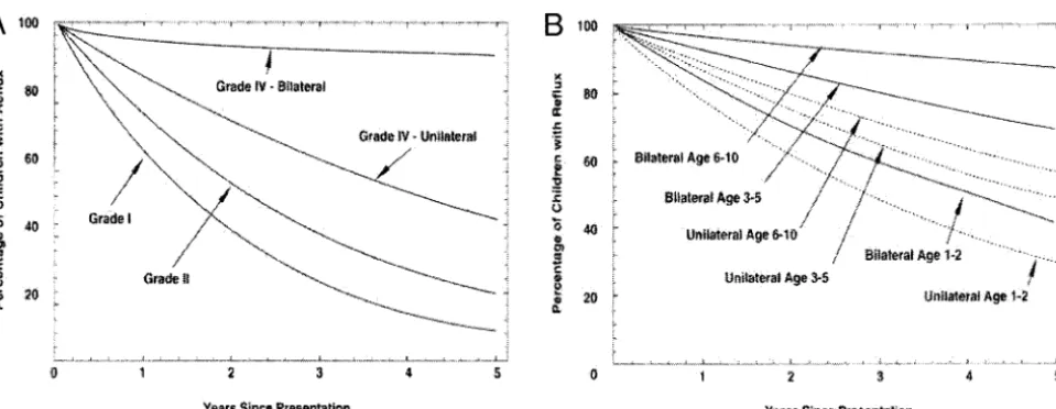
This study stratified variables into predictors of per-sistence of VUR on patients followed between the years 1976 and 1990, presented in Fig 1 in the form of a survival nomogram.16The medical management of
VUR, namely long-term ABX prophylaxis, has been shown to be as effective as surgical management in reducing the risks associated with VUR grades I to IV.7–9,17–22Guidelines regarding the management of
primary VUR in children recommend that, for most children with VUR, initial treatment is comprised of continuous ABX prophylaxis until indication for sur-gery or spontaneous resolution of VUR.16,23–25
How-ever, no specific guidelines are given with regards to From the *Section of Nephrology and ‡Department of Medical Research,
Children’s Mercy Hospital, University of Missouri, Kansas City, Missouri. Accepted for publication Aug 12, 2004.
doi:10.1542/peds.2004-0927
This work was presented in part at the American Society of Nephrology Annual Meeting; November 12–17, 2003; San Diego, CA.
No conflict of interest declared.
Address correspondence to Uri S. Alon, MD, Section of Nephrology, Chil-dren’s Mercy Hospital, 2401 Gillham Rd, Kansas City, MO 64108. E-mail: [email protected]
the timing of follow-up VCUGs to detect VUR reso-lution.
Currently, wide variation exists regarding the fre-quency of obtaining VCUGs after diagnosis of VUR. Some authors recommend VCUGs at intervals of 6 to 18 months.26–28 A VCUG is an invasive procedure
that is a source of significant patient discomfort re-sulting from instrumentation of the urinary tract for the purpose of instilling contrast material through a bladder catheter.29–32 Furthermore, as much as 25%
of exposure to ionizing radiation during childhood may be the result of imaging of the urinary tract.2On
rare occasions the procedure may be followed by an infection.33 Additionally, there are cost
consider-ations regarding the surveillance of VUR. Beyond the expense of the imaging study, there are the costs of work missed by caregivers, travel expenses, etc.1,34–37
However, early detection of VUR resolution by a VCUG may minimize the use of prophylactic anti-microbials, which would result in a reduction in the cost of unnecessary prophylactic treatment and re-duce the risk of potential side effects associated with ABX exposure and the possible emergence of bacte-ria resistant to common antimicrobials.38–42
The ideal medical management of children with primary VUR would require only the minimal num-ber of invasive imaging studies while concomitantly minimizing any unnecessary exposure to antimicro-bial prophylaxis. The timing of follow-up VCUGs should be based on a rational approach guided by the best available data.43 The primary goal of the
present study was to develop a clinically applicable algorithm (CA) for the timing of follow-up VCUGs in children with VUR. A secondary objective was to validate this CA by applying it to a retrospective cohort of children with VUR at an urban pediatric referral institution.
METHODS
This study was considered exempt by the University of Mis-souri (Kansas City) Pediatric Institutional Review Board, accord-ing to criteria 45 CFR 46.101 (b)4because it involved the collection
of existing data, with information recorded by the investigator in
such a manner that subjects could not be identified directly or through identifiers linked to the subjects.
Decision-Tree Model Analysis Structure of Decision-Tree Model Analysis
To develop a CA for the optimal timing of follow-up VCUG in childrenⱕ10 years old with primary VUR, decision-tree model (DTM) analysis was used. Three different strategies were modeled for the timing of VCUG: (1) VCUG conducted once yearly; (2) VCUG conducted every 2 years; and (3) VCUG conducted every 3 years. Grades of VUR were grouped into stratification groups identified by Elder et al16 to be significant predictors of VUR
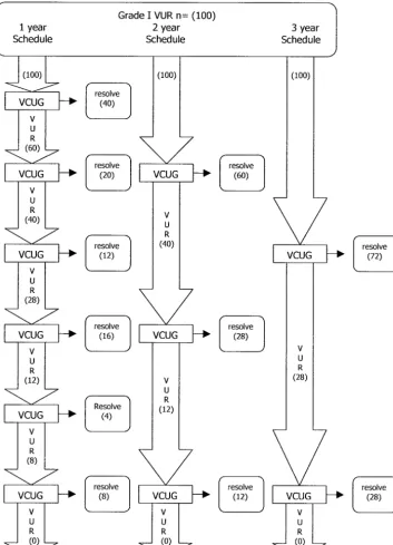
resolution. These stratification groups were based on VUR grade, age in the case of grade III, and laterality in the case of grades III and IV VUR. Mild VUR included grades I and II and unilateral grade III in a child ⱕ2 years old, and moderate/severe VUR included all other grades III and IV. Each stratification group was evaluated by using the 3 timing strategies. Figure 2 demonstrates the DTM.
Assumptions
Assumptions made for the analysis were: all VCUGs occur at yearly intervals; ABX are discontinued at yearly intervals; no patient drop-out occurs due to death, kidney transplant, etc; and any additional costs (ie, costs of risks associated with ABX and complications from VCUGs) are negligible and were not included in the analysis. The cost estimates were assumed to be $475 per study for a VCUG and $100 per year for ABX. This was based on the billed charges for a VCUG and the average generic cost of a prophylaxis dose of trimethoprim/sulfamethoxazole at our insti-tution in 2002. Costs were considered from the societal perspec-tive, not taking into account work missed, travel expenses, etc.
Probabilities
Data regarding the probability of the resolution of VUR was based on nomograms published by Elder et al16(Fig 1).
Outcomes
The following outcomes were estimated: average number of VCUGs per patient; average time receiving ABX prophylactic therapy; and total costs of VCUG and ABX per patient.
The relative change in average number of VCUGs, time of ABX exposure, and costs were analyzed for each different timing strat-egy. A CA was developed based on this analysis.
Validation Using a Retrospective Cohort
Medical records of a retrospective cohort of patients with VUR at an urban pediatric referral center were reviewed for the sec-ondary objective of validating the CA developed from the DTM.
Patients were included if they were diagnosed with VUR after an episode of UTI during the years 1995 and 1998 and were⬍10 years old at diagnosis. Patients were excluded if they had secondary causes for VUR (ie, spina bifida, voiding dysfunction, neurogenic bladder, etc). Excluded also were those diagnosed with VUR as a result of evaluation for prenatal hydronephrosis or due to a sibling screening. Medical records were reviewed, and data were ex-tracted regarding the age, laterality, VUR grade at diagnosis and at follow-up imaging, and the duration of ABX prophylaxis. Co-hort data were analyzed in the mild and moderate/severe strati-fication groups. Kaplan-Meier survival curves were computed by
using SPSS 12.0 software (SPSS Inc, Chicago, IL). Data were cen-sored in cases of loss of follow-up or surgical intervention. From the survival curves the median time to resolution was determined for the cohort and compared with median time to resolution of the Elder et al data.16The actual average number of VCUGs and average
dura-tion of prophylactic ABX were then established for the cohort. Next, based on the cohort’s actual rates of resolution, we determined the average numbers of VCUGs and time of ABX exposure that would have occurred if the CA had been applied to the cohort. Finally, the average numbers of VCUGs, ABX exposure, and estimated costs were compared between the actual cohort values and the CA values.
Fig 2. Example of a decision-tree analysis of 100 hypothetical patients with grade I VUR evaluated by follow-up VCUG under 3 different timing regimens. Probability of resolution is 40% at 1 year, 60% at year 2, 78% at year 3, 88% at year 4, 92% at year 5, and 100% at year 6. With an every-1-year schedule, over the course of the analysis (which needs to be 6 years to allow for VCUG intervals of 1 year, 2 years, and 3 years) 40 patients had 1 VCUG, 20 patients had 2 VCUGs, 12 patients had 3 VCUGs, 16 patients had 4 VCUGs, 4 patients had 5 VCUGs, and 8 patients had 6 VCUGs. Calculations to determine average number of VCUGs:
RESULTS DTM
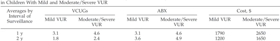
Average numbers of VCUGs, years of ABX expo-sure, and cost are shown in Table 1 and Fig 3.
Mild VUR (Grades I and II and Unilateral Grade III for Thoseⱕ2 Years Old)
The change from once-yearly VCUG to an every-2-year schedule of imaging results in a dramatic decrease (42%) in average VCUGs, with minimal change (16%) in ABX exposure. By further delaying to an every-3-year schedule, compared with a yearly schedule, the decrease in average VCUGs (55%) con-tinues but is less substantial compared with the com-mensurate increase (35%) in ABX exposure. By de-laying follow-up VCUG by 2 and 3 years, overall costs would be reduced by 33% and 39%, respec-tively.
Moderate/Severe VUR (All Other Grades III and IV)
The change from once-yearly VCUG to an every-2-year schedule of imaging results in a dramatic decrease in average VCUGs (48%) with minimal change (7%) in ABX exposure. By further delaying to an every-3-year schedule, compared with a yearly schedule, there is a further decrease in average VCUGs (63%), with a minimal increase (10%) in ABX exposure. By delaying follow-up VCUG by 2 and 3 years, overall costs would be reduced by 38% and 51%, respectively.
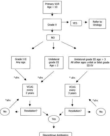
For the CA, a schedule of a VCUG every 2 years in mild VUR and every 3 years for moderate/severe VUR was therefore considered optimal (Fig 4).
Retrospective Cohort Chart Review
The medical charts of 92 patients with primary VUR were reviewed. Sixteen patients were excluded based on diagnosis after evaluation for prenatal hy-dronephrosis (n⫽9), evaluation without a history of UTI (n⫽4), and grade V VUR (n⫽3). A total of 76 patients was included in the analysis. The mean age of the cohort was 1.9 years, and the median age 1.0 year; 10% were male. At the time of diagnosis, 6 patients had VUR grade I (8%), 26 had grade II (34%), 37 had grade III (49%), and 7 had grade IV (9%).
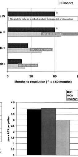
Kaplan-Meier survival curves were created for grades I to III VUR (Figs 5–7). Because of the small sample size, we pooled all grade III patients together. VUR did not resolve in any of the children with grade IV, and therefore a survival curve was not produced (Fig 8). Based on the 3 VUR survival curves, estimates of median months to resolution were calculated. These results demonstrate
compa-rable or somewhat prolonged median time to reso-lution compared with median times to resoreso-lution calculated from survival curves presented by Elder et al16 (Fig 8). Additionally, the pattern of VUR
res-olution, distribution of age, and prevalence of VUR follows that of the largest database (n⫽468) used by Elder et al for the development of the nomograms.15
In their cohort, 62% of children with grades I to IV were⬍2 years old, compared with 60% in our cohort. Also, they had a similar distribution of prevalence of VUR, with 82% of their patients having VUR grades II and III, compared with 83% in our cohort.15
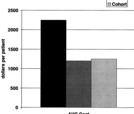
The actual average number of VCUGs in the co-hort was 2.0 with 2.9 years on ABX and a cost of $1250. Applying the CA to the cohort would have reduced the predicted numbers of average VCUGs by 19% (P⫽.001) and the costs by 6% (P⫽.17) and increased ABX exposure by 26% (P⫽.001), as shown in Figs 9 –11.
DISCUSSION
In 1997, Elder et al,16 serving as an ad hoc
com-mittee of expert pediatric urologists and nephrolo-gists, thoroughly reviewed the world literature to establish guidelines for the medical and surgical management of VUR in children. Among these guidelines, they included important nomograms (Fig 1) that illustrated the natural course of VUR resolution in children. However, numerous publica-tions, including the most recent editions of Nelson’s Textbook of Pediatrics27andPediatric Nephrology,28
rec-ommend repeat VCUG anywhere between 6 and 18 months.26 These recommendations are inconsistent
Fig 3. Weighted average VCUG per patient following a yearly schedule (Q1), every-2-year schedule (Q2), or every-3-year sched-ule, as calculated from data published by Elder et al.16
TABLE 1. DTM: Effect of 3 Time Schedules of Follow-up VCUG on Average Numbers of VCUGs, Use of ABX, and Cost Per Patient in Children With Mild and Moderate/Severe VUR
Averages by Interval of Surveillance
VCUGs ABX Cost, $
Mild VUR Moderate/Severe
VUR
Mild VUR Moderate/Severe
VUR
Mild VUR Moderate/Severe
VUR
1 y 3.1 4.6 3.1 4.6 1790 2650
2 y 1.8 2.4 3.6 4.9 1200 1650
with the above-mentioned nomograms and indicate the need for a more rational approach to surveillance of VUR based on the probability of its spontaneous resolution. By using the data presented by Elder et al, we have identified an approach to the timing of repeat VCUG, which moves 1 step closer to the goal of balancing the number of VCUGs, prophylactic ABX exposure, and total costs. Additionally, our analysis of a cohort of patients with VUR strengthens the validity and applicability of the proposed algo-rithm, because the pattern of VUR resolution, distri-bution of age, and prevalence of VUR follows that of the largest database used by Elder et al.15
The DTM analysis suggests that, when balancing exposure to VCUGs, exposure to ABX prophylaxis, and costs, the optimal timing of follow-up VCUG is every 2 years for children with mild VUR (grades I and II as well as those ⱕ2 years old with unilateral grade III). The placement of younger children with unilateral grade III VUR in the mild group is
consis-tent with the findings of Elder et al (Fig 1) and other recent recommendations.27 For those with
moder-ate/severe VUR (all other grade III-IV), the DTM analysis found 3-year intervals to be optimal, based on which we made our recommendations as pre-sented in the form of an algorithm (Fig 4). Our rec-ommendations are consistent with the opinion ex-pressed by Arant12 in an editorial in which he
suggested that VCUG only needs to be performed every 2 to 3 years unless the clinical course is com-plicated.
Retrospective review of a cohort at our institution suggests that we perform follow-up VCUGs on av-erage every 18 months. As a result of this trend toward delaying VCUG, applying the CA to our own cohort yielded less substantial change in average VCUGs, ABX exposure, and costs than would have been predicted by the DTM schedule of yearly VCUG (Figs 9 –11). Although the decrease in VCUGs was statistically significant (P ⫽ .001), so was the
increase in ABX exposure (P ⫽ .001), whereas the cost reduction was not statistically significant (P ⫽
.17). These findings possibly reflect a local recogni-tion that less frequent VCUGs may strike a better balance between invasive imaging procedures and ABX exposure. We did not include nuclear cysto-gram as a surveillance modality in our study;
al-though it is a widely accepted method for VUR fol-low-up that reduces radiation exposure, it is no less invasive and is more costly than standard fluoro-scopic VCUG ($650).
This study does not apply to children with second-ary VUR. The management of secondsecond-ary VUR re-quires additional considerations including anticho-linergics, bladder training, and numerous other specific issues. In regards to primary VUR, we ac-knowledge that there are many variables that play a role in the decision of when to order a follow-up VCUG. Among these factors are parental anxiety surrounding the invasiveness of the procedure, length of antimicrobial treatment, breakthrough in-fections, voiding dysfunction, and cost. Additionally, our analysis only included children diagnosed with VUR after a UTI. However, it seems pathophysi-ologically reasonable to assume that a similar course of resolution of VUR could be expected in children of similar age and severity diagnosed with VUR with-out a history of UTI (eg, as a result of a work-up of prenatal hydronephrosis or during evaluation of sib-lings of an index case with VUR). Nonetheless, ad-ditional research might be required on these specific groups. Therefore, the local application of this algo-rithm should reflect individual physician experi-ences, patient preferexperi-ences, and other factors not mea-sured in this study.
Another important question that should be raised is whether follow-up VCUG should be performed at all in the context of mild VUR. Several recent studies have found that the majority of children with mild VUR do not have recurrence of UTI while off pro-phylaxis,39,40,44 further indicating the need for
addi-tional studies to clarify the best approach to the surveillance and management of VUR in children.
Fig 5. Kaplan-Meier survival curve of patients in the cohort with grade I VUR.
Fig 6. Kaplan-Meier survival curve of patients in the retrospective cohort with grade II VUR
Recently, a meta-analysis by Wheeler et al45
ques-tioned the justification of the need to detect VUR at all and questioned the indication for long-term anti-microbial prophylaxis. Hellerstein and Nickell39
re-cently reported findings to suggest that children with VUR less than grade 3 and without voiding dysfunc-tion are not at significant risk for recurrent UTI and may not need ABX prophylaxis at all. Also, with the advent of new techniques that are proving effective in eliminating VUR in children, such as subureteral injection of dextranomer/hyaluronic acid copoly-mer, there may be a shift in the entire approach to VUR in children.46,47 The issue of the timing of
VCUG will remain pertinent as long as children with VUR are managed along the current management
guidelines for VUR, which call for surveillance im-aging to stage rate of resolution of VUR.1,16,27,28
Limitations of our study include its retrospective and observational design. There were no interven-tions done and no randomization of the algorithm; only a randomized, controlled trial, assigning pa-tients to the different timing strategies, can provide definite evidence of the relative benefits of the dif-ferent timing procedures. Calculated costs were based on US costs, possibly limiting the international applicability of the cost analysis. Limitations not
Fig 8. Median months to resolution of VUR in the retrospective cohort compared with Elder et al16data
(dark bars indicate 95% confidence interval).
Fig 9. Comparison of overall average VCUGs (total number) be-tween a yearly schedule of VCUG (Q1), a schedule following the CA, and actual retrospective cohort data (Cohort).
withstanding, this study provides data that help to lay a foundation for a less arbitrary and more scien-tific approach to the optimal timing of follow-up VCUG in children with VUR.
CONCLUSIONS
Whether for the purpose of reducing unnecessary radiologic imaging or reducing overall costs of man-agement of primary VUR in children, a schedule of surveillance of every 2 or 3 years is preferred to a yearly schedule in children maintained on prophy-lactic ABX until resolution of VUR. In particular, we found the optimal timing of follow-up VCUG in children with primary VUR to be every 2 years for children with grades I and II VUR and for thoseⱕ2 years old with unilateral grade III VUR. For all others with grade III and those with grade IV VUR, the optimal timing of VCUG is every 3 years.
ACKNOWLEDGMENT
This work was supported by the Sam and Helen Kaplan Re-search Fund in Pediatric Nephrology.
REFERENCES
1. Downs, S. Technical report: urinary tract infections in febrile infants and young children. Pediatrics. 1999;103(4). Available at: www. pediatrics.org/cgi/content/full/103/4/e54
2. Berrocal T, Gaya F, Arjonilla A, Lonergan GJ. Vesicoureteral reflux: diagnosis and grading with echo-enhanced cystosonography versus voiding cystourethrography.Radiology.2001;221:359 –365
3. Bosio M. Cystosonography with echocontrast: a new imaging modality to detect vesicoureteric reflux in children. Pediatr Radiol. 1998;28: 250 –255
4. Lohr G, Olbing H, Smellie J, Tamminen-Mobius T. Infection pattern in children with vesicoureteral reflux randomly allocated to operation or long-term antibacterial prophylaxis. The International Reflux Study in Children.J Urol.1992;148:1650 –1652
5. Pattaragarn A, Alon US. Urinary tract infection in childhood. Review of guidelines and recommendations.Minerva Pediatr.2002;54:401– 413 6. Smellie JM, Prescod NP, Shaw PJ, Risdon RA, Bryant TN. Childhood
reflux and urinary infection: a follow-up of 10 – 41 years in 226 adults.
Pediatr Nephrol.1998;12:727–736
7. Smellie JM, Jodal U, Lax H, Mobius TT, Hirche H, Olbing H. Outcome at 10 years of severe vesicoureteric reflux managed medically: Report of the International Reflux Study in Children.J Pediatr.2001;139:656 – 663 8. Smellie JM, Barratt TM, Chantler C, et al. Medical versus surgical treatment in children with severe bilateral vesicoureteric reflux and bilateral nephropathy: a randomized trial.Lancet.2001;357:1329 –1333 9. Olbing H, Claesson I, Ebel KD, et al. Renal scars and parenchymal
thinning in children with vesicoureteral reflux: a 5-year report of the International Reflux Study in Children (European branch).J Urol.1992; 148:1653–1656
10. Wennerstrom M, Hansson S, Jodal U, Stokland E. Disappearance of vesicoureteral reflux in children.Arch Pediatr Adolesc Med.1998;152: 879 – 883
11. Jacobson SH, Hansson S, Jakobsson B. Vesico-ureteric reflux: occurrence and long-term risks.Acta Paediatr Suppl.1999;88(431):22–30
12. Arant BS Jr. Vesicoureteral reflux and evidence-based management.
J Pediatr.2001;139:620 – 621
13. Tamminen-Mobius T, Brunier E, Ebel KD, et al. Cessation of vesi-coureteral reflux for 5 years in infants and children allocated to medical treatment. The International Reflux Study in Children.J Urol.1992;148: 1662–1666
14. Arant BS Jr. Medical management of mild and moderate vesicoureteral reflux: followup studies of infants and young children. A preliminary report of the Southwest Pediatric Nephrology Study Group.J Urol.
1992;148:1683–1687
15. Skoog SJ, Belman AB, Maid M. A nonsurgical approach to the manage-ment of primary vesicoureteral reflux.J Urol.1987;138:941–946 16. Elder JS, Peters CA, Arant BS Jr, et al. Pediatric Vesicoureteral Reflux
Guidelines Panel summary report on the management of primary vesi-coureteral reflux in children.J Urol.1997;157:1846 –1851
17. Smellie JM, Tamminen-Mobius T, Olbing H, et al. Five-year study of medical or surgical treatment in children with severe reflux: radiolog-ical renal findings. The International Reflux Study in Children.Pediatr Nephrol.1992;6:223–230
18. Nielsen JB, Frokiaer J, Rehling M. Jorgensen TM, Djurhuus JC. A 14-year follow-up of conservative treatment for vesico-ureteric reflux.BJU Int.
2000;86:502–507
19. Smellie JM. Commentary: management of children with severe vesi-coureteral reflux.J Urol.1992;148:1676 –1678
20. Smellie JM. Reflections on 30 years of treating children with urinary tract infections.J Urol.1991;146:665– 668
21. Panaretto KS, Knight JF, Howman-Giles R, Sureshkumar P, Roy LP. Risk factors for recurrent urinary tract infection in preschool children.J Paediatr Child Health.1999;35:454 – 459
22. Jakobsson B, Jacobson SH, Hjalmas K. Vesico-ureteric reflux and other risk factors for renal damage: identification of high- and low-risk chil-dren.Acta Paediatr Suppl.1999;88(431):31–39
23. Jodal U, Lindberg U. Guidelines for management of children with urinary tract infection and vesico-ureteric reflux. Recommendations from a Swedish state-of-the-art conference. Swedish Medical Research Council.Acta Paediatr Suppl.1999;88(431):87– 89
24. Elder JS. Guidelines for consideration for surgical repair of vesi-coureteral reflux.Curr Opin Urol.2000;10:579 –585
25. Jodal U, Hansson S, Hjalmas K. Medical or surgical management for children with vesico-ureteric reflux?Acta Paediatr Suppl.1999;88(431): 53– 61
26. Elder JS, Snyder HM, Peters C, et al. Variations in practice among urologists and nephrologists treating children with vesicoureteral re-flux.J Urol.1992;148:714 –717
27. Elder JS. Vesicoureteral reflux. In: Behrman RE, Kliegman RM, Jenson HB, eds.Nelson’s Textbook of Pediatrics. 16th ed. Philadelphia, PA: W. B. Saunders Co; 2000:1625–1629
28. Rushton, GH. Vesicoureteral reflux and scarring. In: Avner ED, Harmon WE, Niaudet P, eds.Pediatric Nephrology. 5th ed. Philadelphia, PA: Lippincott Williams and Wilkins; 2004:1027–1048
29. Salmon K, Price M, Pereira JK. Factors associated with young children’s long-term recall of an invasive medical procedure: a preliminary inves-tigation.J Dev Behav Pediatr.2002;23:347–352
30. Hellstrom M, Jacobsson B. Diagnosis of vesico-ureteric reflux. Acta Paediatr Suppl.1999;88(431):3–12
31. Jodal U. Selective approach to diagnostic imaging of children after urinary tract infection.Acta Paedtr.2000;89:767–768
32. Ogan K, Pohl HG, Carlson D, Belman AB, Rushton HG. Parental pref-erences in the management of vesicoureteral reflux.J Urol.2001;166: 240 –243
33. Goldman M, Rachmiel M, Starinsky R, Mordechay A. Symptomatic
urinary tract infections following voiding cystourethrography [ab-stract].J Am Soc Nephrol.2003;14:453a– 454a
34. Stark, H. Urinary tract infections in girls: the cost-effectiveness of cur-rently recommended investigative routines.Pediatr Nephrol. 1997;11: 174 –177
35. Chambers T. An essay on the consequences of childhood urinary tract infection.Pediatr Nephrol.1997;11:178 –179
36. Nicklasson L, Hojgard S. Cost-analysis of management strategies for children with vesico-ureteric reflux.Acta Paediatr Suppl.1999;8(431): 79 – 86
37. Foxman B. Epidemiology of urinary tract infections: incidence, morbid-ity, and economic costs.Am J Med.2002;113(suppl 1A):5s–13s 38. Magiarotti P, Pizzini C, Fanos V. Antibiotic prophylaxis in children with
relapsing urinary tract infections:J Chemother. 2000;12:115–123 39. Hellerstein S, Nickell E. Prophylactic antibiotics in children at risk for
urinary tract infection.Pediatr Nephrol.2002;17:506 –510
40. Cooper CS, Chung BI, Kirsch AJ, Canning DA, Snyder HM. The out-come of stopping prophylactic antibiotics in older children with vesi-coureteral reflux.J Urol.2000;163:269 –273
41. Bitar CN, Steele RW.Use of Prophylactic Antibiotics in Children.Advances in Pediatric Infectious Diseases. Vol 10. St Louis, MO: Mosby; 227–262 42. Bollgren I. Antibacterial prophylaxis in children with urinary tract
infection.Acta Paediatr Suppl.1999;88(431):48 –52
43. Dick PT, Feldman W. Routine diagnostic imaging for childhood urinary tract infections: a systematic overview.J Pediatr.1996;128:15–22 44. Thompson RH, Chen JJ, Pugach J, Naseer S, Steinhardt GF. Cessation of
prophylactic antibiotics for managing persistent vesicoureteral reflux.
J Urol.2001;166:1465–1469
45. Wheeler D, Vimalachandra D, Hodson EM, Roy LP, Smith G, Craig JC. Antibiotics and surgery for vesicoureteric reflux: a meta-analysis of randomised controlled trials.Arch Dis Child.2003;88:688 – 694 46. Kirsch AJ, Perez-Brayfield MR, Scherz HC. Minimally invasive
treat-ment of vesicoureteral reflux with endoscopic injection of dextranomer/hyaluronic acid copolymer: the Children’s Hospitals of Atlanta experience.J Urol.2003;170:211–215
47. Jodal U. Antibiotics and surgery for vesicoureteric reflux: a meta-analysis of randomised controlled trials.J Pediatr.2004;144:405– 406
“SORRY” SEEN AS A MAGIC WORD TO AVOID SUITS
“It’s a lesson children learn even before their ABCs—say you’re sorry when you hurt someone. But it’s now being taught in the grown-up world of medicine as a surprisingly powerful way to soothe patients and head off malpractice lawsuits. . . . The hospitals in the University of Michigan Health System have been encouraging doctors since 2002 to apologize for mistakes. ‘The system’s annual attorney fees have since dropped from $3 million to $1 million, and malpractice lawsuits and notices of intent to sue have fallen from 262 filed in 2001 to about 130 per year,’ said Rick Boothman, a former trial attorney who launched the practice there.”
Associated Press. November 15, 2004
DOI: 10.1542/peds.2004-0927
2005;115;426
Pediatrics
Matthew Thompson, Stephen D. Simon, Vidya Sharma and Uri S. Alon
Vesicoureteral Reflux: Development and Application of a Clinical Algorithm
Timing of Follow-up Voiding Cystourethrogram in Children With Primary
Services
Updated Information &
http://pediatrics.aappublications.org/content/115/2/426 including high resolution figures, can be found at:
References
http://pediatrics.aappublications.org/content/115/2/426#BIBL This article cites 43 articles, 1 of which you can access for free at:
Subspecialty Collections
http://www.aappublications.org/cgi/collection/urology_sub
Urology
following collection(s):
This article, along with others on similar topics, appears in the
Permissions & Licensing
http://www.aappublications.org/site/misc/Permissions.xhtml in its entirety can be found online at:
Information about reproducing this article in parts (figures, tables) or
Reprints
DOI: 10.1542/peds.2004-0927
2005;115;426
Pediatrics
Matthew Thompson, Stephen D. Simon, Vidya Sharma and Uri S. Alon
Vesicoureteral Reflux: Development and Application of a Clinical Algorithm
Timing of Follow-up Voiding Cystourethrogram in Children With Primary
http://pediatrics.aappublications.org/content/115/2/426
located on the World Wide Web at:
The online version of this article, along with updated information and services, is
by the American Academy of Pediatrics. All rights reserved. Print ISSN: 1073-0397.