5
A Study on Bacterial Contamination od dead in
shell Chicken Embryos and Culled One Day Chicks
M.M. Amer
1*, Kh. M. ELbayoumi
2, Zeinab M.S. Amin Girh
2, Hoda M. Mekky
2and
Nagwa S. Rabie
21Department of Poultry Diseases., Faculty of Veterinary Medicine, Cairo University, Egypt. 2Department of Poultry Diseases, Veterinary Research Division, National Research Centre, Egypt.
ABSTRACT
A total of 360 samples (160 dead in shell and 200 day old chicks) were collected from 10 commercial hatcheries were subjected to microbiological analyses for detection of bacterial contamination. A total bacterial species were isolated from dead in shell and one day old chicks in rate of 21.67% (78/360) including 23.12% from dead in shell and 20.5% from one day old chick. isolation of 9 bacterial species including 2 gram positive Streptococcus and Staphylococcus and 7 gram negative including Salmonella spp. ,E.coli, Citrobacter spp., Proteus spp.,Campylobacter spp., Pseudomonas spp and Klebsiella spp.. The isolated bacterial spp. has been reported to be associated with infection of yolk sac and death of chicken embryos.
The gram positive isolates were 1 Streptococcus (S) and 17 Staphylococcus (Staph) 14 coagulase negative (CoNS) including 4 S. epidermidis, 1 S. haemolyticus, 6 S. xylosus and 3 S. scuiri).and 3 S. auras coagulase positive (CoPS). The Gram negative isolates were 4 Salmonella Enteritidis (S. Enteritidis), 28Escherchia coli (E. coli) ,4 Citrobacter (C.frundi), 9 Proteus (P.vulgaris), 2 Campylobacter (C.jejuni) and 7 Pseudomonas (P.aeruginosa) and 4 Klebsiella (K.pneumonia). Four S. Enteritidis1.11% (oneisolate was obtained from dead in shell and other 3 isolates from chicks). The most isolated strains were E. coli in rate of 9.4% and 6.5 out of dead in shell and culled chicks with total rate of 7.78%. Streptococcus was isolated only from culled 1 day old chicks. Staph.aurous were isolated from both dead in shell and culled chick.
E.coliisolates showed of sensitivity rate 52.1, 39.3, 32.1, 28.6, 60.1, 78.5, 64.3 to Cefatoxaime , Enrofloxacin, Oxytetracycline, Oxacillin, Kanamycin , Calindamycin and Gentamycin; respectively.Isolates of S.enteritidis, P.vulgaris, C.frundii, K. pneumonia, C.jejuni , Staph.aureus, Streptococcus and S. scuiriaresensitive to Cefatoxaime , Enrofloxacin,Kanamycinand Gentamycin with rate 50- 100%.P.aeruginosawas generally resistant to all tested antibacterial, while S. haemolyticus and S.xylosusare sensitive only to Oxytetracycline. Most of tested organisms are resistant to Oxytetracyclineand Oxacillin Trimethoprime+Sulphamethexole still effective on S.enteritidis, P.vulgaris, C.frundii, S. haemolyticus, S. scuiri and Streptococcus.
Therefore we recommended the application of restricted hatchery sanitation together with use of suitable disinfectant to minimize the risk of bacterial contamination and the possible related effect on hatchability and health of produced one day old chicks. Control usage of antibacterial agents to get good effect and avoid drug resistance.
Key Words: Dead in shell, Day old-chicks, Bacterial contamination, Hatchery, Antibiogram
eIJPPR 2017; 7(2):5-11 HOW TO CITE THIS ARTICLE: M.M. Amer, Kh. M. ELbayoumi, Zeinab M.S. Amin Girh, Hoda M. Mekky and Nagwa S. Rabie. (2017). “A study on bacterial contamination of dead in shell chicken embryos and culled one day old chicks.” ,International Journal of Pharmaceutical
and Phytopharmacological Research, 7(2), pp. 5-11.
Corresponding authorM.M. Amer
Address: Department of Poultry Diseases., Faculty of Veterinary Medicine, Cairo University, Egypt.
e-mailprofdramer @ yahoo.com
Relevant conflicts of interest/financial disclosures: The authors declare that the research was conducted in the absence of any commercial
6
INTRODUCTIONHygiene is an important link, not only in terms of health and production performance but also in terms of food safety [1]. Hatchery can be an important source of spread of a variety of pathogenic microorganisms that can cause diseases problems in poultry farm[2],[3]. Hatchery waste: eggshell debris and fluff, infertile eggs, dead embryos, culled chicks, egg fluids, as well as wastewater from cleaning and disinfecting equipment and processing areas. Campylobacteriosis and Salmonellosis are two zoonotic infections that can be transmitted to human by contact with either the poultry itself or their eggs [4].
Eggs can be contaminated by coming in contact with contaminants like dust or droppings in the nest or on the litter floor [5] but in fact, most of Salmonellosis originates from a feeding gradient and can cause gastrointestinal illness in human. E. coli are found naturally in the gastrointestinal tract of all warm blooded
animals.Both yolk sac infection (YSI) and dead-in-shell occur in chicks a few days before hatching, which result in decreased hatchability and increased mortality. Members of the Enterobacteriaceae family, such as E. coli, Salmonella spp. and Klebsiella spp., along with other bacteria such as Staphylococcus spp., Pseudomonas and Clostridia spp., and also Aspergillusfumigatus are common causes of YSI and dead-in-shell [6]. [7] studied on bacteriology, pathology and antimicrobial resistance of YSI in broiler chicks. [8] isolatedKlebsiella spp. in 15% of bacterial from dead-in-shell ostrich embryos of ostrich, Staphylococcus spp. (25%), E. coli (10%) and Proteus spp. (5%). Of 79 pooled samples containing 632 dead-in-shell chicken embryos, cultured from two hatcheries in Nigeria, 13 isolates were Klebsiella spp. [8], [9] detected Gram-negative bacteria among canaries with clinical disease 6 of 88 isolates belonged to Klebsiella spp. in Suleimani district and reported K. pneumonia as 12% of bacterial isolates from yolk sac samples. The most well-known bacterial contaminant chicken eggs are E.coliand Salmonella[10]. S. enterica is worldwide in both the environment and in warm blooded animals. Salmonella usually exists as normal flora for chickens. Bacteria have been isolated from chicken eggs. These including Protus, A. hydrophilia, Campylobacter, staphylococcus and streptococcus have been isolated from chicken eggs [11]. During the period of 39 months (May, 2002 to August, 2005), 330 samples from yolk and visceral organs were taken from chicks suffered from omphalitis. Various bacteria isolated were Escherichia coli (47.93%), Proteus (5.87%), mixed infection (3.59%), Streptococci (2.89%), Klebsiella (1.79%), Salmonella (0.5%),
Staphylococci (0.5%), Pseudomonas (0.5%),
Pasteurella (0.5%) and Yarseinia (0.5%) [12].
Miss using of antimicrobials in poultry production leads to an increase in resistance of pathogenic and commensals [13] and [14].The aim of this study was to evaluate the hygienic conditions of commercial chicken
hatchery by detection of bacterial contamination and bacterial species variety of microorganisms in incubator wastes (dead in shell embryo's and culled day old chicks) as well as sensitivity test of bacterial isolates using the standard disk diffusion method to determine the current situation of their susceptibility to available antibacterial agents,.
MATERIAL AND METHODS Samples:
A total of 360 samples (160 dead in shell and 200 day old chicks) collected from 10 commercial hatcheries Sixteen dead in shell embryos and 20 one day old chicks showing leg deformity or ompholitis were collected at the end of the hatching from each of different hatcheries. The collected samples were kept separately in sterile container and transfers quickly to the laboratory for microbiological evaluation and analyses.
The Culture media:
Fluid media (nutrient broth and selenite-F-broth media) and sold agar media including MacConkey agar media for Enterobacteriaceae, Nutrient and Blood agar media for Gram- positive bacteria as well as Skirrow’s, Butzler, and thioglygolate media for Campylobacter and Nutrient agar medium for P. aeruginosa. were prepared and used according to [15], [16] and [17]. Isolation of organisms:
From the sample collected egg with fully developed dead embryos, the unabsorbed yolk was used. Outer shell was washed thoroughly with a disinfectant (2% tincture iodine) and after dryness they were mopped with alcohol. by 70% alcohol and broken with sterile blade, with using a sterile Pasteur pipette, 0.1ml of the unabsorbed yolk was inoculated separately on bacterial media.
One day old chicks were separately opened and samples from liver and non-absorbed yolk sac were inoculated used bacterial media. Culture media plates were labeled and incubated at the recommended temperature, time and precaution then examined for bacterial growth according to [18] and [15].
Identification of Isolates:
The obtained isolates were identified and characterized on the basis of the results obtained from their colonial, morphological, cultural and biochemical
properties [16],[17]. Biochemical characterization was
done on the basis API identification kits (API System, France) were analyzed using Bergey’s manual of
systematic bacteriology [19] .The results of these
investigations are shown in table (1). Antibiogram:
In vitro sensitivity test for bacterial isolates was
7
(μg) were as follows: Cefatoxaime 30 µg/ml (CTX),Enrofloxacin 5 µg/ml (ENR), Oxytetracycline 30 µg/ml
(T30) , Oxacillin 30 µg/ml (OX), Kanamycin 30 µg/ml
(K), Calindamycin 2 µg/ml (DA),
Trimethoprime+Sulphamethexole 2.25/23.75 µg/ml
(SXT) and Gentamycin 10 µg/ml (CN). The obtained
results are shown in table (2 and 3). RESULTS AND DISCUSSION
Hen’s eggs can be contaminated or infected horizontally (Through the shell) or vertically (transovarially) that makes them a potential source of pathogen sparticipating in the etiology of diseases in
poultry or food borne diseases in human [10] , [22].
Omphalitis or YSI is a common cause of death in chicks during the first week of life and most common with artificially hatched chicks. It is a bacterial infection of the yolk sac. Various bacteria may be involved in yolk sack infection including E.coli, Staphylococci,Proteus,
Clostridia, fecalis and Pseudomonas [10] , [12].Most
chicks with a yolk sac infection die within 24 hours of hatching, peaking at 5 to 7 days.
A total of 9 bacterial genera of gram positive (2 out of 9) and gram negative were isolated from all the examined samples with different percentage (Table 1). Regarding isolates it was related to comes in accordance of [23]. It was found that mostly isolated bacterial contaminant is E.coliin both dead in shell and one day old chicks which was 9.4% and 6.5% respectively when compared with other contaminating microorganism this may be due to its virulence factors including [24] ;[25].
The most isolated strains were E.coli in total rate of 7.78%.Organism motility have an important role in avian pathogenic E.coli virulence including egg penetration.[26]Seven gram negative (Table 1)
including Salmonella spp. ,E.coli, Citrobacter spp.,
Proteus spp.,Campylobacter spp., Pseudomonas spp
and Klebsiella spp. had been isolated from examend
samples. Same Gram-negative bacteria such
asCitrobacter spp., Klebsiella spp., Proteus spp.,
Campylobacter spp, and Pseudomonas spp., and
Salmonella spp. have also been found in eggs with intact
or damaged shells with low proportion which seem to
be in agreement with those reported by[22] and [27]
who found that Escherichia was present on most eggs
examined but in small numbers; while, Pseudomonas,
Proteus, and Serratia were occasionally recovered.
Moreover, [28] correlated the presence of E. coli,
Proteus, Pseudomonas and Aerobacter with different
percentage in tested eggs. [29]isolatedCitrobacter ,
Escherichia, Klebsiella and Salmonella from the shells of
eggs examined. The isolated bacterial species and
isolates were reported by many authors [10] ,[30] ,
[12], [8],[9]. Regarding identified bacterial isolates including the gram positive isolates were 1
Streptococcus and 17Staph.out of them 14 coagulase
negative (CoNS) including 4 S. epidermidis , 1 S.
haemolyticus, 6 S. xylosus and 3 S. scuiri).and 3 S. auras
coagulase positive (CoPS) [31]and[32].
Table(1):Bacterial isolates obtained from examined samples.
Bacterial
spp. Bacterial isolates
dead in shell
(160) 1 day old chicks (200) Total 360
No % No % No %
Salmonella S. Enteritidis 3 1.9 1 0.5 4 1.11
E.coli E.coli 15 9.4 13 6.5 28 7.78
Protus P.vulgaris 5 3.1 4 2.0 9 2.50
Citrobacter C.frundii 1 0.6 3 1.5 4 1.11
Klebsiella K. pneumonia 2 1.2 2 1.0 4 1.11
Pseudomonas P. aeruginosa 2 1.2 5 2.5 7 1.67
Campylobacter C.jejuni 1 0.6 1 0.5 2 0.56
Staphylococcus .
Staph. aureus 2 1.2 1 0.5 3 0.83
S. epidermidis 2 1.2 2 1.0 4 1.11
s. xylosus 2 1.2 4 2.0 6 1.67
S. haemolyticus 1 0.6 - 1 0.28
S. scuiri 1 0.6 2 1.0 3 0.83
Streptococcus Streptococcus - - 1 0.5 1 0.28
un typed un typed - - 2 1.0 2 0.56
Total number bacterial
8
The Gram negative isolates were 4 SalmonellaEnteritidis (S. Enteritidis), 28EscherchiaE.coli,4 C.frundi,
9 P.vulgaris, 2 C.jejuni and 7 P.aeruginosa and 4
K.pneumonia.[33] and [34].
The isolated bacterial spp. has been reported to be associated with infection of yolk sac and death of chicken embryos. The most common of these are Staphylococcus, Streptococcus, Klebsiella, E. coli, Enterobacter, Citrobacter, Proteus, Salmonella and Pseudomonas spp. [35], [36], [37], [38], [39] and [40]. Dead-in-shell embryos and culled chicks are common in chicken hatcheries with high bacterial contamination and it is important to dispose them hygienically to prevent source of spread to the poultry. Hatchery can be an important source of spread of a variety of pathogenic microorganisms that can cause diseases problems in poultry farm [2], [3].
Results of table (3) revealed that bacterial isolate under Egyptian field in 2016 have variable antibiotic sensitivity profile, as S.enteritidis was 100% sensitive to Cefotaxaime, Enrofloxacin and Gentamycin, while
E.coli has variable sensitivity varies from 14.3% to
Trimethoprime+Sulphamethexole to 64.3% sensitivity to Calindamycin this was matched with [41]and[42]who report variable sensitivity to different antibacterial medications for both E.coli and Salmonella
spp..
P.vulgaris found to be 100% sensitive to Cefotaxime and lowest in sensitivity (33.3%) to Calindamycin, C. Frundii was 100% sensitive to Calindamycin and lowest
sensitivity to Oxacillin. K.pneumonia was 100%
senseitive to all used antibiotic except calindamycin
which was 25% sensitivity, P.aeruginosato be resistant
to both Oxytetracycline and Oxacillin and with variable sensitivity varied from 28.6% to Cefotaxime, Enrofloxacin and kanamycin reach 85.7% to
Gentamycin. C.jejunifound to be 100% senseitive to
Cefotaxime, Enrofloxacin and kanamycin while found to be resistant to Oxytetracycline, Oxacillinand
Trimethoprime+Sulphamethexole.Staph.aureusfound
to be 100% senseitive to Cefotaxime and Enrofloxacin while S. epidermidis found to be 100% sensitive only to
Calindamycin , S. xylosus found to be 100% sensitive
only to Oxytetracycline while S. haemolyticus found to be 100% senseitive to both Oxytetracycline and
Trimethoprime+Sulphamethexole, S. scuiri found to be
100% senseitive to all tested antibiotics except Oxytetracycline, Oxacillin and Cefotaxime and finally
Streptococcus found to be 100% senseitive to all tested
antibiotics except Oxytetracycline, Oxacillin and Kanamycin which were resistant. Emerging of resistant bacterial strains to antibacterial agents maybe due to several conditions such as huzzard used of antibiotics in field, lack of new commercial antibiotic development in market by pharmaceutical companies [43].
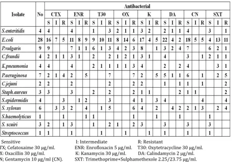
Table (2): Results of antibiogram of bacterial isolated from dead in shell and culled chicks.
S: Sensitive I: Intermediate R: Resistant
CTX; Cefatoxaime 30 µg/ml. ENR: Enrofloxacin 5 µg/ml. T30: Oxytetracycline 30 µg/ml.
OX: Oxacillin 30 µg/ml. K: Kanamycin 30 µg/ml. DA: Calindamycin 2 µg/ml.
9
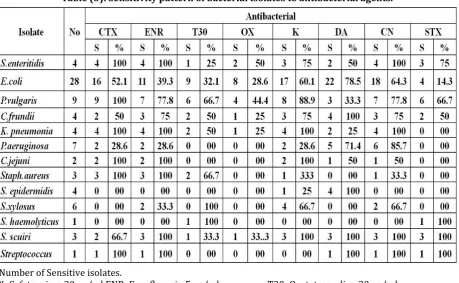
Table (3): Sensitivity pattern of bacterial isolates to antibacterial agents.S: Number of Sensitive isolates.
CTX; Cefatoxaime 30 µg/ml.ENR; Enrofloxacin 5 µg/ml. T30: Oxytetracycline 30 µg/ml.
OX: Oxacillin 30 µg/ml. K: Kanamycin 30 µg/ml. DA: Calindamycin 2 µg/ml.
CN; Gentamycin 10 µg/ml (CN). SXT: Trimethoprime+Sulphamethexole 2.25/23.75 µg/ml.
General speaking, E.coli isolates showed of sensitivity rate 52.1, 39.3, 32.1, 28.6, 60.1, 78.5, 64.3 to Cefatoxaime, Enrofloxacin, Oxytetracycline, Oxacillin, Kanamycin, Calindamycin and Gentamycin; respectively.Isolates of S.enteritidis, P.vulgaris, C.frundii, K. pneumonia, C.jejuni , Staph.aureus, Streptococcus and S. scuiriaresensitive to Cefatoxaime , Enrofloxacin,Kanamycinand Gentamycin with rate 50- 100%. P.aeruginosawas generally resistant to all tested antibacterial, while S. haemolyticus and S.xylosusare sensitive only to Oxytetracycline. Most of tested organisms are resistant to Oxytetracyclineand
Oxacillin. Trimethoprime+Sulphamethexole still effective on S.enteritidis, P.vulgaris, C.frundii, S. haemolyticus, S. scuiri and Streptococcus. Our results indicate the usage of antibacterial agents must be good controlled to get good effect and avoid drug resistance
Therefore we recommended the application of restricted hatchery sanitation together with using suitable disinfectant to minimize the risk of bacterial contamination and the possible related effect on hatchability and health of produced one day old chicks. Usage of antivacterial agents must be used under control and according to sensitivity test.
REFERENCES
[1] Vucemilo, M. ;Vinkovic, B.; Matkovic, K;
stokovic, I.; Jaksic, S.; Radovic, S.;Granic, K. and Stubican ,Đ.(2010) : The influence of housing systems on the air
quality and bacterial eggshell
contamination of table eggs. Czech J. Anim. Sci., 55 (6): 243–249.
[2] Sheldon, W. and Brake, J. (1991): Hydrogen peroxide as alternative
hatching eggs disinfectant. Poult. Sci.,
70(5): 1092-1098.
[3] Berrang, M.E.; Cox, N.A.;Frank, J.F. and
Buhr, R.J. (1999): Bacterial penetration
of the eggshell and shell membranes of the chicken hatching egg: a review. J. Appl. Poult. Res., 8: 499-504.
10
[5] Perry, GC.) (2004b): Welfare of the
Laying Hen.‘CAB International, Oxford shire, p.53.
[6] Khan, KA; Khan, SA; Aslam, A; Rabbani,
M and Tipu, MY (2004):Factors
contributing to yolk retention in poultry: a review. Pakistan Vet. J.,24:46-5
[7] Husseina, SA; Hassanb, AH and
Sulaimanc, RR (2008):Bacteriological and pathological study of yolk sac infection in broiler chicks in Sulaimani district. J. Dohuk Univ., 11: 48-56.
[8] Jahantigh, M (2010): Bacteriological
study of dead-in-shell embryos of ostrich. Iranian J. Vet. Res., 11: 88-90.
[9] Giacopello, C; Foti, M; Fisichella, V and
Lo Piccolo, F (2014): Antibiotic-resistance patterns of gram-negative bacterial isolates from breeder canaries (Serinuscanariadomestica) with clinical disease. J. Exotic Pet. Med., 24: 84-91.
[10] S
aif, Y.M.; Barnes, H.J.; Glisson, J.R.; Fadly, A.M.; McDougald, L.R. and Swayne, D.E. (2003): Diseases of poultry. 11th Ed., Ames, Iowa, Iowa State University Press.
[11] Zohair G.A M and Amer, M.M.
(2014): A study on bacterial contamination of table eggs sold for consumption in Sana’a city. VMJG, Vol. 61 (1) 15-22.
[12] IqbalM , Shah I A, Ali A, Khan
M A and S. Jan S (2006): Prevalence and in vitro antibiogram of bacteria associated with omphalitis in chicks. Pakistan Vet. J., 2006, 26(2): 94-96.
[13] Lukasova, J. and Sustackova, A.
(2003): Enterococci and antibiotic resistance. ActaVeterinaria Brno, 72: 315-323.
[14] Karmi, M. (2013): resistant
Prevalence of methicillin Staphylococcus aureus in poultry meat in Qena, Egypt. Vet. World, 6: 711-715.
[15] Collee, J.G.; Fraser, A.G.;
Marmion, B.P. and Simmons,A.(1996):
Practical Medical Microbiology.14th
Ed.,Chuechill, Livingstone.
[16] Forbes BA, Sahm DF,
Weissfeld AS (2002): Diagnostic microbiology. 11th Edition. Mosby, Inc. USA.
[17] Greenwood D, Slack RC,
Peutherer JF (2005)Medical microbiology. 16th Edition. Churchill Livingstone China.
[18] Q
uinn, P.J.; Carter, M.E.; Markey,B.K. and
Microbiology. WelfePublishing ,Mosbay . Year Book Europe Limited.
[19] Sneath, P.H.A.; Mair, N.S.;
Sharpe, M.E. and Holt, J.G. ( 1986) :Bergey’s Manual of Systematic Bacteriol. Vol. 2.Williams and Wilkins Co. Baltimore.
[20] Bauer, A.W., Kirby, W.M.M.,
Sheris, J.C. and Truk, M. (1966): Antibiotic susceptibility testing by a standardized single disc method. American Journal Clinical Pathology, 145: 225-230.
[21] CLSI (2013): Clinical and
Laboratory Standard Institute; Performance Standards for Antimicrobial Susceptibility Testing. CLSI Approved Standard M100-S23. Wayne, PA: Clinical and Laboratory Standards Institute.
[22] Stępien-Pysniak, D. (2010):
Occurrence of Gram-negative bacteria in hens’ eggs depending on their source and storage conditions. Polish J. of Vet. Sci., 13( 3) 507-513.
[23] Moats, W. A.
(1980):Classification of bacteria from commercial egg washers and washed and unwashed eggs. Appl. Environ. Microbiol. 40:710–714.
[24] Yaguchi K, Ogitani T, Osawa R,
Kawano M, Kokumai N, Kaneshige T, Noro T, Masubuchi K, Shimizu Y.(2007) : Virulence factors of avian pathogenic Escherichia coli strains isolated from chickens with colisepticemia in Japan. Avian Dis. Sep;51(3):656-62.
[25] P
ilattilivia, Jacqueline Boldrin de Paiva, Thaís Cabrera Galvão Rojas, Janaína Luisa Leite ,RogérioArcuriConceição ,GersonNakazato and Wanderley Dias da Silveira (2016): The virulence factor ychO has a pleiotropic action in an Avian Pathogenic Escherichia Coli (APEC) strain. BMC Microbiol. 16:35 DOI 10.1186/s12866-016-0654-2.
[26] GersonNakazato, Tatiana
Amabile de Campos ,ElianaGuedesStehling, Marcelo Brocchi
and Wanderley Dias da Silveira(2009): Virulence factors of avian pathogenic Escherichia coli (APEC). Pesq. Vet. Bras. 29(7):479-486, julho.
[27] Board, R. G.; Ayres, J. C. Kraft, A.
11
[28] Florian, M. L. E. and Trussell, P.
C. (1956): Bacterial spoilage of shell eggs. IV. Identification of spoilage organisms. Food Technol. 11:56–60.
[29] Musgrove, M. T.; Jones, D. R. and
Northcutt.J. K. (2004): Identification of Enterobacteriaceae from washed and unwashed commercial shell eggs. J. Food Prot., 67:2613–2616.
[30] Nazer, A. H. K.; Dadras,
H.andEskandari, S. (2006): Aerobic bacteria isolated from eggs and day-old chicks and their antibacterial resistance in Shiraz, Iran. Iranian J. of Vet. Res., University of Shiraz, 7 (2), Ser. No. 15,20-30.
[31] Cecilia Rosario Cortés,
Guillermo TéllezIsaías, Carlos LópezCuello, Jorge Mateo Villaseca Flores, Robin C. Anderson, Carlos Eslava Campos (2004): Bacterial isolation rate from fertile eggs, hatching eggs, and neonatal broilers with yolk sac infection. Rev LatinoamMicrobiol 2004; 46 (1-2): 12-16.
[32] P
yzik E., Marek A. (2012):Characterization of bacteria of the genus Staphylococcus isolated from the eggs of Japanese quail (Coturnixcoturnix japonica). Polish Journal of Veterinary Sciences Vol. 15, No. 4 (2012), 767-772.
[33] Nasrin S., Islam M.A., Khatun M.,
AkhterL.and Sultana S. (2012): Characterization of bacteria associated with omphalitis in chicks. The Bangladesh Veterinarian (2012) 29(2) : 63 – 68.
[34] Knöbl T, Cappellete CP and
Vigilato MAN (2012):Enterobacteria isolation in ostrich eggs (StruthioCamelus). Rev. Bras. Cienc. Avic. vol.14 no.1
[35] Orajaka LJ and Mohan K
(1985): Aerobic bacterial flora from dead-in-shell chicken embryos from Nigeria. Avian Dis 29: 583-589.
[36] Alaboudi AR, Hammad DA,
Basher HA and Hassen MG (1992): Potential pathogenic bacteria from
dead-in-shell chicken embryos. Iraqi J Vet Sci 5: 109-114.
[37] Bassouni AA, Saad FE, Awaad
MHH, Shalaby NA and Karaman RAA (1987): Microbial agents responsible for embryonic chicken mortality in native hatcheries in Monofia Province. Egypt. Poult. Sci 66: 3.
[38] Gulhan DB, Mehra KN,
Chaturved VK, Dhanesar NS (1999): Bacterial and fungal flora of dead in shell embryos. Indian Vet J 76: 750-751.
[39] Al-Sadi HI, Basher HA and Ismail
HK (2000): Bacteriologic and Pathologic studies on dead in-shell chicken embryos. Iraqi J Vet Sci 13: 297-307.
[40] Babaca ZAL ( 2014 ):
Epidemiological and bacteriological studies on dead-in-shell embryos. J Vet. SciTechnol5:(2) 170 . doi:10.4172/21577579.1000170.
[41] Boris Habrun, GordanKompes,
ŽeljkoCvetnić , Silvio Špičić,
MiroslavBenić, and Mario Mitak (2010): Antimicrobial sensitivity of Antimicrobial sensitivity of Escherichia coli Escherichia coli, , Salmonella Salmonella spp., spp., Pasteurellamultocida, Streptococcus suisPasteurellamultocida, Streptococcus
suis and andActinobacillusActinobacilluspleurop
neumoniaepleuropneumoniae isolated from diagnostic samples from large pig isolated from diagnostic samples from large pig breeding farms in Croatia. VeterinarskiArhiv 80 (5), 571-583.
[42] DalilaAngélicaMoliterno Duarte,
AldemirReginatoRibeiro, Ana Mércia Mendes Vasconcelos, Sylnei Barros Santos, Juliana Vital Domingos Silva, PatríciaLúciaArrudadeAndra de, LúciaSadae Pereira da Costa de ArrudaFalcão (2009): Occurrence of salmonella spp. in broiler chicken carcasses and their susceptibility to antimicrobial agents.Brazilian J of Microbiol., 40: 569-573.
[43] Lee Ventola C. (2015): The
Antibiotic Resistance Crisis Part 1: Causes and Threats. Pharmacy and therapeutics; 40(4): 277–283.