MOLECULAR TECHNIQUES FOR THE DETECTION AND
CHARACTERISATION OF A NOVEL RETROVIRUS
ASSOCIATED WITH MULTIPLE SCLEROSIS
BY
PHILIP WILLIAM TUKE
UNIVERSITY COLLEGE LONDON
Department of Virology
Windeyer Institute of Medical Sciences
Royal Free and University College Medical School
London W IP 6DB
A thesis submitted to the University of London
in fulfilment for the degree of
Doctor of Philosophy
ProQuest Number: 10014309
All rights reserved
INFORMATION TO ALL USERS
The quality of this reproduction is dependent upon the quality of the copy submitted.
In the unlikely event that the author did not send a complete manuscript and there are missing pages, these will be noted. Also, if material had to be removed,
a note will indicate the deletion.
uest.
ProQuest 10014309
Published by ProQuest LLC(2016). Copyright of the Dissertation is held by the Author.
All rights reserved.
This work is protected against unauthorized copying under Title 17, United States Code. Microform Edition © ProQuest LLC.
ProQuest LLC
789 East Eisenhower Parkway P.O. Box 1346
Abstract
Multiple sclerosis (MS) is thought to be an autoimmune disease precipitated in
genetically susceptible individuals by environmental factors. Recent attention has
focused on the possible involvement of retroviruses in its aetiology. Initial
experiments performed to detect the human retrovirus HTLV, in lymphocytes from
12 patients with MS, proved negative.
In an attempt to identify a putative novel human retrovirus, a polymerase chain
reaction (PCR) technique was developed which was capable of detecting a very
diverse range of retroviruses including HIV, HTLV, MPMV and MMLV. This ‘Pan-
Retrovirus’ PCR employed semi-nested, degenerate primers complementary to the
two most highly conserved motifs of the pol gene. Using this technique a novel
retroviral sequence, designated MSRV c-pol, was detected in the serum from a
patient with a 12 year history of MS. This sequence was also present in retroviral
particles which had been isolated from MS patient derived tissue cultures in France.
MSRV is related to the endogenous retrovirus ERV-9, however it remains uncertain
whether MSRV itself is an exogenous or endogenous retrovirus.
By combining the ‘Pan-Retrovirus’ PCR with a hybridisation-based detection assay,
MSRV c-pol RNA was detected in serum from 24 of 40 (60%) patients with MS but
not in 30 controls; and in cerebrospinal fluid from 5 of 10 patients with MS but not
in 10 other neurological disease controls. An MSRV specific RT-PCR assay was
also developed. This detected virion-associated MSRV pol RNA in serum from 9 of
17 (53%) patients with clinically active MS (in all 6 o f those not undergoing
immunosuppressive treatment), compared with only 3 of 44 (7%) controls.
A novel human retroviral sequence has been identified, and an association
demonstrated between the presence of MSRV-RNA and MS. Further work will be
required to determine the significance of MSRV in the aetiopathogenesis of this
Acknowledgements
I would like to thank my supervisor Dr. Jeremy Garson for his enthusiasm, support
and guidance during this project. I am grateful to Dr. Hervé Perron for the
invaluable assistance provided by his collaboration.
I would also like to thank Prof. Richard Tedder for his support and all my colleagues
in the Department of Virology for their help and encouragement over the years, as
well as for providing such a friendly working environment.
Thanks also goes to all my friends for their constant support, encouragement and
understanding especially during the writing of this thesis. Similarly, I am indebted
to my parents for their enduring support and encouragement.
This work was initially funded by the Middlesex Hospital Trustees, subsequently by
the Multiple Sclerosis Society of Great Britain and Northern Ireland and by
Table of contents
Title page 1
Abstract 2
Acknowledgements 3
Table of contents 4
List of figures 11
List of tables 12
Abbreviations 13
PUBLICATIONS ARISING FROM THIS THESIS 16
1 INTRODUCTION 17
1.1 Retroelements 17
1.1.1 Retroviridae 23
1.1.1.1 History 23
1.1.1.2 Classification 24
1.1.1.3 Replication strategy 27
1.1.1.3.1 V irion-receptor interactions 27
1.1.1.3.2 Reverse transcription 30
1.1.1.3.3 Nuclear transport of DNA 31
1.1.1.3.4 Integration 31
1.1.1.3.5 Transcription 32
1.1.1.3.6 Translation 33
1.1.1.3.7 Virion assembly and budding 36
1.1.1.4 Structure 37
1.1.1.5 Morphology 39
1.1.1.6 Disease association 41
1.1.1.6.1 Exogenous retroviruses in human disease 41
1.1.1.6.1.2 Human T-cell lymphotropic virus type I (HTLV-I) 43
1.1.1.6.1.3 Human T-cell lymphotropic virus type II (HTLV-II) 45
1.1.1.6.1.4 Human immunodeficiency virus type 1 (HIV-1) 46
1.1.1.6.1.5 Human immunodeficiency virus type 2 (HIV-2) 47
1.1.1.6.1.6 Retroviruses and autoimmunity 48
1.1.1.6.1.7 Retroviruses and neurological disease 50
1.1.1.6.1.8 Retroviruses and Multiple Sclerosis 51
1.1.1.7 Endogenous retroviruses 54
1.1.1.7.1 Classification of human endogenous retroviruses 54
1.1.1.7.2 Endogenous retroviruses and genome plasticity 55
1.1.1.7.3 Endogenous retroviruses: a functional role? 56
1.1.1.7.4 Endogenous retroviruses in disease 58
1.1.1.8 Diagnosis and detection 63
1.1.1.9 Prevention and treatment of infection 64
1.2 MULTIPLE SCLEROSIS 66
1.2.1 Epidemiology 66
1.2.1.1 Prevalence 66
1.2.1.2 Genetics 69
1.2.2 Clinical manifestations. 70
1.2.2.1 Presentation 71
1.2.2.2 Diagnosis 71
1.2.3 Pathology 74
1.2.3.1 Gross pathology 74
1.2.3.2 Chronic active plaques 75
1.2.3.3 Nascent plaques 77
1.2.3.4 Inactive plaques 78
1.2.4 Pathogenesis 79
1.2.4.1 Viruses 80
1.2.5 Aetiology 85
1.2.5.1 Genes 85
1.2.5.2 Environment 88
1.2.5.2.1 Viruses 89
1.2.6 Treatment 90
1.2.6.1 Corticosteroids 90
1.2.6.2 Cyclophosphamide 91
1.2.6.3 Copolymer 1 91
1.2.6.4 Interferons 92
1.2.6.5 Cladribine 93
1.2.6.6 Linomide 93
1.2.6.7 y-globulin 94
1.2.6.8 Cyclosporine 94
1.2.6.9 Methotrexate 94
1.2.6.10 Plasma Exchange 95
1.2.6.11 Lymphoid irradiation 95
AIMS OF THE PRESENT STUDY 96
2 MATERIALS AND METHODS 97
2.1 Materials 97
2.1.1 Patients, clinical samples, cells, viral cultures and plasmids 97
2.1.2. General sample handling 100
2.1.2.1 Separation and storage of peripheral blood 100
2.1.3 Cell cultures, virus isolation and purification. 101
2.1.3.1 HTLV infected cell lines 101
2.1.3.2 Mycoplasma screening of primary cultures 102
2.1.3.3 Monocyte cultures 102
2.1.3.4 EBV immortalised lines 103
2.1.3.5 Leptomeningeal and choroid plexus cell cultures 104
2.1.3.6 Extracellular virion purification 104
2.1.3.7 Reverse transcriptase (RT) activity assay 105
2.2 DNA Extraction 106
2.2.1 Preparation of buffer equilibrated phenol 106
2.2.2 Phenol chloroform extraction 106
2.2.4 Optical density measurement of concentration of nucleic acids. 108
2.3 RNA Extraction 108
2.3.1 Modification of the method of Chomczynski and Sacchi 108
2.3.1.1 RNA extraction from purified virions, serum, plasma and tissue culture fluid 109
2.3.2 Modified SNAP™ RNA extraction 110
2.3.2 1 SNAP™ extraction of RNA from PBMCs 110
2.3.2.2 SNAP™ extraction of RNA from brain tissue 111
2.3.2.3 SNAP^“ extraction of virion associated RNA from serum 112
2.4 cDNA synthesis 113
2.4.1 “In house” cDNA synthesis method 113
2.5 PCR 114
2.5.1 Theory of PCR 114
2.5.2 Principles of primer design 115
2.5.3 Principles of PCR optimisation 116
2.5.4 Standard PCR protocol 116
2.5.4.1 “Pan Retrovirus” PCR 117
2.5.4.2 Intra-yf/M PCR. 118
2.5.4.3 HTLV rax/rex PCR 118
2.6 Combined RT-PCRs 119
2.6.1 Titan™ RT-PCR 119
2.6.2 Pyruvate dehydrogenase (PDH) Tth RT-PCR 120
2.7 Cloning and sequencing 122
2.7.1 Cloning 122
2.7.1.1 Restriction endonuclease digests 122
2.7.1.2 Gel electrophoresis 122
2.7.1.3 Ligation, transformation and selection of transformants 123
2.7.2 Culture of R408 phage and purification of single stranded DNA for sequencing using
“magic minipreps” 124
2.7.3 Sequencing 125
2.7.3.1 Manual dideoxy sequencing 125
2.8 Hybridisation analysis o f PCR products: MSRV-/?^/ detection by Enzyme Linked
3 RESULTS 130
3.1 HTLV in MS 130
3.1.1 HTLV in MS. Introduction 130
3.1.2 HTLV in MS. PCR with HTLV specific primers 131
3.1.2.1 HTLV in MS. PCR with tax/rex primers 131
3.1.2.1.1 Introduction 131
3.1.2.1.2 Results 132
3.1.2.2 HTLV in MS. PCR with env primers 133
3.1.2.3 HTLV in MS. PCR w i t h p r i m e r s 133
3.1.3 HTLV in MS. Discussion 134
3.2 Development of a “Pan-Retrovirus” PCR system 136
3.2.1 Development of a “Pan-Retrovirus” PCR system. Introduction 136
3.2.2 Development of a “Pan-Retrovirus” PCR system. Results 138
3.2.2.1 Contamination prevention 139
3.2.2.2 Primer design 141
3.2.2.3 Optimisation of PCR parameters 142
3.2.2.4 Ability of the ‘Pan-Retrovirus’ PCR system to detect diverse retroviruses 144
3.2.3 Development of a “Pan-Retrovirus” PCR system. Discussion 145
3.3 “Pan-Retrovirus” PCR studies in multiple sclerosis 147
3.3.1 Detection of a novel/?o/sequence in serum from a patient with MS 147
3.3.1.1 Detection of a novel pol sequence in serum from a patient with MS. Introduction 147
3.3.1.2 Detection of a novel pol sequence in serum from a patient with MS. Methods 147
3.3.1.3 Detection of a novel pol sequence in serum from a patient with MS. Results 148
3.3.2 “Pan-Retrovirus” PCR studies in MS. Studies on purified virions 148
3.3.2.1 “Pan-Retrovirus” PCR studies in MS. Studies on purified virions. Introduction 148
3.3.2.2 “Pan-Retrovirus” PCR studies in MS. Studies on purified virions. Methods 149
3.3.2.3 “Pan-Retrovirus” PCR studies in MS. Studies on purified virions. Results 150
3.3.3 “Pan-Retrovirus” PCR studies in multiple sclerosis. Discussion 152
3.4 “Pan Retrovirus” PCR ELOSA studies in multiple sclerosis 154
3.4.1 “Pan Retrovirus” PCR ELOSA studies in multiple sclerosis. Introduction 154
3.4.2 “Pan Retrovirus” PCR ELOSA studies in multiple sclerosis. Methods 155
3.4.3 “Pan Retrovirus” PCR ELOSA studies in multiple sclerosis. Results 156
3.4.3.1 Prevalence of MSRV-c/»o/ in serum of patients with MS and controls. Results 156
3.4.4 “Pan Retrovirus” PCR ELOSA studies. Prevalence of MSRV-c/?o/ in serum and CSF
of patients with multiple sclerosis and controls. Discussion 158
3.5 MSRV specific PCR studies in multiple sclerosis 159
3.5.1 MSRV specific DNA PCR studies in multiple sclerosis. 159
3.5.1.1 MSRV specific DNA PCR studies in multiple sclerosis. Introduction 159
3.5.1.2 MSRV specific DNA PCR studies in multiple sclerosis. Method 160
3.5.1.3 MSRV specific DNA PCR studies in multiple sclerosis. Results 160
3.5.1.3.1 MSRV LTR specific DNA PCR studies in multiple sclerosis. Results 160
3.5.1.3.2 MSRV gag specific DNA PCR studies in multiple sclerosis. Results 161
3.5.1.3.3 M S R V s p e c i f i c DNA PCR studies in multiple sclerosis. Results 161
3.5.1.3.4 MSRV env specific DNA PCR studies in multiple sclerosis. Results 162
3.5.1.4 MSRV specific DNA PCR studies in multiple sclerosis. Discussion 162
3.5.2 MSRV pol specific RT-PCR studies on cellular RNA from patients with multiple sclerosis
and controls. 164
3.5.2.1 MSRV pol specific RT-PCR studies on cellular RNA from patients with multiple sclerosis
and controls. Introduction. 164
3.5.2.2 MSRV pol specific RT-PCR studies on cellular RNA from patients with multiple sclerosis
and controls. Methods 165
3.5.2.3. MSRV pol specific RT-PCR studies on cellular RNA from patients with multiple sclerosis
and controls. Results 167
3.5.2.4 MSRV pol specific RT-PCR studies on cellular RNA from patients with multiple sclerosis
and controls. Discussion 167
3.5.3 MSRV pol specific RT-PCR studies on serum from patients with multiple sclerosis
and controls 169
3.5.3.1 MSRV pol specific RT-PCR studies on serum from patients with multiple sclerosis
and controls. Introduction 169
3.5.3.2 MSRV pol specific RT-PCR studies on serum from patients with multiple sclerosis
and contols. Method 169
3.5.3.3 MSRV pol specific RT-PCR studies on serum from patients with multiple sclerosis
and controls. Results 171
3.5.3.4 MSRV pol specific RT-PCR studies on serum from patients with multiple sclerosis
4 GENERAL DISCUSSION 174
4.1 Discussion of results 174
4.1.1 Characterisation of a novel retrovirus from MS patients 174
4.1.2 Molecular epidemiology of MSRV 175
4.1.3 Characteristics of the MSRV virus 178
4.1.4 MSRV phylogeny 179
4.1.5 Endogenous or exogenous nature of MSRV 180
4.2 Expression of endogenous retroviruses in autoimmunity 184
4.2.1 Expression of endogenous related retroviruses in multiple sclerosis 185
4.2.2 Cross reactive antibody responses in disease 190
4.3 Possible mechanisms of action of MSRV in the aetiology and pathogenesis of multiple
sclerosis 191
4.3.1 Possible Autoimmune Mechanisms 192
4.3.2 Gliotoxin 193
4.4 Possible models for MSRV pathogenesis 195
4.5 Future work 198
4.5.1 Detection and analysis of virion-associated retroviral RNA 198
4.5.2 Immunoassay development and seroepidemiology 199
4.5.3 Immunohistology on post-mortem central nervous system tissue 200
4.5.4 Pathogenesis and MSRV 201
References 237
Appendices 283
Appendix 1 Buffers and Solutions 283
Appendix 2:
List of figures
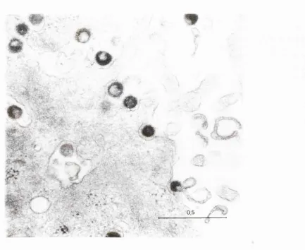
Figure 1 Electron micrograph of MSRV virions in a culture of leptomeningeal cells from a
patient with multiple sclerosis 202
Figure 2 MSRV cpol sequence amplified between the conserved VLPQG YXDD motifs in
p o t alignment with other retroviruses 203
Figure 3 HTLV tax/rex PCR of DNAs from HTLV patients and controls 204
Figure 4 Restriction enzyme analysis of HTLV-I/II for/rex PCR products 205
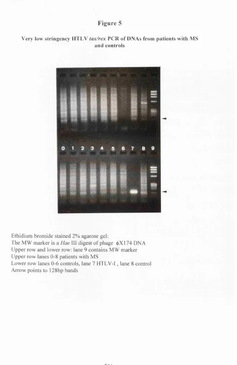
Figure 5 HTLV tax/rex PCR of DNAs from patients with MS and controls 206
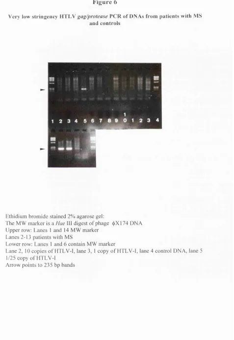
Figure 6 HTLV gag/protease PCR of DNAs from patients with MS and controls 207
Figure 7 “Pan-Retrovirus” PCR products of HIV pBHlO DNA without ‘hot-start’ 208
Figure 8 “Pan-Retrovirus” PCR on HIV pBHlO DNA showing effect of ‘hot-start’ 209
Figure 9 “Pan-Retrovirus” PCR showing sensitivity on the HTLV-I plasmid pMT2 210
Figure 10 “Pan-Retrovirus” PCR showing sensitivity on HIV-1 plasmid pBHlO DNA 211
Figure 11 “Pan-Retrovirus” PCR of MPMV cDNA and controls 212
Figure 12 Hae III digest of “Pan-Retrovirus” PCR product of MPMV cDNA 213
Figure 13 “Pan-Retrovirus” PCR on cDNA of RNA from sera of patients with MS 214
Figure 14 RT activity profile of sucrose density gradients 215
Figure 15 Nucleotide homology of clones with a reference MSRV-cpo/ clone 216
Figure 16 Phylogenetic analysis of the of the YMDD VLPQG region of pol 217
Figure 17 MSRV-po/ ELOSA data from 40 multiple sclerosis patients 218
Figure 18 MSRV-/?o/ ELOSA data from 30 blood donor controls 219
Figure 19 Alu PCR on cDNA samples from patients with MS and controls 220
Figure 20 MSRV gag specific PCR on DNA samples from patients with MS and controls 221
Figure 21 MSRV PT/?o/ AB/EF specific PCR on DNA samples from patients with MS and
controls 222
Figure 22 MSRV env specific PCR on DNA samples from patients with MS and controls 223
Figure 23 Total cellular RNA from PBMCs 224
Figure 24 MSRV pol 4/5/6 and gag RT-PCR and pol 4/5/6 ‘no-RT’ PCR on total cellular
RNA from PBMCs and brain cryosections 225
Figure 25 MSRV ŸTpol AB/EF RT-PCR on RNA from sera of patients with MS and controls 226
Figure 26 MSRV ?Tpol AB/EF ‘no RT’ PCR on RNA from sera of patients with MS and
controls 227
List of tables
Table 1 Detection of MSRV-cpo/ sequence in CSF of patients with MS and other neurological
disease controls 229
Table 1 continued (summary) “Pan-Retrovirus” PCR ELOSA detection of MSRV-RNA in
CSF cells of patients with MS and other neurological disease controls 230
Table 2 PCR Conditions 231
Table 2 continued PCR Conditions 232
Table 2 continued Primer and probe sequences (5' to 3') 233
Table 2 continued MSRV specific primer sequences (5' to 3') 234
Table 3 Sequences detected by ‘Pan-Retrovirus’ PCR in density gradient fractions with peak
RT-activity 235
Table 4 Detection of circulating virion associated MSRV-RNA in patients with MS and
Abbreviations
1° Primary
2° Secondary
A adenine
ALSV Avian Leukosis-Sarcoma Virus
Amp Ampicillin
AMV Avian Myeloblastosis Virus
APC Antigen Presenting Cell
Arg arginine
BAEV Baboon Endogenous Virus
BLV Bovine Leukaemia Virus
bp base pairs
BSA Bovine Serum Albumin
C cytosine
CA capsid
CaMV Cauliflower Mosaic Virus
CAT Chloramphenicol Acetyltransferase
cfu colony forming units
CNS Central Nervous System
CPM Counts Per Minute
CSF Cerebro Spinal Fluid
Dept. Department
DMSO dimethyl sulfoxide
DNA DeoxyriboNucleic Acid
DNase Deoxyribonuclease
DPM Decays Per Minute
Dra\ Deinococcus radiophilius 1
DTT dithiothreitol
EAE experimental allergic encephalitis EDSS expanded disability status scores EDTA disodium ethylenediaminetetra-acetate EIAV Equine Infectious Anaemia Virus ELOSA enzyme linked oligo-sorbent assay
EM electron microscopy
EMBL European Molecular Biology Laboratory
env envelope
ERV endogenous retrovirus
F-MuLV Friend-Murine Leukaemia Virus
FCS foetal calf serum
G guanine
gag group specific antigen
GaLY Gibbon Ape Leukaemia Virus
HaeUl Haemophilus aegyptius III
HBV Hepatitis B Virus
HCV hepatitis C virus
HSERV-9 (ERV-9), Human Sequence of Endogenous Retrovirus
hsps
y
heat shock proteins
HSRV human spuma retrovirus
HTLV-I Human T-cell Leukaemia Viruse type I HTLV-II Human T-cell Leukaemia Viruse type II HUMER41 Human Endogenous Retroviral sequ(
clone 41
I inosine
lAP Intracistemal A-type Particle IDDM insulin-dependent diabetes mellitus
IFN interferon
Ig immunoglobulin
IPTG Isopropylthio-|3-D-galactoside
Jr. junior
kbp kilo base pairs
kDa kilo Daltons
LB Luria-Bertani
LINES long interspersed sequences.
LTR long terminal repeat
Lys lysine
M molar
MAG myelin associated glycoprotein MALDI-TOF matrix assisted laser desorption/
ionisation time of flight
MBP myelin basic protein
MHC major histocompatability complex
min minute
MLV murine leukaemia virus
MMLV Moloney Murine Leukaemia Virus
MMTV Mouse Mammary Tumour Virus
MOI Multiplicity Of Infection
MoMLV Moloney-Murine Leukaemia Virus
MP methylprednisilone
MPMV Mason-Pfizer monkey virus
MRI Magnetic Resonance Imaging
mRNA messenger RNA
MS Multiple Sclerosis
MSRV Multiple Sclerosis associated Retro Virus
MW molecular weight
NC nucleoprotein
MEN New England Nuclear
nm nanometer
OD optical density
OPD o-phenylenediamine
ORF open reading frame
PBMC peripheral blood mononuclear cells
PBS phosphate buffered saline
PCP Pneumocystis carnii pneumonia
PCR polymerase chain reaction
Pfu Pyrococcus furiosus
pol Pwo RA RNA RNase RPMI RR RSV RT RT-PCR RTVL-H S SD secs. SFV SFVcpz SINES SI VI S I V mac SMRV-H SNAP™ SRV-2 Ssp\ STLV-I STLV-II T Tag TCA TcR TE Tm Tth U UCLMS USB YEP uv v/v VISNA w/v X-gal
xg
polymerase Pyrococcus woesei rheumatoid arthritis ribonucleic acid ribonucleaseRoswell Park Memorial Institute relapsing remitting
Rous sarcoma virus reverse transcriptase
combined reverse transcriptase and polymerase chain reaction
Retrovirus-Like Human endogenous sequence
Svedberg unit standard deviation seconds
Simian Foamy Virus
chimpanzee simian foamy virus short interspersed sequences.
Simian Immunodeficiency Virus type 1 simian immunodeficiency virus macaque Simian Monkey Retrovirus H
simple nucleic acid preparation Simian Retrovirus type 2
Sphaerotilus natansX
Simian T-cell lymphotropic virus I Simian T-cell lymphotropic virus II thymine
Thermus aquaticus
trichloroacetic acid T cell receptor Tris EDTA melt temperature
Thermus thermophilus
uracil
University College London Medical School United States Biochemical
PUBLICATIONS ARISING FROM THIS THESIS
Garson, J. A., Tuke, P. W., Giraud, P., Paranhos Baccala, G., and Perron, H. (1998).
Detection of virion-associated MSRV-RNA in serum o f patients with multiple
sclerosis. Lancet 351, 33
Tuke, P. W., Perron, H., Bedin, P., Beseme, P., and Garson, J. A. (1997).
Development o f a pan-retrovirus detection system for multiple sclerosis studies. Acta
neurol. Scand. Suppl. 169, 16-21.
Perron, H., Garson, J. A., Bedin, P., Beseme, P., Paranhos Baccala, G., Komurian
Pradel, P., Mallet, P., Tuke, P. W., Voisset, C., Blond, J. L., Lalande, B.,
Seigneurin, J. M., and Mandrand, B. (1997b). Molecular identification of a novel
retrovirus repeatedly isolated from patients with multiple sclerosis. The
Collaborative Research Group on Multiple Sclerosis. Proc. Natl. Acad. Sci. U. S. A.
94, 7583-7588.
Perron, H., Pirouzi, R., Tuke, P., Garson, J. A., Michel, M., Beseme, P., Bedin, P.,
Mallet, P., Marcel, E., Seigneurin, J. M., and Mandrand, B. (1997). Cell cultures and
associated retroviruses in multiple sclerosis. Collaborative Research Group on MS.
Acta Neurol. Scand. Suppl. 169, 22-31.
Brennan, M., Runganga, J., Barbara, J. A., Contreras, M., Tedder, R. S.,
Garson, J. A., Tuke, P. W., Mortimer, P. P., McAlpine, L., and Tosswill, J. H.
(1993). Prevalence of antibodies to human T cell leukaemia/lymphoma virus in
blood donors in north London. BMJ. 307, 1235-1239.
Tuke, P. W., Luton, P., and Garson, J. A. (1992). Differential diagnosis of HTLV-I
and HTLV-II infections by restriction enzyme analysis o f 'nested' PCR products.
1 INTRODUCTION
1.1 Retroelements
In order to understand the role of the retroviruses, both exogenous and endogenous,
in both the pathogenic and non-pathogenic context, it is necessary to place them
within the setting of retroelements as a whole. The unifying feature of retroelements
is that they have been generated by reverse transcription. This group of entities
forms a continuum from purely repetitive sequences in genomic deoxyribonucleic
acid (DNA) such as short interspersed sequences (SINES), through long interspersed
sequences (LINES) and pseudogenes, to complex fully functional retroviruses.
Earliest pre-cellular life is thought to have been ribonucleic acid (RNA) based,
initially with self replicating RNAs and then later with RNA genomes and their
encoded proteins (Weiner, 1987). The transition to DNA as the genetic material
required the evolution of reverse transcriptase (RT). The evolution of the
retroelement, and hence RT, is considered as one o f the key initial stages in the
evolution of life. It may well be the case that the retrotransposons (described below),
represent molecular fossils of this evolution from RNA based life to DNA based.
The ubiquitous nature of retroelements across kingdoms is consistent with this
The simplest retroelements are highly repetitive non-coding sequences of DNA e.g.
Alu family and SINES. Then come the pseudogenes which have been generated by
reverse transcription and integration of cellular RNAs and hence have coding
capability. Pseudogenes that have acquired an upstream promoter sequence and can
be actively transcribed are termed retrogenes. Pseudogenes and other repetitive non-
long terminal repeat (LTR) sequences e.g. SINES and Alu sequences are thought to
have been generated by LINE encoded RT (Hutchison III et al. 1989; Jenson et al.
1991) rather than by misdirected retroviral co-packaging (Domburg and Temin,
1990; Levine et al. 1990).
Retroelements are found in organisms from Eubacteria to Eukaryotes.
Retroelements which encode their own RT (retrotransposons) are phylogenetically
related and computer alignment of all these sequences has allowed phylogenies to be
constructed (Doolittle et al. 1989; Xiong and Eickbush, 1990). From these
phylogenetic trees one can attempt to reconstruct the history of events leading to the
formation o f these entities. There are also homologous proteins in some plant and
animal DNA viruses (Toh et al. 1985), and their reverse transcriptase is similar to
sequences found in the introns of some fungal mitochondria (Michel and Lang,
1985).
Retrotransposons can be phylogenetically divided into two main groups based upon
their reverse transcriptase (RT) sequences (Xiong and Eickbush, 1990). Firstly,
there is the branch containing the LINE like sequences, or non-LTR retrotransposon
mitochondrial plasmid, RT-like sequences of Chlamydomonas reinhardtii (Boer and
Gray, 1988), multicopy single stranded DNA (msDNA) found in bacteria, and
telomerases (Eickbush, 1997). Secondly, there is the branch containing the LTR
retrotransposons, both the Copia/Tyl group and Gypsy group o f transposable
elements, and also the true retroviruses as well as the hepadnaviruses and
caulimoviruses (both DNA viruses which do not possess LTRs) (Xiong and
Eickbush, 1990).
LINES represent the next level o f complexity being retroposons or retrotransposons
of the non-LTR type. Retroposons encode the RT which is responsible for their own
retrotransposition. They also possess an internal GC rich promoter and a partially
characterised protein, OREL Retrotransposons (of the LTR type) are distinguished
by the possession of a capsid gene, the equivalent of the retroviral gag, and LTRs
which contain the enhancer/promoter sequences responsible for their own
expression.
The most complex type of retroelement is the retrovirus which contains at least one
extra gene, the envelope gene {env), in addition to the genes present in
retrotransposons. The env gene would appear to be the final acquisition necessary
for the formation of a fully infectious viral particle. Retroviruses can be divided into
two types, exogenous and endogenous, according to their mode of transmission. In
general, endogenous retroviruses can be considered to be the molecular fossils of
lesser degree supports this theory. However, an alternative and perhaps equally valid
view, is that exogenous retroviruses represent a currently activated form of an
endogenous retrovirus (Lower et al. 1996). By entering the germ cell line
endogenous retroviruses have escaped from the selection pressure for maintaining
the integrity of their open reading frames (ORFs). Their new mode o f transmission
results in a lack o f necessity for formation o f viral particles.
Recombinatorial exchange between retroelements, as between retroviruses, would
seem to be an extremely rare event, as evidenced by the concordance between the RT
based phylogeny and the genome structure of the element (Xiong and Eickbush,
1990). LTR containing retrotransposons fall on the LTR branch and non LTR
containing retrotransposons fall on the non LTR branch of their RT based
phylogenetic tree. Similarly, there is no evidence for genomic exchange between
members of the different groups of retroelements present in the same species.
However, there is evidence for limited genomic exchange within the retroviruses,
(typically within env), giving rise to new retroviruses (McClure et al. 1988). The
acquisition of new genes and functional abilities during the evolution of
retroelements is also thought to have been due to recombination.
Retrotransposons are present in the genomes of organisms as evolutionarily diverse
as animals, plants, protozoans and fungi (Garfinkel 1992). Closely related
retrotransposons may be present in organisms which are evolutionarily extremely
caulimoviruses are restricted to plants, and the hepadnaviruses and retroviruses to
vertebrates. It is not possible to simply superimpose the deduced evolutionary
histories o f the retroelements on that of the host organisms.
The interpretation of such phylogenies in terms of the evolutionary relatedness o f the
various sequences is fraught with difficulty. Although a phylogeny gives an accurate
representation o f the current degree of homology o f the various entities, the implied
inference of past temporal ancestries may well be misleading.
The high rates o f divergent evolution observed for exogenous retroviruses, in
particular those that are not highly cell associated such as HIV-1 and HIV-2, and the
overall phylogeny and species distribution of retroviral sequences would appear to be
incompatible. If retroviruses diverged at the rate currently observed for HIV then
their common ancestor would only date back 10,000 years or so (Doolittle et al.
1989). The species diversity of retroviral infections alone, (not even accounting for
the endogenous retroviruses), from fish and foul to mammals, would argue against
this. The most likely explanation for this apparent contradiction would be that
exogenous retroviruses and their associated high rate of genetic change, represent a
very short period in the evolutionary history of any given retrovirus. The limited
number o f exogenous retroviruses in comparison to endogenous retroviral sequences
Cross species transfer of retroviruses would also be a feature of this model of
retroviral evolution. HIV may be a classic example in this sense, representing a
recent zoonosis. All the major sub types of HIV 1 (A-G) are thought to have
originated from a single zoonotic transmission event within the last 50 years or so
(Weiss, 1998). Allowing for such zoonoses it has been estimated that retroviruses
evolved after vertebrate evolution and perhaps subsequent to mammalian evolution
(Doolittle et al. 1989). The time period at which the evolution of individual
retroviruses took place can similarly be estimated. Human endogenous retrovirus
(HERV) evolution can also be traced, and the majority o f retrotransposition events
and the most significant expansion seems to date back to early primate evolution
(Haltmeier et a l 1995). Endogenous retroviruses can be used like any other genetic
marker to date the history o f their evolution and incorporation into the genome
(section 1.1.1.10.2) (Arvidsson et al. 1995; Svensson et al. 1996; Andersson et al.
1998; Svensson and Andersson, 1997; Svensson et al. 1995; Widegren et al. 1996).
The majority of HERVs demonstrate sequence homology to murine or primate
retroviruses (Rasmussen et al. 1993) and only a few are related to the exogenous
human retroviruses, HIV (Tonjes et al 1996) (Shih et a l 1989) (Perl et a l 1989),
HTLV (Perl et al. 1989; Shih et al. 1989) and human foamy virus (Cordonnier et al.
1995).
It is interesting to note that the most variable region o f the retroviral genome is the
env gene and this applies to both the endogenous and exogenous forms. Endogenous
retroviruses with defective env genes may be able to use in trans the gene products
pseudotypes (Lusse et al. 1990; Hu and Temin, 1990a; Hu and Temin, 1990b). The
most defective endogenous retroviruses may be considered to be the lone LTR
elements. These are commonly found at high copy levels in the genome and are
thought to be the result of recombination between retroviral LTRs with excision of
the internal sequence.
1.1.1 Retroviridae
1.1.1.1 History
Retroviruses were first isolated at the beginning of the century during studies of the
aetiology of infectious diseases. Equine infectious anaemia, which is now known to
be due to a mammalian C-type retrovirus, was one of the first diseases shown to have
a viral aetiology by the filterable nature of the causative agent (Vallee and Carre
1904). In 1911 Peyton Rous isolated Rous sarcoma virus (RSV) as the cause of a
spontaneous chicken sarcoma (Rous 1911). The virus was observed under EM in
1947 by Claude and colleagues (Claude et al 1947) but the morphology of the viral
particle was not determined until thin section methods were developed (Gaylord
1.1.1.2 Classification
The classification of retroviruses into a single family is warranted by the unique
character o f their genome and mode of replication, and by their common physical,
biochemical and morphological properties. Nucleotide sequence analysis of their
genomes has confirmed that retroviruses represent an individual family of viruses
and has enabled further phylogenetic analysis.
Retroviruses are classified as a family within the taxon of reverse transcribing
viruses, which also includes the Caulimoviridae and Hepadnaviridae families of
viruses (Mayo and Pringle 1997) as detailed below.
Family Retroviridae
Genus Spumavirus
This includes Human spuma retrovirus (HSRV) and Simian spuma retrovirus
Genus unnamed, mammalian B-type retroviruses
Mouse mammary tumour virus (MMTV)
Genus unnamed, mammalian and reptilian C-type retroviruses
This includes many murine leukaemia viruses (MLV)-related e.g. Abelson, AKR
sarcoma viruses (e.g. Harvey, Kirsten, Moloney murine sarcoma viruses); feline
leukaemia virus; feline sarcoma viruses; gibbon ape leukaemia virus; woolly monkey
sarcoma virus; porcine type C virus; guinea pig type C virus; and viper type C virus.
Genus unnamed, avian type C retroviruses
This includes Rous sarcoma virus (RSV); avian carcinoma viruses; avian sarcoma
viruses; avian leukosis viruses; avian myeloblastosis viruses (AMV); avian
reticuloendotheliosis viruses; and duck spleen necrosis virus.
Genus unnamed, type D retroviruses
This includes Mason-Pfizer monkey virus (MPMV); simian type D virus 1; Langur
type D virus; squirrel monkey type D virus; and ovine pulmonary adenocarcinoma
virus (Jaagsiekte).
Genus unnamed, HTLV/BLV viruses
This includes human T-cell lymphotropic viruses (HTLV-I and HTLV-II), simian T-
cell lymphotropic viruses (STLV-I and STLV-II) and bovine leukaemia virus (BLV).
Genus Lentivirus
This includes human immunodeficiency viruses (HIV-1 and HIV-2), simian
immunodeficiency viruses (SIV) (African green monkey, sooty mangabey, stump
tailed macaque, pig-tailed macaque. Rhesus, chimpanzee, and mandrill viruses);
Alternatively, retroviruses can be divided into four groups according to their RT
sequence based phytogenies; the MLV group (which includes the spumaviruses), the
MMTV/RSV group, the HTLV/BLV group and the lentiviral group (Xiong and
Eickbush, 1990).
Retroviruses can also be divided into two subgroups according to their mode of
transmission. Exogenous retroviruses can be regarded as those where transmission
occurs via an extracellular intact viral particle, whereas endogenous retroviruses are
inherited in Mendelian fashion. The division of retroviruses into these two
categories is not straightforward, since families of retroviruses exist with both
exogenous and endogenous members. Also an endogenous retrovirus may be
infectious in a different host context (Weiss, 1984).
Retroviruses are further classified on the basis o f the range of host species they can
infect. This is a reflection of the species distribution of the cellular receptor to which
the virus binds. Xenotropic viruses are ones which are normally endogenous in their
natural host species, where they do not usually replicate, but have a wide range of
heterologous species which they can infect. Ecotropic viruses will only grow in cells
of the species from which they were isolated. Amphotropic viruses are capable of
both growing in cells of the species from which they were isolated and have a wide
range o f heterologous species which they can infect (Teich, 1984; Weiss, 1984).
The criteria used to classify retroviruses may also include genome structure as well
1.1.1.3 Replication strategy
L I .1.3.1 V irion-receptor interactions
The virus binds to a specific cell surface molecule which acts as a receptor. To date
only a few retroviral receptors have been identified but they vary greatly in structure,
function and tissue distribution. Their only common feature is that they are all cell
surface proteins with transmembrane domains.
The principal HIV-1 receptor is CD4, which is part of the immunoglobulin
superfamily and is involved in the interaction of helper T cells with antigen
presenting cells. Recently two of the long searched for co-receptors for HIV-1 were
identified and found to be chemokine receptors. The CXCR4 molecule is the major
co-receptor for cellular entry by T-cell tropic HIV-1 strains and the CCR5 molecule
is the major co-receptor for macrophage tropic strains (Doranz et al. 1997).
The receptor for the ecotropic murine leukaemia virus is Rec-1, the receptor for the
rat amphotropic virus is Ram-1 and the receptor for the gibbon ape leukaemia virus
is Glvr-1. All of these three proteins have the numerous transmembrane domains
characteristic of membrane transport systems. Rec-1 is a transporter of basic amino
acids (Kim et a l 1991; Wang et a l 1991) and Ram-1 and Glvr-1 are phosphate
transporters (Johann et a l 1993; Kavanaugh et a l 1994). Glvr-1 also acts as a
sarcoma virus (Tailor et a l 1993). It is interesting to note that these viruses are all
related members of the mammalian C-type genus of retroviruses.
The bovine leukaemia virus receptor has a single transmembrane domain but it is
unrelated to any of the other known retroviral receptors and its function remains to
be determined (Ban et a l 1993).
The receptor for Rous sarcoma virus and subgroup A avian leukosis-sarcoma virus
has been identified as the protein Tva. This resembles a single membrane spanning
domain of the much more complex low density lipoprotein receptor (Bates et a l
1993; Young et a l 1993).
A putative cellular receptor for the feline immunodeficiency virus has been identified
as the feline homologue of CD9. This identification was achieved by using a
monoclonal antibody which blocks infection (Hosie et a l 1993; Willett et a l 1994).
Initial reports of a similar approach identifying a putative cellular receptor for
HTLV-I (Gavalchin et a l 1993) have not been confirmed. Recently a novel MMTV
receptor has been identified which has no homology with any known membrane
proteins (Golovkina et a l 1998b). Once a retrovirus has bound to a cell, the exact
Virus interference
The infection of a cell by a given retrovirus results in resistance to superinfection
with that retrovirus and with any others which utilise the same receptor molecule.
This phenomenon is termed interference and arises from the interaction of the
retroviral Env protein with the receptor. Virion formation is not necessary for
interference to occur, indicating that the Env protein produced by an infected cell can
interact in this manner with the receptor from the same cell. The exact mechanism
of this process is not known but it has been shown for Rec-1, the ecotropic MLV
receptor, that the important step is the binding and continued blocking o f the receptor
by the Env protein. This blocking of the receptor does not interfere with the normal
cellular functioning of the protein (Coffin 1996).
The viral glycoproteins encoded by the env genes o f endogenous retroviral genomes
are frequently expressed as host antigen at the cell surface. The expression of these
endogenous retroviral env genes may thus protect the host organism against infection
by a closely related pathogenic exogenous retrovirus by receptor interference (Lower
et al. 1996). This subject is reviewed in further detail by Coffin (1996) and Weiss
1.1.1.3.2 Reverse transcription
Retroviruses replicate by means of their virally encoded reverse transcriptase (RT).
This enzyme is packaged within the virion and is present at between 15 and 50
molecules (Pyra et al. 1994). Reverse transcription takes place within the core
particle, whether this occurs entirely in the cytoplasm of the newly infected cell, or at
least partially within that of t^ ^ r o g e n itc ^ s uncertain. Extracellular viral particles
may contain replicative intermediates which possess at least partial cDNA copies of
the viral genome (Zhu and Cunningham, 1993; Lori et al. 1992; Trono, 1992). In
addition, other retroviral RNAs (predominately those transcripts including a
packaging signal, i|/) and even various transfer RNAs (tRNAs), 5 Svedberg units (S)
rRNAs and cellular messenger RNAs (mRNAs) (e.g. globin) may be co-packaged
within the virion (Coffin, 1996).
Infection of a cell by a retrovirus results in the synthesis o f a cDNA copy of the viral
genome by the viral RT, the RNA within the DNA:RNA hybrid is then degraded by
the RNase H activity of the enzyme and the synthesis o f the second strand of the
DNA is initiated. The linear double stranded DNA copy is completed prior to
integration into the host’s genome. Circularised forms of the double stranded DNA
are also formed, these may possess one or two LTRs (Famet and Haseltine, 1991).
Initially the two LTR form was thought to be the pre-integration form of the virus
(Panganiban and Temin, 1984) but these DNA circles are now thought to be
1.1.1.3.3 Nuclear transport of DNA
The precise mechanism of nuclear transport of the pre-integration complex of viral
proteins and double stranded DNA has not been fully elucidated but the size of the
complex would seem to preclude entry through the nuclear pores (Bowerman et al.
1989). This conclusion would appear to be supported by the observation that cell
division and the preceding breakdown of the nuclear membrane are a prerequisite for
integration (Roe et al. 1993) with only one of the daughter cells becoming infected
(Hajihosseini et al. 1993). However this is not true for all retroviruses, lentiviruses
seem to employ a separate mechanism not requiring mitosis (Lewis and Emerman,
1994).
1.1.1.3.4 Integration
Integration of the retroviral DNA genome into that of the host is generally
considered to be a prerequisite for viral replication. The site o f integration is precise
with respect to the viral genome, but essentially random with respect to that of the
host. Integration occurs by a strand transfer mechanism which has an absolute
requirement for the viral integrase protein (IN) but may also normally include other
proteins. There is a reported preference for transcriptionally active regions i.e. where
the DNA within the chromatin structure is more accessible, generally these are non
The integrated provims can become part of the inherited genetic material of the host
genome if it infects germ line cells. All animal genomes are known to contain
retroviral sequences. The majority of these sequences are quiescent, forming a
substantial proportion of the repetitive genomic sequences. Frequently they are
located in the methylated sections of the genome and consequently not usually
expressed, although some endogenous retroviruses are capable of forming intact
retroviral particles. Endogenous retroviruses can be activated by certain chemicals
such as mutagens or carcinogens (Irons et al. 1987) e.g. the chemical carcinogens 5-
iododeoxyuridine (lUdR) and 5-bromodeoxyuridine (BUdR) (Lowy et al. 1971,
Stoye and Moroni, 1983) or by mitogens (Stoye and Moroni, 1985), by radiation
with UV and X-rays (Weiss et al. 1971) and y-rays, and by other mechanisms such
as DNA-viruses or physiological processes (e.g. ageing) to express antigens or to
form infectious virus particles.
1.1.1.3.5 Transcription
Once the DNA provirus has integrated into the host genome the expression of the
virus is controlled by the enhancer and promoter elements o f the 5’ LTR. These are
contained within the U3 region of the LTR and frequently have duplicated enhancer
motif regions, as is also often found in cellular genes. Typical cellular transcription
factor binding motifs are frequently found in these U3 regions e.g. AP-1, SPl,
CCAAT and TATA boxes, and hormonally and tissue specific responsive elements
ordinary cellular transcription machinery, the RNA being transcribed by the cellular
RNA polymerase II. The full length RNA transcripts are consequently capped at
their 5’ terminus and cleaved and polyadenylated at the 3’ end of R following the
polyadenylation signal AAUAAA. These full length genomic RNAs then have a
variety o f subsequent fates, a proportion are destined to become new viral genomes
and are transported from the nucleus to the cytoplasm and assembled into the nascent
viral particles. The precise cellular location of this process depends on the type of
virus (Section 1.1.1.5). Another fraction is also transported to the cytoplasm but
functions as the polycistronic mRNA for the Gag and Pol proteins. Finally, some of
the transcripts are spliced at various splice junctions to form the mRNAs for the Env
proteins and any accessory regulatory proteins (e.g. Tax/Rex o f HTLV, Tat/Rev of
HIV) (Coffin, 1996).
1.1.1.3.6 Translation
The full length genomic RNA and the spliced viral mRNAs all maintain the same 5’
LTR structure. This serves as the initiation site for translation, ribosomal subunits
binding initially to the capping group and progressively scanning the RNA 5’ to 3’,
with protein synthesis commencing at the gag AUG initiation codon. The gag, pro,
and pal genes form a single translational unit. This is expressed in differing lengths
to different extents. Hence Gag, Gag-Pro, and Gag-Pro-Pol proteins are synthesised
on free polyribosomes from the same genomic length mRNAs. Only between one
the enzymatic verses structural roles of these products. Most retroviruses produce
either Gag or Gag-Pro rather than both. The precise mechanisms by which this
proportionality is ensured differ between the retroviruses but all consist of a partial
block to translational read-through. (Hatfield et a l 1992).
The first mechanism identified was the nonsense suppression which occurs in the
murine leukaemia virus (MLV). The gag and pro-pol genes are in the same reading
frame, but there is an amber stop codon (UAG) separating them. This provides a
weak translational stop codon, which is occasionally read-through by the
translational machinery of the cell by the mis-incorporation o f a glutamine residue
(CUG or CUA) (Yoshinaka et a l 1985). Also involved are a purine rich sequence
immediately downstream of the UAG, a 49 bp sequence which forms a stem loop
structure, and a sequence which base pairs with this loop to form a structure termed a
pseudoknot. This pseudoknot is thought to impede the passage of the ribosome.
(Feng era/. 1992).
The second mechanism is ribosomal frame-shifting, where for the Pro or Pol protein
to be synthesised, the ribosome has to slip since the protein is coded for in a different
reading frame. As with the nonsense suppression mechanism there is a stop codon,
but this is overcome by frame-shifting rather than by simple mis-incorporation. A
shift to the -1 frame requires, in all viruses studied, the stop codon and a specific (but
unique) seven base A-U rich sequence at the frame-shift site, followed by a
pseudoknot. The efficiency of ribosomal frameshifting is controlled so that the
(ALSV) where only one ribosomal frame-shift takes place, the probability o f it
occurring is -5% (Arad et a l 1995). In HTLV-II and MMTV where two ribosomal
ffameshifts are required the probability of the first occurring is -25% and the second
-10% hence the ratio of Gag to Pol is -2.5% (Hizi et a l 1987; Jacks et a l 1987;
Mador et a l 1989). Spumaviruses require a +1 frame shift to occur but the
mechanism behind this is not clear (Jacks et a l 1988; Kupiec et a l 1991).
The Env protein is synthesised from spliced viral RNA on polyribosomes of the
rough endoplasmic reticulum (RER), in the same manner as ordinary cell surface
proteins (Lake, 1981) . The Env proteins contain a signal peptide sequence at their
amino terminus which directs their synthesis to the RER, and as with cellular
proteins it is subsequently cleaved. The Env precursor proteins are anchored in the
RER by their hydrophobic trans-membrane domain which is located toward the
carboxy terminus of the protein. Deletion of this sequence results in the synthesis of
a soluble truncated form of the protein which is exported and released from the cell
(Perez et a l 1987). Glycosylation of the Env protein occurs shortly after translation.
Subsequent modification of the carbohydrate and cleavage o f the Env precursor
protein into surface (SU) and trans-membrane (TM) subunits occurs in the Golgi
apparatus. The surface (SU) and trans-membrane (TM) subunits remain associated
with each other subsequent to their cleavage. This occurs at a conserved amino acid
sequence which follows three or four basic amino acid residues (typically Arg/Lys-
X-Lys-Arg) (Dong et a l 1992). Failure of this cleavage to occur results in the
1.1.1.3.7 Virion assembly and budding
The details o f retroviral particle assembly are poorly understood, but some general
observations can be made. The Gag precursor protein associates with the retroviral
genome via the carboxy-terminal nucleoprotein (NC) region and the packaging
signal psi (v|/) located 3’ of the 5’ LTR. The middle capsid (CA) regions o f the
precursor proteins self aggregate, and the amino-terminal matrix (MA) region
associates with the cell membrane. The same interactions of the polyproteins Gag-
Pro and Gag-Pro-Pol brings these protein domains into the virion in the requisite
proportion and location. The interaction with the cell membrane and acquisition of
the viral envelope, occurs after particle formation where A-type particles are
observed, (as is the case for the A-type, B-type and D-type retroviruses), but occurs
simultaneously for C-type retroviruses (Wills and Craven, 1991).
Subsequent to assembly o f the viral particles the domains o f the polyprotein are
cleaved by the protease to yield the individual proteins. This results in the formation
of a virion capable of performing the reverse transcription of its genome. The Env
proteins are incorporated into the envelope of the virion presumably by interactions
with the matrix protein. The functional proteins during virion assembly are all
polyproteins. Gag (MA-CA-NC), Gag-Pro, and Gag-Pro-Pol, but are individual
separate proteins during the early stages of infection. Virion assembly requires the
polyproteins, whilst the uncleaved precursor to RT shows no or very reduced reverse
transcriptase activity. Although all of the major genes and proteins are required to
of viral particles. Discernible viral particles can even be produced by just the first
180 of the 701 amino acids of RSV Gag, hence there is an aggregation domain
within the MA domain. These viral particles were much less dense than the
authentic retrovirions but were still capable of budding and release from the plasma
membrane (Weldon, Jr. and Wills, 1993).
1.1.1.4 Structure
Retrovirus particles have a common structure. Their genome is packaged within an
icosahedral core or nucleoid which is formed by the capsid protein of the Gag
polyprotein. This is in turn surrounded by the matrix (MA) protein fragment which
directly underlies the envelope. The amino terminal residue o f MA is usually
myristilated. The matrix protein seems to determine the intracellular location of
virion formation. Finally, the extracellular virion is contained within an outer
envelope made of a unit membrane derived from the host cell membrane, but
containing in addition the retro virally derived envelope protein, which is often
clearly visible as projecting spikes under electron microscopy (Teich, 1984).
Retroviruses are diploid single stranded RNA viruses and possess a genome of 60 to
70 Svedberg units composed of identical subunits 7 to 10 thousand bases in size.
Each RNA molecule is effectively an mRNA, being the positive sense strand, having
a 5’ methylated cap structure and a poly A 3’ tail approximately 200 bases in length.
contact has been localised to near the packaging signal (\\f) and the primer binding
site (PBS). The reason for this diploidy is also unknown but it may be involved in
replication or enable complementation of deleterious mutations. In addition to the
viral genome, the nucleocapsid contains the virally encoded reverse transcriptase
(RT), and base paired with the RNA genome, a host encoded specific tRNA
molecule. The genomic structure of all retroviruses is essentially similar. The RNA
genome o f simple retroviruses consists of three major coding regions, gag, pol and
e m and possesses long terminal repeat (LTR) sequences at either end. The gag
region encodes the capsid proteins, the pol region the protease, reverse transcriptase,
RNase H and the integrase, and the env region the envelope glycoprotein. The LTRs
o f the RNA genome consist of identical direct terminal repeat (R) sequences with
unique sequences internal to these at the 5’ (U5) and 3’ (U3) ends of the genome.
Complex retroviruses have accessory genes which play a role in the regulation of the
viral life cycle (Coffin, 1996).
The typical chemical composition of retrovirus particles is 60-70% protein, 30-40%
lipid, 2-4% carbohydrate, ~1% RNA with trace amounts of DNA. Their
physicochemical properties include a buoyant density between 1.16 and 1.18 g/ml in
sucrose and 1.16 and 1.21 g/ml in caesium chloride, susceptibility to inactivation by
organic solvents, detergents, formaldehyde and heat (56 °C for 30 minutes), but high
resistance to inactivation by UV- and X-ray irradiation (Teich, 1984).
All retroviruses have an essentially similar general morphology but their detailed
are spherical enveloped virions (80-120 nm in diameter) possessing variable surface
projections. Internal to the envelope is an icosohedral capsid containing a
ribonucleoprotein complex within a core shell (Figure 1).
1.1.1.5 Morphology
A-type
These particles were initially viewed as the immature forms of MMTV, but the term
now applies to viruses which produce core particles that remain strictly intracellular.
They are typically spherical, 60-90 nm diameter, with a hollow double walled
appearance. The intracellular location of these particles has led to two subdivisions,
intracistemal and intracytoplasmic. Both B-type and D-type retroviruses pass
through stages of maturation where intracytoplasmic A-type particles can be
observed. Intracistemal A type particles (lAPs) are also produced by certain
endogenous retroviral sequences.
B-type
The mature extracellular intact viral particles of MMTV are the archetypal B-type
retroviral particles. During the budding process at the cell membrane, toroidal
(doughnut shaped) cores -75 nm in diameter are evident. After budding, the electron
dense core is eccentrically located within the 125-130 nm diameter enveloped
C-type
Extracellular particles are 80-110 nm in diameter and have a centrally located core.
Immature extracellular particles possess an electron-lucent core whereas mature
particles are electron dense. Some C-type particles have discernible membrane
spikes (although these are generally not as prominent as those of B-type particles),
while others do not. Early intracytoplasmic precursor forms (e.g. A-type particles)
are not observed. The first discernible feature of immature particles is an electron
dense crescent shaped core which forms at the cell membrane prior to budding, viral
assembly and budding occurring synchronously at the cell membrane.
D-type
MPMV is the archetype for the D-type retroviruses. Mature extracellular D-type
particles are 100-120 nm in diameter, have an electron dense eccentrically located
core and possess discernible membrane spikes although these are shorter than those
of B-type particles. Early intracytoplasmic precursor forms, A-type particles, are
observed. These are toroidal 60-95 nm in diameter and are most frequently observed
near the plasma membrane.
Viruses which lie outside this strict morphological A-D type classification include
both the lentiviruses and spumaviruses and the BLV/HTLV group. Lentiviruses
resemble C-type viruses but have a distinctive truncated cone shaped nucleocapsid.
Spumaviruses pass through an intracellular A-type particle stage to develop as
C-type viruses and possess a central nucleocapsid but have different envelope
projections.
For further details on retroviral morphology see Weiss, (1984).
1.1.1.6 Disease association
1.1.1.6.1 Exogenous retroviruses in human disease
There are only four established exogenous human retroviruses associated with
disease, these are the HTLVs I and II, and the HI Vs 1 2. The classical disease
associations o f these viruses are, HTLV with adult T-cell leukaemia (ATL) and HIV
with the Acquired Immune Deficiency Syndrome (AIDS). The origin and evolution
of these four human exogenous retroviruses is now widely accepted to be the result
of four separate zoonotic infections (Weiss, 1998). The human foamy virus (HFV)
(also known as the human spumaretrovirus (HSRV)) has not been ascribed any
1.1.1.6.1.1 Human spumaretrovirus (HSRV)
Although the human spumaretrovirus (HSRV) was isolated from the lymphoblastoid
cells o f a patient with nasopharyngeal carcinoma, it has not been shown to have a
definite aetiological role in any disease. Humans are now not thought to be natural
hosts o f foamy virus infections (Schweizer et al. 1995; Ali et al. 1996). HSRV
would appear to be the result of a zoonotic infection, HSRV having high homology
with the chimpanzee simian foamy virus (SFVcpz) (Herchenroder et al. 1995).
Indeed there is evidence for ongoing zoonotic infections of SFVs into man
(Schweizer et al. 1997; Heneine et al. 1998). It has long been established that both
endogenous retroviruses (if activated) and non-pathogenic retroviral infections when
transferred to a new host species can become lethal (e.g. SIV, African mangabeys to
Asian macaques) but with the SFV zoonoses there is no evidence o f disease or of
secondary transmission to regular contacts or sexual partners (Schweizer et al. 1997;
1.1.1.6.1.2 Human T-cell lymphotropic virus type I (HTLV-I)
Disease recognition
Adult T-cell leukaemia (ATL) was first described by Uchiyama and colleagues in
1977 (Uchiyama et al. 1977). Acute ATL is an aggressive disease which leads to
death usually 3-6 months after diagnosis. The disease is characterised by peripheral
lymph node enlargement, hepatomegaly, splenomegaly, skin lesions and
hypercalcaemia. Variations in the clinical course of ATL have resulted in the need
for sub-classification of the disease into five types, acute, chronic, smouldering,
crisis and lymphoma (Takatsuki et al. 1985).
The term tropical spastic paraparesis (TSP) was first used by Roman and colleagues
in 1985 to describe a chronic myelopathy of unknown aetiology (Roman et al. 1985).
The disease itself had been recognised almost three decades prior to this as a chronic
neurological disorder of Jamaican adults. It was sub-divided into a spastic and an
ataxic group, the spastic group corresponded to TSP. In Japan HTLV-I associated
myelopathy, was initially described as a new clinical entity (Osame et al. 1986) but
Discovery o f causative agent
HTLV-I was first detected in 1979 by van der Loo and colleagues (van der Loo et al.
1979). The virus was named by Poiesz and colleagues (Poiesz et al. 1980) who
isolated it from cultured T-lymphocytes derived from the lymph nodes and
peripheral blood of a 28 year old black American with a cutaneous T-cell lymphoma
diagnosed as mycosis ftingoides. This was the first isolation o f a human retrovirus.
The link with ATL was confirmed by several studies. Miyoshi and colleagues
(Miyoshi et al. 1981) demonstrated production of C-type virus particles in a cell line
derived from co-cultivating leukaemic T-cells from a Japanese ATL patient.
Hinuma and colleagues (Hinuma et al. 1981) showed that sera from 44 patients with
ATL reacted with a cytoplasmic antigen from this cell line. Gallo and colleagues
(Gallo et al. 1982) confirmed that this cell line was producing HTLV-I and
Yoshikura and colleagues (Yoshikura et al. 1984) characterised an independent
HTLV-I virus isolate from a Japanese ATL patient.
The involvement of HTLV-I in the aetiology o f TSP was a serendipitous finding.
Gessain and colleagues (Gessain et al. 1985) performed a large retrospective
serological survey of haematological, infectious, and related diseases in Martinique.
They found HTLV-I antibodies in two patients with TSP. This led them to
undertake a systematic study o f TSP patients which revealed that 59% of patients
with no systemic symptoms were HTLV-I positive, and all TSP patients with