CONCLUSION
Vit B12 deficiency may be seen in infants and present with distinct clinical and MRI findings.
Zekai Avci, MD Department of Pediatrics
Fatih University School of Medicine Ankara, Turkey
Tuba Turul, MD Sabiha Aysun, MD Department of Pediatrics
Hacettepe University School of Medicine Ankara, Turkey
Is¸ıil U¨ nal, MD
Department of Neurology
Hacettepe University School of Medicine Ankara, Turkey
REFERENCES
1. Schenck UV, Bender-Go¨tze C, Koletzko B. Persistence of neurological damage induced by dietary vitamin B-12 deficiency in infancy.Arch Dis Child. 1997;77:137–139
2. Kalaycıi O¨ , C¸etin M, Kıirel B, et al. Neurologic findings of vitamin B-12 deficiency: presentation of 7 cases.Turk J Pediatr. 1996;38:67–72 3. Graham SM, Arvela OM, Wise GA. Long-term neurologic consequences
of nutritional vitamin B12 deficiency in infants. J Pediatr. 1992;121:
710 –714
4. Renault F, Verstıchel P, Ploussard JP, Costil J. Neuropathy in two cobalamin-deficient breast-fed infants of vegetarian mothers. Muscle Nerve. 1999;22:252–254
5. Grattan-Smith PJ, Wilcken B, Procopis PG, Wise GA. The neurological syndrome of infantile cobalamin deficiency: developmental regression and involuntary movements.Mov Disord. 1997;12:39 – 46
6. Jadhav M, Webb JKG, Vaishnava S, Baker SJ. Vitamin-B12deficiency in
Indian infants.Lancet.1962;ii:903–907
7. Louwman MW, van Dusseldorp M, van de Vijver FJ, et al. Signs of impaired cognitive function in adolescent with marginal cobalamin status.Am J Clin Nutr. 2000;72:762–769
8. Goebels N, Soyka M. Demantia associated with vitamin B12deficiency:
presentation of two cases and review of the literature.J Neuropsychiatry Clin Neurosci. 2000;12:389 –394
9. Emery SE, Homans AC, Colletti RB. Vitamin B12deficiency: a cause of
abnormal movements in infants.Pediatrics. 1997;99:255–256
10. Frenkel H. Abnormal fatty acid metabolism in peripheral nerve of patients with pernicious anemia.J Clin Invest. 1973;52:1237–1245 11. Cooper BA, Rosenblatt DS. Inherited defects of vitamin B12 metabolism.
Annu Rev Nutr. 1987;7:291–320
12. Carmel R, Wathuis D, Goodman SI, Rosenblatt DS. Heriditary defects of cobalamin metabolism (CbIG-mutation) presenting as a neurological disorder in childhood.N Engl J Med. 1988;318:1738 –1741
13. Allen RH, Stabler SP, Savage DG, Lindenbaum J. Metabolic abnormal-ities in cobalamin (vitamin B12) and folate deficiency.FASEB J. 1993;7:
1344 –1353
14. Higginbottom MC, Sweetman L, Nyhan WL. A syndrome of methyl-malonic aciduria, homocystinuria, megaloblastic anemia and neuro-logic abnormalities in a vitamin B12-deficient breast-fed infant of a strict
vegetarian.N Engl J Med. 1978;299:317–323
15. Kuhne T, Bubl R, Baumgartner R. Maternal vegan diet causing a serious infantile neurologic disorder due to vitamin B12deficiency.Eur J Pediatr.
1991;150:205–208
16. Funada U, Wada M, Kawata T, et al. Changes in CD4⫹CD8⫺/ CD4⫺CD8⫹ratio and humoral immune functions in vitamin B12
-deficient rats.Int J Vitam Nutr Res. 2000;70:167–171
17. Sharma S, Khilnani GC, Berry M. Case of season. Megaloblastic anemia with subacute combined degeneration (SCD) of the spinal cord.Semin Roentgenol. 1999;34:2– 4
18. Bassi SS, Bulundwe KK, Greeff GP, Labuscagne JH, Gledhill RF. MRI of the spinal cord in myelopathy complicating vitamin B12deficiency: two
additional cases and review of the literature.Neuroradiology. 1999;41: 271–274
Steroid-Responsive Encephalopathy
Associated With Hashimoto’s
Thyroiditis in an Adolescent With
Chronic Hallucinations and
Depression: Case Report and Review
ABBREVIATIONS. MRI, magnetic resonance imaging; SPECT, single-photon emission computed tomography; TSH, thyroid-stimulating hormone; EEG, electroencephalogram background ac-tivity; TPO, thyroperoxidase; CSF, cerebrospinal fluid; SREHT, steroid responsive encephalopathy associated with Hashimoto’s thyroiditis.
W
e describe the case of a 14-year-old girl who presented with a 5-year history of halluci-nations and depression. She had signifi-cantly elevated thyroperoxidase (TPO) antibody ti-ters consistent with the diagnosis of Hashimoto’s thyroiditis. A magnetic resonance imaging (MRI) scan of the brain showed white matter changes af-fecting the frontal lobe, and cerebral hypoperfusion deficits were observed on serial single-photon emis-sion computed tomography (SPECT) scans. The pa-tient had significant clinical improvement and showed resolution on neuroimaging after corticoste-roid treatment. Stecorticoste-roid responsive encephalopathy associated with Hashimoto’s thyroiditis (SREHT) is a more accurate description of the previously named “Hashimoto’s encephalopathy.” This is a condition with neuropsychiatric symptoms associated with high anti-thyroid antibody titers which shows marked improvement following corticosteroid treat-ment.The medical evaluation of adolescents who present with psychiatric symptoms requires a full clinical assessment to exclude organic disease. The list of potential etiologies in these patients can be long, particularly if presenting symptoms do not fall into recognized patterns. We present the case of an adolescent with recent disclosure of long-standing hallucinations and depression who was found to have elevated thyroid-stimulating hormone (TSH) and anti-thyroid antibody titers. This case reinforces the importance of evaluating thyroid function in pe-diatric patients who present with ill-defined neuro-psychiatric symptoms.
CASE REPORT
The patient is a 14-year-old girl with no significant past medical history who described visual and auditory hallucinations begin-ning at age 9. Her visual hallucinations comprised seeing animals and unknown people on walls often engaged in violent acts and she began hearing commanding and denigrating voices. Her symptoms caused significant distress associated with decreased mood and energy.
After normal electroencephalogram background activity (EEG), the patient was treated with numerous psychotropic medications over a 6-month period by her psychiatrist including valproic acid,
Received for publication Nov 18, 2002; accepted Feb 20, 2003.
Address correspondence to Farid H. Mahmud, MD, Mayo Clinic, 200 First St SW, Rochester, MN 55905. E-mail: [email protected]
sertraline, and quetiapine fumarate. These medications reduced the intensity, but not the frequency, of her hallucinations and resulted in intolerable adverse effects including marked weight gain and hypersomnolence.
The patient had menarche at 11 years. The patient’s father suffers from epilepsy, her mother has rheumatoid arthritis, and her twin sister has also been diagnosed with Hashimoto’s thyroid-itis. She was an above-average eighth grade student whose school performance had recently declined.
On evaluation, the patient had an intact mental status exami-nation with a score of 38/38 on the Kokman scale. Temperature was 37.4°C, heart rate 90 beats per minute, respiratory rate 19 breaths per minute, blood pressure 110/78 mm Hg. Height 168.3 cm (25%), weight 129 kg (⬎⬎97%), and body mass index 45.5 kg/m2. Her formal neurologic examination did not reveal any focal findings. Thyroid examination was normal. Aside from flesh-colored striae on the abdomen, the remainder of the exami-nation was unremarkable.
A thorough laboratory and multidisciplinary evaluation was undertaken. Initial results revealed a TSH of 77.4 mIU/mL (0.3–5.0 mIU/mL), free thyroxine of 0.5 ng/dL (0.8 –1.8), and TPO anti-body titer of 6320 (normal⬍20 IU/mL). Hemoglobin was 10.7 g/dL, MCV 79 fL, with iron studies and peripheral blood smear indicative of a hypochromic, microcytic anemia. Other laboratory studies including white blood cells, platelets, erythrocyte sedi-mentation rate (3 mm/hour), electrolytes, liver and renal function, ceruloplasmin, lactate, AM cortisol, corticotropin, C-reactive pro-tein, complement, glutamic acid decarboxylase, antinuclear anti-body, anti-endomysial antianti-body, muscle acetylcholine receptor antibody, heavy metal screen, urine-free cortisol, angiotensin-con-verting enzyme, and syphilis serology were normal. A chest film, electrocardiograph, and echocardiogram were also normal. Cere-brospinal fluid (CSF) analysis revealed normal cell count, protein, glucose, and was negative for oligoclonal banding. MRI of the head showed scattered foci of T2 hyperintensity in the white matter of the frontal lobe. A brain perfusion scan (SPECT) was also abnormal with patchy decreased perfusion most notable in the left temporal lobe, inferior aspect of the frontal lobes as well as de-creased perfusion to the right basal ganglia. Short and prolonged video electroencephalography (24-hour) did not reveal epilepti-form activity despite hallucinatory episodes.
The patient was started on 100 g of l-thyroxine daily in addition to iron supplementation and her previous psychiatric medications. Because the patient was having some gradual im-provement in symptoms and CSF analysis did not reveal elevated protein suggestive of an inflammatory process, steroids were held pending the patient returning for repeat thyroid function testing and SPECT imaging.
On follow-up visit to the clinic 1 month after her initial assess-ment, the patient noted mild improvements in concentration and memory but remained hallucinatory, fatigued, and dysthymic. Herl-thyroxine dose was increased after repeat thyroid testing showed an elevated TSH, and her TPO antibodies remained ele-vated. A second SPECT scan remained abnormal. Cortical uptake was decreased in both frontal and fronto-parietal regions, worse on the right. Improvement was noted in temporal lobe and right basal ganglia perfusion. In light of the minimal improvement in symptoms of the patient on thyroid replacement, steroid therapy was initiated with methylprednisolone 1 g intravenously per day for 3 days, followed by prednisone at a dose of 60 mg orally per day.
At the outset, the patient had a partial response to prednisone treatment with resolution of visual hallucinations, but persistence of auditory hallucinations. In an effort to lose weight, the patient stopped taking her prednisone 3 months into her steroid course. She immediately noticed an increase in frequency and intensity of her hallucinations and depressed mood, which were eliminated on resumption of corticosteroids. The patient returned for evalu-ation at 6 months after prednisone was initiated. She denied any hallucinations and described improved mood. Her school perfor-mance improved significantly and her weight stabilized on re-sumption of a regular exercise routine. At her latest visit, she had normal thyroid function tests (TSH⫽2.1 mIU/L, free thyroxine⫽ 1.2 ng/dL) and a decreased TPO antibody titer (2300 IU/mL). A repeat SPECT scan showed complete resolution of her prior per-fusion deficits affecting the temporal lobe with continued de-creased uptake in the frontal and parietal lobes. The steroid was decreased and the patient was advised to taper her dose monthly.
DISCUSSION
Hashimoto’s thyroiditis is the most common cause of acquired hypothyroidism in children and adoles-cents.1 It is a chronic autoimmune thyroiditis char-acterized by diffuse lymphocytic infiltration, which may result in firm goiter with a high titer of circu-lating anti-thyroid antibodies. Although a minority of cases present with thyrotoxicosis, most children are euthyroid or hypothyroid at diagnosis. Clinical manifestations in hypothyroid patients may include impaired growth, myxedema, cold intolerance, and lethargy as well as behavioral changes. Psychiatric symptoms include impaired short-term memory and other cognitive dysfunction in 66% to 90% and de-pression in 40%.2 The term “myxedema madness,” coined in 1949, described a florid psychotic illness associated with profound hypothyroidism.3,4 This condition has become rare with the advent of sensi-tive thyroid screening tests and effecsensi-tive treatment.5 The first description of neuropsychiatric disease as-sociated with autoimmune thyroid dysfunction was by Brain et al6 in 1966. He described the case of a 40-year-old coachbuilder with known thyroid-anti-body positive Hashimoto’s disease who subse-quently developed focal neurologic deficits and coma successfully treated with steroids and thyrox-ine replacement. This patient was noted to have ele-vated CSF protein as well as slowing on EEG. Shaw and colleagues7 further reported 5 cases with sub-acute onset of encephalopathy with elevated titers of anti-thyroid antibodies and proposed the term “Hashimoto’s encephalopathy” as a distinct clinical entity.
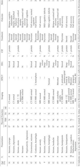
SREHT is a clinical condition encompassing per-sistent or relapsing seizures, myoclonus, focal neu-rologic deficits, and delusions or hallucinations along with elevated thyroid antibody titers.8,9 A workup must exclude other etiologies and the neu-ropsychiatric deficits show significant clinical im-provement with corticosteroid treatment. The pa-tient, typically female, may be euthyroid or hypothyroid and anti-thyroid antibody titers do not correlate to the degree of encephalopathy.7–9 Al-though heterogeneous in presentation, 2 subtypes have been described. The vasculitic type is charac-terized by acute stoke-like episodes associated with focal neurologic features and seizures. The diffuse progressive type is associated with insidious onset with progressive impairment of mental status with confusion, somnolence, and psychosis.8Neither pre-sentation is exclusive and significant overlap may occur. Furthermore, EEG, computerized tomogra-phy, MRI, and CSF analysis cannot distinguish be-tween the 2 types.
year-old patient with longstanding attentional difficulties which improved with steroid treatment.9 Our case is unique because of the long (5-year) his-tory of hallucinations and depressed mood.
The MRI findings observed in our patient, with small T2 weighted white matter changes, has been described in other pediatric patients.10,11 Neverthe-less, cranial imaging performed in most pediatric cases reported normal computerized tomography or MRI scans of the head. This differs from the adult literature, as a recent review of patients with SREHT followed at the Mayo Clinic revealed abnormal MRI scans in 57% of patients, a minority (45%) of which resolve with treatment.12
An interesting aspect of our case relates to the changes seen in serial SPECT imaging studies over the course of the patient’s illness. When treated with thyroid hormone alone, the SPECT showed im-proved temporal lobe perfusion mirroring clinical improvement in her memory and cognition. These deficits completely resolved on initiation of steroids, although decreased perfusion in the frontal and pa-rietal lobes persisted. Hypothyroid patients with “Hashimoto’s encephalitis” with abnormal SPECT scans at diagnosis have been described with full resolution of symptoms on treatment with thyroid hormone alone.13–15
Although formal treatment studies are absent, most reports have shown that glucocorticoids are effective. We recommend use of high-dose steroids, initially with intravenous methylprednisolone, fol-lowed by a prednisone (1–2 mg/kg/days, max 60 mg/days) for 6 to 8 weeks. The dose should be gradually reduced with expected improvement in symptoms over 3 to 6 months. Despite treatment, patients may experience relapses, and adolescents with this condition may experience residual cogni-tive deficits.7,15–17
Etiologically, SREHT is a controversial diagnosis, as it lacks a precise pathophysiologic basis. We are not aware of any evidence indicative of a causative link between thyroid autoimmunity and encephali-tis. CSF analysis has not shown the presence of TPO antibodies or thyroglobulin antibodies in symptom-atic adolescent and adult cases.11,18,19This condition best describes an association that is postulated to include aspects of endocrine dysfunction, cerebral vasculitis, and impaired autoimmunity.8,9,17,20,21 In the case of our patient, subclinical hypothyroidism and abnormalities on SPECT scanning, indicative of altered cerebral microvascular perfusion, were found. Thus far, our patient has not shown any evi-dence of other autoimmune disease, which has been reported in a significant proportion of patients with Hashimoto’s thyroiditis.22
Despite the speculative origin of SREHT, it re-mains a treatable encephalopathy. Pediatricians must be aware that younger patients may present with a diverse clinical picture, including seizures, myoclonus, focal neurologic deficits, delusions, and hallucinations. This case is unique as it illustrates that the neuropsychiatric component of this condi-tion may be chronic. SREHT requires a high index of suspicion, and our case stresses the importance of
evaluating thyroid function and thyroid antibody titers in adolescent patients with psychiatric presen-tations.
Farid H. Mahmud, MD Aida N. Lteif, MD
Division of Pediatric Endocrinology and Metabolism Department of Pediatric and Adolescent Medicine Mayo Clinic
Rochester, MN 55905
Deborah L. Renaud, MD
Division of Child and Adolescent Neurology Department of Pediatrics and Neurology Mayo Clinic
Rochester, MN 55905
Ann M. Reed, MD Division of Rheumatology
Department of Pediatrics and Medicine Mayo Clinic
Rochester, MN 55905
Chad K. Brands, MD
Division of Community Pediatric and Adolescent Medicine
Department of Pediatric and Adolescent Medicine Mayo Clinic
Rochester, MN 55905
REFERENCES
1. Rallison MK, Dobyns BM, Keating FR, Rall JE, Tyler FH. Occurrence and natural history of chronic lymphocytic thyroiditis in childhood.
J Pediatr. 1976;86:675– 682
2. Leigh H, Kramer SI. The psychiatric manifestations of endocrine disor-ders.Adv Intern Med. 1984;29:413– 445
3. Asher R. Myxedema madness.BMJ. 1949;2:555–562
4. Garrard P, Hodges JR, De Vries PJ, et al. Hashimoto’s encephalopathy presenting as myxoedematous madness.J Neurol Neurosurg Psychiatry. 2000;68:102–103
5. Hall RCW. Psychiatric effects of thyroid hormone disturbance. Psycho-somatics. 1983;24:7–18
6. Brain L, Jellinick EH, Ball K. Hashimoto’s disease and encephalopathy.
Lancet. 1966;2:512–514
7. Shaw PJ, Walls TJ, Newman PK, Cleland PG, Cartlidge NEF. Hashimo-to’s encephalopathy: a steroid-responsive disorder associated with high anti-thyroid antibody titers—report of 5 cases.Neurology. 1991;41(Suppl 1):228 –233
8. Kothbauer-Margreiter I, Sturzenegger M, Komor J, Baumgartner R, Hess EW. Encephalopathy associated with Hashimoto thyroiditis: di-agnosis and treatment.J Neurol. 1996;243:585–593
9. Vasconcellos E, Pina-Garza JE, Takoury F, Fenichel GM. Pediatric man-ifestations of Hashimoto’s encephalopathy. Pediatr Neurol. 1999;20: 394 –398
10. Takahashi S, Mitamura R, Itoh Y, Suzuki N, Okuno A. Hashimoto encephalopathy: etiologic considerations. Pediatr Neurol. 1994;11: 328 –331
11. Maydell BV, Kopp M, Komorski GV, Jeugling FD, Korinthenberg R. Hashimoto encephalopathy—is it underdiagnosed in pediatric pa-tients?Neuropediatrics. 2002;33:86 – 89
12. Castillo PR, Broeve PF, Caselli RJ, et al. Steroid-responsive encephalop-athy associated with thyroid autoimmunity; clinical and laboratory findings.Neurology. 2002;58(Suppl 3):A248
13. Sybesma CA, Pinxtreren-Nogler EV, Sinnige LGF, van Oostrom JCH, deVrieses TW. Hashimoto encephalopathy in a 12 year old girl.Eur J Paediatr. 1999;158:876 – 868
14. Querol Pascual MR, Aguirre Sanchez JJ, Velicia Mata MR, Gahete Jimenez C, Duran Herrera MC, Gonzalez Dorrego F. Encephalitis de Hashimoto: un nueva caso con remission espontanea.Neurologia. 2000; 15:313–316
in compensated Hashimoto’s encephalopathy: a clinical expression of autoimmune cerebral vasculitis.Brain Dev. 1986;8:60 – 64
17. Byrne OC, Zuberi SM, Madigan CA, King MD. Hashimoto’s thyroid-itis—a rare but treatable cause of encephalopathy in children.Eur J Paediatr Neurol. 2000;4:279 –282
18. Seipelt M, Zerr I, Nau R, et al. Hashimoto’s encephalitis as a differential diagnosis of Creutzfeldt-Jakob disease.J Neurol Neurosurg Psychiatry. 1999;66:172–176
19. Archambeaud F, Galinat S, Regouby Y, et al. Encephalopathe de Hashi-moto. Analyse de quatre observations.Rev Med Interne. 2001;22:653– 659 20. Canton A, de Fabregas O, Tintore M, Mesa J, Codina A, Simo R. Encephalopathy associated to autoimmune thyroid disease: a more appropriate term for an underestimated condition?J Neurol Sci. 2000; 176:65– 69
21. Boers PM, Colebatch JG. Hashimoto’s encephalopathy responding to plasmapheresis.J Neurol Neurosurg Psychiatry. 2001;70:132–135 22. Becker KL, Ferguson RH, McConahay WM. Connective tissue diseases
and symptoms associated with Hashimoto’s thyroiditis.N Engl J Med. 1963;268:277–280
23. Forchetti CM, Katsamakis G, Garron DC. Autoimmune thyroiditis and a rapidly progressive dementia: global hypoperfusion on SPECT scan-ning suggests a possible mechanism.Neurology. 1997;49:623– 626 24. Gucuyener K, Serdaroglu A, Bideci A, et al. Tremor and myoclonus
heralding Hashimoto’s encephalopathy.J Pediatr Endocrinol Metab. 2000; 13:1137–1141
25. Balestri P, Grosso S, Garibaldi G. Alternating hemiplegia of childhood or Hashimoto’s encephalopathy?J Neurol Neurosurg Psychiatry. 1999;66: 548 –549
IS DIVERSITY OVERRATED?
“One of the most comprehensive studies ever undertaken of diversity in higher education indicates that this contention is at least questionable. The study’s find-ings show that college diversity programs fail to raise standards, and that a majority of faculty members and administrators recognize this when speaking anonymously. . . To find out, in 1999 we surveyed a random sample of ⬎1600 students and 2400 faculty members and administrators at 140 American colleges and universities, asking them to evaluate the quality of education at their institu-tion, the academic preparation and work habits of the student body, the state of race relations on campus, and their own experiences of discrimination. . . If diver-sity works as advertised, we surmised, then those at institutions with higher proportions of black enrollment should rate their educational and racial milieus more favorably than their peers at institutions with lower proportions. . . The results contradict almost every benefit claimed for campus diversity. Students, faculty members, and administrators all responded to increasing racial diversity by registering increased dissatisfaction with the quality of education and the work ethic of their peers. Students also increasingly complained about discrimination. . . One cannot help but wonder why the public and private views of higher educa-tion’s leadership differ so greatly.”
Rothman S.New York Times.March 30, 2003
DOI: 10.1542/peds.112.3.686
2003;112;686
Pediatrics
Farid H. Mahmud, Aida N. Lteif, Deborah L. Renaud, Ann M. Reed and Chad K. Brands
Adolescent With Chronic Hallucinations and Depression: Case Report and Review
Steroid-Responsive Encephalopathy Associated With Hashimoto's Thyroiditis in an
Services
Updated Information &
http://pediatrics.aappublications.org/content/112/3/686
including high resolution figures, can be found at:
References
http://pediatrics.aappublications.org/content/112/3/686#BIBL
This article cites 25 articles, 6 of which you can access for free at:
Subspecialty Collections
http://www.aappublications.org/cgi/collection/endocrinology_sub
Endocrinology
collection(s):
This article, along with others on similar topics, appears in the following
Permissions & Licensing
http://www.aappublications.org/site/misc/Permissions.xhtml
its entirety can be found online at:
Information about reproducing this article in parts (figures, tables) or in
Reprints
http://www.aappublications.org/site/misc/reprints.xhtml
DOI: 10.1542/peds.112.3.686
2003;112;686
Pediatrics
Farid H. Mahmud, Aida N. Lteif, Deborah L. Renaud, Ann M. Reed and Chad K. Brands
Adolescent With Chronic Hallucinations and Depression: Case Report and Review
Steroid-Responsive Encephalopathy Associated With Hashimoto's Thyroiditis in an
http://pediatrics.aappublications.org/content/112/3/686
on the World Wide Web at:
The online version of this article, along with updated information and services, is located
American Academy of Pediatrics. All rights reserved. Print ISSN: 1073-0397.