Surface Exposure and Packing of Lipoproteins into Outer
Membrane Vesicles Are Coupled Processes in
Bacteroides
Ezequiel Valguarnera,
aNichollas E. Scott,
bPhilippe Azimzadeh,
aMario F. Feldman
aaDepartment of Molecular Microbiology, Washington University in Saint Louis, Saint Louis, Missouri, USA
bDepartment of Microbiology and Immunology, The University of Melbourne, Victoria, Australia
ABSTRACT
Outer membrane vesicles (OMVs) are spherical structures derived from
the outer membranes (OMs) of Gram-negative bacteria.
Bacteroides
spp. are
promi-nent compopromi-nents of the human gut microbiota, and OMVs produced by these
spe-cies are proposed to play key roles in gut homeostasis. OMV biogenesis in
Bacte-roides
is a poorly understood process. Here, we revisited the protein composition of
Bacteroides thetaiotaomicron
OMVs by mass spectrometry. We confirmed that OMVs
produced by this organism contain large quantities of glycosidases and proteases,
with most of them being lipoproteins. We found that most of these OMV-enriched
lipoproteins are encoded by polysaccharide utilization loci (PULs), such as the
sus
operon. We examined the subcellular locations of the components of the Sus system
and found a split localization; the alpha-amylase SusG is highly enriched in OMVs,
while the oligosaccharide importer SusC remains mostly in the OM. We found that
all OMV-enriched lipoproteins possess a lipoprotein export sequence (LES), and we
show that this signal mediates translocation of SusG from the periplasmic face of
the OM toward the extracellular milieu. Mutations in the LES motif caused defects in
surface exposure and recruitment of SusG into OMVs. These experiments link, for the
first time, surface exposure to recruitment of proteins into OMVs. We also show that
surface-exposed SusG in OMVs is active and rescues the growth of bacterial cells
in-capable of growing on starch as the only carbon source. Our results support the role
of OMVs as “public goods” that can be utilized by other organisms with different
metabolic capabilities.
IMPORTANCE
Species from the
Bacteroides
genus are predominant members of the
human gut microbiota. OMVs in
Bacteroides
have been shown to be important for
the homeostasis of complex host-commensal relationships, mainly involving immune
tolerance and protection from disease. OMVs carry many enzymatic activities
in-volved in the cleavage of complex polysaccharides and have been proposed as
pub-lic goods that can provide growth to other bacterial species by release of
polysac-charide breakdown products into the gut lumen. This work shows that the presence
of a negatively charged rich amino acid motif (LES) is required for efficient packing
of the surface-exposed alpha-amylase SusG into OMVs. Our findings strongly suggest
that surface exposure is coupled to packing of
Bacteroides
lipoproteins into OMVs.
This is the first step in the generation of tailor-made probiotic interventions that can
exploit LES-related sequences to generate
Bacteroides
strains displaying proteins of
interest in OMVs.
KEYWORDS
Bacteroides
, OMV, hydrolases, lipoproteins, vesicles
O
uter membrane vesicles (OMVs) are small spherical structures derived from the
outer membranes (OMs) of most Gram-negative bacterial species. OMVs are
composed of phospholipids, lipopolysaccharide (LPS), or lipooligosaccharide (LOS) and
OM and periplasmic proteins (1, 2). OMVs can mediate host-microbe interactions by
Received9 October 2018Accepted15
October 2018 Published7 November 2018
CitationValguarnera E, Scott NE, Azimzadeh P,
Feldman MF. 2018. Surface exposure and packing of lipoproteins into outer membrane vesicles are coupled processes inBacteroides. mSphere 3:e00559-18.https://doi.org/10.1128/ mSphere.00559-18.
EditorCraig D. Ellermeier, University of Iowa
Copyright© 2018 Valguarnera et al. This is an
open-access article distributed under the terms of theCreative Commons Attribution 4.0 International license.
Address correspondence to Mario F. Feldman, [email protected].
RESEARCH ARTICLE
Molecular Biology and Physiology
crossm
on September 8, 2020 by guest
http://msphere.asm.org/
facilitating long-distance delivery of virulence factors, by modulating the host immune
response, and by contributing to antibiotic resistance (1, 3–8). Despite these key roles
in bacterial physiology, OMV biology is poorly understood. Recent research indicates
that OMVs are produced by diverse mechanisms. For example, OMVs are proposed to
be generated by LPS remodeling in
Porphyromonas gingivalis
,
Salmonella enterica
, and
Pseudomonas aeruginosa
(9–12). In contrast, OMVs from
Haemophilus influenzae
and
Vibrio cholerae
are thought to be the result of an accumulation of phospholipids in the
OM outer leaflet mediated by their specialized VacJ/Yrb transporter (13). Thus, it
appears that there is not a universal mechanism of OMV biogenesis. For most species,
including
Bacteroides
spp., OMV biogenesis remains poorly understood (5).
Species from the phylum
Bacteroidetes
compose a major part of the human gut
microbiota (14, 15). OMVs from these organisms are proposed to play important roles
in the commensal-host relationship, including the delivery of immunomodulatory
molecules to host immune cells, an interaction that appears to help prevent colitis
flare-ups in the context of inflammatory bowel disease (IBD) (7, 8). Furthermore,
Bacteroides
OMVs have been proposed to interfere with intracellular Ca
2⫹signaling in
host cells (16). Most studies focus primarily on two predominant species in the human
gut,
Bacteroides thetaiotaomicron
and
Bacteroides fragilis
. These species produce large
amounts of uniformly sized OMVs that have a protein composition distinct from that of
the OM, indicating that these OMV particles are not by-products of bacterial lysis (5).
Most
Bacteroides
OMV-exclusive proteins are putative acidic lipoproteins with hydrolase
activity, suggesting that proteins with similar structural and physicochemical properties
are selectively sorted to OMVs (5). Many
Bacteroides
enzymatic lipoproteins are
en-coded on polysaccharide utilization loci (PULs), which constitute
⬃
20% of the
B.
thetaiotaomicron
genome and are essential for the breakdown and acquisition of plant,
fungus, and mucin complex polysaccharides (17–19). PULs typically consist of at least
one TonB-dependent receptor, or SusC-like protein, and one nutrient binding accessory
protein, or SusD, and they can also present other accessory proteins (20). PULs can also
carry two-component systems that sense nutrient variations in the medium, with
subsequent induction of polysaccharide utilization genes required for the utilization of
complex carbon sources (17–19, 21–24). Hence,
Bacteroides
cells can modify the
enzymatic compositions of OMVs according to available carbon sources (5). The
enzy-matic arsenal carried by
Bacteroides
OMVs appears to carry a “social” function, as the
products of OMV-mediated hydrolysis can be utilized by other bacteria within the gut
(25, 26).
Here, we further characterized the protein composition of
B. thetaiotaomicron
OMVs.
We confirmed that OMVs produced by this organism contain mainly putative acidic
lipoproteins with hydrolytic or carbohydrate-binding activities. Many of these
OMV-enriched lipoproteins were found to be encoded by PULs. We examined the subcellular
localization of the components of the archetypical PUL, the starch utilization system
(Sus) (20, 27), and found that the alpha-amylase SusG and other Sus lipoproteins are
highly enriched in OMVs. In contrast, the oligosaccharide importer SusC remains mostly
in the OM, as previously characterized (20, 27, 28). We show that the presence of a
lipoprotein export sequence (LES) mediates translocation of SusG from the periplasmic
face onto the extracellular milieu and is both required and sufficient for SusG to localize
preferentially to OMVs. Our results support the role of OMVs as “public goods” that can
be utilized by other organisms with different metabolic capabilities.
RESULTS
Proteomics analysis of membrane and OMV fractions from
B. thetaiotaomicron.
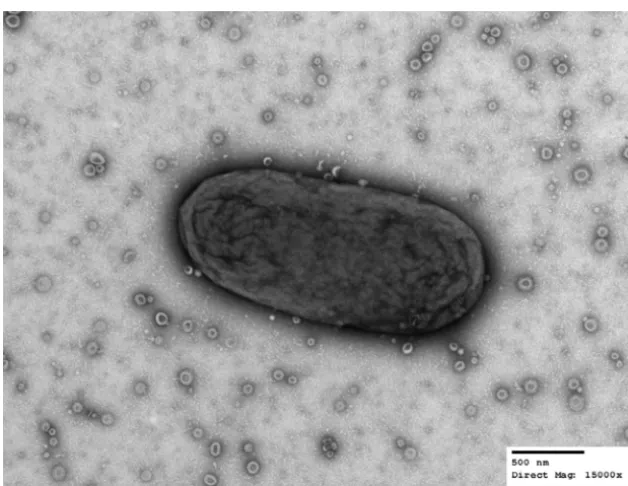
Electron micrograph analysis confirmed that
B. thetaiotaomicron
produces large
amounts of uniform OMV particles (Fig. 1). We previously performed a proteomic
analysis of
B. thetaiotaomicron
OMs and OMVs by employing Triton X-100 for the
purification of OM proteins (5). In this work, we employed
N
-lauroylsarcosine (Sarkosyl),
which has been widely used for the separation of inner membrane (IM) and OM
fractions in Gram-negative bacteria (29–31) to verify that the apparent selective
on September 8, 2020 by guest
http://msphere.asm.org/
tionation of proteins into OM and OMV was not due to the use of a specific detergent.
Using early-stationary-phase cultures from
B. thetaiotaomicron
, we prepared the
differ-ent membrane fractions as indicated in Fig. S1 in the supplemdiffer-ental material. In our
previous study, OMVs were not treated with the detergent employed to extract the IM
proteins from total membrane preparations. To rule out possible detergent effects, we
added an additional step consisting of incubating OMVs in 1% Sarkosyl prior to the
ultracentrifugation of the samples to recover an OMV supernatant (OMV-S) and an OMV
pellet (OMV-P). Samples were lyophilized for proteomic analysis by liquid
chromatography-tandem mass spectrometry (LC-MS/MS), and aliquots were visualized by Coomassie blue
staining (Fig. S1).
Our new data set with annotations and predicted functions and locations for
proteins in all fractions is provided as Table S1 in the supplemental material. We
confirmed our previous findings showing an enrichment of lipoproteins in the OMV
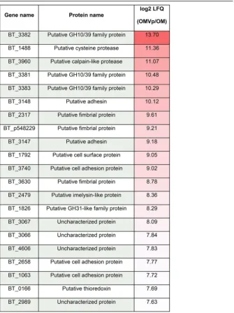
fraction in comparison with their level in the OM fraction (Table 1; Table S2) (5). Figure 2
highlights the top enriched proteins in the OMV. We found that 18 out of the 23
OMV-exclusive lipoproteins from our previous study are also enriched in the new OMV
preparations (5). We then cloned three OMV- and three OM-enriched proteins identified
in our studies into the expression vector pFD340 as C-terminal 6
⫻
His-tagged proteins
to confirm their localization by Western blotting. Figure 3a shows that a putative cell
surface protein (BT_1488), as well as a putative calpain-like protease (BT_3960) and a
putative zinc peptidase (BT_3237), is highly enriched in OMVs. Figure 3b shows that
BT_0418, BT_2844, and BT_2817, identified as OM-enriched proteins by MS, are
re-tained at the OM and are not present in OMVs. BT_0418 is a porin F ortholog
(8-

-strand protein with a peptidoglycan-binding domain), BT_2817 is a
TonB-dependent receptor (22-

-strand protein), and BT_2844 is a lipoprotein-containing
tetratricopeptide repeat (TPR) motif. Both BT_0418 and BT_2817 are not predicted to be
lipoproteins, they carry a signal peptidase I (SPI) cleavage site, and they do not contain
the lipoprotein attachment cysteine residue. Before cloning this group of OMV- and
OM-enriched proteins, we observed that the automated start codon annotation for
FIG 1 B. thetaiotaomicronproduces outer membrane vesicles (OMVs). Transmission electron microscopy of a singleB. thetaiotaomicroncell together with OMVs in the extracellular milieu.B. thetaiotaomicron cells were swabbed from a solid-medium plate, suspended in PBS, and processed for TEM. Images were acquired at a direct magnification of⫻15,000. The scale bar represents 500 nm. (Image courtesy of Wandy Beatty, Molecular Microbiology Imaging Facility, WUSTL.)
Secretion of SusG intoBacteroidesOMVs
on September 8, 2020 by guest
http://msphere.asm.org/
some open reading frames (ORFs) (BT_0418 and BT_2844) seemed incorrect. Any
mistakes in start codon-predicted annotations were corrected accordingly (see
Mate-rials and Methods). These sets of experiments demonstrate that lipoproteins are indeed
differentially sorted between OMs and OMVs.
Common features of OMV-enriched lipoproteins.
Consistently with our previous
results, we found that lipoproteins enriched in OMVs are acidic, with an average
isoelectric point of 4.86 (5). We also determined that OMV-enriched lipoproteins
possess a negatively charged rich amino acid motif, S(D/E)
3, adjacent to the cysteine
residue required for lipoprotein attachment (Fig. 4; Table S2). A similar motif [K-(D/E)
2or Q-A-(D/E)
2] in the oral pathogen
Capnocytophaga canimorsus
, a member of the
Bacteroidetes
phylum, has recently been described. The report showed that this motif
functions as a lipoprotein export signal (LES) required for surface exposure of OM
lipoproteins (32). The authors also showed that proteins carrying a LES from
B. fragilis
TABLE 1Top 20 most OMVp-enriched proteins, including protein name and enrichment valuea
aLFQ, label-free quantification.
on September 8, 2020 by guest
http://msphere.asm.org/
were exposed when expressed in
C. canimorsus
. We found that many OMV-enriched
B.
thetaiotaomicron
lipoproteins are putative protein and sugar hydrolases required for
the breakdown of complex nutritional sources. The presence of a LES on OMV-enriched
proteins is consistent with their annotated functions, as they are required to face the
extracellular milieu to access their cognate substrates. Conversely, OM-enriched
lipo-proteins like BT_2844 do not carry a LES motif. These lipo-proteins have lower sequence
conservation and lower frequency of negatively charged amino acids along the
resi-dues adjacent to the lipoprotein attachment cysteine and are therefore expected to be
oriented toward the periplasmic face of the OM (Fig. 4b; Table S2).
SusG and other Sus lipoproteins are enriched in OMV, and SusC is retained at
the OM.
In
B. thetaiotaomicron
, hydrolytic lipoproteins are encoded mainly in PULs. PUL
and PUL-like operons account for approximately 20% of the
B. thetaiotaomicron
ge-nome. Thirty-six lipoproteins encoded in PUL and PUL-like operons were found in our
OMV-enriched proteins. One of the most studied lipoproteins from
B. thetaiotaomicron
is the
␣
-amylase SusG, encoded by the
sus
operon and essential for starch catabolism
(33, 34). The
sus
operon has been shown to be induced by starch and
maltooligosac-charides (35). Our MS data show that even under noninducing conditions, two
lipo-proteins encoded by the
sus
operon, SusD and SusE, are enriched in OMVs (Table S2).
Given the importance of starch in the mammalian diet, we investigated the subcellular
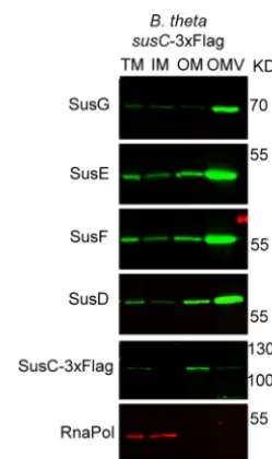
localization of the components of the Sus operon. We found that all Sus lipoproteins,
in particular SusG, are enriched in the OMV. The only exception was the porin SusC,
which is retained mostly at the OM (Fig. 5). We also observed considerable levels of
FIG 2 TheB. thetaiotaomicronOMV pellet (OMVp) protein content is different from that of the OM. Three hundred micrograms of each preparation and biological replicates of purified OM and OMVp proteins were digested with trypsin. The resulting peptides were enriched and then analyzed via liquid chroma-tography coupled to tandem mass spectrometry (LC-MS/MS) as explained in Materials and Methods, followed by protein identification with the Mascot search engine using the UniProt database. Volcano plot shows OM and OMVp protein populations. Red labels indicate the proteins with the highest OMVp enrichment in comparison to that of OM.
Secretion of SusG intoBacteroidesOMVs
on September 8, 2020 by guest
http://msphere.asm.org/
SusD in the OM, concomitant with its strong functional interaction with SusC (28). This
result suggests that PUL components can exert their biological effect not only at the
level of the outer membrane but also through OMVs. These findings represent a
localization split for the components of the
sus
operon exclusively and need to be
further validated for other
sus
-like systems through the
B. thetaiotaomicron
genome,
especially because we observed other SusC-like proteins enriched in both OMs and
OMVs (Table S2).
FIG 3 Validation of OMV- and OM-enriched proteins. Candidate ORFs that encode proteins identified in our MS analysis as OMV or OM enriched were cloned into pFD340 with a C-terminal 6⫻His tag. Constructs were introduced intoB. thetaiotaomicronby conjugation, generated strains were grown in TYG medium, and fractions were prepared. Ten micrograms of each fraction was subjected to 12% SDS-PAGE and analyzed by Western blotting using anti-His polyclonal antibodies. (a) OMV-enriched proteins; (b) OM-enriched proteins. The isoelectric point as well as the residues following the lipoprotein attachment cysteine (for lipoproteins) are indicated below the protein name.
FIG 4 OMV-enriched proteins show a conserved N-terminal LES motif. The top 91 OMV-enriched proteins (a) and top 24 OM-enriched proteins (b) were aligned using the lipoprotein attachment cysteine at the⫹1 position (not shown in the logo), followed by the 9 C-terminal contiguous residues. The lipoprotein export sequence (LES) consensus was generated using WebLogo (https://weblogo.berkeley .edu/logo.cgi) (56).
on September 8, 2020 by guest
http://msphere.asm.org/
LES is required for SusG exposure and packing into OMVs.
All
B.
thetaiotaomi-cron
endoglycanases involved in the first step of polysaccharide breakdown, including
SusG, are surface-exposed lipoproteins (33, 36). Because all OMV-enriched lipoproteins
contain a LES motif, we investigated the link between surface exposure and packing
into OMVs. SusG contains only two Asp residues following the
⫹
2 Ser residue, and
therefore, for the purpose of this work, we provisionally define its LES motif as CSDD.
We performed a mutational analysis on the LES motif of SusG and analyzed the
localization of the protein by fractionation and Western blotting (Fig. 6). We cloned
wild-type (WT) His-tagged SusG and a set of SusG derivatives carrying amino acid
FIG 5 Sus lipoproteins are OMV enriched, while SusC is OM enriched. B. thetaiotaomicron cells containing a tagged genomic copy ofsusCwith a 3⫻Flag tag were grown in TYM medium (TYG recipe with 0.5% maltose instead of glucose for induction of thesusoperon). Fractions were prepared and analyzed by 12% SDS-PAGE and Western blotting using specific anti-Sus, anti-Flag, and anti-RNApol antibodies.
FIG 6 SusG LES is required for efficient packing into OMVs. (a) SusG LES point mutants were generated on pFD340/susG-6⫻His using Quick site-directed mutagenesis. (b) Constructs were introduced in the ΔsusGbackground, and generated strains were grown in minimal medium with glucose as the only carbon source. Fractions were prepared and analyzed by SDS-PAGE and Western blotting using anti-SusG antibodies. (c) OMV/OM ratios were calculated using the fluorescence signal values for each protein and are plotted as a percentage of the WT OMV/OM ratio (100%). Statistical significance was determined by unpairedttest of each LES variant in comparison with values for the wild-type LES strain.*,Pvalues⬍ 0.03. NS, not significant. The experimental results are representative of those of three biological replicates; shown are mean values with standard deviations (SD) for two technical replicates.
Secretion of SusG intoBacteroidesOMVs
on September 8, 2020 by guest
http://msphere.asm.org/
replacements in the LES (Fig. 6a) and expressed them in the Δ
susG
strain (37).
Replacement of the lipid attachment site (C23) by Ala resulted in abrogation of SusG
recruitment into OMVs. Furthermore, substitution of a single Asp residue with Ala
(CSAD, CSDA, CAAD) decreased OMV packaging (
⬃
50% relative to that in the WT),
while mutation of both Asp residues by either Ala or Lys (CSAA, CAAA, CAKK) had a
more dramatic effect (
⬃
15% relative to that of the WT) (Fig. 6c). Conservative
replace-ment of Asp by Glu (CSEE) displayed a WT-like behavior. These results indicate that the
LES motif CS(D/E)
2is required for SusG packaging into OMVs.
The LES motif has been defined as a surface exposure tag in
C. canimorsus
(32). We
used the WT LES SusG construct as well as the non-Asp variants (CSAA, CAAA) to
determine whether this motif is also required for surface exposure in
B.
thetaiotaomi-cron
. Whole cells and OMVs from the different strains were subjected to proteinase K
(ProK) sensitivity analysis. Only the WT SusG was degraded by ProK (Fig. 7). As a control,
we employed a periplasmic soluble protein that localizes into the lumens of OMVs
(BT_0766) and is therefore protected from ProK degradation. Taken together, these
experiments confirm that LES mediates both SusG surface exposure and enrichment in
OMVs.
OMVs containing SusG rescue the
⌬
susG
strain’s growth in starch.
Our
exper-iments demonstrated that SusG is surface exposed and packed into OMVs. OMVs
carrying certain glycosyl hydrolases can digest complex polysaccharides, providing
essential nutrients to bacteria unable to degrade these substrates (25, 26). The Δ
susG
strain is unable to grow on minimal media with starch as the sole carbon source (Fig. 8).
We investigated whether surface exposure of OMV-delivered SusG can rescue the
Δ
susG
growth phenotype on starch. For this, we employed OMVs produced by the
Δ
susG
strain expressing WT SusG and SusG with a mutated LES (ASDD, CSAA, and
CAAA). The ASDD mutation renders soluble SusG in the periplasm, while the CSAA and
CAAA SusG variants face the periplasmic side of the OM (Fig. 7). Purified OMVs were
added to the Δ
susG
strain in minimal media containing starch. Only WT OMVs restored
the growth of the Δ
susG
strain to wild-type levels (Fig. 8). OMVs from the
susG
deletion-carrying vector control (pFD340) or expressing the nonlipoprotein mutant
(ASDD) or the CSAA mutant were unable to rescue the expression of SusG, while the
CAAA mutant displayed an intermediate phenotype. All together, our results indicate
FIG 7 The LES is required for SusG surface exposure. WT SusG and the LES mutants were assessed for their surface exposure by proteinase K assays using whole cells (a) and OMVs (b). Proteinase K (PK) was added and incubated at 37°C; at different times (in minutes), aliquots were TCA precipitated and analyzed by SDS-PAGE and Western blotting using anti-SusG antibodies. We used 3⫻Flag-tagged BT_0766, a periplasmic soluble protein found in OMVs, as an outer membrane and OMV integrity control.
on September 8, 2020 by guest
http://msphere.asm.org/
that OMVs carrying only surface-exposed SusG can mediate cross-feeding of other
bacteria.
DISCUSSION
The human gut microbiome is composed largely of species from the
Bacteroides
genus.
Bacteroides
spp. are important for gut homeostasis (14, 15). They establish
ecological interactions with each other and are also involved in host-commensal
relationships, especially regarding the development of the immune system (7, 8, 16).
Here, we show that
B. thetaiotaomicron
produces large amounts of uniformly sized
OMVs. Using an optimized methodology for the purification of different membrane
fractions, we confirmed that OMVs are highly enriched with lipoproteins, particularly
glycosyl hydrolases. Our MS data identified the presence of a lipoprotein exposure
sequence (LES) in all OMV-enriched lipoproteins. Employing the
␣
-amylase SusG as a
model, we confirmed that the LES is required for surface exposure and showed that it
also mediates recruitment into OMVs. To our knowledge, this is the first identification
of coupling surface exposure to protein sorting into OMVs.
A few lipoprotein surface transport mechanisms in Gram-negative bacteria have
been described (38–41). Shuttling of a group of
Neisseria meningitidis
lipoproteins to
the surface is dependent on proteins Slam1 and Slam2, although the sorting
mecha-nism has not been defined (41, 42). Moreover, the well-studied Bam system for folding
of beta-barrel proteins in the outer membrane has been shown to export specific
lipoproteins (39). We have not identified orthologs of Slam1 or Slam2 by sequence
similarity in any
Bacteroides
genomes. The existence of LES motifs in
B. thetaiotaomicron
lipoproteins, together with the discovery of the LES in
C. canimorsus
, suggests the
existence of a conserved phylum-wide mechanism that flips specific lipoproteins
toward the extracellular milieu among
Bacteroidetes
. Defining a functional LES in
Bacteroides
spp. constitutes the first step toward the identification of the machinery
that mediates lipoprotein surface exposure in these organisms. Our experiments
con-firmed that the two Asp residues in positions
⫹
3 and
⫹
4 respecting the lipid
attach-ment site (
⫹
1) are essential components of SusG LES. The proposed
B. thetaiotaomicron
LES motif CS(D/E)
2is similar but not identical to the K-(D/E)
2or Q-A-(D/E)
2LES proposed
for
C
.
canimorsus
. An exhaustive mutagenesis analysis of multiple proteins from several
species will be required to exactly define a consensus sequence for the LES motif
among
Bacteroidetes
.
MS results show that OMV-enriched lipoproteins contain a LES motif, suggesting
FIG 8 OMVs displaying WT SusG can rescue a strain with a ΔsusGgrowth phenotype on starch as the carbon source. WT and ΔsusGstrains were grown overnight in TYG medium. Cultures were washed with minimal medium without any carbon source and normalized by OD. Minimal medium with 0.5% starch as the only carbon source was inoculated with WT or ΔsusGstrains to a final OD600of 0.05. OMVs purified from the ΔsusGstrain containing different pFD340 derivatives were added to the ΔsusG cultures in minimal medium with starch (1-g/ml final concentration of OMVs). Aliquots were taken at different times, and the OD600was measured to determine growth. Statistical significance when two growth curves were compared was determined by performing one pair of an unpairedttest analysis per each time point.*,Pvalues⬍0.01. The experiment is representative of three biological replicates; shown are mean values with SD for three technical replicates.
Secretion of SusG intoBacteroidesOMVs
on September 8, 2020 by guest
http://msphere.asm.org/
that most if not all OMV-enriched lipoproteins are surface exposed in a LES-dependent
manner (Fig. 9). We can speculate with two models to account for this correlation
between surface exposure and OMV recruitment of lipoproteins. One possibility is that
the LES sequence is somehow recognized by a putative OMV sorting machinery,
packaging them preferentially into vesicles. Alternatively, exposed lipoproteins might
be recruited into OMVs based on their biophysical properties. Uncoupling surface
exposure with OMV packaging is currently not possible, and future work will be
required to unravel the molecular basis of the link between these two processes.
In the current models for PUL systems, the hydrolytic enzymes and the
oligosac-charide importers are present at the outer membrane (20, 36). However, we determined
that functional glycosyl hydrolases are packed mainly into OMVs. We also found that
the glycosidases present in OMVs can provide substrates to support the growth of
other bacteria, in agreement with the previously proposed function of OMVs as public
goods (25, 26). We propose a model in which, in a process involving the LES, the
glycosidases are sequentially surface exposed and sorted into OMVs (Fig. 9). The
secreted OMVs, armed with an arsenal of hydrolases, can digest diverse dietary
poly-saccharides and host glycoconjugates, making the mono- and oligopoly-saccharides
avail-able to all members of the microbiota. Depending on the diet composition,
Bacteroides
spp. can induce and package different enzyme repertoires, and therefore, members of
the microbiota can act as donors or acceptors in their ecological niches. In addition,
pathogens such as
Campylobacter
,
Salmonella
, and
Clostridium
can also benefit from the
glycosidic activity contained in OMVs (43, 44). From an evolutionary and social
stand-point, it is not clear how beneficial OMV-mediated hydrolysis is for the producing
organisms. On one side, it is expected that hydrolytic OMVs make the nearby gut
environment more mixed in its nutritional composition, rendering the producers more
susceptible to cheating by consumers. On the other side, it is tempting to speculate
that OMVs might have counteracting benefits to the producers, such as a higher
efficiency of hydrolysis than that of OM-anchored enzymes (by increasing the
enzy-matic surface) and, potentially, an increase in the intestinal surface that could be
colonized by its being able to access substrates through a longer stretch of the colonic
niche.
We have identified a set of OMV- and OM-enriched proteins that may be employed
FIG 9 A model for SusG LES-mediated surface exposure and packing into OMV. The SusG lipoprotein is probably transported to the OM by a machinery homologous to the LOL system. The presence of a LES sequence mediates the surface exposure of SusG and its incorporation into OMVs. The LES might have a dual role, where it is required for both surface exposure and OMV packing, or it might be involved only in surface exposure; biophysical properties of lipoproteins would be determinant for packing into OMVs. Surface-exposed SusG in OMVs as well as in the OM hydrolyzes starch molecules into oligosaccharides that can be imported by TonB-dependent receptors by the OMV-producing cell, as well as other commensal and pathogen organisms.
on September 8, 2020 by guest
http://msphere.asm.org/
as markers to investigate the process of OMV biogenesis through the visualization of
OMV in the mammalian gut. For example, these markers would allow the differentiation
in vivo
between bona fide OMV and cell lysis-derived material. Furthermore, the
identification for the first time of a role for the LES as a signal for protein transport into
OMVs constitutes a starting point for the design of novel probiotic interventions in
animal and human health. In the future, the LES of SusG or other OMV proteins might
be employed to engineer
Bacteroides
strains to secrete OMVs packed with medically
relevant proteins in the mammalian gut.
MATERIALS AND METHODS
Bacterial strains and growth conditions.Oligonucleotides, strains, and plasmids are described in Table S3 in the supplemental material.Bacteroidesstrains were grown in an anaerobic chamber (Coy Laboratories) using an atmosphere of 10% H2, 5% CO2, 85% N2. For liquid growth, tryptone-yeast extract-glucose (TYG), TYM (TY medium supplemented with 0.5% maltose instead of 0.5% glucose), or minimal media supplemented with 0.5% glucose or 0.5% potato starch were prepared as previously described (17, 45). Brain heart infusion (BHI) agar with 10% defibrinated horse blood was used as a solid medium. Antibiotics were used as follows: ampicillin at 100g/ml, erythromycin at 25g/ml, and bromodeoxyuridine at 200g/ml.
Construction of plasmids, mutagenesis, and generation of thesusC-3ⴛFlag strain.Constructs for overexpression were built on pFD340 (46). Inserts obtained by PCR were digested with restriction enzymes, purified, and ligated onto the digested vector pFD340 as previously reported. For all 6⫻ His-containing constructs, the tag was added to the C terminus of the ORF product by including the 6⫻His sequence in the reverse primer used for cloning into pFD340 (Table S3). In the cases of BT_0418 and BT_2844, we detected that the putative correct start codons extend 9 and 10 amino acids into the N terminus of the sequence, respectively, and included that information for the cloning of both genes. For cloning of BT_0766-Flag into pFD340, purified PCR inserts were integrated into PCR-amplified pFD340 using an In-Fusion cloning kit (Clontech). Mutagenesis of pFD340/susG-6⫻His was carried out by inverse PCR with PfuTurbo (Agilent) using overlapping oligonucleotides carrying the mismatch required as described elsewhere. Generation of asusC-3xFlag strain was carried out using theB. thetaiotaomicron Δtdkstrategy as previously described (47). Briefly, 1,000-bp upstream and downstream fragments of a C-terminalsusC-3xFlag translational fusion were cloned into pExchange-tdk. Constructs were conjugated intoB. thetaiotaomicronΔtdkcells using previously transformedEscherichia coliS17-1piras a donor, and strain plating and selection were performed as previously described (47).
OMV preparations.Outer membrane vesicles were purified by ultracentrifugation of filtered spent media as previously described by our group (5). For MS analysis, OMV preparations were resuspended in 50 mM HEPES, pH 7.4, and N-lauroyl sarcosine was added to a 1% final concentration in 1.5 ml polyallomer tubes (Beckman Coulter). Samples were incubated with gentle rocking for 1 h at room temperature (RT) and ultracentrifuged at 100,000⫻gfor 2 h at RT. Supernatants were recovered (OMV-S) and pellets (OMV-P) were resuspended in 50 mM HEPES, pH 7.4. Protein content was quantified using a DC protein assay kit (Bio-Rad). Fractions were lyophilized for MS analysis.
Membrane preparations.Total membrane preparations were performed by cell lysis and ultracen-trifugation as previously described (5). For separation of the inner membrane (IM) and OM, total membranes were resuspended in 50 mM HEPES, pH 7.4, using a 2-ml glass tissue grinder with a polytetrafluoroethylene (PTFE) pestle (VWR), andN-lauroyl sarcosine was added to a 1% final concen-tration in 1.5-ml polyallomer tubes (Beckman Coulter). Samples were incubated with gentle rocking for 1 h at RT and ultracentrifuged at 100,000⫻gfor 2 h at RT. Supernatants were recovered (IM) and pellets were resuspended in 50 mM HEPES, pH 7.4. Protein content was quantified using a DC protein assay kit (Bio-Rad). Fractions were lyophilized for MS analysis.
Mass spectrometry analysis. (i) Protein cleanup and in-solution digestion.Lyophilized protein preparations were solubilized in lysis buffer (4% SDS, 10 mM, 100 mM Tris, pH 8.5) by boiling them for 10 min, and the protein content was assessed by the bicinchoninic acid (BCA) protein assay according to the manufacturer’s instructions. Three hundred micrograms of each preparation and biological replicate was adjusted to a volume of 200l and precipitated overnight using 800l of ice-cold acetone (1:4, vol/vol). Samples were spun down at 16,000⫻gfor 10 min at 0°C to the resulting protein precipitate, and acetone was removed. Residue acetone was driven off at 70°C for 5 min. Protein precipitants were resuspended in 6 M urea, 2 M thiourea, 40 mM NH4HCO3and reduced/alkylated prior to digestion with Lys-C (1/200, wt/wt) and then trypsin (1/50, wt/wt) overnight as previously described (48). Digested samples were acidified to a final concentration of 0.5% formic acid and desalted with using C18stage tips (49, 50).
Label-free quantification (LFQ)-based quantitative proteomic LC-MS.Prepared purified peptides were resuspended in buffer A* (2% acetonitrile, 0.01% trifluoroacetic acid) and separated using a two-column chromatography setup composed of a PepMap100 C1820-mm by 75-m trap column and a PepMap C18500-mm by 75-m analytical column (ThermoFisher Scientific). Samples were concen-trated onto the trap column at 5l/min for 5 min and infused into an Orbitrap Q-Exactive Plus mass spectrometer (ThermoFisher Scientific) at 300 nl/min via the analytical column using a Dionex Ultimate 3,000 ultrahigh-performance liquid chromatograph (UPLC) (ThermoFisher Scientific). One-hundred-twenty-five-minute gradients were run, altering the buffer composition from 1% buffer B to 28% buffer B over 95 min, from 28% buffer B to 40% buffer B over 10 min, and then from 40% buffer B to 100% buffer
Secretion of SusG intoBacteroidesOMVs
on September 8, 2020 by guest
http://msphere.asm.org/
B over 2 min; the composition was held at 100% buffer B for 3 min and then dropped to 3% buffer B over 5 min and held at 3% buffer B for another 10 min. The Q-Exactive mass spectrometer was operated in a data-dependent mode, automatically switching between the acquisition of a single Orbitrap MS scan (60,000 resolution) and 15 MS-MS scans (Orbitrap HCD; 35,000 resolution; maximum fill time, 110 ms; and AGC [automatic gain control] at 2⫻105).
Mass spectrometry data analysis.Identification and LFQ analysis were accomplished using Max-Quant (v1.5.3.1) (51). Searches were performed against theB. thetaiotaomicron(strain ATCC 29148/VPI-5482) proteome (UniProt proteome identifier UP000001414, downloaded 20 May 2017, 4,782 entries) with carbamidomethylation of cysteine set as a fixed modification and with variable modifications of oxidation of methionine and acetylation of protein N termini. Searches were performed with trypsin cleavage specificity allowing 2 miscleavage events, with a maximum false discovery rate (FDR) of 1.0% set for protein and peptide identifications. To enhance the identification of peptides between samples, the Match Between Runs option was enabled with a precursor match window set to 2 min and an alignment window of 10 min. For label-free quantitation, the MaxLFQ option within MaxQuant (52) was enabled in addition to the requantification module. The resulting protein group output was processed within the Perseus (v1.4.0.6) (53) analysis environment to remove reverse matches and common protein contaminates prior. For LFQ comparisons, missing values were imputed using Perseus. Visualization was done using Perseus and R. Predicted localization and topology analysis for proteins identified by MS was performed using LipoP and TOPCONS (54, 55).
SDS-PAGE and OMV/OM ratio determination.Membrane and OMV fractions were analyzed by standard 10 to 12% Tris-glycine SDS-PAGE, as described elsewhere. Briefly, 10g of each fraction (transmembrane [TM]/IM/OM/OMV) was loaded onto an SDS-PAGE gel and transferred onto a nitrocel-lulose membrane, and Western blotting was performed using the LI-COR system. Membranes were blocked using Tris-buffered saline (TBS)-based Odyssey blocking solution (LI-COR). Primary antibodies used in this study were rabbit polyclonal anti-His (ThermoFisher), mouse monoclonal anti-Flag M2 (Sigma), mouse monoclonal anti-E. coliRNApol subunit alpha (Biolegend), and mouse polyclonal anti-SusD/E/F/G (Nicole Koropatkin/Eric Martens, University of Michigan). Secondary antibodies used were IRDye anti-rabbit 780 and IRDye anti-mouse 680 antibodies (LI-COR). Imaging was performed using an Odyssey CLx scanner (LI-COR).
For validation of MS data, duplicate SDS-PAGE gels were stained with Coomassie blue as described elsewhere, and gel images were acquired to determine fraction quality and relative abundance (Fig. S2 and S3). For OMV/OM determination of SusG-6⫻His experiments, cells were grown in minimal medium with glucose and fractions were prepared as described previously for OMV and membrane preparations in this section. After transference of the preparations to SDS-PAGE gels, nitrocellulose membranes were incubated with Revert total protein stain as described by the manufacturer (LI-COR) and imaged immediately at 680 nm (Fig. S4). After the imaging, Western blotting using SusG antibodies was carried out and membranes were scanned at 780 nm. Total intensity values were calculated for each lane using the Odyssey scanner software (Image Studio; LI-COR). Total intensity values for each fraction, determined by scanning each lane of the Revert stain image, were used to relativize each SusG signal. Relativized OMV and OM SusG fluorescence signals were used to calculate an OMV/OM SusG ratio. Such a ratio was considered 100% for the WT LES SusG construct. Statistical significance between different OMV/OM ratios was determined by an unpairedttest for each pair of SusG LES variants.
Proteinase K assays.Strains fromB. thetaiotaomicronwere grown in minimal medium with glucose, and cells were washed with phosphate-buffered saline (PBS) and normalized to an optical density at 600 nm (OD600) of 9/ml. PBS (540l) and 10l of a proteinase K (ProK) solution (20 mg/ml) were added to 450l of the cell suspension. Tubes were incubated at 37°C, and 200-l aliquots were removed at different time points and precipitated using trichloroacetic acid (TCA; final concentration, 20% [vol/wt]). Precipitated aliquots were washed twice with acetone, and pellets were resuspended into Laemmli buffer for Western blot analysis. A nontreated control of the cell suspension was incubated for the longest time point of the experiment and TCA precipitated as described above. A similar procedure was followed for OMV ProK treatments; 450l of purified OMVs was treated under the same conditions as whole cells.
Growth curves and OMV complementation.For growth curves, wild-type or ΔsusGstrains were grown overnight in TYG medium. Cultures were washed with minimal medium (MM) without any carbon source and normalized by their OD600values. Minimal medium with 0.5% starch as the only carbon source was inoculated with WT or ΔsusGstrains to a final OD600of 0.05. OMVs were purified from ΔsusG strains containing different pFD340/susG-6⫻His derivatives grown in minimal medium with glucose and were added to the ΔsusGcultures in minimal medium with starch (1g/ml final OMV concentration). Aliquots were taken at different times, and the OD600was measured to determine growth. Statistical significance when two growth curves were compared was determined by performing one pair of an unpairedttest analysis per each time point.
Transmission electron microscopy.For negative staining and analysis by transmission electron microscopy, bacterial suspensions in PBS were allowed to absorb onto freshly glow-discharged Formvar/ carbon-coated copper grids for 10 min. Grids were washed in distilled H2O and stained with 1% aqueous uranyl acetate (Ted Pella, Inc., Redding, CA) for 1 min. Excess liquid was gently wicked off, and grids were allowed to air dry. Samples were viewed on a JEOL 1200EX transmission electron microscope (JEOL United States, Peabody, MA) equipped with an 8-megapixel digital camera (Advanced Microscopy Techniques, Woburn, MA).
Data availability.The mass spectrometry proteomics data have been deposited in the Proteome Xchange Consortium via the PRIDE partner repository with the data set identifierPXD011378(57).
on September 8, 2020 by guest
http://msphere.asm.org/
SUPPLEMENTAL MATERIAL
Supplemental material for this article may be found at
https://doi.org/10.1128/
mSphere.00559-18
.
FIG S1,
TIF file, 2.2 MB.
FIG S2,
TIF file, 1.8 MB.
FIG S3,
TIF file, 1.5 MB.
FIG S4,
TIF file, 1 MB.
TABLE S1,
XLSX file, 2.1 MB.
TABLE S2,
XLSX file, 0.1 MB.
TABLE S3,
XLSX file, 0.01 MB.
ACKNOWLEDGMENTS
We thank our colleagues from the Feldman laboratory for critical reading of the
manuscript and comments. We also express our gratitude to Nicole Koropatkin and Eric
Martens (University of Michigan, USA) for their generosity in providing us with the
Δ
susG
strain as well as with the anti-Sus-specific antibodies. We also thank the
Mel-bourne Mass Spectrometry and Proteomics Facility of The Bio21 Molecular Science and
Biotechnology Institute at The University of Melbourne for the support, maintenance,
and access to mass spectrometry infrastructure.
REFERENCES
1. Haurat MF, Elhenawy W, Feldman MF. 2015. Prokaryotic membrane vesicles: new insights on biogenesis and biological roles. Biol Chem 396:95–109.https://doi.org/10.1515/hsz-2014-0183.
2. Schwechheimer C, Kuehn MJ. 2015. Outer-membrane vesicles from Gram-negative bacteria: biogenesis and functions. Nat Rev Microbiol 13:605– 619.https://doi.org/10.1038/nrmicro3525.
3. Alaniz RC, Deatherage BL, Lara JC, Cookson BT. 2007. Membrane vesicles are immunogenic facsimiles of Salmonella typhimurium that potently activate dendritic cells, prime B and T cell responses, and stimulate protective immunity in vivo. J Immunol 179:7692–7701.https://doi.org/ 10.4049/jimmunol.179.11.7692.
4. Bomberger JM, Maceachran DP, Coutermarsh BA, Ye S, O’Toole GA, Stanton BA. 2009. Long-distance delivery of bacterial virulence factors by Pseudomonas aeruginosa outer membrane vesicles. PLoS Pathog 5:e1000382.https://doi.org/10.1371/journal.ppat.1000382.
5. Elhenawy W, Debelyy MO, Feldman MF. 2014. Preferential packing of acidic glycosidases and proteases into Bacteroides outer membrane vesicles. mBio 5:e00909 – e00914.https://doi.org/10.1128/mBio.00909-14.
6. Ellis TN, Kuehn MJ. 2010. Virulence and immunomodulatory roles of bacterial outer membrane vesicles. Microbiol Mol Biol Rev 74:81–94.
https://doi.org/10.1128/MMBR.00031-09.
7. Hickey CA, Kuhn KA, Donermeyer DL, Porter NT, Jin C, Cameron EA, Jung H, Kaiko GE, Wegorzewska M, Malvin NP, Glowacki RW, Hansson GC, Allen PM, Martens EC, Stappenbeck TS. 2015. Colitogenic Bacteroides thetaiotaomicron antigens access host immune cells in a sulfatase-dependent manner via outer membrane vesicles. Cell Host Microbe 17:672– 680.https://doi.org/10.1016/j.chom.2015.04.002.
8. Shen Y, Giardino Torchia ML, Lawson GW, Karp CL, Ashwell JD, Mazma-nian SK. 2012. Outer membrane vesicles of a human commensal medi-ate immune regulation and disease protection. Cell Host Microbe 12: 509 –520.https://doi.org/10.1016/j.chom.2012.08.004.
9. Haurat MF, Aduse-Opoku J, Rangarajan M, Dorobantu L, Gray MR, Curtis MA, Feldman MF. 2011. Selective sorting of cargo proteins into bacterial membrane vesicles. J Biol Chem 286:1269 –1276. https://doi.org/10 .1074/jbc.M110.185744.
10. Elhenawy W, Bording-Jorgensen M, Valguarnera E, Haurat MF, Wine E, Feldman MF. 2016. LPS remodeling triggers formation of outer mem-brane vesicles in Salmonella. mBio 7:e00940-16.https://doi.org/10.1128/ mBio.00940-16.
11. Bonnington KE, Kuehn MJ. 2016. Outer membrane vesicle production facilitates LPS remodeling and outer membrane maintenance in Salmo-nella during environmental transitions. mBio 7:e01532-16.https://doi .org/10.1128/mBio.01532-16.
12. Kadurugamuwa JL, Beveridge TJ. 1995. Virulence factors are released from Pseudomonas aeruginosa in association with membrane
vesi-cles during normal growth and exposure to gentamicin: a novel mechanism of enzyme secretion. J Bacteriol 177:3998 – 4008.https:// doi.org/10.1128/jb.177.14.3998-4008.1995.
13. Roier S, Zingl FG, Cakar F, Durakovic S, Kohl P, Eichmann TO, Klug L, Gadermaier B, Weinzerl K, Prassl R, Lass A, Daum G, Reidl J, Feldman MF, Schild S. 2016. A novel mechanism for the biogenesis of outer mem-brane vesicles in Gram-negative bacteria. Nat Commun 7:10515.https:// doi.org/10.1038/ncomms10515.
14. Gilbert JA, Blaser MJ, Caporaso JG, Jansson JK, Lynch SV, Knight R. 2018. Current understanding of the human microbiome. Nat Med 24:392.
https://doi.org/10.1038/nm.4517.
15. Lozupone CA, Stombaugh JI, Gordon JI, Jansson JK, Knight R. 2012. Diversity, stability and resilience of the human gut microbiota. Nature 489:220 –230.https://doi.org/10.1038/nature11550.
16. Stentz R, Osborne S, Horn N, Li AW, Hautefort I, Bongaerts R, Rouyer M, Bailey P, Shears SB, Hemmings AM, Brearley CA, Carding SR. 2014. A bacterial homolog of a eukaryotic inositol phosphate signaling enzyme mediates cross-kingdom dialog in the mammalian gut. Cell Rep 6:646 – 656.https://doi.org/10.1016/j.celrep.2014.01.021.
17. Martens EC, Chiang HC, Gordon JI. 2008. Mucosal glycan foraging en-hances fitness and transmission of a saccharolytic human gut bacterial symbiont. Cell Host Microbe 4:447– 457.https://doi.org/10.1016/j.chom .2008.09.007.
18. Bjursell MK, Martens EC, Gordon JI. 2006. Functional genomic and metabolic studies of the adaptations of a prominent adult human gut symbiont, Bacteroides thetaiotaomicron, to the suckling period. J Biol Chem 281:36269 –36279.https://doi.org/10.1074/jbc.M606509200. 19. Martens EC, Lowe EC, Chiang H, Pudlo NA, Wu M, McNulty NP, Abbott DW,
Henrissat B, Gilbert HJ, Bolam DN, Gordon JI. 2011. Recognition and deg-radation of plant cell wall polysaccharides by two human gut symbionts. PLoS Biol 9:e1001221.https://doi.org/10.1371/journal.pbio.1001221. 20. Foley MH, Cockburn DW, Koropatkin NM. 2016. The Sus operon: a model
system for starch uptake by the human gut Bacteroidetes. Cell Mol Life Sci 73:2603–2617.https://doi.org/10.1007/s00018-016-2242-x. 21. Schwalm ND, III, Townsend GE, II, Groisman EA. 2016. Multiple signals
govern utilization of a polysaccharide in the gut bacterium Bacte-roides thetaiotaomicron. mBio 7:e01342-16.https://doi.org/10.1128/ mBio.01342-16.
22. Miyazaki K, Hirase T, Kojima Y, Flint HJ. 2005. Medium- to large-sized xylo-oligosaccharides are responsible for xylanase induction in Pre-votella bryantii B14. Microbiology 151:4121– 4125. https://doi.org/10 .1099/mic.0.28270-0.
23. Sonnenburg ED, Zheng H, Joglekar P, Higginbottom S, Firbank SJ, Bolam DN, Sonnenburg JL. 2010. Specificity of polysaccharide use in intestinal
Secretion of SusG intoBacteroidesOMVs
on September 8, 2020 by guest
http://msphere.asm.org/
Bacteroides species determines diet-induced microbiota alterations. Cell 141:1241–1252.https://doi.org/10.1016/j.cell.2010.05.005.
24. Sonnenburg JL, Xu J, Leip DD, Chen CH, Westover BP, Weatherford J, Buhler JD, Gordon JI. 2005. Glycan foraging in vivo by an intestine-adapted bacterial symbiont. Science 307:1955–1959.https://doi.org/10 .1126/science.1109051.
25. Rakoff-Nahoum S, Coyne MJ, Comstock LE. 2014. An ecological network of polysaccharide utilization among human intestinal symbionts. Curr Biol 24:40 – 49.https://doi.org/10.1016/j.cub.2013.10.077.
26. Rakoff-Nahoum S, Foster KR, Comstock LE. 2016. The evolution of co-operation within the gut microbiota. Nature 533:255–259.https://doi .org/10.1038/nature17626.
27. Shipman JA, Berleman JE, Salyers AA. 2000. Characterization of four outer membrane proteins involved in binding starch to the cell surface of Bacteroides thetaiotaomicron. J Bacteriol 182:5365–5372.https://doi .org/10.1128/JB.182.19.5365-5372.2000.
28. Glenwright AJ, Pothula KR, Bhamidimarri SP, Chorev DS, Basle A, Firbank SJ, Zheng H, Robinson CV, Winterhalter M, Kleinekathofer U, Bolam DN, van den Berg B. 2017. Structural basis for nutrient acquisition by dom-inant members of the human gut microbiota. Nature 541:407– 411.
https://doi.org/10.1038/nature20828.
29. Wilson MM, Anderson DE, Bernstein HD. 2015. Analysis of the outer membrane proteome and secretome of Bacteroides fragilis reveals a multiplicity of secretion mechanisms. PLoS One 10:e0117732.https://doi .org/10.1371/journal.pone.0117732.
30. Ottman N, Huuskonen L, Reunanen J, Boeren S, Klievink J, Smidt H, Belzer C, de Vos WM. 2016. Characterization of outer membrane pro-teome of Akkermansia muciniphila reveals sets of novel proteins ex-posed to the human intestine. Front Microbiol 7:1157.https://doi.org/ 10.3389/fmicb.2016.01157.
31. Renzi F, Manfredi P, Mally M, Moes S, Jenö P, Cornelis GR. 2011. The N-glycan glycoprotein deglycosylation complex (Gpd) from Capnocy-tophaga canimorsus deglycosylates human IgG. PLoS Pathog 7:e1002118.https://doi.org/10.1371/journal.ppat.1002118.
32. Lauber F, Cornelis GR, Renzi F. 2016. Identification of a new lipoprotein export signal in Gram-negative bacteria. mBio 7:e01232-16.https://doi .org/10.1128/mBio.01232-16.
33. Shipman JA, Cho KH, Siegel HA, Salyers AA. 1999. Physiological charac-terization of SusG, an outer membrane protein essential for starch utilization by Bacteroides thetaiotaomicron. J Bacteriol 181:7206 –7211. 34. Koropatkin NM, Smith TJ. 2010. SusG: a unique cell-membrane-associated alpha-amylase from a prominent human gut symbiont tar-gets complex starch molecules. Structure 18:200 –215.https://doi.org/ 10.1016/j.str.2009.12.010.
35. Spence C, Wells WG, Smith CJ. 2006. Characterization of the primary starch utilization operon in the obligate anaerobe Bacteroides fragilis: regulation by carbon source and oxygen. J Bacteriol 188:4663– 4672.
https://doi.org/10.1128/JB.00125-06.
36. Luis AS, Briggs J, Zhang X, Farnell B, Ndeh D, Labourel A, Basle A, Cartmell A, Terrapon N, Stott K, Lowe EC, McLean R, Shearer K, Schuckel J, Venditto I, Ralet MC, Henrissat B, Martens EC, Mosimann SC, Abbott DW, Gilbert HJ. 2018. Dietary pectic glycans are degraded by coordi-nated enzyme pathways in human colonic Bacteroides. Nat Microbiol 3:210 –219.https://doi.org/10.1038/s41564-017-0079-1.
37. Cameron EA, Kwiatkowski KJ, Lee BH, Hamaker BR, Koropatkin NM, Martens EC. 2014. Multifunctional nutrient-binding proteins adapt hu-man symbiotic bacteria for glycan competition in the gut by separately promoting enhanced sensing and catalysis. mBio 5:e01441-14.https:// doi.org/10.1128/mBio.01441-14.
38. Hooda Y, Shin HE, Bateman TJ, Moraes TF. 2017. Neisserial surface lipoproteins: structure, function and biogenesis. Pathog Dis 75:ftx010. 39. Konovalova A, Perlman DH, Cowles CE, Silhavy TJ. 2014. Transmembrane
domain of surface-exposed outer membrane lipoprotein RcsF is threaded through the lumen of beta-barrel proteins. Proc Natl Acad Sci U S A 111:E4350 –E4358.https://doi.org/10.1073/pnas.1417138111. 40. Konovalova A, Silhavy TJ. 2015. Outer membrane lipoprotein biogenesis:
Lol is not the end. Philos Trans R Soc B Biol Sci 370:20150030.https:// doi.org/10.1098/rstb.2015.0030.
41. Hooda Y, Moraes TF. 2018. Translocation of lipoproteins to the surface of gram negative bacteria. Curr Opin Struct Biol 51:73–79.https://doi.org/ 10.1016/j.sbi.2018.03.006.
42. Hooda Y, Lai CC, Judd A, Buckwalter CM, Shin HE, Gray-Owen SD, Moraes TF. 2016. Slam is an outer membrane protein that is required for the surface display of lipidated virulence factors in Neisseria. Nat Microbiol 1:16009.https://doi.org/10.1038/nmicrobiol.2016.9.
43. Stahl M, Friis LM, Nothaft H, Liu X, Li J, Szymanski CM, Stintzi A. 2011. L-Fucose utilization provides Campylobacter jejuni with a competitive advantage. Proc Natl Acad Sci U S A 108:7194 –7199.https://doi.org/10 .1073/pnas.1014125108.
44. Ng KM, Ferreyra JA, Higginbottom SK, Lynch JB, Kashyap PC, Gopi-nath S, Naidu N, Choudhury B, Weimer BC, Monack DM, Sonnenburg JL. 2013. Microbiota-liberated host sugars facilitate post-antibiotic expansion of enteric pathogens. Nature 502:96 –99.https://doi.org/ 10.1038/nature12503.
45. Bacic MK, Smith CJ. 2008. Laboratory maintenance and cultivation of bacteroides species. Curr Protoc Microbiol Chapter 13:Unit 13C 1. 46. Smith CJ, Rogers MB, McKee ML. 1992. Heterologous gene expression in
Bacteroides fragilis. Plasmid 27:141–154. https://doi.org/10.1016/0147 -619X(92)90014-2.
47. Koropatkin NM, Martens EC, Gordon JI, Smith TJ. 2008. Starch catab-olism by a prominent human gut symbiont is directed by the recog-nition of amylose helices. Structure 16:1105–1115.https://doi.org/10 .1016/j.str.2008.03.017.
48. Scott NE, Parker BL, Connolly AM, Paulech J, Edwards AV, Crossett B, Falconer L, Kolarich D, Djordjevic SP, Hojrup P, Packer NH, Larsen MR, Cordwell SJ. 2011. Simultaneous glycan-peptide characterization using hy-drophilic interaction chromatography and parallel fragmentation by CID, higher energy collisional dissociation, and electron transfer dissociation MS applied to the N-linked glycoproteome of Campylobacter jejuni. Mol Cell Proteomics 10:M000031MCP201. https://doi.org/10.1074/mcp.M000031 -MCP201.
49. Ishihama Y, Rappsilber J, Mann M. 2006. Modular stop and go extraction tips with stacked disks for parallel and multidimensional peptide frac-tionation in proteomics. J Proteome Res 5:988 –994.https://doi.org/10 .1021/pr050385q.
50. Rappsilber J, Mann M, Ishihama Y. 2007. Protocol for micro-purification, enrichment, pre-fractionation and storage of peptides for proteomics using StageTips. Nat Protoc 2:1896 –1906.https://doi.org/10.1038/nprot.2007.261. 51. Cox J, Mann M. 2008. MaxQuant enables high peptide identification rates, individualized p.p.b.-range mass accuracies and proteome-wide protein quan-tification. Nat Biotechnol 26:1367–1372.https://doi.org/10.1038/nbt.1511. 52. Cox J, Hein MY, Luber CA, Paron I, Nagaraj N, Mann M. 2014. Accurate
proteome-wide label-free quantification by delayed normalization and maximal peptide ratio extraction, termed MaxLFQ. Mol Cell Proteomics 13:2513–2526.https://doi.org/10.1074/mcp.M113.031591.
53. Tyanova S, Temu T, Carlson A, Sinitcyn P, Mann M, Cox J. 2015. Visual-ization of LC-MS/MS proteomics data in MaxQuant. Proteomics 15: 1453–1456.https://doi.org/10.1002/pmic.201400449.
54. Tsirigos KD, Peters C, Shu N, Kall L, Elofsson A. 2015. The TOPCONS web server for consensus prediction of membrane protein topology and signal peptides. Nucleic Acids Res 43:W401–W407. https://doi.org/10 .1093/nar/gkv485.
55. Juncker AS, Willenbrock H, Von Heijne G, Brunak S, Nielsen H, Krogh A. 2003. Prediction of lipoprotein signal peptides in Gram-negative bacte-ria. Protein Sci 12:1652–1662.https://doi.org/10.1110/ps.0303703. 56. Crooks GE, Hon G, Chandonia JM, Brenner SE. 2004. WebLogo: a
se-quence logo generator. Genome Res 14:1188 –1190.https://doi.org/10 .1101/gr.849004.
57. Vizcaíno JA, Csordas A, del-Toro N, Dianes JA, Griss J, Lavidas I, Mayer G, Perez-Riverol Y, Reisinger F, Ternent T, Xu QW, Wang R, Hermjakob H. 2016. 2016 update of the PRIDE database and related tools. Nucleic Acids Res 44:D447–D456.https://doi.org/10.1093/nar/gkv1145.