www.impactjournals.com/oncotarget/
Oncotarget, 2017, Vol. 8, (No. 17), pp: 29442-29457
Generation of induced cardiac progenitor cells via somatic
reprogramming
Jianyong Xu
1,2,3, Wei Lian
1,2,3, Lingyun Li
1,2,3and Zhong Huang
1,2,31 Institute of Biological Therapy, Shenzhen University, Shenzhen, China
2 Department of Pathogen Biology and Immunology, Shenzhen University School of Medicine, Shenzhen, China 3 Shenzhen City Shenzhen University Immunodiagnostic Technology Platform, Shenzhen, China
Correspondence to: Jianyong Xu, email: [email protected] Correspondence to: Zhong Huang, email: [email protected]
Keywords: cardiac progenitor cell, CPC, somatic reprogramming, induced CPC
Received: November 30, 2016 Accepted: January 24, 2017 Published: February 10, 2017
Copyright: Xu et al. This is an open-access article distributed under the terms of the Creative Commons Attribution License (CC-BY), which permits unrestricted use, distribution, and reproduction in any medium, provided the original author and source are credited.
ABSTRACT
It has been demonstrated that cardiac progenitor cells (CPCs) represent a more effective cell-based therapy for treatment of myocardial infarction. Unfortunately, their therapeutic application is limited by low yield of cell harvesting, declining quality and quantity during the ageing process, and the need for highly invasive heart biopsy. Therefore, there is an emerging interest in generating CPC-like stem cells from somatic cells via somatic reprogramming. This novel approach would provide an unlimited source of stem cells with cardiac differentiation potential. Here we would firstly discuss the different types of CPC and their importance in stem cell therapy for treatment of myocardial infarction; secondly, the necessity of generating induced CPC from somatic cells via somatic reprogramming; and finally the current progress of somatic reprogramming in cardiac cells, especially induced CPC generation.
Cardiovascular diseases (CVD) are the most
prevalent diseases in the world and are associated with
significant morbidity and mortality [1]. Myocardial
infarction (MI) due to blockade of coronary arteries
causing myocardial injuries is the most common cause of
CVD. After MI, there is progressive cardiac remodelling,
which can lead to left ventricular dilatation and heart
failure [2].
Cell-based therapy has been proposed as a
promising strategy for treating MI and adverse heart
remodelling. Transplantation of healthy and functional
cells would replenish the damaged cells and repair the
injured heart [3]. Different types of cells, including
skeletal myoblasts, bone marrow stem cells (BMSCs),
mesenchymal stem cells (MSCs), endothelial progenitor
cells (EPCs), cardiomyocytes, and cardiac progenitor
cells (CPCs), have been studied for treating MI [4-8].
Because of the potential arrhythmia risk of skeletal
myoblasts and cardiomyocytes for treating MI, they are
not discussed here [9, 10]. BMSCs, MSCs and EPCs have
been demonstrated effective for treating MI. However,
their direct involvement in cardiac regeneration with
cardiomyocyte differentiation is controversial [6]. On the
other hand, CPCs are safer and more effective compared to
BMSCs, MSCs and EPCs for treating MI, with evidences
of direct cardiac differentiation [6].
CARDIAC PROGENITOR CELLS
CPCs (Cardiac Progenitor Cells) are localised in
the heart. They have the abilities of self-renewing and
differentiating into cardiomyocytes, endothelial cells and
smooth muscle cells (the three major cell types of the
heart) [11, 12]. CPCs have become an important player in
cardiac homeostasis under both physiological (continual
cellular turnover) and pathological (proliferative activity
and regenerative potential) conditions. Since the first
demonstration of CPCs as the c-Kit
+Lin
-population [11],
different kinds of CPCs have been identified (Figure
1, Table 1), including Flk1
+[13, 14], Sca1
+[15], side
population [16, 17], Mesp1
+[18, 19], Isl1
+[20-22],
Nkx2.5
+[23], Wt1
+[24, 25] and cardiospheres [26].
Review
c-Kit
+Lin- population
The first report of purification and characterisation
of CPCs was published in 2003 and defined as c-Kit
+Lin
−cells [11]. To prove the existence of adult CPCs in
the rat heart, Antonio et al. analysed three stem cell
surface markers, including c-Kit, Sca1, and Flk1, which
are commonly expressed by other adult stem cells.
Immunohistological analysis showed that some c-Kit
+Lin
−,
Sca1
+Lin
−, and Flk1
+Lin
−cells localised in the heart with
a high nucleus/cytoplasm ratio. Finally, they focused on
the c-Kit
+Lin
-population for further investigation because
of the therapeutic effect of the c-Kit
+Lin
-cell
population in
treating MI [27]. The purified c-Kit
+Lin
-cells were positive
for cardiac markers Nkx2.5 and MEF2C, but negative for
leukocyte marker CD45 and the hematopoietic progenitor
marker CD34. They were self-renewing, clonogenic,
and multi-potent, with the ability to differentiate into
cardiomyocytes, endothelial cells, and smooth muscle
cells both in vitro and in vivo. The cardiac regeneration
role of c-kit
+Lin
-cells in the animal model was proven to
not result from cell fusion [11].
Table 1: Comparison of resident cardiac progenitor cells
Cell Type Markers Developmental Origin Self Renewal Clonality Differentiation Potential Functional Characterization in vitro Function in vivo Reference cKit+Lin-cKit+; Nkx2.5low; Gata4low; Mef2clow; CMC marker low; CD34-; CD45-; CD20-; CD8-CD20-; EC Marker-CD20-; SMC Marker-;
Lin-ND Yes Yes CMC; SMC; EC CMC sarcomere markers; markers; action potential Function improvement of the infarcted heart with regeneration [11]
Flk1+ Flk1+; Mesp1+; Isl1+; Nkx2.5+ ND Yes Yes CMC; SMC; EC
CMC markers; sarcomere markers; action potential; spontaneous contraction ND [13; 14] Sca1+ Sca1+; Gata4+; CD38+; CD31+; Mef2c+; CD34-; Nkx2.5-; cKit-; Flk1/Flt1-cKit-; CD45-cKit-; vWF-; Lin-; CMC
Marker-ND ND ND CMC CMC sarcomere markersmarkers; Engrafted into the infarcted myocardium [15] Side Population Abcg2+; Sca1+; CD31+; Mef2c+; cKit-; CD45-; CD34-; Nkx2.5-; Gata4-Nkx2.5-; CMC Marker- ND Yes Yes CMC CMC markers; sarcomere markers; calcium transient; spontaneous contraction Engrafted into the infarcted myocardium [16, 17] Mesp1+ Mesp1+; Nkx2.5+; Hand2+; Gata4+; CXCR4+; Flk1+; PDGFRa+ Mesp1+ cells contribute to the whole heart development by lineage tracing analysis
Yes Yes CMC; SMC; EC CMC sarcomere markers; markers; Engrafted into the infarcted myocardium [18, 19] Isl1+ Isl1+; Nkx2.5+;Gata4+; Sca1-; cKit-; CD31-; CMC Markers-; SMC markers-Isl1+ cells contribute to the second heart field development by lineage tracing analysis Yes Yes CMC; SMC; EC CMC markers; sarcomere markers; action potential; calcium transient; spontaneous contraction ND [20, 22] 21,
Nkx2.5+ Nkx2.5+; Isl1+; cKit+; Sca1+; Lin-; EC
Marker-Nkx2.5+ cells contribute to the whole heart development by lineage tracing analysis Yes Yes CMC; SMC CMC markers; sarcomere markers; action potential; spontaneous contraction Engrafted into the infarcted myocardium [23] Wt1+ Wt1+; Isl1+; Nkx2.5+ Wt1+ cells contribute to the whole heart development by lineage tracing analysis ND ND CMC; SMC; EC CMC markers; sarcomere markers; action potential; calcium transient; spontaneous contraction Engrafted into the infarcted myocardium [24, 25] Cardiosphere Flk1+; cKit+; CD34+; Sca1+; vWF+; CD31low; cTnT+; MHC+; CD105+ ND Yes Yes CMC; SMC; EC CMC markers; sarcomere markers; action potential; calcium transient; spontaneous contraction Function improvement of the infarcted heart with regeneration [26]
CMC: Cardiomyocyte; SMC: Smooth Muscle Cell; EC: Endothelial Cell; ND: Not Determined
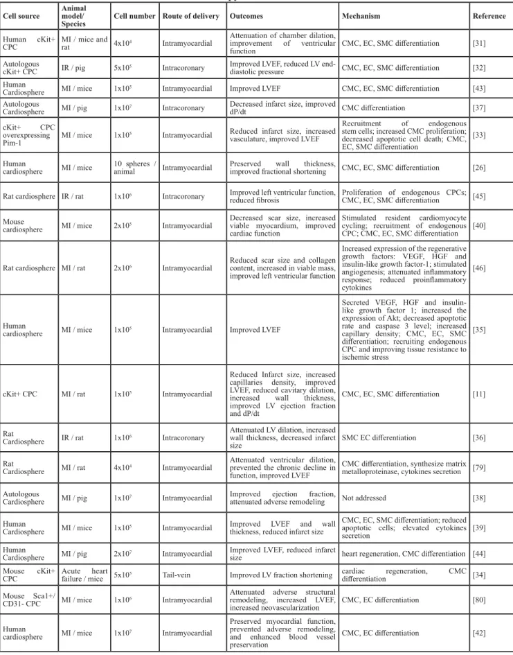
Table 2: Pre-clinical studies of CPC based cell therapy in heart diseases
Cell source Animal model/Species Cell number Route of delivery Outcomes Mechanism Reference
Human cKit+
CPC MI / mice and rat 4x10⁴ Intramyocardial
Attenuation of chamber dilation, improvement of ventricular
function CMC, EC, SMC differentiation [31] Autologous
cKit+ CPC IR / pig 5x10⁵ Intracoronary Improved LVEF, reduced LV end-diastolic pressure CMC, EC, SMC differentiation [32] Human
Cardiosphere MI / mice 1x10⁵ Intramyocardial Improved LVEF CMC, EC, SMC differentiation [43] Autologous
Cardiosphere MI / pig 1x10⁷ Intracoronary Decreased infarct size, improved dP/dt CMC differentiation [37] cKit+ CPC
overexpressing
Pim-1 MI / mice 1x10
5 Intramyocardial Reduced infarct size, increased vasculature, improved LVEF
Recruitment of endogenous stem cells; increased CMC proliferation; decreased apoptotic cell death; CMC, EC, SMC differentiation
[33] Human
cardiosphere MI / mice 10 spheres /animal Intramyocardial Preserved improved fractional shorteningwall thickness, CMC, EC, SMC differentiation [26] Rat cardiosphere IR / rat 1x10⁶ Intracoronary Improved left ventricular function,
reduced fibrosis Proliferation of endogenous CPCs; CMC, EC, SMC differentiation [45] Mouse
cardiosphere MI / mice 2x10⁵ Intramyocardial
Decreased scar size, increased viable myocardium, improved cardiac function
Stimulated resident cardiomyocyte cycling; recruitment of endogenous CPC; CMC, EC, SMC differentiation [40]
Rat cardiosphere MI / rat 2x10⁶ Intramyocardial Reduced scar size and collagen content, increased in viable mass, improved left ventricular function
Increased expression of the regenerative growth factors: VEGF, HGF and insulin-like growth factor-1; stimulated angiogenesis; attenuated inflammatory response; reduced proinflammatory cytokines
[46]
Human
cardiosphere MI / mice 1x10⁵ Intramyocardial Improved LVEF
Secreted VEGF, HGF and insulin-like growth factor 1; increased the expression of Akt; decreased apoptotic rate and caspase 3 level; increased capillary density; CMC, EC, SMC differentiation; recruiting endogenous CPC and improving tissue resistance to ischemic stress
[35]
cKit+ CPC MI / rat 1x10⁵ Intramyocardial
Reduced Infarct size, increased capillaries density, improved LVEF, reduced cavitary dilation, increased wall thickness, improved LV ejection fraction and dP/dt
CMC, EC, SMC differentiation [11]
Rat
Cardiosphere IR / rat 1x10⁶ Intracoronary
Attenuated LV dilation, increased wall thickness, decreased infarct
size SMC EC differentiation [36] Rat
Cardiosphere MI / rat 4x10⁴ Intramyocardial
Attenuated ventricular dilation, prevented the chronic decline in function, improved LVEF
CMC differentiation, synthesize matrix metalloproteinase, cytokines secretion [79] Autologous
Cardiosphere MI / pig 1x10⁷ Intramyocardial Improved ejection fraction, attenuated adverse remodeling Not addressed [38] Human
Cardiosphere MI / mice 1x10⁵ Intramyocardial Improved LVEF and wall thickness, reduced infarct size
CMC, EC, SMC differentiation; reduced apoptotic cells; elevated cytokines
secretion [39]
Human
Cardiosphere MI / pig 2x10⁷ Intramyocardial Improved LVEF, reduced infarct size heart regeneration, CMC differentiation [44] Mouse cKit+
CPC Acute heart failure / mice 5x10⁵ Tail-vein Improved LV fraction shortening cardiac differentiationregeneration, CMC [34] Mouse Sca1+/
CD31- CPC MI / mice 1x10⁶ Intramyocardial
Attenuated adverse structural remodeling, increased LVEF,
increased neovascularization CMC, EC differentiation [80] Human
cardiosphere MI / mice 1x10⁷ Intramyocardial
Preserved myocardial function, prevented adverse remodeling, and enhanced blood vessel preservation
CMC, EC differentiation [42]
CPC: cardiac progenitor cell; MI: myocardial infarction; IR: ischemia re-perfusion; LV: left ventricular; LVEF: left ventricular
ejection fraction; CMC: cardiomyocyte; EC: endothelial cell; SMC: smooth muscle cell
Flk1
+population
By using the GFP (Green Fluorescent Protein)-Bry
mouse embryonic stem cell (mESC) line, in which GFP is
expressed under the brachyury (Bry) promoter, it has been
demonstrated that a GFP
+/Flk1
+cell population during
mESC differentiation has the ability to differentiate into
beating cardiomyocytes in vitro. GFP
+/Flk1
+cells express
cardiac progenitor markers (Mesp1, Isl1, and Nkx2.5) with
clonogenic capability and can also be differentiated into
endothelial cells and smooth muscle cells. To determine
whether this cell population exist in vivo, different regions
and different stages of mouse embryos were cultured
in vitro, and it was found that the colonies generated
from anterior neural plate embryos and head-fold-stage
embryos could become beating colonies. Of these, the
head-fold-stage embryos produced more beating colonies.
Then, the Flk1
+cells were purified from the
head-fold-stage embryos and they had the ability to differentiate into
cardiomyocytes, endothelial cells, and smooth muscle
cells [13, 14]. This strategy was also applied to human
ESC and a similar Flk1
+cell population with cardiac
differentiation abilities (cardiomyocytes, endothelial cells,
and smooth muscle cells) was discovered [13, 14].
Sca1
+population
Oh et al. analysed the stem cell markers Sca1
and c-Kit in adult mouse cardiac cells, in which the
cardiomyocytes were depleted via enzyme digestion. It was
found that 14-17% of them were Sca1
+. The Sca1
+cells
did not express blood cell lineage markers, hematopoietic
Figure 1: Timeline of the discovery of CPCs and their applications.
A. Timeline of the discovery of different populations ofCPCs. B. Timeline of the pre-clinical studies of CPCs. C. Timeline of the clinical studies of CPCs. CPC: cardiac progenitor cell; MI:
stem cell markers, or endothelial cell markers. They
expressed cardiogenic genes, but not mature cardiac
structural genes. After cardiomyocyte differentiation, they
started to express mature cardiac structural genes. By
using a lineage tracing system, the transplanted Sca1
+cells
were recruited into the infarct region in a mouse MI model
and expressed cardiomyocytes markers [15].
Side population
Side population cells are defined by their capacity
to efflux Hoechst dye through an ATP (Adenosine
Triphosphate)-binding cassette transporter. After depleting
the cardiomyocytes, there was a population of
Hoechst-low cells existing in the mouse heart-derived cells. The
cardiac side population cells are capable of self-renewal
and differentiating functional cardiomyocytes with
spontaneous contracting [16, 17]. And the Hoechst efflux
ability of cardiac side population cells was completely
inhibited by the ATP-binding cassette transporter
inhibitor. They were negative for CD45, CD34, CD44,
and c-Kit, but positive for CD31 and Sca1. The cardiac
side population cells formed colonies, indicating their
multi-potency characteristics. And their cardiomyocytes
derivatives coupled with adult cardiomyocytes in vitro via
the co-culture system without cell fusion events [16, 17].
Under physiologic conditions, the cardiac side population
cells maintained their cell pool through cell proliferation
without recruiting extra-cardiac stem cells. After MI, the
cardiac side population cells were depleted quickly, and
then the cell pool was reconstituted later through cell
proliferation and recruiting stem cells from bone marrow
[16, 17].
Mesp1
+population
Mesp1 is the earliest marker in heart development,
and almost all of the heart and related vessels are developed
Figure 2: Potential mechanisms of stem cell therapy for myocardial infarction.
The potential mechanisms have been proposedas direct cardiac differentiation (cardiomyocytes, endothelial cells and smooth muscle cells), paracrine effects (immune regulation, gene transfer, angiogenesis cytokines, anti-apoptosis cytokines, anti-inflammation cytokines, MMP, collagen deposit), and cell fusion. CPC: cardiac progenitor cell; BMSC: bone marrow stem cell; MSC: mesenchymal stem cell; EPC: endothelial progenitor cell; MMP: matrix metalloproteinase. Dash lines indicate that the evidences are controversial.
from the Mesp1
+cells through lineage tracing studies [18,
19]. Transient expression of Mesp1 dramatically enhanced
CPC generation and also cardiomyocyte differentiation in
mouse ESC. Through whole-genome expression profiling
and chromatin immunoprecipitation (ChIP) analysis,
it has been shown that Mesp1 could directly upregulate
cardiac transcription factors, such as Hand2 and Nkx2.5,
and the Wnt pathway. In addition, Mesp1 suppressed the
expression of genes related to pluripotent, endoderm,
and early mesoderm [18, 19]. Then, the ESC cell line
with GFP expression driven by the Mesp1 promoter
was established to purify the Mesp1
+cells. The purified
Mesp1
+cells enriched CPCs with abilities to differentiate
into cardiomyocytes, endothelial cells, and smooth muscle
cells. Transplanting these Mesp1
+cells into the kidney
capsule of immunodeficient mice showed that they mainly
differentiated into cardiomyocytes in vivo and, to a lesser
extent, endothelial cells and smooth muscle cells [18, 19].
Isl1
+population
Isl1 is a transcription factor modulating heart
development; lack of Isl1 results in heart abnormalities
[20-22]. Using a lineage tracing strategy, the Isl1
+cells
represent a new population of CPCs involved in heart
development. Approximately 30-40% cardiomyocytes
originated from Isl1
+cells during heart development.
Purified Isl1
+cells showed functional ability of
cardiomyocyte differentiation [20-22]. Using the
mouse ESC cell line, Isl1
+cells were further proven as
a CPC population with the ability to differentiate in
cardiomyocytes, endothelial cells, and smooth muscle
cells [20-22].
Nkx2.5
+population
By using transgenic mice with GFP expression
driven by the cardiac-specific Nkx2.5 enhancer, it was
demonstrated that Nkx2.5 expression overlapped partially
with Isl1 and completely overlapped with the sarcomeric
myosin heavy chain [23]. Isolated Nkx2.5
+cells from
embryos showed cardiomyocyte, conduction system cell,
and smooth muscle cell differentiation ability. Purified
Nkx2.5
+cell during mouse ESC differentiation also
showed cardiomyocyte and smooth muscle differentiation
ability in vitro and in vivo [23]. These cells were positive
for c-Kit and Sca1, but negative for hematopoietic and
endothelial markers [23]. Later study also showed that
NKX2.5 positive CPCs could be generated from human
ESC [28].
Wt1
+population
By knocking-in GFP after the gene Wt1 (Wilms
tumour 1), it was demonstrated that one population
of CPCs located within the epicardium expressed the
transcription factor Wt1. The data showed that some
of the Wt1
+cells migrated and differentiated into
functional cardiomyocytes during heart development.
The cardiomyocytes originated from Wt1
+progenitor
cells were located in all four chambers of the heart.
Furthermore, these progenitor cells originated from early
CPCs that expressed Nkx2.5 and Isl1. The purified Wt1
+cells also had the capability of differentiating into beating
cardiomyocytes, endothelial cells, and smooth muscle
cells [24, 25]. The Wt1
+CPCs were activated after MI
or thymosin beta4. Transplanting these Wt1
+cells into
the heart after MI showed functional cardiomyocyte
differentiation and integration into the resident
myocardium [24, 25].
Cardiosphere
Cardiospheres are composed of sphere-forming cells
isolated from human heart biopsy (atrial and ventricular)
and mouse heart (embryo, fetal, and postnatal). These
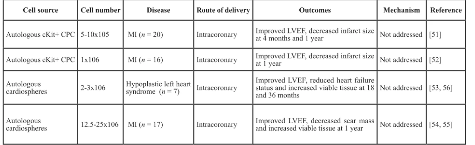
Table 3: Clinical studies of CPC-based cell therapy in heart diseases
Cell source Cell number Disease Route of delivery Outcomes Mechanism Reference
Autologous cKit+ CPC 5-10x105 MI (n = 20) Intracoronary Improved LVEF, decreased infarct size at 4 months and 1 year Not addressed [51] Autologous cKit+ CPC 1x10⁶ MI (n = 16) Intracoronary Improved LVEF, decreased infarct size at 1 year Not addressed [52] Autologous
cardiospheres 2-3x10⁶ Hypoplastic left heart syndrome (n = 7) Intracoronary
Improved LVEF, reduced heart failure status and increased viable tissue at 18
and 36 months Not addressed [53, 56] Autologous
cardiospheres 12.5-25x106 MI (n = 17) Intracoronary Improved LVEF, decreased scar mass and increased viable tissue at 1 year Not addressed [54, 55]
CPC: cardiac progenitor cell; MI: myocardial infarction; LVEF: left ventricular ejection fraction.
sphere-forming cells originate from small, round, and
phase-bright cells that migrated from the heart explants
[26]. Cardiospheres generated from mouse heart could
beat spontaneously after sphere formation. However, the
human cardiospheres did not have this capability unless
they were co-cultured with rat neonatal cardiomyocytes.
Cardiospheres could attach onto fibronectin-coated
plates. They formed spheres when growing on
poly-D-lysine-coated plates. They contained Flk1, CD31, CD34,
c-Kit and Sca1 positive cells. When transplanting these
cardiospheres into the dorsal subcutaneous region or the
heart after MI, they had the capability to differentiate
into cardiomyocytes, endothelial cells, and smooth
muscle cells in vivo [26]. Although the cardiac stem cell
property of cardiospheres has been questioned [29], the
cardiosphere concept has been widely accepted.
PRE-CLINICAL AND CLINICAL
STUDIES OF CARDIAC PROGENITOR
CELLS
The therapeutic potential of CPCs has been
intensively studied in animal models [30]. And the safety
and practicability are further demonstrated by clinical
studies. Among all types of CPCs, only two of them are
widely studied for their therapeutic application in treating
MI, c-Kit
+CPCs and cardiosphere-derived cells [31]
(Figure 1, Table 2, Table 3 and Supplementary Table 1).
Transplanting the c-Kit
+CPCs into animal
models with injured hearts promoted myocardial
regeneration, heart function improvement, adverse heart
remodelling attenuation, and cell death reduction with
cardiomyocytes differentiation (Table 2) [11, 32-35].
In addition to the role of improving heart function and
cardiac cell differentiation, transplanting cardiospheres
or cardiosphere-derived cells also showed the effects of
anti-apoptotic, anti-fibrotic, activating endogenous CPCs,
cytokine secretion, and inflammation modulation (Table 2)
[26, 36-47]. Furthermore, transplanting the cardiospheres
in the form of the sphere was more effective than the
cardiosphere-derived cells, indicating that the
three-dimensional structure maintained the niche for stem cells
[48]. Combing the MSCs with cardiosphere-derived cells
also showed increased efficacy in improving heart function
and cardiac regeneration [49-51].
Because of the promising results in pre-clinical
animal models, clinical trials were conducted to assess
the safety, efficacy, and feasibility of CPCs in treating
patients (Table 3 and Supplementary Table 1) [52-57]. The
first phase 1 clinical trial using CPCs was conducted to
investigate their safety and feasibility. Autologous c-Kit
+/
Lin
−CPCs were harvested during coronary artery bypass
graft surgery and then used to treat heart failure patients
with intracoronary infusion. Heart function was improved
as early as 4 months after cell transplantation, and this
effect continued up to 1 year later. During this period, the
control group did not show any evidence of functional
improvement. Furthermore, after CPCs transplantation,
infarct size decreased, ventricular mass increased, and no
adverse effects were observed (Table 3) [52, 53].
Transplanting autologous cardiospheres also showed
cardiac function improvement, scar size reduction without
any adverse events and tumour formation, indicating
their safety and feasibility (Table 3) [54-56]. The
ongoing clinical trials are designed to further address the
effectiveness, cell type, cell number, delivery strategy,
time window, and other factors (Table 4).
MECHANISMS OF CELL THERAPY
AND THE KEY ROLE OF CARDIAC
PROGENITOR CELLS
Two goals should be achieved in MI treatment.
The first is to prevent cardiomyocyte death and adverse
heart remodelling. The second is to promote cardiac
repair or regeneration and preserve and recover cardiac
function. So far, the potential mechanisms of stem cell
therapy for treating MI have been proposed as direct
cardiomyocyte differentiation, blood vessel formation
(endothelial and smooth muscle cell differentiation), cell
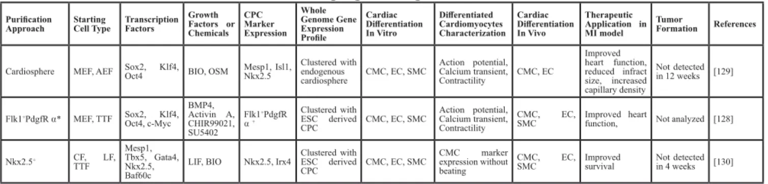
Table 4: Generation of induced CPC via somatic reprogramming
Purification
Approach Starting Cell Type Transcription Factors Growth Factors or Chemicals CPC Marker Expression Whole Genome Gene Expression Profile Cardiac Differentiation In Vitro Differentiated Cardiomyocytes Characterization Cardiac Differentiation In Vivo Therapeutic Application in MI model Tumor Formation References
Cardiosphere MEF, AEF Sox2, Klf4, Oct4 BIO, OSM Mesp1, Isl1, Nkx2.5 Clustered with endogenous
cardiosphere CMC, EC, SMC Action potential, Calcium transient, Contractility CMC, EC Improved heart function, reduced infract size, increased capillary density Not detected in 12 weeks [129] Flk1+PdgfR ⍺* MEF, TTF Sox2, Klf4, Oct4, c-Myc BMP4, Activin A, CHIR99021, SU5402 Flk1+PdgfR ⍺ + Clustered with ESC derived CPC CMC, EC, SMC Action potential, Calcium transient, Contractility CMC, EC,
SMC Improved heart function, Not analyzed [128]
Nkx2.5+ CF, LF, TTF Mesp1, Tbx5, Gata4, Nkx2.5, Baf60c
LIF, BIO Nkx2.5, Irx4 Clustered with ESC derived
CPC CMC, EC, SMC
CMC marker expression without beating
CMC, EC,
SMC Improved survival Not detected in 4 weeks [130]
MEF: mouse embryonic fibroblast; AEF: adult mouse ear fibroblast; TTF: mouse tail-tip fibroblast; CF: mouse cardiac
fibroblast; LF: mouse lung fibroblast; OSM: oncostatin M; ESC: mouse embryonic stem cell; CPC: cardiac progenitor cell;
CMC: cardiomyocyte; EC: endothelial cell; SMC: smooth muscle cell.
fusion, and paracrine effects (endogenous CPC activation,
neovascularisation, and apoptosis inhibition) (Figure 2) [7,
58].
The efficacy of BMSCs, MSCs, and EPCs in treating
myocardial infarction is evident. However, the underlying
mechanism is believed to be paracrine effects and cell
fusion, but not direct cardiomyocyte differentiation
[58-63]. The paracrine effects include promoting angiogenesis
[64-66], preventing apoptosis [67, 68], suppressing
inflammation [69-73], modulating extracellular matrix
dynamics [74] and transferring genes into the local
cardiomyocytes [75]. However, there are some other
studies that have argued that direct cardiomyocyte
differentiation also contributes to improvement in heart
function [42, 76-79].
CPCs also have shown effectiveness in improving
heart function, increasing neovascularisation, reducing
infarct size, and attenuating adverse remodelling.
Differing from BMSCs, MSCs and EPCs, for which the
paracrine effect is the main mechanism of heart function
improvement, it has shown strong evidences of direct
cardiac differentiation (cardiomyocytes, endothelial cells,
and smooth muscle cells) of CPCs [11, 26, 32-41,
43-47, 80, 81]. Furthermore, cardiosphere transplantation
also shows paracrine effects (Table 2). For example,
they secrete cytokines (such as vascular endothelial
growth factor, hepatocyte growth factor and insulin-like
growth factor), activate endogenous stem cells, decrease
cell apoptosis, remodel extracellular matrix, and inhibit
inflammation response [34, 36, 40, 41, 46, 47, 80].
In a rat MI model, the c-Kit
+CPCs migrated into the
infarct region through collagen type I and type III bundles.
Analysis of matrix metalloproteinases (MMP; responsible
for degrading extracellular matrix) and its tissue inhibitors
of MMP (TIMP) showed that CPCs transplantation
strongly promoted the expression level of MMP2, MMP9,
and MMP14, and inhibited TIMP4 expression. These
data indicated that CPCs have invasive ability through
modulating extracellular matrix [80]. Transplanted CPCs
(c-Kit
+CPCs and cardiospheres) continued to proliferate
in vivo and activated endogenous CPCs [36, 41, 46].
Overexpression of Pim1 in c-Kit
+CPC (component of
AKT pathway that showed cardiac-protective activity)
further enhanced the activity of CPCs [34]. Furthermore,
miRNA could be transferred from CPC to cardiomyocytes
[82-86].
MI triggers the resident cardiomyocytes to
re-enter the cell cycle, up-regulating cell cycle-related
genes. This effect was further amplified by cardiosphere
transplantation [41]. Cardiosphere transplantation reduced
pro-inflammatory cytokines (tumour necrosis factor-α,
interferon-γ, interleukin-6, and interleukin-1β), which
are normally activated by MI, and produced regenerative
growth factors (VEGF, Vascular Endothelial Growth
Factor; HGF, Hepatocyte Growth Factor; and
insulin-like growth factor) [36, 47]. In a rat ischemia reperfusion
(IR) model, cardiosphere transplantation was effective
in reducing heart injury only when the transplantation
was conducted in 20 minutes, but not after 2 days [87].
Cardiospheres could reduce the infarct size and prevent
cardiomyocyte death through reducing pro-inflammatory
factor secretion and the number of CD68
+macrophages.
Cardiosphere cells activated the macrophage polarization,
shifting them away from M1 macrophages, resulting in
a reduction of the inflammation response. Cardiospheres
that activated M2 macrophages polarization showed
anti-oxidative and anti-apoptotic effects, and transplanting
them into the rat IR model showed a reduction in heart
injury [87]. More interestingly, the cardiospheres had
been shown to be much more effective than c-Kit
+CPC
for improving heart function because they secreted more
cytokines [40].
CPCs in cell therapy for MI are important not
only because they could differentiate into cardiac cells
(cardiomyocytes, endothelial cells, and smooth muscle
cells) and are involved in cardiac function preservation
and recovery but also because they mediate the efficacy
of other types of stem cells (BMSCs and MSCs, Figure
2). Therefore, CPCs are the most important player
in heart function recovery after MI [35]. MSCs and
BMSCs could stimulate endogenous CPC proliferation
and differentiation, and the activated CPCs contribute to
cardiac function improvement after MI [88, 89]. The cell
mixture of MSCs and CPCs had a synergistic effect on
treating MI [50]. And the cell hybrids through cell fusion
of cardiac progenitors and MSCs showed better efficacy in
myocardial repair. However, abolishing endogenous CPCs
would eliminate the regeneration process [49].
Therefore, it is becoming much more obvious that
the therapeutic effects of cell therapy for preventing
adverse heart remodelling and promoting cardiac
regeneration might work through cardiac progenitor
activity. Accounting for cardiac differentiation abilities,
the CPCs play a central role in cell therapy of MI (Figure
2).
ENDOGENOUS CARDIAC PROGENITOR
CELLS ARE NOT SUFFICIENT FOR
MYOCARDIAL REGENERATION
The fact that most survivors of MI would eventually
develop and die from congestive heart failure makes it
clear that although there is some level of cardiomyocyte
turnover in the adult heart, it is not sufficient to compensate
for the cell loss [63]. In mice, cardiomyocyte regeneration
occurs at a very low rate, and this is decreased during
ageing and increased after injury [90]. Furthermore, both
cell number and cell activity of cardiomyocytes and CPCs
decline during ageing [91]. Cardiospheres derived from
neonatal human atrium are more cardiomyogenic and
more effective in the improvement of heart function in the
mouse MI model [43].
Therefore, the body could not regenerate the
heart by itself after the MI. After MI, the heart is in a
high-inflammatory, fibrotic, low-nutrient, and hypoxia
environment, which impairs the function and efficacy
of cardiomyocytes, bone marrow stem cells, and local
CPCs. Isolated CPCs from the heart could be used for
transplantation after expansion in vitro. Considering that
the procedures for cardiac cell harvesting are invasive
and would cause more injury to patients with MI, the
generation of induced CPCs via somatic reprogramming
from somatic cells, which are more easily available, is
necessary.
CELL FATE CONVERSION VIA SOMATIC
REPROGRAMMING IN CARDIAC CELLS
The embryo is developed in a temporal and spatial
manner and is precisely controlled by cytokines and
genes. Stem cells with differentiation abilities play a
crucial role in the development process. Normally, the
differentiation ability of stem cells would become more
and more committed during development, and this
process is difficult to reverse. Eventually, cells become
terminally differentiated in different tissues with different
characteristics and functions. This process is called cell
fate determination. Long ago, people believed that once the
cell fate is determined, it is difficult to change. However,
this has been challenged with the development of somatic
reprogramming technologies. Somatic reprogramming
is used to convert the terminally differentiated somatic
cells to a more progenitor cell state or even pluripotent
stem cells state with more differentiation abilities
[92-95]. After the breakthrough in somatic reprogramming
with the development of induced pluripotent stem cell
(iPSC) technology, the somatic reprogramming field
has grown rapidly [96, 97]. iPSC are generated through
overexpressing transcription factors (Sox2, Klf4, Oct4,
and c-Myc), which are important for pluripotent stem cell
maintenance, in terminal differentiated somatic cells, and
this converts the somatic cells into pluripotent stem cells
[98-101].
Since then, the strategy of cell fate conversion
through transcription factor overexpression has been
widely used in the generation of different types of stem
cells and terminally differentiated cells from somatic
cells. There are two approaches to achieving cell fate
conversion, lineage-specific transcription factors and
Yamanaka factors (Sox2, Klf4, Oct4, and c-Myc)
[102-104]. Cardiomyocytes could be generated through
overexpressing cardiac-specific transcription factor
(Gata4, Mef2c, and Tbx5 or cardiac microRNAs) [102,
105-125] or Yamanaka factors in fibroblast cells [111, 123]
(Supplementary Table 2).
Although the mechanism of cardiac cell fate
conversion still remains unclear, genome-wide gene
expression profile analysis showed that the cell fate
conversion process induced cardiac gene expression and
silenced the fibroblast-related genes. Direct cardiomyocyte
generation from fibroblasts through cardiac-specific
factors, such as GTM (Gata4, Tbx5, and Mef2c), did
not pass a CPC stage [105, 109, 120, 125]. However,
the combination of Sox2, Klf4, and Oct4 reprogrammed
fibroblasts into cardiomyocytes through a CPC stage
with Mesp1 and Isl1 expression [111, 123], indicating the
possibility of generating induced CPCs from somatic cells
via somatic programming.
GENERATION OF INDUCED CARDIAC
PROGENITOR CELLS VIA SOMATIC
REPROGRAMMING
The reprogrammed cardiomyocytes have very
low conversion efficiency and no proliferation capacity.
Therefore, expansion in vitro for transplantation is not
feasible. The therapeutic application of this strategy only
relies on reprogramming in vivo [106, 108, 110, 114].
In addition to the low conversion efficiency in vivo,
disturbing the function of cardiac fibroblasts with virus
and gene overexpression might also affect the regenerative
effects of cardiac fibroblasts that play a central role in
heart remodelling and regeneration [126-128]. Therefore,
it is necessary to generate CPC-like stem cells with the
ability to regenerate the heart which might provide an
unlimited source of stem cells with cardiac differentiation
potentials [129-131] (Table 4).
We developed a novel approach to generate
induced cardiospheres (iCS) from adult skin fibroblasts
via somatic reprogramming [130]. After infection with
Sox2, Klf4 and Oct4, iCS were generated from mouse
adult skin fibroblasts treated with Gsk3β
inhibitor-(2’Z, 3’E) - 6-Bromoindirubin-3’-oxime (BIO) and
Oncostatin M. They resembled endogenous cardiospheres
(eCS) with whole genome gene expression analysis,
but contained a higher percentage of cells expressing
Mesp1, Isl1 and Nkx2.5. They were differentiated
into functional cardiomyocytes in vitro with similar
electrophysiological properties, calcium transient and
contractile function to eCS and mouse embryonic stem
cell-derived cardiomyocytes. Transplantation of iCS into
mouse myocardium following MI had similar effects to
transplantation of eCS but significantly better than saline
or fibroblast in improving left ventricular ejection fraction,
increasing anterior/septal ventricular wall thickness and
capillary density in the infarcted region 4 weeks after
transplantation [130].
Zhang et al. also found that the induced CPC
(Flk1
+PdgfR
⍺
+population) could be generated from
mouse fibroblasts with overexpressing Sox2, Klf4, Oct4
and c-Myc plus BMP4, Activin A, CHIR99021 (GSK3beta
inhibitor) and SU5402 (FGF, VEGF and PDGF inhibitor)
stimulation [129]. The Flk1
+PdgfR
⍺
+induced CPC
(iCPC) could be expandable and differentiated into
cardiomyocytes, endothelial cells and smooth muscle
cells. The differentiated cardiomyocytes had functional
action potential and calcium transient activities. They
could be stimulated by caffeine and isoproterenol. Whole
genome expression analysis showed that they are similar
with ESC derived CPCs. Transplanting Flk1
+PdgfR
⍺
+iCPC into MI mice model improved heart function [129].
On the other hand, the iCPC could be generated
through lineage specific transcription factors Mesp1,
Tbx5, Gata4, Nkx2.5 and Baf60c (MTGNB) plus LIF
(leukemia inhibitory factor) and BIO stimulation [131].
Using a mouse model containing Nkx2.5-EYFP (enhanced
yellow fluorescent protein) reporter system, the
Nkx2.5-EYFP positive iCPC could be purified and expanded in
vitro without expressing pluripotent markers. Whole
genome expression analysis showed that they are similar
with ESC derived CPCs. They could be differentiated into
cardiomyocytes, endothelial cells and smooth muscle cells
in vitro and cardiomyocytes in vivo. Transplanting these
iCPC into MI mice model improved heart function without
tumor formation [131].
FUTURE PERSPECTIVES
The iCPC could be generated from somatic cells
via somatic reprogramming strategy with lineage specific
transcription factors or Yamanaka factors overexpression.
The Nkx2.5
+and Flk1
+PdgfR
⍺
+iCPC are similar to mouse
ESC derived CPCs, but whether they are also similar to
endogenous CPCs or not remains unclear. The induced
cardiosphere is similar to the endogenous cardiosphere
but they both contain mixed cell populations, including
CPCs and other supporting cells [132]. Therefore, more
specific CPC surface markers and CPC purification
methods should be developed. Furthermore, the iCPC are
generated through a virus based method and the genome
integration impairs their potential therapeutic applications.
Thus integration-free methods should be applied in iCPC
generation in the future.
CONFLICTS OF INTEREST
The authors declare that there are no conflicts of
interest.
FUNDING
Novel technology development project of Shenzhen.
No. CXZZ2015093105220591.
REFERENCES
1. Go AS, Mozaffarian D, Roger VL, Benjamin EJ, Berry JD, Borden WB, Bravata DM, Dai S, Ford ES, Fox CS, Franco
S, Fullerton HJ, Gillespie C, et al. Executive summary: heart disease and stroke statistics—2013 update: a report from the American Heart Association. Circulation. 2013; 127:143-152.
2. Heusch G, Libby P, Gersh B, Yellon D, Bohm M, Lopaschuk G, Opie L. Cardiovascular remodelling in coronary artery disease and heart failure. Lancet. 2014; 383:1933-1943.
3. Willerson JT. Stem cell therapy for cardiovascular diseases: a progressive journey. Current opinion in cardiology. 2015. 4. Martin-Puig S, Wang Z, Chien KR. Lives of a heart cell:
tracing the origins of cardiac progenitors. Cell stem cell. 2008; 2:320-331.
5. Segers VF, Lee RT. Stem-cell therapy for cardiac disease. Nature. 2008; 451:937-942.
6. Behfar A, Crespo-Diaz R, Terzic A, Gersh BJ. Cell therapy for cardiac repair—lessons from clinical trials. Nature reviews Cardiology. 2014; 11:232-246.
7. Sanganalmath SK, Bolli R. Cell therapy for heart failure: a comprehensive overview of experimental and clinical studies, current challenges, and future directions. Circulation research. 2013; 113:810-834.
8. Deutsch MA, Sturzu A, Wu SM. At a crossroad: cell therapy for cardiac repair. Circulation research. 2013; 112:884-890.
9. Menasche P, Hagege AA, Scorsin M, Pouzet B, Desnos M, Duboc D, Schwartz K, Vilquin JT, Marolleau JP. Myoblast transplantation for heart failure. Lancet. 2001; 357:279-280. 10. Zhang YM, Hartzell C, Narlow M, Dudley SC Jr. Stem cell-derived cardiomyocytes demonstrate arrhythmic potential. Circulation. 2002; 106:1294-1299.
11. Beltrami AP, Barlucchi L, Torella D, Baker M, Limana F, Chimenti S, Kasahara H, Rota M, Musso E, Urbanek K, Leri A, Kajstura J, Nadal-Ginard B, et al. Adult cardiac stem cells are multipotent and support myocardial regeneration. Cell. 2003; 114:763-776.
12. Sturzu AC, Wu SM. Developmental and regenerative biology of multipotent cardiovascular progenitor cells. Circulation research. 2011; 108:353-364.
13. Kattman SJ, Huber TL, Keller GM. Multipotent flk-1+ cardiovascular progenitor cells give rise to the cardiomyocyte, endothelial, and vascular smooth muscle lineages. Developmental cell. 2006; 11:723-732.
14. Yang L, Soonpaa MH, Adler ED, Roepke TK, Kattman SJ, Kennedy M, Henckaerts E, Bonham K, Abbott GW, Linden RM, Field LJ, Keller GM. Human cardiovascular progenitor cells develop from a KDR+ embryonic-stem-cell-derived population. Nature. 2008; 453:524-528.
15. Oh H, Bradfute SB, Gallardo TD, Nakamura T, Gaussin V, Mishina Y, Pocius J, Michael LH, Behringer RR, Garry DJ, Entman ML, Schneider MD. Cardiac progenitor cells from adult myocardium: homing, differentiation, and fusion after infarction. Proceedings of the National Academy of Sciences of the United States of America. 2003;
100:12313-12318.
16. Mouquet F, Pfister O, Jain M, Oikonomopoulos A, Ngoy S, Summer R, Fine A, Liao R. Restoration of cardiac progenitor cells after myocardial infarction by self-proliferation and selective homing of bone marrow-derived stem cells. Circulation research. 2005; 97:1090-1092. 17. Pfister O, Mouquet F, Jain M, Summer R, Helmes M, Fine
A, Colucci WS, Liao R. CD31- but Not CD31+ cardiac side population cells exhibit functional cardiomyogenic differentiation. Circulation research. 2005; 97:52-61. 18. Bondue A, Lapouge G, Paulissen C, Semeraro C, Iacovino
M, Kyba M, Blanpain C. Mesp1 acts as a master regulator of multipotent cardiovascular progenitor specification. Cell stem cell. 2008; 3:69-84.
19. Bondue A, Tannler S, Chiapparo G, Chabab S, Ramialison M, Paulissen C, Beck B, Harvey R, Blanpain C. Defining the earliest step of cardiovascular progenitor specification during embryonic stem cell differentiation. The Journal of cell biology. 2011; 192:751-765.
20. Cai CL, Liang X, Shi Y, Chu PH, Pfaff SL, Chen J, Evans S. Isl1 identifies a cardiac progenitor population that proliferates prior to differentiation and contributes a majority of cells to the heart. Developmental cell. 2003; 5:877-889.
21. Laugwitz KL, Moretti A, Lam J, Gruber P, Chen Y, Woodard S, Lin LZ, Cai CL, Lu MM, Reth M, Platoshyn O, Yuan JX, Evans S, et al. Postnatal isl1+ cardioblasts enter fully differentiated cardiomyocyte lineages. Nature. 2005; 433:647-653.
22. Moretti A, Caron L, Nakano A, Lam JT, Bernshausen A, Chen Y, Qyang Y, Bu L, Sasaki M, Martin-Puig S, Sun Y, Evans SM, Laugwitz KL, et al. Multipotent embryonic isl1+ progenitor cells lead to cardiac, smooth muscle, and endothelial cell diversification. Cell. 2006; 127:1151-1165. 23. Wu SM, Fujiwara Y, Cibulsky SM, Clapham DE, Lien
CL, Schultheiss TM, Orkin SH. Developmental origin of a bipotential myocardial and smooth muscle cell precursor in the mammalian heart. Cell. 2006; 127:1137-1150.
24. Zhou B, Ma Q, Rajagopal S, Wu SM, Domian I, Rivera-Feliciano J, Jiang D, von Gise A, Ikeda S, Chien KR, Pu WT. Epicardial progenitors contribute to the cardiomyocyte lineage in the developing heart. Nature. 2008; 454:109-113. 25. Smart N, Bollini S, Dube KN, Vieira JM, Zhou B,
Davidson S, Yellon D, Riegler J, Price AN, Lythgoe MF, Pu WT, Riley PR. De novo cardiomyocytes from within the activated adult heart after injury. Nature. 2011; 474:640-644.
26. Messina E, De Angelis L, Frati G, Morrone S, Chimenti S, Fiordaliso F, Salio M, Battaglia M, Latronico MV, Coletta M, Vivarelli E, Frati L, Cossu G, et al. Isolation and expansion of adult cardiac stem cells from human and murine heart. Circulation research. 2004; 95:911-921. 27. Orlic D, Kajstura J, Chimenti S, Jakoniuk I, Anderson SM,
Li B, Pickel J, McKay R, Nadal-Ginard B, Bodine DM,
Leri A, Anversa P. Bone marrow cells regenerate infarcted myocardium. Nature. 2001; 410:701-705.
28. Elliott DA, Braam SR, Koutsis K, Ng ES, Jenny R, Lagerqvist EL, Biben C, Hatzistavrou T, Hirst CE, Yu QC, Skelton RJ, Ward-van Oostwaard D, Lim SM, et al. NKX2-5(eGFP/w) hESCs for isolation of human cardiac progenitors and cardiomyocytes. Nature methods. 2011; 8:1037-1040.
29. Andersen DC, Andersen P, Schneider M, Jensen HB, Sheikh SP. Murine “cardiospheres” are not a source of stem cells with cardiomyogenic potential. Stem cells. 2009; 27:1571-1581.
30. Samanta A, Dawn B. Meta-Analysis of Preclinical Data Reveals Efficacy of Cardiac Stem Cell Therapy for Heart Repair. Circulation research. 2016; 118:1186-1188. 31. Zwetsloot PP, Vegh AM, Jansen of Lorkeers SJ, van Hout
GP, Currie GL, Sena ES, Gremmels H, Buikema JW, Goumans MJ, Macleod MR, Doevendans PA, Chamuleau SA, Sluijter JP. Cardiac Stem Cell Treatment in Myocardial Infarction: A Systematic Review and Meta-Analysis of Preclinical Studies. Circulation research. 2016; 118:1223-1232.
32. Bearzi C, Rota M, Hosoda T, Tillmanns J, Nascimbene A, De Angelis A, Yasuzawa-Amano S, Trofimova I, Siggins RW, Lecapitaine N, Cascapera S, Beltrami AP, D’Alessandro DA, et al. Human cardiac stem cells. Proceedings of the National Academy of Sciences of the United States of America. 2007; 104:14068-14073. 33. Bolli R, Tang XL, Sanganalmath SK, Rimoldi O, Mosna
F, Abdel-Latif A, Jneid H, Rota M, Leri A, Kajstura J. Intracoronary delivery of autologous cardiac stem cells improves cardiac function in a porcine model of chronic ischemic cardiomyopathy. Circulation. 2013; 128:122-131. 34. Fischer KM, Cottage CT, Wu W, Din S, Gude NA,
Avitabile D, Quijada P, Collins BL, Fransioli J, Sussman MA. Enhancement of myocardial regeneration through genetic engineering of cardiac progenitor cells expressing Pim-1 kinase. Circulation. 2009; 120:2077-2087.
35. Ellison GM, Vicinanza C, Smith AJ, Aquila I, Leone A, Waring CD, Henning BJ, Stirparo GG, Papait R, Scarfo M, Agosti V, Viglietto G, Condorelli G, et al. Adult c-kit(pos) cardiac stem cells are necessary and sufficient for functional cardiac regeneration and repair. Cell. 2013; 154:827-842. 36. Chimenti I, Smith RR, Li TS, Gerstenblith G, Messina
E, Giacomello A, Marban E. Relative roles of direct regeneration versus paracrine effects of human cardiosphere-derived cells transplanted into infarcted mice. Circulation research. 2010; 106:971-980.
37. Dawn B, Stein AB, Urbanek K, Rota M, Whang B, Rastaldo R, Torella D, Tang XL, Rezazadeh A, Kajstura J, Leri A, Hunt G, Varma J, et al. Cardiac stem cells delivered intravascularly traverse the vessel barrier, regenerate infarcted myocardium, and improve cardiac function. Proceedings of the National Academy of Sciences of the United States of America. 2005; 102:3766-3771.
38. Johnston PV, Sasano T, Mills K, Evers R, Lee ST, Smith RR, Lardo AC, Lai S, Steenbergen C, Gerstenblith G, Lange R, Marban E. Engraftment, differentiation, and functional benefits of autologous cardiosphere-derived cells in porcine ischemic cardiomyopathy. Circulation. 2009; 120:1075-1083, 1077 p following 1083.
39. Lee ST, White AJ, Matsushita S, Malliaras K, Steenbergen C, Zhang Y, Li TS, Terrovitis J, Yee K, Simsir S, Makkar R, Marban E. Intramyocardial injection of autologous cardiospheres or cardiosphere-derived cells preserves function and minimizes adverse ventricular remodeling in pigs with heart failure post-myocardial infarction. Journal of the American College of Cardiology. 2011; 57:455-465. 40. Li TS, Cheng K, Malliaras K, Smith RR, Zhang Y, Sun
B, Matsushita N, Blusztajn A, Terrovitis J, Kusuoka H, Marban L, Marban E. Direct comparison of different stem cell types and subpopulations reveals superior paracrine potency and myocardial repair efficacy with cardiosphere-derived cells. Journal of the American College of Cardiology. 2012; 59:942-953.
41. Malliaras K, Zhang Y, Seinfeld J, Galang G, Tseliou E, Cheng K, Sun B, Aminzadeh M, Marban E. Cardiomyocyte proliferation and progenitor cell recruitment underlie therapeutic regeneration after myocardial infarction in the adult mouse heart. EMBO Mol Med. 2013; 5:191-209. 42. Rota M, Kajstura J, Hosoda T, Bearzi C, Vitale S, Esposito
G, Iaffaldano G, Padin-Iruegas ME, Gonzalez A, Rizzi R, Small N, Muraski J, Alvarez R, et al. Bone marrow cells adopt the cardiomyogenic fate
in vivo
. Proceedings of the National Academy of Sciences of the United States of America. 2007; 104:17783-17788.43. Simpson DL, Mishra R, Sharma S, Goh SK, Deshmukh S, Kaushal S. A strong regenerative ability of cardiac stem cells derived from neonatal hearts. Circulation. 2012; 126:S46-53.
44. Smith RR, Barile L, Cho HC, Leppo MK, Hare JM, Messina E, Giacomello A, Abraham MR, Marban E. Regenerative potential of cardiosphere-derived cells expanded from percutaneous endomyocardial biopsy specimens. Circulation. 2007; 115:896-908.
45. Takehara N, Tsutsumi Y, Tateishi K, Ogata T, Tanaka H, Ueyama T, Takahashi T, Takamatsu T, Fukushima M, Komeda M, Yamagishi M, Yaku H, Tabata Y, et al. Controlled delivery of basic fibroblast growth factor promotes human cardiosphere-derived cell engraftment to enhance cardiac repair for chronic myocardial infarction. Journal of the American College of Cardiology. 2008; 52:1858-1865.
46. Tang XL, Rokosh G, Sanganalmath SK, Yuan F, Sato H, Mu J, Dai S, Li C, Chen N, Peng Y, Dawn B, Hunt G, Leri A, et al. Intracoronary administration of cardiac progenitor cells alleviates left ventricular dysfunction in rats with a 30-day-old infarction. Circulation. 2010; 121:293-305. 47. Tseliou E, Pollan S, Malliaras K, Terrovitis J, Sun B,
Galang G, Marban L, Luthringer D, Marban E. Allogeneic
cardiospheres safely boost cardiac function and attenuate adverse remodeling after myocardial infarction in immunologically mismatched rat strains. Journal of the American College of Cardiology. 2013; 61:1108-1119. 48. Li TS, Cheng K, Lee ST, Matsushita S, Davis D, Malliaras
K, Zhang Y, Matsushita N, Smith RR, Marban E. Cardiospheres recapitulate a niche-like microenvironment rich in stemness and cell-matrix interactions, rationalizing their enhanced functional potency for myocardial repair. Stem cells. 2010; 28:2088-2098.
49. Quijada P, Salunga HT, Hariharan N, Cubillo J, El-Sayed F, Moshref M, Bala KM, Emathinger JM, De La Torre A, Ormachea L, Alvarez R, Gude NA, Sussman MA. Cardiac Stem Cell Hybrids Enhance Myocardial Repair. Circulation research. 2015.
50. Williams AR, Hatzistergos KE, Addicott B, McCall F, Carvalho D, Suncion V, Morales AR, Da Silva J, Sussman MA, Heldman AW, Hare JM. Enhanced effect of combining human cardiac stem cells and bone marrow mesenchymal stem cells to reduce infarct size and to restore cardiac function after myocardial infarction. Circulation. 2013; 127:213-223.
51. Zakharova L, Mastroeni D, Mutlu N, Molina M, Goldman S, Diethrich E, Gaballa MA. Transplantation of cardiac progenitor cell sheet onto infarcted heart promotes cardiogenesis and improves function. Cardiovascular research. 2010; 87:40-49.
52. Chugh AR, Beache GM, Loughran JH, Mewton N, Elmore JB, Kajstura J, Pappas P, Tatooles A, Stoddard MF, Lima JA, Slaughter MS, Anversa P, Bolli R. Administration of cardiac stem cells in patients with ischemic cardiomyopathy: the SCIPIO trial: surgical aspects and interim analysis of myocardial function and viability by magnetic resonance. Circulation. 2012; 126:S54-64. 53. Bolli R, Chugh AR, D’Amario D, Loughran JH, Stoddard
MF, Ikram S, Beache GM, Wagner SG, Leri A, Hosoda T, Sanada F, Elmore JB, Goichberg P, et al. Cardiac stem cells in patients with ischaemic cardiomyopathy (SCIPIO): initial results of a randomised phase 1 trial. Lancet. 2011; 378:1847-1857.
54. Ishigami S, Ohtsuki S, Tarui S, Ousaka D, Eitoku T, Kondo M, Okuyama M, Kobayashi J, Baba K, Arai S, Kawabata T, Yoshizumi K, Tateishi A, et al. Intracoronary autologous cardiac progenitor cell transfer in patients with hypoplastic left heart syndrome: the TICAP prospective phase 1 controlled trial. Circulation research. 2015; 116:653-664. 55. Makkar RR, Smith RR, Cheng K, Malliaras K, Thomson
LE, Berman D, Czer LS, Marban L, Mendizabal A, Johnston PV, Russell SD, Schuleri KH, Lardo AC, et al. Intracoronary cardiosphere-derived cells for heart regeneration after myocardial infarction (CADUCEUS): a prospective, randomised phase 1 trial. Lancet. 2012; 379:895-904.
56. Malliaras K, Makkar RR, Smith RR, Cheng K, Wu E, Bonow RO, Marban L, Mendizabal A, Cingolani E,
Johnston PV, Gerstenblith G, Schuleri KH, Lardo AC, et al. Intracoronary cardiosphere-derived cells after myocardial infarction: evidence of therapeutic regeneration in the final 1-year results of the CADUCEUS trial (CArdiosphere-Derived aUtologous stem CElls to reverse ventricUlar dySfunction). Journal of the American College of Cardiology. 2014; 63(2):110-122.
57. Tarui S, Ishigami S, Ousaka D, Kasahara S, Ohtsuki S, Sano S, Oh H. Transcoronary infusion of cardiac progenitor cells in hypoplastic left heart syndrome: Three-year follow-up of the Transcoronary Infusion of Cardiac Progenitor Cells in Patients With Single-Ventricle Physiology (TICAP) trial. J Thorac Cardiovasc Surg. 2015; 150:1198-1207, 1208 e1191-1192.
58. Gnecchi M, Zhang Z, Ni A, Dzau VJ. Paracrine mechanisms in adult stem cell signaling and therapy. Circulation research. 2008; 103:1204-1219.
59. Balsam LB, Wagers AJ, Christensen JL, Kofidis T, Weissman IL, Robbins RC. Haematopoietic stem cells adopt mature haematopoietic fates in ischaemic myocardium. Nature. 2004; 428:668-673.
60. Murry CE, Soonpaa MH, Reinecke H, Nakajima H, Nakajima HO, Rubart M, Pasumarthi KB, Virag JI, Bartelmez SH, Poppa V, Bradford G, Dowell JD, Williams DA, et al. Haematopoietic stem cells do not transdifferentiate into cardiac myocytes in myocardial infarcts. Nature. 2004; 428:664-668.
61. Nygren JM, Jovinge S, Breitbach M, Sawen P, Roll W, Hescheler J, Taneera J, Fleischmann BK, Jacobsen SE. Bone marrow-derived hematopoietic cells generate cardiomyocytes at a low frequency through cell fusion, but not transdifferentiation. Nature medicine. 2004; 10:494-501. 62. Gnecchi M, He H, Liang OD, Melo LG, Morello F, Mu
H, Noiseux N, Zhang L, Pratt RE, Ingwall JS, Dzau VJ. Paracrine action accounts for marked protection of ischemic heart by Akt-modified mesenchymal stem cells. Nature medicine. 2005; 11:367-368.
63. Hayashi E, Hosoda T. Myocyte renewal and therapeutic myocardial regeneration using various progenitor cells. Heart failure reviews. 2014; 19:789-797.
64. Pober JS, Min W, Bradley JR. Mechanisms of endothelial dysfunction, injury, and death. Annual review of pathology. 2009; 4:71-95.
65. Fazel S, Cimini M, Chen L, Li S, Angoulvant D, Fedak P, Verma S, Weisel RD, Keating A, Li RK. Cardioprotective c-kit+ cells are from the bone marrow and regulate the myocardial balance of angiogenic cytokines. The Journal of clinical investigation. 2006; 116:1865-1877.
66. Zeng L, Hu Q, Wang X, Mansoor A, Lee J, Feygin J, Zhang G, Suntharalingam P, Boozer S, Mhashilkar A, Panetta CJ, Swingen C, Deans R, et al. Bioenergetic and functional consequences of bone marrow-derived multipotent progenitor cell transplantation in hearts with postinfarction left ventricular remodeling. Circulation. 2007; 115:1866-1875.
67. Xu JY, Lee YK, Wang Y, Tse HF. Therapeutic application of endothelial progenitor cells for treatment of cardiovascular diseases. Current stem cell research & therapy. 2014; 9:401-414.
68. Iekushi K, Seeger F, Assmus B, Zeiher AM, Dimmeler S. Regulation of cardiac microRNAs by bone marrow mononuclear cell therapy in myocardial infarction. Circulation. 2012; 125:1765-1773, S1761-1767.
69. Psaltis PJ, Zannettino AC, Worthley SG, Gronthos S. Concise review: mesenchymal stromal cells: potential for cardiovascular repair. Stem cells. 2008; 26:2201-2210. 70. Williams AR, Hare JM. Mesenchymal stem cells: biology,
pathophysiology, translational findings, and therapeutic implications for cardiac disease. Circulation research. 2011; 109:923-940.
71. Nombela-Arrieta C, Ritz J, Silberstein LE. The elusive nature and function of mesenchymal stem cells. Nature reviews Molecular cell biology. 2011; 12:126-131. 72. Karantalis V and Hare JM. Use of mesenchymal stem cells
for therapy of cardiac disease. Circulation research. 2015; 116:1413-1430.
73. Lee RH, Pulin AA, Seo MJ, Kota DJ, Ylostalo J, Larson BL, Semprun-Prieto L, Delafontaine P, Prockop DJ. Intravenous hMSCs improve myocardial infarction in mice because cells embolized in lung are activated to secrete the anti-inflammatory protein TSG-6. Cell stem cell. 2009; 5:54-63.
74. Mias C, Lairez O, Trouche E, Roncalli J, Calise D, Seguelas MH, Ordener C, Piercecchi-Marti MD, Auge N, Salvayre AN, Bourin P, Parini A, Cussac D. Mesenchymal stem cells promote matrix metalloproteinase secretion by cardiac fibroblasts and reduce cardiac ventricular fibrosis after myocardial infarction. Stem cells. 2009; 27:2734-2743. 75. Burghoff S, Ding Z, Godecke S, Assmann A, Wirrwar A,
Buchholz D, Sergeeva O, Leurs C, Hanenberg H, Muller HW, Bloch W, Schrader J. Horizontal gene transfer from human endothelial cells to rat cardiomyocytes after intracoronary transplantation. Cardiovascular research. 2008; 77:534-543.
76. Kajstura J, Rota M, Whang B, Cascapera S, Hosoda T, Bearzi C, Nurzynska D, Kasahara H, Zias E, Bonafe M, Nadal-Ginard B, Torella D, Nascimbene A, et al. Bone marrow cells differentiate in cardiac cell lineages after infarction independently of cell fusion. Circulation research. 2005; 96:127-137.
77. Thal MA, Krishnamurthy P, Mackie AR, Hoxha E, Lambers E, Verma S, Ramirez V, Qin G, Losordo DW, Kishore R. Enhanced angiogenic and cardiomyocyte differentiation capacity of epigenetically reprogrammed mouse and human endothelial progenitor cells augments their efficacy for ischemic myocardial repair. Circulation research. 2012; 111:180-190.
78. Condorelli G, Borello U, De Angelis L, Latronico M, Sirabella D, Coletta M, Galli R, Balconi G, Follenzi A,
Frati G, Cusella De Angelis MG, Gioglio L, Amuchastegui S, et al. Cardiomyocytes induce endothelial cells to trans-differentiate into cardiac muscle: implications for myocardium regeneration. Proceedings of the National Academy of Sciences of the United States of America. 2001; 98:10733-10738.
79. Badorff C, Brandes RP, Popp R, Rupp S, Urbich C, Aicher A, Fleming I, Busse R, Zeiher AM, Dimmeler S. Transdifferentiation of blood-derived human adult endothelial progenitor cells into functionally active cardiomyocytes. Circulation. 2003; 107:1024-1032. 80. Rota M, Padin-Iruegas ME, Misao Y, De Angelis A,
Maestroni S, Ferreira-Martins J, Fiumana E, Rastaldo R, Arcarese ML, Mitchell TS, Boni A, Bolli R, Urbanek K, et al. Local activation or implantation of cardiac progenitor cells rescues scarred infarcted myocardium improving cardiac function. Circulation research. 2008; 103:107-116. 81. Wang X, Hu Q, Nakamura Y, Lee J, Zhang G, From AH,
Zhang J. The role of the sca-1+/CD31- cardiac progenitor cell population in postinfarction left ventricular remodeling. Stem cells. 2006; 24:1779-1788.
82. Das S, Halushka MK. Extracellular vesicle microRNA transfer in cardiovascular disease. Cardiovascular pathology. 2015; 24:199-206.
83. McManus DD, Freedman JE. MicroRNAs in platelet function and cardiovascular disease. Nature reviews Cardiology. 2015.
84. Dangwal S, Thum T. microRNA therapeutics in cardiovascular disease models. Annual review of pharmacology and toxicology. 2014; 54:185-203.
85. Quiat D, Olson EN. MicroRNAs in cardiovascular disease: from pathogenesis to prevention and treatment. The Journal of clinical investigation. 2013; 123:11-18.
86. Hata A. Functions of microRNAs in cardiovascular biology and disease. Annual review of physiology. 2013; 75:69-93. 87. de Couto G, Liu W, Tseliou E, Sun B, Makkar N,
Kanazawa H, Arditi M, Marban E. Macrophages mediate cardioprotective cellular postconditioning in acute myocardial infarction. The Journal of clinical investigation. 2015; 125:3147-3162.
88. Hatzistergos KE, Quevedo H, Oskouei BN, Hu Q, Feigenbaum GS, Margitich IS, Mazhari R, Boyle AJ, Zambrano JP, Rodriguez JE, Dulce R, Pattany PM, Valdes D, et al. Bone marrow mesenchymal stem cells stimulate cardiac stem cell proliferation and differentiation. Circulation research. 2010; 107:913-922.
89. Loffredo FS, Steinhauser ML, Gannon J, Lee RT. Bone marrow-derived cell therapy stimulates endogenous cardiomyocyte progenitors and promotes cardiac repair. Cell stem cell. 2011; 8:389-398.
90. Senyo SE, Steinhauser ML, Pizzimenti CL, Yang VK, Cai L, Wang M, Wu TD, Guerquin-Kern JL, Lechene CP, Lee RT. Mammalian heart renewal by pre-existing cardiomyocytes. Nature. 2013; 493:433-436.
91. Torella D, Rota M, Nurzynska D, Musso E, Monsen A, Shiraishi I, Zias E, Walsh K, Rosenzweig A, Sussman MA, Urbanek K, Nadal-Ginard B, Kajstura J, et al. Cardiac stem cell and myocyte aging, heart failure, and insulin-like growth factor-1 overexpression. Circulation research. 2004; 94:514-524.
92. Gurdon JB. From nuclear transfer to nuclear reprogramming: the reversal of cell differentiation. Annu Rev Cell Dev Biol. 2006; 22:1-22.
93. Wilmut I, Beaujean N, de Sousa PA, Dinnyes A, King TJ, Paterson LA, Wells DN, Young LE. Somatic cell nuclear transfer. Nature. 2002; 419:583-586.
94. Cowan CA, Atienza J, Melton DA, Eggan K. Nuclear reprogramming of somatic cells after fusion with human embryonic stem cells. Science. 2005; 309:1369-1373. 95. Noggle S, Fung HL, Gore A, Martinez H, Satriani KC,
Prosser R, Oum K, Paull D, Druckenmiller S, Freeby M, Greenberg E, Zhang K, Goland R, et al. Human oocytes reprogram somatic cells to a pluripotent state. Nature. 2011; 478:70-75.
96. Takahashi K, Yamanaka S. Induction of pluripotent stem cells from mouse embryonic and adult fibroblast cultures by defined factors. Cell. 2006; 126:663-676.
97. Takahashi K, Tanabe K, Ohnuki M, Narita M, Ichisaka T, Tomoda K, Yamanaka S. Induction of pluripotent stem cells from adult human fibroblasts by defined factors. Cell. 2007; 131:861-872.
98. Buganim Y, Faddah DA, Jaenisch R. Mechanisms and models of somatic cell reprogramming. Nat Rev Genet. 2013; 14:427-439.
99. Nishikawa S, Goldstein RA, Nierras CR. The promise of human induced pluripotent stem cells for research and therapy. Nature reviews Molecular cell biology. 2008; 9:725-729.
100. Robinton DA, Daley GQ. The promise of induced pluripotent stem cells in research and therapy. Nature. 2012; 481:295-305.
101. Yamanaka S. Induced pluripotent stem cells: past, present, and future. Cell stem cell. 2012; 10:678-684.
102. Fu JD, Srivastava D. Direct reprogramming of fibroblasts into cardiomyocytes for cardiac regenerative medicine. Circulation journal : official journal of the Japanese Circulation Society. 2015; 79:245-254.
103. Sadahiro T, Yamanaka S, Ieda M. Direct cardiac reprogramming: progress and challenges in basic biology and clinical applications. Circulation research. 2015; 116:1378-1391.
104. Sahara M, Santoro F, Chien KR. Programming and reprogramming a human heart cell. The EMBO journal. 2015; 34:710-738.
105. Ieda M, Fu JD, Delgado-Olguin P, Vedantham V, Hayashi Y, Bruneau BG, Srivastava D. Direct reprogramming of fibroblasts into functional cardiomyocytes by defined factors. Cell. 2010; 142:375-386.