51
HISTO-ANATOMICAL AND PRELIMINARY TLC ANALYSIS ON
TEUCRIUM CHAMAEDRYS L. (LAMIACEAE) SPECIES
ANDREI BIŢĂ1, CORNELIA BEJENARU2, OLIVIU MIHAI SCOREI3,
LUDOVIC EVERARD BEJENARU1*, GEORGE DAN MOGOŞANU1
1Department of Pharmacognosy & Phytotherapy, Faculty of Pharmacy, University of Medicine
and Pharmacy of Craiova, e-mail: [email protected]
2Department of Vegetal & Animal Biology, Faculty of Pharmacy, University of Medicine
and Pharmacy of Craiova
3BioBoron Research Institute, University of Craiova
keywords: Teucrium chamaedrys L., histo-anatomy, polyphenols, thin-layer chromatography.
ABSTRACT
For Teucrium chamaedrys L. (Lamiceae) species, harvested in July 2016, from Dolj County (southwestern Romania), the paper presents the histo-anatomical investigation of roots, rhizomes, stolons, aboveground stems and leaves, and the preliminary TLC analysis of the poyphenols from the aerial parts (Teucrii herba). Chlorogenic acid was identified starting from the 11 fingerprint chromatographic bands.
INTRODUCTION
Teucrium chamaedrys L., Germander, Wall germander, Lamiaceae family, is an old medicinal plant spontaneous in meadows, sunny shrubs and arid zones, from Europe (Mediterranean region), Northern Africa and Asia. It is an undergrowth species, often used for ornamental purposes [4].
Numerous active principles were evidenced for the aerial parts of T. chamaedrys, as follows: essential oil (β-caryophyllene, nonacosane, germacrene D, caryophyllene oxide, α -pinene, β-pinene, α-farnesene) [7, 14], neo-clerodane diterpenoids (teucrin A, dihydro-teugin, teucroxide, syspirensin A) [14], iridoids (ajugol and harpagide derivatives) [14], phenylpropanoid glycosides (verbascoside, teucrioside) [14], flavonoids (luteolin, quercetin, rutin, isoquercitrin, quercitrin) [13], polyphenolcarboxylic acids (caftaric, gentisic, caffeic, p -coumaric, chlorogenic and ferulic acids) [13, 14].
For the above-mentioned active principles, different pharmacological properties were highlighted: in vitro cytotoxic, antiproliferative and proapoptotic effects on P388 lymphocytic leukemia in mice and HCT-116 cells (diterpenoids, polyphenols) [11], antioxidant (essential oil, flavonoids, polyphenolic acids) [11, 13, 14], antihyperglycemic [14], analgesic and inflammatory (flavonoids, iridoids) [10], antimicrobial (essential oil, polyphenols) [13], anti-amoebian [14]. T. chamaedrys extracts containing high concentrations of teucrin A (a neo -clerodane diterpenoid) are potentially hepatotoxic [9].
In the specialty papers, there are short and fragmentary information about the histo-anatomy of T. chamaedrys species [6, 8].
In this paper, we reported the histo-anatomical investigation of the root, rhizome, stolon, aboveground stem and leaf of T. chamaedrys species, as well as the preliminary TLC analysis of the polyphenols content from the aerial parts (Teucrii herba).
MATERIAL AND METHOD Histo-anatomical investigation
52
Fixation and preservation of the biological material (roots, rhizomes, stolons, above-ground stems, leaves) were performed in 70% ethanol. Cross-sections were made with the aid of a botanical razor.
After washing with distilled water, the sections were clarified in 10% solution of sodium hypochlorite (Javel water). The clarifying agent was removed through washing with distilled water. For the staining of sections, Genevese reagent (Congo red–chrysoidine mixture) was applied, getting various colors, depending on the chemical composition of cell membranes: pink–red for cellulose and mucilages, pale red for cytoplasm, yellow for suberin, brown for lignin [2].
For the analysis of stained and mounted sections, Krüss binocular photon microscope was used (objectives ×4, ×10, ×20, ×40). The photos were obtained on a Nikon Eclipse 55i binocular microscope coupled with Nikon DS–Fi1 high definition video camera. For images acquisition, Image-Pro Plus ver. 6.0 software package was applied.
The description of microscopic sections was made taking into account one classical Romanian work [12].
Thin-layer chromatography (TLC) analysis
The preliminary analysis of polyphenols from the aerial parts of T. chamaedrys species (Teucrii herba) was made using a CAMAG (Muttenz, Switzerland) system, in the following experimental conditions [1, 3, 5]:
▪ stationary phase: TLC silica gel G 60 F254, 20×10 cm precoated glass plates (Merck, Darmstadt, Germany) pre-washed with chloroform–methanol (1:1, v/v) and activated by oven drying (1100C, 30 min.);
▪ mobile phase: chloroform–ethyl acetate–toluene–formic acid–methanol (15:20:10: 10:1, in volumes), 10 mL in the chromatographic tank (20×10 cm twin trough chamber, CAMAG), without oversaturation;
▪ sample: 20% methanolic extract of Teucrii herba;
▪ standards: 0.05% methanolic solutions of caffeic acid, chlorogenic acid, quercetin and rutin (Merck);
▪ sample (1–10 μL) and standards (2 μL) applications: CAMAG Linomat 5 semiautomatic system (spray gas nitrogen, syringe volume 100 μL, predosage volume 0.2 μL, dosage speed 150 nL/s, band length 8 mm);
▪ migration distance: 80 mm (sample application line – 10 mm, solvent front – 90 mm); ▪ detection: CAMAG TLC Scanner 3 photodensitometer, without derivatization, UV 254 nm (deuterium–wolfram lamp, scanning speed 20 mm/s, resolution 100 μm/step);
▪ measurement mode and spectra acquisition, processing and quantification analysis: absorption, winCATS ver. 1.4.3 software package.
RESULTS AND DISCUSSIONS Histo-anatomical investigation
Root
53
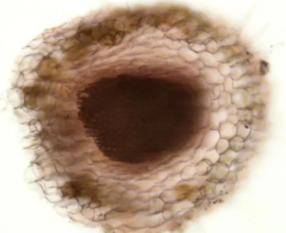
pericycle, and the metaxylem and metaphloem in the central part. The metaxylem has reticulate thickenings and occupies the central part of the root, replacing the medullary parenchyma (Figure 1).
Figure 1. Cross-section through T. chamaedrys root: overview (Congo red–chrysoidine staining, ×200).
Rhizome
54
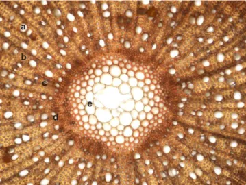
Figure 2. Cross-section through T. chamaedrys rhizome: overview (Congo red–chrysoidine staining, ×40).
Figure 3. Cross-section through T. chamaedrys rhizome: (a) metaxylem; (b) libriform tissue; (c) medullary ray; (d) protoxylem; (e) medullary parenchyma
(Congo red–chrysoidine staining, ×100).
Figure 4. Cross-section through T. chamaedrys rhizome: (a) metaxylem; (b) libriform tissue; (c) medullary ray (Congo red–chrysoidine staining, ×400).
Stolon
55
the outer wall bulged, thickened and covered by a thick cuticle. The epidermal cells are tangentially elongated, with thin radial walls and tangential outer and inner thickened walls. Long multi-cellular tector hairs are found in patches. The cortex consists of two areas: the outer area made up of 3–4 layers of angular collenchyma and the inner part of cortical parenchyma. In the inner part of the parenchymatic zone, there is one layer of endodermis exhibiting large, antero-posterior flattened cells, impregnated with suberin, as well as some sclerenchyma fiber bundles with periphloemic disposition. The conducting tissues are placed in two concentric rings, due to the activity of the libero-ligneous cambium. The phloem tissue is the thin, outer ring, consisting of sieve tubes, phloem parenchyma and annex cells. The medullary rays are multi-cellular, uniseriate, cellulosic. The xylem tissue forms the inner ring made up of metaxylem vessels of various calibers, scattered into the well-represented libriform tissue. The xylem vessels have spiral and reticulate thickenings. At this level, the medullary rays are multi-cellular, uniseriate, lignified. The primary xylem tissue is poorly represented, consisting of few primary xylem vessels and xylem parenchyma. The medullary parenchyma is well developed, of meatus type, and occupies the central area of the stolon (Figure 5).
Figure 5. Cross-section through T. chamaedrys stolon: (a) epidermis; (b) tector hair; (c) angular collenchyma; (d) cortical parenchyma; (e) endodermis; (f) sclerenchyma
fibers; (g) phloem tissue; (h) xylem tissue (Congo red–chrysoidine staining, ×200).
Aboveground stem
56
vessels and xylem parenchyma. The meatus-type well-developed medullary parenchyma occupies the entire central area of the aboveground stem (Figure 6).
Figure 6. Cross-section through T. chamaedrys aboveground stem: (a) epidermis; (b) tector hair; (c) glandular hair; (d) angular collenchyma; (e) cortical parenchyma;
(f) endodermis; (g) sclerenchyma fibers; (h) phloem tissue; (i) xylem tissue; (j) medullary parenchyma (Congo red–chrysoidine staining, ×100).
Leaf’s limb
57
Figure 7. Cross-section through T. chamaedrys leaf’s limb: (a) upper epidermis; (b) angular collenchyma; (c) libero-ligneous conducting fascicle; (d) sclerenchyma cap;
(e) leaf’s parenchyma; (f) lower epidermis (Congo red–chrysoidine staining, ×200).
Figure 8. Cross-section through T. chamaedrys leaf’s limb: (a) upper epidermis; (b) palisade parenchyma; (c) lacunose parenchyma; (d) lower epidermis; (e) glandular
hair (Congo red–chrysoidine staining, ×400).
TLC analysis
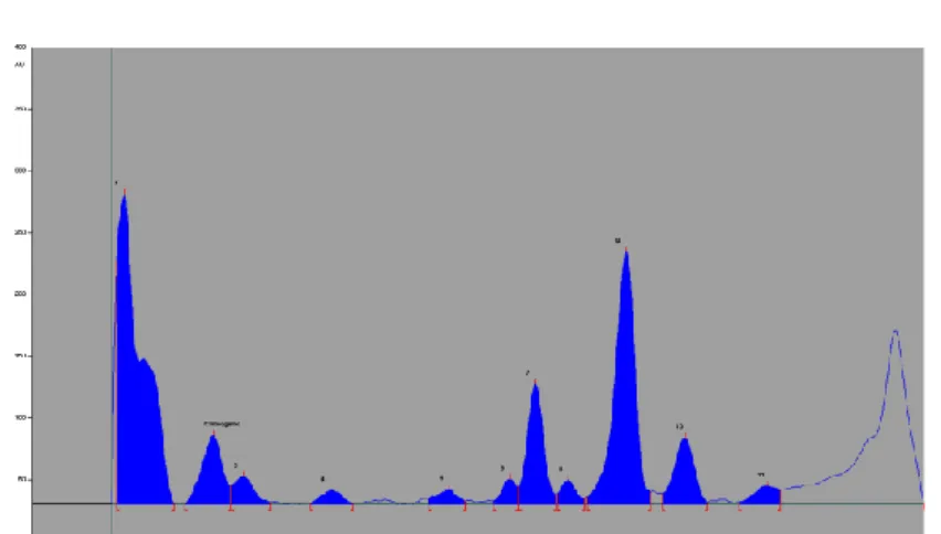
Figures 9–11 highlighted the experimental data on the preliminary TLC analysis of polyphenols from Teucrii herba. Chlorogenic acid (Rf 0.13, 22.37 mg/100 g of dried vegetal product) was identified starting from the 11 fingerprint chromatographic bands.
Figure 9. TLC chromatogram of polyphenols from Teucrii herba methanolic extract (UV 254 nm, without derivatization). From left to right: first five applications –
58
Figure 10. Densitogram of polyphenols (UV 254 nm) separated from Teucrii herba methanolic extract. From left to right, No. of peak/Rf: 1/0.02, 2/0.13 – chlorogenic
acid, 3/0.16, 4/0.27, 5/0.42, 6/0.49, 7/0.52, 8/0.56, 9/0.63, 10/0.71, 11/0.81.
Figure 11. In situ UV spectra of chlorogenic acid standard and compound separated from the analyzed sample.
CONCLUSIONS
The histo-anatomical investigations of roots, rhizomes, stolons, aboveground stems and leaves of T. chamaedrys species and the preliminary TLC analysis of the polyphenols from the aerial parts (Teucrii herba) were achieved. The root has circular shape and primary structure. The rhizome has circular-sinuous shape and secondary structure. The stolon and the aboveground stem have quadratic shape and secondary structure (libero-ligneous cambium). The leaf’s limb has bifacial, dorsiventral, hypostomatic structure. Chlorogenic acid was identified from the 11 TLC fingerprint bands.
BIBLIOGRAPHY
1. Altemimi, A., Watson, D.G., Kinsel, M., Lightfoot, D.A., 2015 – Simultaneous extraction,
optimization, and analysis of flavonoids and polyphenols from peach and pumpkin extracts using a TLC-densitometric method, Chem. Cent. J. 9:39.
2. Andrei, M., Paraschivoiu, R.M., 2003 – Microtehnică botanică, Ed. Niculescu, Bucureşti,
2003, 222 pag.
3. Bojić, M., Simon Haas, V., Sarić, D., Maleš, Z., 2013 – Determination of flavonoids,
phenolic acids, and xanthines in mate tea (Ilex paraguariensis St.-Hil.), J. Anal. Methods Chem. 2013:658596.
4. Ciocârlan V., 2000 – Flora ilustrată a României. Pteridophyta et Spermatophyta, ediţia a
59
5. Gîrd, C.E., Nencu, I., Costea, T., Duţu, L.E., Popescu, M.L., Ciupitu, N., 2014 –
Quantitative analysis of phenolic compounds from Salvia officinalis L. leaves, Farmacia 62(4):649–657.
6. Jurišić Grubešić, R., Vladimir-Knežević, S., Kremer, D., Kalođera, Z., Vukovic, J., 2007
– Trichome micromorphology in Teucrium (Lamiaceae) species growing in Croatia, Biologia Sect. Botany (Bratislava) 62(2):148–156.
7. Kaya, A., Demirci, B., Başer, K.H., 2009 – Compositions of essential oils and trichomes of
Teucrium chamaedrys L. subsp. trapezunticum Rech. fil. and subsp. syspirense (C. Koch) Rech. fil., Chem. Biodivers. 6(1):96–104.
8. Lakušić, B., Stevanović, B., Jančić, R., Lakušić, D., 2010 – Habitat-related adaptations
in morphology and anatomy of Teucrium (Lamiaceae) species from the Balkan peninsula (Serbia and Montenegro), Flora 205(10):633–646.
9. Nencini, C., Galluzzi, P., Pippi, F., Menchiari, A., Micheli, L., 2014 – Hepatotoxicity of
Teucrium chamaedrys L. decoction: role of difference in the harvesting area and preparation method, Indian J. Pharmacol. 46(2):181–184.
10. Pourmotabbed, A., Farshchi, A., Ghiasi, G., Khatabi, P.M., 2010 – Analgesic and
Anti-inflammatory activity of Teucrium chamaedrys leaves aqueous extract in male rats, Iran. J. Basic Med. Sci. 13(3):119–125.
11. Stankovic, M.S., Curcic, M.G., Zizic, J.B., Topuzovic, M.D., Solujic, S.R., Markovic,
S.D., 2011 – Teucrium plant species as natural sources of novel anticancer compounds: antiproliferative, proapoptotic and antioxidant properties, Int. J. Mol. Sci. 12(7):4190–4205.
12. Toma, C., Rugină, R., 1998 – Anatomia plantelor medicinale. Atlas, Ed. Academiei
Române, Bucureşti, 1998, 320 pag.
13. Vlase, L., Benedec, D., Hanganu, D., Damian, G., Csillag, I., Sevastre, B., Mot, A.C.,
Silaghi-Dumitrescu, R., Tilea, I., 2014 – Evaluation of antioxidant and antimicrobial activities
and phenolic profile for Hyssopus officinalis, Ocimum basilicum and Teucrium chamaedrys. Molecules 19(5):5490–5507.
14. Zlatić, N.M., Stanković, M.S., Simić, Z.S., 2017 – Secondary metabolites and metal