Received 16 Aug 2016|Accepted 8 Nov 2016|Published 9 Jan 2017
Hepatitis C virus has a genetically determined
lymphotropism through co-receptor B7.2
Chia-Lin Chen
1, Jeffrey Y. Huang
1, Chun-Hsiang Wang
1, Stanley M. Tahara
1, Lin Zhou
1, Yasuteru Kondo
1,
Joel Schechter
2, Lishan Su
3, Michael M.C. Lai
1,4, Takaji Wakita
5, Franc
¸
ois-Loı¨c Cosset
6, Jae U. Jung
1& Keigo Machida
1B-cell infection by hepatitis C virus (HCV) has been a controversial topic. To examine whether HCV has a genetically determined lymphotropism through a co-receptor specific for the infection by lymphotropic HCV, we established an infectious clone and chimeric virus of hepatotropic and lymphotropic HCV strains derived from an HCV-positive B-cell lymphoma. The viral envelope and 50-UTR sequences of the lymphotropic HCV strain were responsible for the lymphotropism. Silencing of the virus sensor, RIGI, or overexpression of microRNA-122 promoted persistent viral replication in B cells. By cDNA library screening, we identified an immune cell-specific, co-stimulatory receptor B7.2 (CD86) as a co-receptor of lymphotropic HCV. Infection of B cells by HCV inhibited the recall reaction to antigen stimulation. Together, a co-receptor B7.2 enabled lymphotropic HCV to infect memory B cells, leading to inhibition of memory B-cell function and persistent HCV infection in HCV-infected hosts.
DOI: 10.1038/ncomms13882 OPEN
1Department of Molecular Microbiology and Immunology, Keck School of Medicine, University of Southern California, 2011 Zonal Avenue, Los Angeles,
California 90033, USA.2Department of Cell and Neurobiology, Keck School of Medicine, University of Southern California, 2011 Zonal Avenue, Los Angeles, California 90033, USA.3Department of Microbiology and Immunology, Lineberger Comprehensive Cancer Center, University of North Carolina at Chapel Hill, Chapel Hill, North Carolina 27599-7290, USA.4Institute of Molecular Biology, Academia Sinica, Taipei 115, Taiwan.5Department of Virology II, National
Institute of Infectious Diseases, Tokyo 162-8640, Japan.6International Center for Infectiology Research, Team EVIR, Inserm, U1111, Universite´ Claude Bernard
H
epatitis C virus (HCV) infection often persists despite robust host immune responses, consequently leading to chronic hepatitis, liver cirrhosis and hepatocellular carcinoma. However, HCV infection and replication in immune cells remains controversial and is not universally accepted. Even though experimental and clinical evidence accumulated during the last two decades are compelling, the issue remains controversial mainly due to insufficient information and deeply fragmented knowledge. Another potentially serious complication of HCV infection is the possible infection of peripheral blood mononuclear cells (PBMC) by HCV leading to B-lymphocyte proliferative disorders, including mixed cryoglobulinemia, oligoclonal proliferation of B cells1,2, and B-cell non-Hodgkin’s lymphoma2–5. Still, HCV infection of B cells and its possible association with B-cell disorders remains a controversial subject6,7.It was reported from McKeating’s group that HCV replication in lymphocytes is relatively rare and attachment of HCV particles to B lymphocytes did not lead to productive HCV replication7. HCV promoted the adhesion of primary B cells to Huh-7 cells for retention of B cells on infected hepatocytes, thus implying that B cells may provide a vehicle for HCV persistence by transmission to the liver. Additionally, lymphotropism of HCV (SB strain: patient splenoma B-cell-derived isolated by our group) is not limited to B cells since we have identified HCV infection (SB strain) of T cells and subsequent alterations in their functions8,9. These studies, however, did not provide conclusive evidence that other molecules on other immune cell types serve as HCV co-receptors.
Cellular surface receptors have been identified as factors promoting viral tropism. HCV uses cell surface factors (LDL-R and HSPG) (ref. 10) for attachment and additional entry factors for infection of hepatocytes. The entry factors include the Scavenger Receptor class B type I (SRB1 or SR-BI) (ref. 11), the tetraspanin CD81 (ref. 12), the tight junction proteins CLDN1 (ref. 13) and the receptor tyrosine kinases EGFR and EphA2 (ref. 14). More recently, the Niemann-Pick C1-like 1 (NPC1L1) cholesterol absorption receptor and the iron uptake receptor transferrin receptor 1 (TfR1) have also been shown to play a role in HCV entry15. Among these, four co-receptors (Claudin-1, Occludin, CD81 and SR-BI) are potentially involved in HCV entry12,16–18, while sulfated homologues of heparin inhibit HCV entry into mammalian cells19. These co-receptors are associated with the viral envelope glycoprotein of HCV. The viral envelope proteins include E1 and E2, which assemble as heterodimers in the prebudding virion form20.
Mutations in the 50-UTR (50-untranslated region) of a hepatotropic HCV strain (H77) cultured in T lymphoid cell lines enhanced viral replication specifically in T lymphoid cells (MOLT-4) (ref. 21). The presence of different, strain-specific 50-UTR sequences or sequence heterogeneities in the region
coding for E1 or E2 can result in altered lymphotropism when compared to hepatotropic strains22,23. However, the lymphotropism of these viruses and the significance of these sequence variations were not fully established since only three nucleotide substitutions found in the 50-UTR in hepatotropic JFH1 strain and variant H77 strain passaged in lymphocytes are unchanged. The sequence variations in the 50-UTR region are
usually associated with B and T lymphocyte replication of HCV (refs 21,24,25). It has been shown that the B-cell specific 50-UTR has a lower translation difference observed between lymphotropic and hepatotropic strains26. (The lymphotropic strain may have a less efficient 50-UTR for translation).
The viral envelope protein is frequently mutated in chronically infected subjects, whereas the 50-UTR of HCV RNA in B cells is
not frequently mutated22, suggesting that B cells suppress
replication of less competent viral sequences whereas liver cells are not as stringent. In SCID mice inoculated with experimentally mutated HCV strains, the HCV mutations in the 50-UTR
(specifically the IRES) or C-E1-E2 regions have different consequences on genome replication and/or translational activity as a function of cell type26,27. Moreover, the 30-UTR
also contributes to efficient RNA replication of HCV (ref. 28). The 30-end of the NS5B-coding region has been shown to form part of a secondary structure (kissing loop) involving the 30-UTR
(ref. 28). This fact may explain why the HCV pseudoparticle (HCVpp) bearing HCV envelope alone did not confer robust replication ability in B cells.
MicroRNA-122 (miR-122) is the most abundant miRNA species in liver and binds the 50-UTR of HCV genomic RNA to facilitate HCV replication. Chronic liver damage results in release of miR-122 into the blood stream and exposes miR-122 to immune cells. However, the role of miR-122 for HCV replication in immune cells is not documented. MicroRNA-122 is not only expressed in the liver but also in malignant and chemotherapy-treated lymphocytes29,30, and positively regulates HCV replication in hepatocytes31–33. Circulating miR-122 can be taken up by several cell types, including lymphocytes34,35 thereby facilitating HCV replication. We hypothesized that this uptake of miR-122 facilitates HCV replication in B cells. Indeed, circulating plasma miR-122 in exosomes is elevated in early stage fibrosis induced by various aetiologies, including HCV infection, hepatocyte injury and inflammation36–39.
In this study, we hypothesized that the ability of certain HCV strains to infect B cells was determined by the sequence variation of the viral genome. We used genetic approaches to show that different HCV strains vary in their relative lymphotropism or hepatotropism. The B-cell infection by lymphotropic HCV may play a role in HCV pathogenesis. These results are crucial for studying the natural history of HCV infections in order to gauge the full scope of its subsequent pathological consequences resulting from HCV infections. We investigated the roles of plasma miR-122 in HCV pathogenesis, especially in lympho-cytes31 and the role of exosome-mediated delivery of tropic factors in creating a permissive environment for viral infection and replication.
Results
Lymphotropic SB-HCV strain encodes a unique E1-E2.
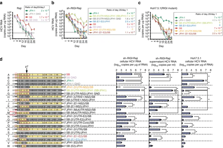
We have previously isolated an HCV clone (SB-HCV) from an HCV-infected (þ) B-cell lymphoma line40. This virus variant can infect B cellsin vitro40; however, virus production was low for reasons unclear. To answer this question we used a genetic approach and assembled the full-length SB viral genomic RNA (genotype 2b) cloned from HCV infected SB cells40. Viral RNA was cloned in 14 overlapping cDNA fragments using primer sets based on the consensus sequences (Supplementary Fig. 1). Sequence determination of the resulting full-length, HCV cDNA showed that the SB-HCV strain represented a unique HCV isolate.
decreased over three weeks of incubation. By comparison, the RNA titer of the JFH-1 strain quickly declined in Raji cells. These results indicated that the SB-HCV strain, but not the JFH-1 strain, was able to replicate or was unstable RNA in Raji cells. Conversely, in Huh7.5.1 cells (Fig. 1c), the SB-HCV strain was less efficiently replicated by day 28 post-transfection when compared to Raji cells (Fig. 1a); this indicated that Raji cells are more permissive for SB strain replication than Huh7.5.1. cells.
During the chronic HCV infection stage, the RIGI RNA sensor pathway is impaired because HCV protease NS3/4A cleaves the RIGI adaptor protein MAVS (IPS-1) (refs 42,43). Furthermore, chronic inflammatory cytokines induce negative feedback pathways through induction of NLRX1 (refs 44,45) to moderate the excessive activation of RIGI pathways. To study how HCV interacts with B cells, Raji cells were infected with a lentiviral vector expressing shRNA targeting RIGI to reduce the antiviral defences (Fig. 1b). Indeed, silencing RIGI in Raji cells significantly increased HCV RNA levels and reduced interferon (IFN)-b
induction probably due to defective viral RNA recognition and induction of RIGI downstream genes (that is, IFN-b). In contrast, silencing of MDA5, another innate immune receptor, did not have any further effect (Supplementary Fig. 2), which was consistent with the finding that HCV RNA binds to RIGI, but not to MDA5 (ref. 46). The highly permissive hepatotropic liver cell line (Huh7.5.1), which is highly permissive for hepatotropic
HCV, has an amino acid replacement (T55I) in the first CARD-homology domain47 of RIGI, resulting in inactivation of RIGI function. Therefore, the tropism of HCV-SB and hepatotropic strains was compared under identical RIGI-deficient conditions (Fig. 1b,c).
To identify the viral gene or genome region(s) responsible for the differential cellular tropism of these viral strains, we constructed chimeric virus genomes from the lymphotropic SB-HCV and hepatotropic JFH-1 HCV strains (Fig. 1d). These constructs were then converted into full-length infectious RNA for transfection into recipient cells (Fig. 1d). From the results of the various viral genome chimeras, it was apparent that all chimeric genomes containing the SB 50-UTR and homologous
structural proteins replicated well in sh-RIGI-Raji cells and released viral RNA (some of them are associated with virion) into the cell culture supernatant. In particular, a chimera containing the JFH1 backbone and the entire SB 50
-UTR-Core-E1-E2-p7-NS2 (construct F: at the second transmembrane domain of NS2) replicated 50-fold more robustly than the parental SB virus in sh-RIGI-Raji cells, producing nearly 107 HCV RNA copies ml1. By contrast replication-incompetent SB/JFH1 GND mutant virus RNA levels were significantly lower (construct G). The corresponding reciprocal genome constructs containing E1-E2 and the 50-UTR from JFH1 (construct E) replicated poorly in sh-RIGI-Raji cells. In Huh7.5.1 cells,
sh-RIGI-Raji cellular HCV RNA (log10 copies per μg of RNA)
sh-RIGI-Raji supernatant HCV RNA
(log10 copies per ml)
Huh7.5.1 cellular HCV RNA (log10 copies per μg of RNA)
d
5' 3′
5' 3′
5′ 3′
5′ 3′
5′ 3′
5′ 3′
C E1 E2 2 3 4A 4B 5A 5B
5' 3′
5' 3′
5' 3′
3′SB
SB GND JFH-1 JFH-1 GND 5' A B C D E F G H I J K L
C E1 E2 2 3 4A 4B 5A 5B
E1 E2 2 3 4A 4B 5A 5B C E1 E2 2 3 4A 4B 5A 5B
C E1 E2 2 3 4A 4B 5A 5B
C E1 E2 2 3 4A 4B 5A 5B
E1 E2 2 3 4A 4B 5A 5B C E1 E2 2 3 4A 4B 5A 5BGND
C E1 E2 2 3 4A 4B 5A 5B C E1 E2 2 3 4A 4B 5A 5B
GND
JFH1 (5′UTR-NS2)/SB SB (5′UTR-NS2)/JFH1 (SB/JFH1)
JFH1 (UTR/E1-NS2)/SB SB (UTR/E1-NS2)/JFH1 JFH1 (E1-NS2)/SB SB (E1-NS2)/JFH1 p7 p7 p7 p7 p7 p7 p7 p7 p7 p7
5′ 3′
5′ 3′
C E1 E2 2 3 4A 4B 5A 5B
C E1 E2 2 3 4A 4B 5A 5B JFH1 (MutUTR-NS2)/SB SB (MutUTR-NS2)/JFH1 p7 p7 C C Mutations Mutations
5′ 3′
5′ 3′
5′ 3′
5′ 3′
5′ 3′
5′ C E1 E2 2 3 4A 4B 5A 5B 3′
5′ 3′
5′ 3′
5′ 3′
5′ 3′
M N O P Q R S T U V W
C E1 E2 2 3 4A 4B 5A 5B
C E1 E2 2 3 4A 4B 5A 5B C E1 E2 2 3 4A 4B 5A 5B
C E1 E2 2 3 4A 4B 5A 5B
C E1 E2 2 3 4A 4B 5A 5B C E1 E2 2 3 4A 4B 5A 5B C E1 E2 2 3 4A 4B 5A 5B C E1 E2 2 3 4A 4B 5A 5B C E1 E2 2 3 4A 4B 5A 5B
JFH1 (5′UTR-E2)/SB SB (5′UTR-E2)/JFH1 JFH1 (5′UTR-Core)/SB SB (5′UTR-Core)/JFH1 JFH1 (5′UTR)/SB SB (5′UTR)/JFH1
JFH1 (E1-E2)/SB SB (E1-E2)/JFH1 JFH1 (E2)/SB SB (E2)/JFH1 p7 p7 p7 p7 p7 p7 p7 p7 p7 p7 3
2 4 5 6 7 8 2 3 4 5 6 7 8 2 3 4 5 6 7 8
5′ C E1 E2 p7 2 3 4A 4B 5A 5B 3′SB (5′UTR-NS2)/JFH1 GND p7
C E1 E2 2 3 4A 4B 5A 5B
sh-RIGI-Raji
a
Raji Ratio of day20/day1
Ratio of day 20/day 1
Ratio of day 20/day 1
4 Day JFH-1 JFH-1 GND JFH-1 GND SB SB GND 2 x 10⁻⁶
0 1.7 x 10⁻³
3 x 10⁻⁶
SB GND HCV RNA (copies per μ g RNA) HCV RNA (copies per μ g RNA) HCV RNA (copies per μ g RNA) b 0 101 102 103 104 105 106 107 108 109 0 101 102 103 104 105 106 107 108 109 0 101 102 103 104 105 106 107 108 109 c JFH-1
SB (5′UTR-NS2)/JFH1 (SB/JFH1) SB (5′UTR-NS2)/JFH1 GND SB (E1-E2)/JFH1 SB
JFH1 (5′UTR-NS2)/SB
JFH1 (E1-E2)/SB
8 x 10⁻⁶ 2 x 10⁻⁵
1 x 10⁻¹
1.6 x 10⁻⁷ 2.5 x 10⁻⁵
3 x 10⁻³
0
1.1 x 10⁻⁶
JFH-1
SB (5′UTR-NS2)/JFH1 (SB/JFH1) SB (E1-E2)/JFH1
SB
JFH1 (5′UTR-NS2)/SB
JFH1 (E1-E2)/SB
4.3 x 10⁻⁷ 7 x 10⁻²
0 0 7 x 10⁻⁶
2 x 10⁻⁵
2.5 x 10⁻²
Day Day Huh7.5.1(RIGI mutant) 1 * * * * * * * * * * * * * * * * * * * * * * * * * * * * * * * 28 24 20 16 8
12 1 4 8 12 16 20 24 28 1 4 8 12 16 20 24 28
this result was reversed: JFH-1 RNA replicated to a level of 107 RNA copies ml1, whereas SB RNA replication was decreased to a significantly lower level (Po0.05, t-test). Furthermore, mutations in the 50-UTR regions of SB and JFH1 strains (constructs L and M) resulted in significantly reduced HCV RNA levels in lymphocytes. These results suggested that there are distinct hepatotropic and lymphotropic HCV strains, whose tropism is largely determined by the envelope proteins (E1-E2) and their respective 50-UTRs. This dramatic increase
was probably due to the sequence changes that occurred at a crucial junction between the second transmembrane segments of NS2 as previously suggested48.
To further ascertain the importance of E1-E2 proteins in the lymphotropism of SB virus, we produced HCVpp using an envelope-defective HIV-1 proviral vector49–51. This HCVpp bears the E1 and E2 envelope proteins of HCV and contains a luciferase reporter gene in the lentivector. As shown in Fig. 2a, HCVpp bearing the SB E1-E2 envelope proteins could efficiently infect Raji cells, whereas its infectivity of Huh7.5.1 cells was significantly lower. As a control, HCVpp assembled with either
E1 or E2 proteins alone were not infectious. The infectivity of HCVpp with SB envelope protein in Raji cells was substantially lower (more than 3-logs lower) than that of HIV vector pseudotyped with MLV Env. Addition of neutralizing anti-E2 antibody (CBH5 clone) specifically antagonized HCVpp entry into Raji cells (Fig. 2a, right). The ability of HCVpp-SB to infect Raji cells gives further support that the SB strain is lymphotropic while HCVpp-JFH1 do not infect Raji cells. Indeed, in the CD81-interacting domain of the E2 region52–54, the SB-HCV amino acid sequence is homologous to other HCV strains, but does have several unique residues that may be responsible for the observed lymphotropism (Fig. 2c). Another genotype 2b strain (AB 30907) has the highest homology to the SB strain among all HCV genotypes while the H77 genotype 1 strain has lesser homology with the SB strain (Fig. 2b,c). Amino acids G496 and N514 of SB HCV E2 are unique amino acids in comparison to other HCV E2 sequences, which contain S514 (ref. 55). The G496C substitution has been shown to form a disulfide bond with C567 (ref. 55). Substitution of G496 by Cys significantly reduced SB E2 binding to B cells while this
100 1,000 10,000 100,000
SB E1 Mock Isotype α
-E2
Mock
Isotype α
-E2
Luciferase
activity (RLU)
SB E2
SB E1E2
SB E1E2
SB E1E2p7
SB E2p7
JFH1 E1E2 Con1 E1E2
N E1E2
1/100 MLV gp
1/100 MLV gp
1/100 VSV gp
No env
Raji Huh7.5.1
a
Transfection
HCV pseudoparticle system (HCVpp)
HEK293T HCV E1/E2
GAG-POL Luciferase
Infection of target cells
Luciferase assay HCVpp entry
Raji or Huh7.5.1
Luciferase expression ,
b
2b SB
E2 HVR3 N514
E2 HVR2
0 1 2 3 4 0 1 2 3
5 6
*
2a JFH1 1b P29846.3 1a H77 2b AB30907 2b SB 2a JFH1 1b P29846.3 1a H77 2b AB30907
2b SB 2a JFH1 1b P29846.3 1a H77 2b AB30907
E2 CD81-II YPP sequence in E2
B7.2-CD28 binding sequence: MYPPP
G496
C496 C567
L641 G658 M680
0 1,000 10,000 100,000
c
d
L641V G658D M680L
G496C
HCVpp(SB) into Raji
HCVpp(SB) into Huh7.5.1
*
** ** **
** ***
*** *** ***
Fold difference
of entry
Fold difference
of entry
WT
G496V N514S
C–C disulfide bond
C–C disulfide bond No bond
B Lymphocyte CD81
JFH1 E2 SB E2
Hepatotropic co-receptor
HCV(SB) E1/E2
CD81
Unknown recepor
Lymphotropic co-recepor Unknown
recepor
Hepatocyte
B Lymphocyte
Less entry CD81
C C
C496
G
G496
C567
C
C567
HCV(SB)
E1/E2 YPP
Particle production
481
480
478
478
480
561
560
558
558
560
641
640
636
636
640
560
559
557
557
559
640
639
635
635
639
720
719
715
715
719
Figure 2 | Lymphotropic pseudoparticles (HCVpp) can infect B lymphocytes more efficiently than hepatic cells.(a) HIV-luc vector was pseudotyped with envelope proteins of HCV SB strain. Pseudoparticles (HCVpp) efficiently infected B lymphocytes, but not hepatic cells. Infectivity of HCV
pseudo-particles was measured by luciferase assay. HIV vector containing a luciferase reporter pseudotyped with E1 and/or E2 of SB strain, Con1, or HCV-N strain and MLV glycoprotein (MLV gp), or no envelope (no env) was used to infect Raji (black bars) or Huh7.5.1 (grey bars). Luciferase activity was assayed at 4 days after infection. Addition of neutralizing anti-E2 antibody blocked HCVpp entry (a, right). Experiments were repeated at least three times (*Po0.05,
substitution significantly increased virus entry into Huh7 cells (Fig. 2d). Formation of the disulfide bonding between G496C and C567 may restore or stabilize a conformation favourable of E2 for entry into hepatocyte HCV co-receptors (Fig. 2d). These results confirmed that the E1-E2 envelope proteins conferred the observed lymphotropism exhibited by the SB strain in addition to the properties of strain-specific 50-UTRs.
Envelope and 50-UTR determine lymphotropism of SB virus.
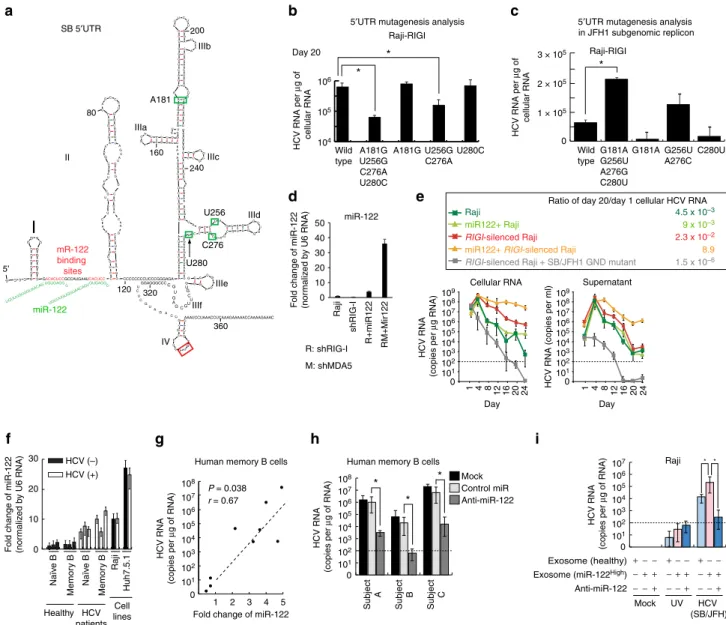
The predicted secondary structure of the SB 50 UTR includes
several possible RNA stem-loop structures (Fig. 3a). Five nucleotide differences between the SB and JFH1 strains are found within 50-UTR and their positions are indicated (Fig. 3a). To address the significance of these genetic differences, and potential effects on RNA secondary structure, four out of five nucleotides were changed from the SB strain to those found in the JFH1 strain. Interestingly, the four-nucleotide substitutions significantly reduced HCV RNA levels in sh-RIGI-Raji cells at 20 days post transfection of HCV RNA (Fig. 3b). The A181G and U280C substitutions did not affect HCV RNA levels, but combined substitutions (A181G, U256G, C276A and U280C), and to a lesser extent, U256G and C276A substitutions reduced HCV RNA levels. However, it cannot be excluded that these differences may be due to changes in RNA stability. Therefore, we constructed a mutant subgenomic JFH1 replicon to see if substitutions of the 50-UTR sequence promoted replication in B
cells. The 50-UTR sequences were replaced with SB
50-UTR sequences of the JFH1 subgenomic HCV replicon. We observed that combined substitutions in G181A, G256U, A276C and C280U increased HCV RNA levels of the JFH1 subgenomic replicon (Fig. 3c) in sh-RIGI-Raji cells. These results indicated that nucleotides A181, U256, C276 and U280 of the 50-UTR have key roles in regulating HCV levels in sh-RIGI
-Raji cells.
To prove that SB-HCV recombinants produced infectious virus particles, the culture supernatant from the transfected cells was used to infect naı¨ve sh-RIGI-Raji or Huh7.5.1 cells by using a batch system to ensure a cell-free transfer (Supplementary Fig. 3). SB RNA-transfected Raji cells produced infectious virus that infected naı¨ve sh-RIGI-Raji cells (Supplementary Fig. 3A and B), but was less efficient at replication in Huh7.5.1 cells (Supplementary Fig. 3C). The converse was true for the JFH strain in Huh7.5.1 cells (Supplementary Fig. 3C). Infectivity of SB/JFH1 virus was directly quantified by limiting dilution assay, demonstrating that HCV-infected cells have infectivity of 40% (6/15 clones) at day 13 and 38% (6/16 clones) at day 20 (Supplementary Fig. 3D). Infection of HCV did not significantly affect cell proliferation (Supplementary Fig. 3E).
The role of miR-122 in HCV lymphocyte infection was examined by transduction of miR-122 into B lymphocytes. A miR-122 expressing lentivirus was transduced into sh-RIGI -Raji cells and cells were challenged with HCV (Fig. 3d). The results showed that miR-122 transduction signi-ficantly enhanced HCV replication, producing virus titers of 2106 copies ml1 and 108 copiesmg1 of cellular RNA (387-fold increase:Po0.001,t-test) after 3 weeks post-transduc-tion (Fig. 3e). Cellular miR-122 levels were significantly reduced 4 days post-transduction while levels of miR-122 significantly increased in the supernatant (Supplementary Fig. 4). Indeed, HCV RNA levels in culture supernatant significantly increased by day 7 and further increased at day 14 post-transfection (Po0.05,t-test, Supplementary Fig. 4, right), indicating that miR-122 may be secreted from HCV-infected B cells. Furthermore, miR-122 levels were measured in immune cells isolated from
HCV patients. Consistent with in vitro results, circulating miR-122 levels were significantly increased in HCV-infected patients (Po0.05, t-test, Fig. 3f). The increased expression of miR-122 in B cells was confirmed in memory B cells from HCV patients (n¼9). The higher miR-122 levels in memory B cells showed a positive correlation with higher cellular HCV RNA levels (r¼0.67,Po0.05,t-test, Fig. 3g).
In order to determine if reduction of miR-122 levels had a negative effect on HCV replication in B cells, an antagomir of miR-122 was transfected into human memory B cells isolated from HCV patients and examined for effect on cellular levels of HCV RNA in B cells. These primary cells were incubated
ex vivo to allow HCV replication. Interestingly, transfection of miR-122 antagomir into patients’ memory B cells significantly lowered HCV replication (Po0.05,t-test; Fig. 3h).
Transfer of miR-122 via exosomes was testedin vitrofor effect on HCV replication. Exosomes derived from HCV patients or healthy individuals were added to the supernatant of HCV-infected Raji cells and examined for effect on HCV replication. Addition of exosomes containing high miR-122 levels from HCV patients indeed enhanced HCV replication (14-fold increase of HCV RNA) in Raji cells (Fig. 3i). These results indicated that miR-122 promotes HCV replication in B lymphocytes.
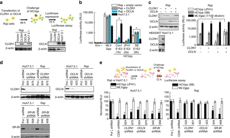
CLDN1/OCLN can rescue hepatotropic HCV infection.
We next studied the cellular basis for the lymphotropism of the SB virus by focusing on the role of CLDN1 and OCLN, which have been shown to be the co-receptors for HCV infection of Huh7.5.1 cells13,16. CLDN1 and OCLN are normally expressed at very low or undetectable levels in Raji cells (Fig. 4a) (ref. 56). To test whether overexpression of either CLDN1 or OCLN could render Raji cells more susceptible to infection by hepatotropic JFH-1, we overexpressed CLDN1 or OCLN in Raji cells. As shown in Fig. 4b, overexpression of CLDN1 or OCLN in Raji cells resulted in only a marginal increase of viral entry and not to the extent observed for SB (E1-E2). By comparison, HCVpp bearing E1-E2 of Con1 or JFH-1 could not infect Raji cells. It is not clear if lower infectivity was due to inability to adsorb and enter cells or due to lower efficiency of viral gene expression and replication. To test if HCV cell entry required both OCLN and CLDN1 co-receptors, both OCLN and CLDN1 were simultaneously transfected into Raji cells prior to challenge with HCVpp-JFH1. The extent of successful HCV intracellular entry was assessed by luciferase assay (Fig. 4c). As four human co-receptors (OCLN, CLDN1, CD81 and SR-BI) are critical for HCV entry into hepatocytes, the effect of OCLN and CLDN1 overexpression was examined in Raji cells, which normally lack their expression. Interestingly, Raji cells expressing both OCLN and CLDN1 were found to promote entry of hepatotropic JFH1 HCV (Fig. 4c), confirming that viral entry required co-expression of both OCLN and CLDN1. The intrinsic ability of HCVpp-SB to infect Raji cells, but not HCVpp-JFH1, in the absence of OCLN and CLDN1, to infect Raji cells gives further support to our contention that the SB strain is lymphotropic (while Huh7 cells express all four receptors CLDN1, OCLN, CD81 and SR-BI).
SR-BI-dependent but CLDN1- and OCLN-independent pathway for infection of B cells.
B7.2 is a co-receptor for lymphotropic HCV of B cells. Inability
of SB to infect hepatocytes indicated that some other cell surface co-receptor proteins were needed. In an effort to identify the potential novel co-receptor specific for lymphotropic HCV
infection, we performed a lentivirus-based screen of a cDNA library, derived from the highly HCV-permissive B lymphoma cell lines Raji and Daudi. This screen was designed for identifi-cation of genes that rendered the non-permissive CD81þ SR-BIþ HEK293T cell line susceptible to infection by HIV-luc reporter vector pseudotyped with HCVgp (HCVpp) (Fig. 5a). The result of this functional screen identified ten co-receptor candidate genes (Supplementary Fig. 5), which were tested in SB 5′UTR
mR-122 binding sites
a
HCV RNA per
μ
g of
cellular RNA
HCV RNA per
μ g of cellular RNA A181G U256G C276A U280C A181G Wild type U256G C276A U280C G181A G256U A276G C280U G181A Wild type G256U A276C C280U 5′UTR mutagenesis analysis 5′UTR mutagenesis analysis
in JFH1 subgenomic replicon
104 105 106 101 105 104 103 102 106 107 108 109 b c
G C C C G C C C C C U G A U G G G G G C 5’ C C 120 320 360 GACACUCCGCCAUGAAUCACUCC CCCCCCCCUCCCGGGAGAA
G A G C C A U A G U G G U C U G C C C C C C C G G G G G G G G G G G G G G G G G G G A A A A A A A A A A A A A
A CC
G G U G A G UA
CACCGG A A UU G C C G G A A A G A C U G G G U C C U
UU CU
U G G AU A A A C C C A C U C U A U G U U U U U U U U U U U U U U U U U U U U U C C G G U C A
GGG CG U G C C C C C G C A A G G G G A A A A C C C C C C C C C C C G CU A
GCC UA
G UAGC
GU U G G G U U G C G A A C G G C C U U G U G G U A C U 160 200 240 80
GCC
C C C C C U G A U A G G G G G G G GGAGGGCCC U G C U U G C G A G U G C U C U C G U A
GAC
IIId miR-122 IIIc IIIa IIIb II
I
IIIf IV IIIe U A U A U A A AAAUCCUAAACCUCAAAGAAAAACCAAAAGAAAC U256 C276 U280 A181 UGUGAGG GUGAGG UGUUUGUGGU AACA G UGUUUGUGGU AACAGU U U * * * Day 20 Raji-RIGI Raji-RIGI gf h i
d Cell lines HCV patients HCV (–) HCV (+) 0 10 20 30 Naïv e B Memor y B Huh7.5.1 Raji
Human memory B cells Human memory B cells
Subject A Subject B Subject C
Mock UV HCV (SB/JFH) Mock
Control miR Anti-miR-122
Fold change of miR-122
P = 0.038
r = 0.67
HCV RNA
(copies per
μ
g of RNA)
HCV RNA
(copies per
μ
g of RNA)
HCV RNA
(copies per
μ
g of RNA)
1 2 3 4 5
Exosome (healthy) − + + − + +−− − + − + Raji − − + + −−+ −−+ − − − + + Anti-miR-122 Exosome (miR-122High)
* * * Healthy Naïv e B Memor y B * * e Cellular RNA 0 101 105 104 103 102 106 107 108 0 101 105 104 103 102 106 107 108 0 101 105 104 103 102 106 107 0 101 105 104 103 102 106 107 108 109 0 4 1 8 12 16 20 24
Day
Raji
RIGI-silenced Raji
miR122+ RIGI-silenced Raji
RIGI-silenced Raji + SB/JFH1 GND mutant
miR122+ Raji
4.5 x 10⁻³
2.3 x 10⁻²
8.9
1.5 x 10⁻⁶
9 x 10⁻³
HCV RNA (copies per μ g RNA) 4 1 8 12 16 20 24
Day Supernatant
HCV RNA (copies per ml)
F
old change of miR-122
(nor maliz ed b y U6 RNA) F
old change of miR-122
(nor maliz ed b y U6 RNA) 0 1 × 10⁵ 2 × 10⁵ 3 × 10⁵
0 10 20 30 40 50 Raj i sh R IG-I R+m iR122 RM + M ir 1 2 2 miR-122 R: shRIG-I M: shMDA5
Ratio of day 20/day 1 cellular HCV RNA
shRNA-transduced Raji cells for HCVpp entry. As a result of this screen and confirmatory knockdown experiments, B7.2 (or CD86) was identified as a potential entry factor. B7.2 did not affect HEK293T susceptibility to HIV-luc particles either lacking envelope proteins (Envpp) or pseudotyped with unrelated vesicular stomatitis virus G protein (VSV-Gp); these served as negative and positive infection controls, respectively. Accordingly, HCVpp-E1-E2 (SB strain) infection of Raji cells was inhibited by anti-B7.2 and anti-CD81/anti-SR-BI (known HCV co-receptors), but not by a control isotype antibody (Fig. 5b).
To further determine if B7.2 is required for HCV-SB infection of B cells, Raji cells were infected with lentiviruses expressing shRNAs targeting different regions of B7.2. The B7.2 knockdown results showed that HCVpp-SB infection was markedly reduced in B7.2-silenced cells as compared to cells treated with a scrambled shRNA (Fig. 5c). When using distinct shRNAs of variable potency against B7.2, HCVpp infectivity was inversely correlated to the degree of silencing (Fig. 5d). By comparison, B7.2 knockdown in Huh7.5.1 cells did not inhibit HCVpp (JFH1) infection (Supplementary Fig. 5A). Next, to determine if the entry step of virus infection required B7.2, synchronized infections were
performed on Raji or Huh7.5.1 cells in the presence of blocking antibodies (Fig. 5e). The kinetics of inhibition by anti-B7.2 antibody was significantly slower than that for anti-CD81 antibody, with half-maximal inhibition observed at 128 min post-temperature shift while the half-maximal inhibition time of anti-CD81 was 32 min. In contrast, anti-B7.2 antibody did not inhibit HCVpp (JFH1) infection of Huh7.5.1 cells. The time course of inhibition was similar to that observed for the Claudin-1 antibody16. Thus, these results were consistent with the interpretation that anti-B7.2 antibody inhibited a step after virus binding.
We next examined which domains of B7.2 are responsible for interaction with SB envelope proteins. While exogenous B7.2 allowed HCVpp to infect B7.2-silenced Raji cells, neither B7.1 nor ICAM supported HCVpp infection (Fig. 5f). Furthermore replacement of the IgV domain of B7.2 with that from B7.1 abrogated the HCVpp infection of B7.2-silenced Raji cells (Fig. 5f). The importance of the B7.2 cytoplasmic domain was underscored since replacement of this domain with that from CD8 also resulted in the loss of HCVpp infectivity. To test if a direct interaction occurred between E2 and B7.2, we performed 100
1,000 10,000 100,000
Env –
HCVpp
Luciferase activity (RLU)
Luciferase activity (RLU)
MLV Gpp
SB E1E2
(2b) JFH1 E1E2 (2a) Con1 E1E2 (1b)
Raji + empty vector Raji + CLDN1 Raji + OCLN
0 25 50 75 100 125 150
CD81 shRNA
525 921
Raji Raji
CLDN1
shRNA Huh7.5.1
Huh7.5.1
422 423
OCLN
shRNA
OCLN
shRNA
OCLN
OCLN
shRNA
Pol
ζ
shRNA
Pol
ζ
shRNA
Pol
ζ
shRNA
Pol
ζ
shRNA
624 875 624 875 624 875
CLDN1
shRNA
CLDN1
shRNA
CLDN1
shRNA
CLDN1
CLDN1
CLDN1
β-actin
β-actin
β-actin CLDN1
β-actin
OCLN
OCLN
β-actin
OCLN
β-actin
624 875
OCLN
shRNA 0
25 50 75 100 125 150
Normalized RLU
Huh7.5.1
Vector CLDN1 OCLN Huh7.5.1
HCVpp (SB) MLVgpp HCVpp (JFH1)
MLVgpp
945 958
SR-BI
shRNA Raji Raji
HCVpp (SB) HCVpp (JFH1) MLVgpp (1/100 dilution)
Raji
Vector CLDN1 OCLN Huh7.5.1
Raji
− + + + + − − −
OCLN
CLDN1 − + +
+ + − − −
Raji or Huh7.5.1 72 h 72 h Luciferase assay
Raji Huh7.5.1
Pol
ζ
shRNA
Pol
ζ
shRNA
945 958 945 958
SR-BI
shRNA
SR-BI
shRNA
SR-BI
Raji Huh7.5.1
422 423 422 423
' Knockdown of CLDN1 or OCLN : ; *- '
Raji cells 48 h 72 h
Luciferase assay Luciferase expression Challenge
of HCVpp Transfection of
CLDN1 or OCLN
*
*
* *
* * * * * *
945 958
SR-BI
shRNA
* * *
Challenge of HCVpp
0 1,000 10,000 100,000
CLDN1 OCLN HEK293T Huh7.5.1
β-actin
Pol
ζ
shRNA
CD81 shRNA Pol
ζ
shRNA
a b c
d e
Figure 4 | HCV pseudo-particles (HCVpp) with envelope proteins of SB strain infected B lymphocytes, but not hepatic cells.(a) Claudin1 (CLDN1) or Occuludin (OCLN) expression confers weak susceptibility of Raji cells to JFH1 infection (Upper panel). Immunoblot analysis demonstrated that Claudin-1 and Occludin proteins were expressed by transfection of plasmids. (b) The respective expression vectors of CLDN1 and OCLN were transfected into both Raji and Huh7.5.1 cells. Luciferase-encoding pseudo-particles bearing the indicated envelope proteins were used to infect recipient Raji cells transfected with control vector (white), CLDN1-expressing vector (grey), or OCLN-expressing vector, or control Huh7.5.1 cells (black). For HCVpp, the respective E1E2 genes are indicated in parentheses. Luciferase assay was performed at 3 days after transduction (*Po0.05,t-test). Values are normalized to the RLU background measured in mock-infected cells (mean ofn¼3; error bars, s.d.). RLU: relative luminescence units. (c) The respective expression vectors of CLDN1 and OCLN were transfected and their protein expression levels were confirmed in both Raji and Huh7.5.1 cells. (Left panel) Expression of both CLDN1 and OCLN (tight junction molecules) promoted HCVpp(SB) entry (right panel) (*Po0.05,t-test,n¼3). Error bars represent s.d. (d) Cells were transfected to express shRNA against CLDN1 or OCLN and examined for silencing effects by immunoblots. shRNAs against DNA polymerase
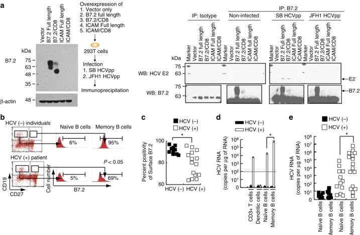
coimmunoprecipitation with anti-B7.2, followed by immunoblot for E2 proteins and showed an interaction between B7.2 and HCV E2 proteins (Fig. 6a and Supplementary Fig. 9). Reciprocal immunochemical analysis further confirmed E2-B7.2 interactions (Supplementary Fig. 5C and Supplementary Fig. 9). These results indicated that B7.2 is a co-receptor for HCV-SB infection of Raji cells.
We further examined whether the level of B7.2 expression in B cells of hepatitis C patients correlated with the extent of HCV infection. Naı¨ve B cells (CD19þCD27) were found to have low levels of B7.2 expression, while memory B cells (CD19þCD27þ) showed higher expression levels (Fig. 6b), in agreement with previous reports58. Interestingly, memory B cells in HCV patient-derived PBMCs expressed significantly lower levels of surface B7.2 when compared to those in HCV () individuals (Fig. 6c). Significantly, HCV RNA levels were two logs higher in memory
B cells than in naı¨ve B cells isolated from HCV patients, which is correlated with increased protein levels of B7.2 (Fig. 6d). These findings were confirmed by examination of 18 different hepatitis C patients (Fig. 6e). Lower surface B7.2 levels were correlated with lower cellular HCV RNA levels (Supplementary Fig. 5D). Activation of B cells (CD40LþIL-4 treatment) is known to promote surface B7.2 expression in naı¨ve B cells59. Interestingly, activation of B cells resulted in higher levels of cellular HCV RNA 8 days post-HCV (SB/JFH1) infection (Supplementary Fig. 5E). Anti-CD81, anti-SR-BI or anti-E2 neutralizing antibody treatment blocked HCV infection in sh-RIGI-Raji cells (Supplementary Fig. 5F). B7.2 expression in B and T cells and dendritic cells was confirmed by immunoblot (Supplementary Fig. 5G). These results indicated that CD81, SR-BI and E2 are involved in critical steps for lymphotropic HCV entry process into B cells. This conclusion is limited to B cells, but not in T-cell
Gag
CMV VSV Gp CMV cDNA library
Pur-HCVppr
Puromycin Test for HCVpp entry
cDNA
Luc Luc Luc Luc-HCVpp
Luc-VSVpp
Luc-Env (–)
0 20 40 60 80 100 120 Synchronized
infection
Blocking of CD81 or B7.2
α-CD81 or
α-B7.2 Ab
Raji and Huh7.5.1
?
Luciferase expression
Luciferase assay
Blocking of entry?
4°C 2 hr 37°C
37°C
Virion binding
α-CD81 blocking antibody
Time after α-B7.2 antibody addition (min) (Half-maximal inhibition: 128 min)
Time after α-CD81 antibody addition (min) (Half-maximal inhibition: 32 min) HCVpp (SB) in Raji
HCVpp (JFH1) in Huh7.5.1 Isotype IgG in Raji None in Raji
0 0
30 60
120 180 240 300
0 20 40 60 80 100 120
−
180
−
120 30 60 120 180 240 300
−
180
−
120
HCVpp entry (%)
α-B7.2 blocking antibody
Raji
Scrambled sh-CD81
7645
11093
7647
sh-B7.2 0
25 50 75 100 125 150
Normalized RLU
HCVpp (SB) MLVpp
*
* * *
100 1,000 10,000 100,000
Luciferase activity
(RLU)
SB E1E2
JFH1 E1E2 Con1 E1E2
VSV gp
No env
Selected HEK293T cells with B cell cDNA library
SB E1E2 +
α
--B7.2
SB E1E2 + Isotype
SB E1E2 +
α
-SR-BI
SB E1E2 +
α
-CD81
* * *
Raji
P = 0.042
r = −0.79
Relative infectivity (%)
0 0 20 40 60 80 100 120
150 23
1 131
Signal peptide
Extracellular domain TM
IgC IgC
IgV IgV
Cytoplasmic domain
IgC
IgV CD8
CD8
ICAM-1 ICAM-1
B7.2
IgC
IgV
B7.2/B7.1/B7.2
IgC IgV
B7.1/B7.2/B7.1 B7.2/CD8
IgC
IgV
B7.1
ICAM-1 ICAM-1/CD8
IgC IgV
B7.1
Normalized RLU HCVpp VSV-Gp 225 269 329
B7.2
0 10 100 1000
Purr Pol
20 40 60 80 100 Reduction in B7.2+ cell population (%)
a b c d
e f
lines since SR-B1 involvement is known for HCV infection of primary T cells. Interestingly, primary T lymphocytes do not display detectable levels of SR-B1 protein, although T-cell lines do60.
Characterization of HCV particles produced from SB strain.
The concentrated virus particles had a density of 1.13 g ml1in sucrose (Fig. 7a), which is in agreement with previous report (1.12 g ml1) of HCV virions isolated from the human T-and B-cell lines HPBMa10-2 T-and Daudi25, but slightly less than that of HCV virions isolated from hepatocytes (ca. 1.15 g ml1) (refs 41,61). To further characterize SB viral RNA, SB-transfected cells were metabolically labelled with [3H]uridine in the presence of actinomycin D. The viral particles produced from transfected cells were concentrated from the culture supernatant and separated by sucrose gradient sedimentation. A specific
3H-uridine-labelled peak, which was resistant to protease
treatment, was detected in the culture supernatant of Raji cells (Fig. 7a). RNA isolated from this material yielded a major RNA species of approximately 9.7 kb and a minor RNA species. The latter could be defective interfering RNA (DI RNA), although further work will be needed to ascertain this (Fig. 7b).
Electron microscopy demonstrated that the particles were approximately 50-60 nm in diameter and possessed an envelope that was immunoreactive with anti-E2 antibodies (Fig. 7c). Furthermore,35S-amino acid-labelled virus particles at the peak
fraction contained several viral proteins, corresponding to core, E1, and E2, all of which are associated with HCV particles (Supplementary Fig. 6A). The putative identities of 35S-labelled core and envelope protein E2 were confirmed by immunopreci-pitation, followed by autoradiography (Supplementary Fig. 6B). It was notable that the virion contained relatively more envelope proteins than core proteins. Treatment with NP-40 disrupted the virus particles, consistent with its identity as an enveloped virion (Supplementary Fig. 6C). The sum of these analyses confirmed that the viral particles produced from the SB-transfected cells in this system had outer cell surface markers consistent with authentic HCV particles.
To determine whether the viral particles released were infectious, we harvested the culture media from SB RNA-transfected Raji cells (sh-RIGI-Raji) which in turn was used to infect naı¨ve Raji cells. The HCV RNA isolated from infected cells was then analysed 8 days later. UV irradiation of virus-containing media prior to infection prevented viral RNA production (Fig. 7d). Also, pretreatment of the transfected cells with IFN-aor -g, which inhibits viral RNA replication but not viral attachment, lowered HCV RNA levels in a dose-dependent manner (Fig. 7d). Finally, treatment with Telaprevir62, an NS3 (viral protease) inhibitor, or 20-modified nucleosides as inhibitors of HCV replication (20-C-methy adenosine) also reduced HCV
RNA levels in a dose-dependent manner (Fig. 7e).
Active viral RNA replication was also confirmed by the detection of () strand RNA in SB-transfected but not
HCV (–) individuals
HCV (+) patient
CD27
CD19 Cell number B7.2
Memory B cells
6%
5% 69%
95%
P < 0.05
Percent positivity of Surface B7.2 60 80 100
HCV (–)
HCV (–) HCV (+)
HCV (+)
*
HCV RNA
(copies per
μ
g of RNA)
Dendritic cells
Memory B cells
CD3+ T cells
HCV (–) HCV (+)
0
106
105
104
103
102
101
*
0
106
105
107
108
104
103
102
101
HCV (–) HCV (+)
Memory B cells Memory B cells * B7.2 Full length B7.2/CD8 ICAM Full length ICAM/CD8
B7.2 E2 Vector B7.2 full length B7.2/CD8 ICAM Full length ICAM/CD8 Marker Vector B7.2 full length B7.2/CD8 ICAM full length ICAM/CD8 Marker B7.2 Full length B7.2/CD8 ICAM Full length ICAM/CD8 Marker Vector
Marker B7.2 Full length B7.2/CD8 ICAM Full length ICAM/CD8 75
kDa
75 63
63 35
48 63 75
kDa 293T cells
1. Vector only 2. B7.2 full length 3. B7.2/CD8 4. ICAM Full length
48
β-actin
B7.2
Vector
Overexpression of
5. ICAM/CD8
Infection
2. JFH1 HCVpp 1. SB HCVpp
Immunoprecipitation
WB: HCV E2
WB: B7.2
IP: Isotype Non-infected SB HCVpp JFH1 HCVpp
IP: B7.2
Vector
Naïve B cells
Naïve B cells
Naïve B cells
HCV RNA
(copies per
μ
g of RNA)
Naïve B cells
a
b c d e
JFH1-transfected sh-RIGI-Raji cells (Supplementary Fig. 6D). Addition of anti-E2 neutralizing antibody prevented () strand RNA synthesis whereas control, non-specific isotype antibody treatment had no effect on HCV replication (Supplementary Fig. 6D, Lower panel). A quantitative estimate of (þ) and () strand HCV RNA synthesis showed the former was tenfold higher in abundance than () genomic RNA. These results clearly demonstrated that active SB HCV RNA replicates in B cells.
By immunofluorescence studies, we also showed that NS3-positive cells could be detected in lymphotropic SB-transfected Raji cells, but not in Huh7.5.1 cells. In contrast, NS3 was detected in hepatotropic JFH-1-transfected Huh7.5.1 cells, but not Raji cells (Fig. 7f). Transfection of replication incompetent strain SB/JFH1 (GND) led to significantly lower levels of NS3 staining at days 7 and 10 post-transfection (Fig. 7g). As a specificity control, SB chimera containing NS5B GND mutations, which cannot replicate in Raji cells (Construct G), did not yield any HCV RNA. These results indicated that the RNA signal was indeed due to
viral infection and replication and not due to trapping of viral particles on the outer cell surface. This finding was confirmed by immunoblot (Supplementary Fig. 6E). Taken together, these results demonstrated that HCV SB preferentially infects, replicates and produces infectious virions in sh-RIGI-Raji cells, whereas JFH-1 preferentially infects and replicates in Huh7.5.1 cells.
Persistent HCV infection of PBMC in a humanized mouse model.
We extended our study of the lymphotropic properties of HCV-SB in vivo by using humanized mice. For this purpose we generated mice with stably engrafted human CD34þ
hematopoietic progenitor cells into an immune-compromised background line [Rag2/;Il2rg/ (RG)-hu HSC mice]. Analysis of these animals (RG-hu HSC mice) following engraft-ment indicated that 6–37% of immune cells were positive for CD45þ of human origin that was maintained through 10 weeks post-engraftment. After human peripheral blood leukocytes
Mock
SB
strain
JFH-1 strain
Huh7.5.1
sh-RIGI
Raji
NS3
NS3 NS3
NS3
NS3 NS3
Act. D + + +
0 50,000 100,000 150,000 200,000 250,000 1.25
1.20
1.15 1.10
1.05 1.00
Density
g ml–1
Fraction no. HCV RNA copies per ml
SB
N SB GND
1 2 3 4 5 6 7 8 9 101112
HCV RNA 10 kb
E2-immunogold 6 nm
*
E2-immunogold particle/virus Isotype control
50 nm
50 nm
0 50 100
Cellular HCV RNA % of untreated
5 50
UV
HCV
Mock
Raji
105
106
107
NS3 protease
inhibitor
(Telaprevir: μM)
UV
HCV
Mock DMSO
(μM)
* * *
* * *
*
0 1 2 3 4 5 6
Isotype control E2-immuno
-gold
(Day 12)
Day 0
SB/JFH1 SB/JFH1(GND)
Day 4
Day 7
Day 10
NS3
NS3
NS3
NS3
NS3
NS3
NS3
Merge
Merge
Merge
Merge
Merge
Merge
Merge
HCV RNA per
μ
g of
cellular RNA
0.1 1 0.01 0.1 1 0.01 0.1 1 0.01
2’-C-methyl adenosine
(μM)
sh-RIGI-Raji
IFN-α
(IU per ml)
IFN-γ
(IU per ml)
500 5 50 500
a b c d
e f g
were detected at 16 weeks post-engraftment, the mice were intravenously injected with either cell culture-derived HCV-SB virus, HCV-JFH1 virus or a chimeric SB/JFH1 virus, the latter of which contains the SB 50-UTR-NS2 genes of the JFH1 genomic backbone (construct F). The HCV RNA levels in the PBMC of
these mice were then analysed by RT-PCR at different time points after infection. As shown in Table 1, five of the six mice infected with either SB or SB-JFH1 chimeric virus tested positive for HCV RNA at 4 weeks post-inoculation; four of these six mice remained HCV RNA positive for more than 10 weeks after
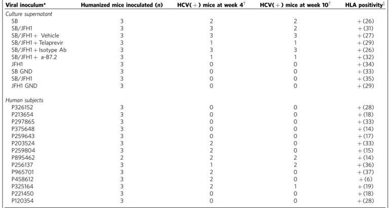
Table 1 | HCV infection analysis on immunocompromised mice transplanted with human hematopoietic stem cells (HSCs).
Viral inoculum* Humanized mice inoculated (n) HCV(þ) mice at week 4w HCV(þ) mice at week 10w HLA positivityz
Culture supernatant
SB 3 2 2 þ(26)
SB/JFH1 3 3 2 þ(31)
SB/JFH1þ Vehicle 3 3 3 þ(27)
SB/JFH1þTelaprevir 3 1 1 þ(29)
SB/JFH1þIsotype Ab 3 3 3 þ(26)
SB/JFH1þ a-B7.2 3 1 1 þ(32)
JFH1 3 0 0 þ(34)
SB GND 3 0 0 þ(33)
SB/JFH1 3 0 0 þ(35)
JFH1 GND 3 0 0 þ(29)
Human subjects
P326152 3 0 0 þ(28)
P213654 3 0 0 þ(18)
P297865 3 0 0 þ(33)
P375648 3 0 0 þ(14)
P259643 3 0 0 þ(17)
P203524 3 2 0 þ(33)
P259804 3 2 0 þ(15)
P895462 2 2 2 þ(14)
P256137 3 1 2 þ(36)
P965701 3 2 0 þ(37)
P458612 3 2 0 þ(6)
P325164 3 2 1 þ(19)
P221450 3 0 0 þ(18)
P120354 3 0 0 þ(28)
*Viral inocula were obtained from nine different chronic HCV patients (coded) or from cell culture-derived HCV (that is, SB, SB/JFH1 chimera and JFH1). wHCV positivity was determined by RT-PCR for HCV positive-strand RNA.
zHLA DNA was determined by FACS using antibody that is specific to human HLA.
Dendritic cells
Monocyte
Memory B cels
Naïve B cells
CD8+ T cells
CD4+ T cells Granulocytes SB GND HCV SB HCV
0 106
105
104
103
102
101
P120354
P221450
P325164
P458612
P965701
P256137
P895462
P259804
P203524
P259643
P375648
P297865
P213654
P326152
JFH1 GND
SB/JFH1 GND
SB GND
JFH1
SB
SB/JFH1
SB/JFH1 + vehicle
SB/JFH1 + Tela
SB/JFH1 + Isotype Ab SB/JFH1 +
α
-B7.2 Ab
Serum
HCV RNA
(copies per ml)
Cellular HCV RNA (copies per
μ
g RNA)
0 101
102
103 104 105 106 107 0 101
102
103
104 105 106 107
* *
* *
*
HCV RNA (copies per
μ
g of RNA)
HCV (−) HCV (+)
a b
virus injection. HCV RNA was detected in PBMCs of these mice (up to 2.7105 particles ml1: Fig. 8a). In contrast, mice injected with JFH1 virus or the replication-incompetent strains (GND mutant) of SB or JFH1 strains were negative for HCV RNA.
We infected humanized mice with sera from 14 human subjects, nine of whom tested positive for HCV. None of the HCV-negative sera led to infection in these mice (Table 1). In contrast, 13 of 24 mice inoculated with the HCV-positive sera tested positive for HCV RNA at week 4, of which five mice remained positive up to week 10. Injection of anti-B7.2 antibody reduced both internal and cell surface associated HCV RNA levels (Fig. 8a). Human PBMC from the infectedRag2/;Il2rg/
(RG) mice were further sorted into T and B lymphocytes, monocytes, and dendritic cells and examined separately for cellular HCV RNA. As shown in Fig. 8b, the HCV RNA was preferentially detected in memory B cells. HCV RNA was also detected in CD4þ and CD8þ T cells in humanized mice (Fig. 8b). These results indicated that HCV was capable of persistently infecting human B cells in a humanized mouse model.
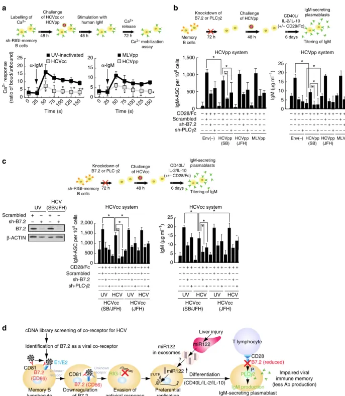
HCV binding downregulates B7.2 and inhibits Ig production.
As CD27þ memory B cells in HCV patients are anergic (exhausted) B cells63,64, the functions of memory B cells infected with HCV particles were examined accordingly. To determine if HCV-infected CD27þ B cells had attenuated signalling upon BCR ligation, Ca2þ mobilization was measured after immunoglobulin (Ig)M stimulation with anti-human IgM. The HCV-infected CD27þ B cells had diminished Ca2þ mobilization (Fig. 9a), indicating that HCV-infected CD27þ
B cells are anergic to BCR-mediated stimulation. To test if HCV-infected cells were prone to apoptosis, HCV-infected primary memory B cells isolated from patients were stimulated with CD40LþIL2þIL-10 or vehicle control ex vivo
(Supplementary Fig. 9A). Treated memory B cells were stained with annexin V (to measure apoptosis) and anti-CD27. After incubation with vehicle control or CD40LþIL2þIL-10 treatment, the percentage of annexin Vþ cells was higher in the CD40LþIL4-stimulated HCV-infected B-cell subset than that of UV-irradiated supernatant-treated cells (Supplementary Fig. 9A). As CD40L stimulation transactivates pro-apoptotic molecule Bax (ref. 65), Bax mRNA levels were also examined. The treatment was found to induce BAX after cross-linking B7.2 on HCV-infected memory B cells (Supplementary Fig. 9A, right). These data suggested that, in the absence of survival signals, HCV-infected CD27þB cells were prone to apoptosis.
B7.2 is a costimulatory molecule that interacts with CD28 and CTLA-4 on T cells. In addition, B7.2 has a cytoplasmic tail that transduces signals in antigen-presenting cells66,67. B7.2 stimulation results in the phosphorylation of phospholipase Cg2 (PLCg2) and protein kinase C ab (PKCa/b), leading to activation of transcription factor Oct-2 and eventual increased IgG1 transcription and protein production68. Since binding of the HCV E2 protein to CD81 can perturb the biological activities of CD81 (ref. 69), we tested if the binding of HCV E2 to B7.2 also affected B7.2 and its signalling pathways. To determine whether HCVpp-SB infection reduced CD28/Fc-mediated activation of PLCg2 and PKCa/b, we analysed the activation of PLCg2 and PKCa/b. As shown in Supplementary Fig. 9B, CD28/Fc-mediated phosphorylation of PLCg2 (Tyr1217) and PKCa/b (Thr638/641) was reduced in HCVpp-infected memory B cells.
To test if the observed effects on signalling were due to the E2–B7.2 interaction, we used purified recombinant E2 (SB strain) purified from Baculovirus expression system instead
of HCVpp-SB. To test if E2 (SB strain) binds to B7.2, His-tagged E2 (without the transmembrane domain) from SB strain (genotype 2b) and H77 strain (genotype 1a) were expressed in the baculovirus expression system and partially purified (Supplementary Fig. 7A). The intracellular E2 was detected in both monomeric and aggregated forms (Supplementary Fig. 7B). Immunoblot analysis confirmed expression of HCV E2 protein (Supplementary Fig. 7C). The partially purified E2 was used for
in vitro binding to various cell lines as determined by flow
cytometry. The results showed that E2 from the SB strain bound to Raji cells very efficiently. E2 from the genotype 2a JFH1 strain does not bind to HepG2 cells (Supplementary Fig. 7D). The binding strengths of E2 from SB and JFH1 strains were further compared by using different amounts of E2 (normalized using an anti-His6 antibody). E2 did not bind to HepG2 cells, which is
among the few human cell lines that do not express CD81 or B7.2. In contrast, E2 bound to the B7.2-overexpressing HepG2 cells efficiently (Supplementary Fig. 7D). The binding was partially blocked by a B7.2-specific antibody but not by an isotype-matched control antibody (Supplementary Fig. 7D). As shown in Supplementary Fig. 7E, E2 of the SB strain bound more than that of the H77 strain at every protein concentration used. These results indicated that recombinant E2 (SB lymphotropic strain), but not E2 (H77 hepatotropic strain), bound to B cells in a B7.2-dependent manner.
Furthermore, to rule out that the observed effects were not a consequence of CD81 engagement by E2-SB, endogenous CD81 was knocked out by CRISPR and replaced with exogenous full-length CD81 or extracellular-domain-truncated CD81 expression vectors. CD81 overexpression partially restored immunoglobulin production in CRISPR-mediated CD81 knock-out cells (Supplementary Fig. 8A). To further test if another co-receptor SR-BI stimulated immunoglobulin production, SR-BI was knocked out by CRISPR and replaced with transfected full-length SR-BI or intracellular-domain-truncated mutant of SR-BI and examined for immunoglobulin production. Both C-terminus truncation mutant and full-length SR-BI showed no effect on IgM production in the CRISPR SR-BI knockout cells expressing C-terminus truncation mutant of SR-BI or full-length SR-BI (Supplementary Fig. 8B). B2.7 restoration promoted IgM production while B7.2/CD8 chimera, which lacks the signal transduction domain, failed to restore the IgM production (Supplementary Fig. 8C, left). These results indicated that HCV E2(SB)-mediated downregulation of B7.2 reduced CD28/Fc-mediated signalling pathways.
HCV-infected B cells do not differentiate into plasmablasts.
Memory B lymphocyte CD81 B7.2 (CD86) E1/E2 CD81 B7.2 (CD86)
5′UTR miR122
miR122 in exosomes cDNA library screening of co-receptor for HCV
Scrambled sh-B7.2 HCVcc
α-IgM α-IgM
UV-inactivated HCVcc system * * * * * * * * * *
Identification of B7.2 as a viral co-receptor
RIG-I Unknown recepor Unknown recepor Preferential replication Differentiation (CD40L/IL-2/IL-10) Downregulation of B7.2 Impaired viral immune memory (less Ab production) Evasion of
antiviral response
PLCγ2 Liver injury T lymphocyte CD28 B7.2 (reduced) miR122 ? b a d 0 5 10 15 20 25 Time (s) Time (s)
0 25 50
75 100 125 150
0 25 50
75 100 125 150
0 5 10 15 20 25 0 5 10 15 20 * HCVpp MLVpp &H4' &))$1/ : ; *- ' sh-RIGI-memory B cells
48 h 72 h
Ca2+
release Challenge
of HCVcc or HCVpp Labelling of Ca2+ 48 h Stimulation with human IgM sh-RIGI-memory B cells
72 h 48 h 6 days
Titering of IgM Knockdown of
B7.2 or PLC γ2
Scrambled sh-B7.2 HCVpp system * * * *
sh-PLCγ2
c 0 5 10 15 20 25 Memory B cells
72 h 6 days
Titering of IgM Knockdown of
B7.2 or PLCγ2
IgM-secreting plasmablasts IgM-secreting plasmablasts 48 h CD40L/ IL-2/IL-10 (+/– CD28/Fc) CD40L/ IL-2/IL-10 (+/– CD28/Fc)
UV HCV UV HCV
– + + – + – – – + – + + – + – – – + – + + – + – – – + – + + – + – – – + + – – + – – + – – + – – CD28/Fc – – –+– – –+– – –+– – – + Env(–) HCVpp (SB) HCVpp (JFH) MLVpp Env(–) HCVpp (SB) HCVpp (JFH) MLVpp – + + – + – – – + – + + – + – – – + – + + – + – – – + – + + – + – – – + + – – + – – + – – + – – CD28/Fc – – –+– – –+– – –+– – – + Challenge of HCVcc Challenge of HCVpp * * * HCVpp system 0 500 1,000 1,500 – + + – – + – + + – – + – + + – – + – + + – – + + – + – + – + – + ––– + ––– + ––– + – – – – – –+– – –+– – –+– – – + UV * * * HCVcc system
HCV UV HCV 0 500 1,000 1,500 2,000 – + + – + – – – + + – – + – – + – – + – – – + + – + – – – + – + + – + – – – + – + + – + – – – + – – – +– – –+– – –+– – –+ * HCVcc (SB/JFH) HCVcc (JFH) HCVcc (SB/JFH) HCVcc (JFH) IgM production IgM-secreting plasmablast P HCV (SB/JFH) UV Scrambled sh-B7.2 B7.2 β-ACTIN + − − + + − − + Ca 2+ response
(ratio of boud/unbound)
IgM-ASC per 10
5 cells
IgM (
μ
g ml
–1
)
Ca2+mobilization
assay
sh-PLCγ2
IgM-ASC per 10
5 cells
IgM (
μ
g ml
–1
)
Figure 9 | HCV infection impairs antigen recall responses in memory B cell.(a) HCV-infected CD27þB cells, compared to uninfected CD27þB cells, have attenuated Ca2þresponses after BCR cross-linking. B cells from healthy donor were loaded with Indo-1-AM, stained with anti-CD27 and warmed to 37°C. After establishing a baseline for 30 s, cells were stimulated with 10mg ml1goat F(ab0)2 anti-human IgM. Kinetic graphs represent ratios of bound/ unbound Indo-1 AM over time for CD27þ B-cell populations. Arrows indicate addition time of F(ab0)2a–human IgM (*Po0.05,t-test,n¼3). Error bars represent s.d. (b) Signals by anti-B7.2 Ab induce the production of IgM by memory B cells. Memory B cells (5103cells per well) were cultured with
different concentrations of anti-B7.2 (0.5mg ml1); after 5 days, supernatants from control and experimental wells were collected and IgM levels were
measured. Data are presented as the fold-increase above memory B cells activated with a CD40LþIL-2þIL-10, and a species- and isotype-matched control Ab (*Po0.05,t-test,n¼3). Error bars represent s.d. (c) Similar assays were performed using HCVcc as described with HCVpp above (*Po0.05,
following CD40L/IL-2/IL-10 stimulation (Fig. 9b, left). This suggested differentiation to IgM-secreting plasmablasts upon CD40L/IL-2/IL-10 stimulation was impaired in HCVpp-infected memory B cells, compared to unHCVpp-infected memory B-cells (Fig. 9b, left). These results indicated that HCVpp-SB-mediated downregulation of B7.2 reduced CD28/Fc-mediated signalling pathways. Silencing B7.2 or PLCg2 significantly reduced IgM production in HCV-infected memory B cells (Fig. 9b, right). In contrast, HCVpp-JFH1, which does not bind to B7.2, did not inhibit IgM production. To confirm the biological relevance of the pseudotyped HCV particle study, similar results were also obtained when we used the HCV-SB virus (Fig. 9c). Class switch did not account for the decreased IgM in these subsets, as we consistently detectedo140 ng ml1 of IgM in cell culture supernatants. Taken together, these results indicated that infection with both HCVcc and HCVpp increased IgM production and by downregulation of B7.2, attenuated CD28-mediated IgM production; thus virus infection enhanced the differentiation of memory B cells into IgM-secreting plasmablasts.
Discussion
In this study, we have established the genetic basis for lymphotropism of HCV infection. A comprehensive set of data indicated that B7.2 is a co-receptor for observed HCV SB strain tropism towards memory B cells. Previous reports have also indicated that some HCV strains can infect human T cells: the H77 strain of HCV can infect and replicate in established T-cell lines, such as Molt-4 and Jurkat cells, in long-term culture21. Furthermore, transfection of SB strain RNA, but not JFH1, led to its replication in Molt-4 cells70. Here we provide evidence indicating that B7.2 is involved in HCV SB strain tropism towards memory B cells (additional discussion is described in Supplementary Note 1).
B7.2 belongs to the immunoglobulin superfamily, and is highly expressed on memory B cells and germinal centre B cells71. Its ligand includes CD28 and CTLA4 on the surface of T cells. B7.2 and B7.1 are expressed by antigen-presenting cells including B cells. The result of ligation of B7.2 leads to the following: B7.2-CD28 interaction co-stimulates T-cell activation and enhances T-cell survival, B7.2-CTLA4 interaction inhibits T-cell activation and leads to T-cell tolerance. The infection of B cells by HCV likely causes significant alteration of these functions72. Furthermore, expression of HCV viral proteins in B cells can result in B-cell lymphoma in transgenic mice73. The expression levels of B7.2 are not correlated with cellular HCV RNA levels in certain primary cells, such as dendritic cells—which are known to be associated with HCV74—indicating that other co-receptor(s) may exist for lymphocyte infection (Supplementary Fig. 5G). Indeed, CD5 expression is linked to HCV infection of lymphocytes75. Another possibility is that inhibitors of HCV infection, such as Ewi-2wint, a partner of CD81, may be associated with this lymphotropism76. Other studies have shown ApoE to be a tropism determinant in a late assembly step and viral cell-to-cell transmission of HCV JFH1 strain77.
In conclusion, HCV tissue tropism is determined primarily by the properties of viral envelope proteins and the 50-UTR, in
addition to the ability of HCV to evade antiviral responses (Fig. 7f). The existence of lymphotropic HCV strains indicates that lymphoid cell infection may be an important facet of HCV infection in some patients. These studies on the genetic basis of lymphotropism of HCV will open up an avenue for studying extrahepatic infection of HCVin vivoandin vitro. The SB strain may be representative of lymphotropic quasispecies that arise from an HCV infection that is primarily hepatotropic.
Methods
Cells.Raji (Cat# CCL-86), HEK293T (Cat# CRL-3216) and HepG2 (Cat# HB-8065) cells were obtained from ATCC. HepG2 cells were transfected with pCDNA3.1-CD81 or B7.2 expression plasmid, selected with G418
(0.5mg ml1) and surviving cell colonies were picked after drug selection.
Individual cell clones were further isolated and characterized for CD81 expression
and E2 binding. To generate sh-RIGI-Raji cells, Raji cells were transduced by
multiple lentivirus expressing shRNAs against RIG-I and MDA5 (Open
Biosys-tems) and/or miR122. The culture media for Raji and sh-RIGI-Raji cells is RPMI
media 1640, GlutaMAX (Cat#61870-036, Life Technologies) containing 20% FBS (Omega, Inc.). PBMC were isolated by Ficoll-Paque Plus (Amersham Biosciences) density gradient centrifugation. Informed consent was obtained. Institutional Review Board at University of Southern California approved the procedures. PBMC were maintained in RPMI 1640 containing 10% FBS. Infection of cells, generation of HCV pseudo-particles and generation of HCV clone ar described in Supplementary Methods.
Vector.The Claudin-1 expression vector was a gift from Dr Weeraratna from National Institute of Aging (NIA/NIH). WT B7.2, B7.2-CD8, ICAM-1/CD8 and WT ICAM-1 expression vectors were previously described (gifts from Dr Jung, USC) (ref. 78).
Sucrose density gradient analysis.The viral particles produced from the transfected cells were concentrated from the culture supernatant and separated by
sucrose gradient sedimentation. Culture medium derived from sh-RIGI-Raji cells
was harvested for sucrose density gradient analysis 6 days after transfection of full-length SB RNA. Collected culture medium was cleared by low speed centrifugation
at 670gfor 10 min, and passed through a filter with 0.45-mm pore size (Millipore).
Filtered culture medium was layered on a stepwise sucrose gradient (60 to 10%, wt/vol) and centrifuged for 16 h in a SW41 rotor (Beckman) at 40,000 or
50,000 rpm (273,865gor 427,914g) at 4°C. After centrifugation, 8 or 16 fractions
were harvested from the bottoms of the tubes. Core protein concentration in each
fraction was determined by an immunoassay using 100ml of the fraction. HCV
RNA titer was determined by RTD-PCR using RNA isolated from 100ml of the
fraction. The35S labelling was performed by standard procedures.
Lentivirus production.To generateRIGIorMDA5shRNA or amiR-122
lenti-virus, the transfer vector expressingmiR-122or shRNA targets sequence on the
RIG-I or MDA5 cDNA (RHS4531-NM_022168, 98851910,
RHS4430-99158156, RHS4430-99161339: MDA5so calledDDX58, orRHS4531-NM_014314,
RHS4430-98910525, RHS4430-99294017, RHS4430-99619590: RIG-I so called DDX58; Human GIPZ Lentiviral shRNAmir target gene set, Open Biosystems) were co-transfected with pPAX2 and pVSV-Gp expression vector in HEK293T cells.
Cell entry assay.Virus infection was carried out at 4°C (no endocytosis) and then
shifted to 37°C (active endocytosis). Antibody-treated and untreated cultures were
infected with HCVpp(SB) or HCVpp(JFH1) for 1 h at 4°C. Plates were then
brought to 37°C and assayed at indicated time points over a 300 min time
course. Controls are normalized to the value for anti-CD81 added at 4 h. Antibodies directed against CD81 (JS81) or B7.2 were added at various time points
after temperature was shifted to 37°C, and the infectivity was determined at 12
days after infection as previously described. The concentration of B7.2 and
CD81 we used in the experiments are both 50mg ml1. Half-maximal inhibition
time of anti-B7.2 was 132 min while half-maximal inhibition time of anti-CD81 was 44 min.
Immunoprecipitation followed by immunoblot for B7.2.Full-length of B7.2, B7.2/CD8, full-length of ICAM or ICAM/CD8 were overexpressed in 293T cells. After 24 h, the expression level of B7.2 was determined by western blots. For infection, the culture media was replaced with fresh media containing either SB HCVpp or JFH1 HCVpp and incubated an additional 24 h. Cell lysates were used for mmunoprecipitation with anti-B7.2, followed by immunoblot for B7.2 proteins in 293T cells.
After incubation, the cells were harvested and lysed for immunoprecipitation. B7.2 only interacted with HCV E2 after SB infection, but not JFH1 infection. These data indicate that SB E2 protein, but not JFH1 E2 protein, directly interacted with B7.2.