EFFECTS OF SMALL MOLECULE BINDING TO mRNA ON
GENE EXPRESSION
Julie Marie Sullivan
A dissertation submitted to the faculty of the University of North Carolina at Chapel Hill in partial fulfillment of the requirements for the degree of Doctor of Philosophy in the
Department of Chemistry.
Chapel Hill 2008
ABSTRACT
Julie Marie Sullivan: Effects of Small Molecule Binding to mRNA on Gene Expression (Under the direction of H. Holden Thorp)
RNA plays significant roles in many cellular processes and exhibits attractive features for use as a small molecule targeted therapeutic. This study focuses on using RNA as a drug target, specifically investigating the process by which small molecules can regulate gene expression through their interaction with secondary structural elements found in the 5’ untranslated region (UTR) of two cellular messenger RNA (mRNA) targets, human ferritin and human preproinsulin.
The 5’UTR of the human preproinsulin mRNA exists in two forms, the native form, and a splice variant which is up-regulated in response to chronic glucose stimulation. Small molecules were screened for their ability to bind to, or change the secondary structure of, the 5’UTR of the preproinsulin mRNAs using RNase cleavage, and the transition metal cleavage agents Ru(tpy)(bpy)O2+ (RuO2+, bpy = 2,2’-bipyridine, tpy = 2,2’,2’’-terpyridine), and Ru(bpy)32+. Two small molecules, neomycin B and kanamycin B, have been identified as binding to the mRNAs using these screening methods. Binding of neomycin B, and
ACKNOWLEDGEMENTS
Five years ago I moved from Buffalo to Chapel Hill. At the time I was unsure if I would adjust, if grad school was really the place I wanted to be. It was hard to move from the only place I had lived, away from all my family and friends. Five years later, I know that it was one of the best decisions I’ve ever made. I’ve learned more about myself and about chemistry that I’d ever known before. Learning chemistry could happen anywhere, but I’m happy that it happened here.
panicking, and constantly supported and took interest in what I was doing. Without these three people, I wouldn’t have made it through the last 4 years.
Working for and with Holden was an experience that I know I couldn’t have gotten anywhere else. He let me gain independence, always listened to my interpretation of data, supported me through anything I wanted to do or try, and understood that the amount of time spent in lab wasn’t as important as getting results and overall being happy with what you were doing. He encouraged me to do things outside of the lab, to travel to Europe, to become involved with the entrepreneurship class he was teaching, to try new things. Who else would call you in the middle of the day to go be part of a “focus group” for the planetarium to make sure their Pink Floyd laser show was appropriate, or invite you to see him perform in a musical, or his band play at Weaver Street Market? It helped make him less of a “boss” and more someone who really wanted to see you succeed, which of course was the ultimate goal.
It takes much more than getting experimental results to make it through graduate school, and it is something that I could not have done by myself. At UNC I’ve made great friends, and without them these past five years would have been incredibly boring. I’ve been lucky to have great housemates who have made day to day issues easier. The past two years, Abby and Stephanie, have been great companions, fellow TV watchers, and provided
out with. Even though Heather moved to Boston, we kept in touch through constant email conversations, and trips, and she is my number one cheerleader. I also have been fortunate to have met a great group of girls who have made girls nights that much more fun.
Through random chance I met Pete while working in the Schoenfisch lab on SPR experiments. While the actual experiments didn’t yield any good results, I met someone who has become a more than a great friend, who has provided endless support, and is the person I look forward to spending my future with.
Along with my friends at UNC, I’ve been lucky to have support from friends from Buffalo, specifically Heather and Jenn. Heather took the first 12 hour drive down here, through the scary mountains of West Virginia, to help me move. She has always been someone that I could count on, that I can call up in any situation and she’ll be there. Jenn is my travel companion, and she encourages me to do things I otherwise wouldn’t think to do. Additionally, I have made friends along the way, who have been there to add a little more happiness into my life. Rob I met at UB and we have kept in touch while finishing our respective programs. He has been helpful with all aspects of laboratory life, and has helped most recently with my search for what I want to do after graduate school. He was also always up for getting outdoors and was a much needed visitor during my early time here. I met Sid at U of R as a freshman. We became good friends, and I enjoy the funny anecdotes and stories that he provides, and am thankful for all the times he has been there for me.
and never fails to remind me of who I am. My younger brothers and sisters provide
entertainment when I visit, and I am amazed by what and who they are becoming each time I return. Mary always reminds me how far I have come, and my Dad is always there to keep me on track. My Uncles, Gary and Rob, are the ones who I remember introducing me to science, and have always shown me that science is exciting, even at its most basic level. There are no words to describe my mother, how much she means to me, how I think of her everyday, and how I hope to one day be as wonderful a person as she was.
TABLE OF CONTENTS
LIST OF TABLES... xiii
LIST OF FIGURES ... xiv
LIST OF ABBREVIATIONS AND SYMBOLS ... xviii
Chapter 1. Introduction: Targeting RNA with Small Molecules...1
1.1 RNA as a Drug Target ...1
1.2 RNA Structure ...2
1.3 Chemical and Enzymatic Mapping Techniques for RNA ...5
1.3.1 Enzymatic Mapping Techniques...5
1.3.2 Chemical Mapping Techniques...6
1.3.2.1 Nucleic Acid Reactivity to Oxidation...6
1.3.2.2 Metallonucleases as RNA Mapping Agents...8
1.3.2.3 Ru(tpy)(bpy)O2+ as an RNA Chemical Mapping and Screen for Small Molecule Binding...9
1.3.2.4 Ru(bpy)32+ as a Chemical Mapping Agent: The Flash-Quench technique...11
1.4 Molecular RNA Targets...12
1.4.1 Antiviral Targets: Transactivation Response RNA (TAR RNA) ...12
1.4.2 mRNA Targets: Inhibiting Ribosome Screening...14
1.5 Principles of RNA Binding...15
1.7 Figures...18
1.8 References...25
Chapter 2. In Vitro Up-regulation of Ferritin Protein Synthesis by Small Molecules that Target the Iron Responsive Element in Ferritin mRNA...34
2.1 Abstract...34
2.2 Introduction...35
2.2.1 Cellular Iron Homeostasis...35
2.2.1.1 Iron Absorption and Transport...35
2.2.1.2 Cellular Iron Uptake: Transferrin-mediated Endocytosis...36
2.2.1.3 Cellular Iron Storage: Ferritin...37
2.2.2 Regulation of Cellular Iron Homeostasis: Regulation of of Transferrin and Ferritin by Iron Regulatory Proteins (IRPs) and Iron Responsive Elements (IREs)...38
2.2.3 Identification of Small Molecules that Disrupt the IRP:IRE Interaction...43
2.3 Experimental...45
2.3.1 Materials...45
2.3.2 Methods...46
2.3.2.1 Transcription of IRE RNA (50-mer)...46
2.3.2.2 5’ RNA End-labeling...47
2.3.2.3 3’ RNA End-labeling...47
2.3.2.4 Internal Labeling of RNA with [α-32P]UTP...48
H-chain Ferritin, IRE-luciferase, and pGEM®-luciferase...51
2.3.2.9 In Vitro Translation of mRNA: Rabbit Reticulocyte Lysate Translation Assay...53
2.4 Results and Discussion ...54
2.4.1 Small Molecule:Ferritin mRNA Interaction: RNase Cleavage Assay ...54
2.4.2 Effect of Promazine on the IRP:IRE Interaction: Electrophoretic Mobility Shift Assay...55
2.4.3 Regulation of Protein Synthesis by Small Molecules: In Vitro Translation Assay...57
2.4.3.1 Ferritin Translation...57
2.4.3.2 IRE-luciferase and Wild-type Luciferase Translation...58
2.4.3.3 Ferritin and Wild-type Luciferase Competition Assay...58
2.4.4 Binding Constant Determination...59
2.5 Conclusions...60
2.6 Tables...64
2.7 Figures...65
2.8 References...79
Chapter 3. Identification of Small Molecules that Target RNA and Mediate Human Preproinsulin Protein Synthesis...86
3.1 Abstract...86
3.2 Introduction...87
3.2.1 Regulation of Glucose Homeostasis...87
3.2.2 Insulin...88
3.2.2.1 Human Insulin Biosynthesis...88
3.2.2.3 Mechanism of Insulin Action in the Body...90
3.2.3 Control of Insulin Gene Expression by Glucose...91
3.2.4 Identification of Small Molecules that Bind to Human Preproinsulin mRNA...93
3.3 Experimental...94
3.3.1 Materials...95
3.3.2 Methods...95
3.3.2.1 Cloning of the Full-length Human Preproinsulin Splice Variant (SPV)...97
3.3.2.2 Cloning of the 5’UTR of Human Preproinsulin Splice Variant (SPV Hairpin) ...99
3.3.2.3 Creation of the Full-length and 5’UTR Native Human Preproinsulin Constructs...100
3.3.2.4 Transcription of NAT and SPV Hairpin RNA...100
3.3.2.5 RNA End-labeling...101
3.3.2.6 RNase Cleavage Assay...102
3.3.2.7 RuO2+ Small Molecule Binding Assay...103
3.3.2.8 Flash-Quench Oxidation...104
3.3.2.9 Transcription of Full-length mRNA: Human NAT Preproinsulin and Human SPV Preproinsulin...104
3.3.2.10 In Vitro Translation of mRNA...104
3.4 Results and Discussion ...105
3.4.2 Screening Compounds to Find a Human Preproinsulin
Hairpin RNA Binder...108
3.4.3 Comparison of Methods Used to Identify a Human Preproinsulin Hairpin RNA Binder...110
3.4.4 Regulation of Protein Synthesis by Small Molecules: In Vitro Translation Assay...111
3.4.4.1 Human Preproinsulin Native and Splice Variant mRNA Constructs...111
3.4.4.2 Wild-type Luciferase mRNA Construct...111
3.5 Conclusions...112
3.6 Future Directions ...114
3.7 Tables...115
3.8 Figures...118
LIST OF TABLES
Table 2.1 Names and structures of compounds found to interact with the
IRE through the RuO2+ assay...64
Table 3.1 Oligonucleotides used in human preproinsulin cloning ...115
Table 3.2 PCR conditions used in human preproinsulin cloning...116
Table 3.3 Names and basic structures of compounds tested in the
LIST OF FIGURES
Figure 1.1 Schematic representation of four general classes of RNA secondary
structure...18 Figure 1.2 Structures of the two metal complexes used as metallonucleases in
this work ...19 Figure 1.3 Proposed mechanism of guanine oxidation in DNA...20 Figure 1.4 Common aromatic ligands used to change the binding properties
of nucleases...21 Figure 1.5 Schematic representations of the secondary structure of RNAs
mapped with metallonucleases ...22 Figure 1.6 Proposed secondary structure of tRNAPhe...23 Figure 1.7 Flash-quench reaction scheme and structures of Ru(bpy)32+
and Co(NH3)5Cl2+...24 Figure 2.1 Cellular iron transport: the transferrin cycle...65 Figure 2.2 Sequence and structures of the human ferritin iron responsive
element (IRE)...66 Figure 2.3 Schematic representation of ferritin translation regulation by
IRP binding ...67 Figure 2.4 Schematic representation of transferrin receptor translation
regulation by IRP binding...68 Figure 2.5 Effect of promazine on RNA cleavage by RNase I in human
IRE RNA...69 Figure 2.6 Effect of yohimbine on RNA cleavage by RNase I in human
IRE RNA...70 Figure 2.7 Effect of promazine (A) and yohimbine (B) on RNase I cleavage
Figure 2.10 Effect of yohimbine on the IRP1:IRE interaction...74 Figure 2.11 Effect of yohimbine on the IRP2:IRE interaction...75 Figure 2.12 In vitro translation assay to determine the effect of promazine
on translation...76 Figure 2.13 Effect of promazine on IRE-dependent mRNA translation in
cell-free extracts as measured by incorporation of [35S]methionine...77 Figure 2.14 Effect of promazine on the translation of a mixture of ferritin
and luciferase mRNA in cell-free extracts as measured by
incorporation of [35S]methionine ...78 Figure 3.1 Schematic of the human insulin sequence and secondary
structure...118 Figure 3.2 Human insulin biosynthesis ...119 Figure 3.3 Proposed secondary structures of the 5’UTRs of the human
preproinsulin mRNA...120 Figure 3.4 Schematic representation of preproinsulin full-length and
hairpin RNA constructs...121 Figure 3.5 Preproinsulin full-length mRNA and hairpin RNA constructs...122 Figure 3.6 Comparison of guanine cleavage sites in the native and splice
variant preproinsulin RNA hairpins...123 Figure 3.7 Analysis of the native preproinsulin mRNA hairpin structure
using RNase cleavage ...124 Figure 3.8 Analysis of the native preproinsulin mRNA hairpin structure
using Ru(tpy)(bpy)O2+ mediated guanine oxidation...125 Figure 3.9 Analysis of the native preproinsulin mRNA hairpin structure
using the flash-quench technique...126 Figure 3.10 Analysis of the preproinsulin splice variant hairpin RNA
Figure 3.12 Analysis of the preproinsulin splice variant hairpin RNA
structure by guanine oxidation using the flash-quench technique ...129 Figure 3.13 Effect of promazine and yohimbine on RNase cleavage of
the native human preproinsulin hairpin RNA...130 Figure 3.14 Effect of promazine and yohimbine on the RNase
cleavage of the human preproinsulin splice variant hairpin RNA...131 Figure 3.15 Effect of neomycin B and Hoechst 33258 on the RNase
cleavage of the native human preproinsulin hairpin RNA...132 Figure 3.16 Effect of neomycin B on RNase cleavage of the native
preproinsulin hairpin RNA ...133 Figure 3.17 Effect of neomycin B on the RNase cleavage of the human
preproinsulin splice variant hairpin RNA ...134 Figure 3.18 Effect of neomycin B on RNase cleavage of the human
preproinsulin splice variant hairpin RNA ...135 Figure 3.19 Effect of neomycin B on guanine oxidation in the native
preproinsulin hairpin RNA ...136 Figure 3.20 Effect of neomycin B on guanine oxidation using the RuO2+
assay for native human preproinsulin hairpin RNA ...137 Figure 3.21 Effect of neomycin B on guanine oxidation in the preproinsulin
hairpin splice variant RNA ...138 Figure 3.22 Effect of neomycin B on guanine oxidation using the RuO2+
assay for human preproinsulin splice variant hairpin RNA...139 Figure 3.23 Effect of kanamycin B on the RNase cleavage of the native
human preproinsulin hairpin RNA...140 Figure 3.24 Effect of kanamycin B on RNase cleavage of the native
human preproinsulin hairpin RNA...141 Figure 3.25 Effect of kanamycin B on the RNase cleavage of the
human preproinsulin splice variant hairpin RNA ...142 Figure 3.26 Effect of kanamycin B on RNase cleavage of the human
Figure 3.27 Effect of kanamycin B on guanine oxidation in the native
preproinsulin hairpin RNA ...144 Figure 3.28 Effect of kanamycin B on guanine oxidation using the RuO2+
assay for native human preproinsulin hairpin RNA ...145 Figure 3.29 Effect of kanamycin B on guanine oxidation in the
preproinsulin hairpin splice variant RNA ...146 Figure 3.30 Effect of kanamycin B on guanine oxidation using the RuO2+
assay for human preproinsulin splice variant hairpin RNA...147 Figure 3.31 Effect of Hoechst 33258 on the RNase cleavage of the
human preproinsulin splice variant hairpin RNA ...148 Figure 3.32 Effect of hygromycin B on the RNase cleavage of the
native human preproinsulin hairpin RNA...149 Figure 3.33 Effect of hygromycin B on the RNase cleavage of the
human preproinsulin splice variant hairpin RNA ...150 Figure 3.34 Effect of neomycin B on guanine oxidation using the
flash-quench technique in the preproinsulin hairpin splice
variant RNA ...151 Figure 3.35 In vitro translation assay to determine the effect of
neomycin B and kanamycin B on protein translation...152 Figure 3.36 Effect of neomycin B and kanamycin B on human preproinsulin
mRNA translation in cell-free extracts as measured by
incorporation of [35S]methionine ...153 Figure 3.37 Effect of promazine and yohimbine on human preproinsulin
mRNA translation in cell-free extracts as measured by
incorporation of [35S]methionine ...154 Figure 3.38 Effect of neomycin B and kanamycin B on luciferase mRNA
translation in cell-free extracts as measured by incorporation of
LIST OF ABBREVIATIONS AND SYMBOLS
[5’-32P]pCp Cytidine 3’, 5’-bis(phosphate) 8-oxo-guanine 7,8-dihydro-8-oxoguanine Å angstrom
A absorbance; adenine; binding equation variable
aa amino acid
ADP adenosine diphosphate
ATP adenosine triphosphate
γ32P-ATP adenosine 5’-gamma 32P-triphosphate β beta
bp basepair bpy 2,2'-bipyridine
BSA bovine serum albumin
oC degrees Celsius
C cytosine
Ca2+ calcium ion
cDNA complimentary deoxyribonucleic acid cm centimeter
CTP cytidine triphosphate
Cys cysteine
DMT-1 divalent metal transporter 1 protein
ds double strand DTT dithiothreitol
ε extinction coefficient
E. coli Escherichia coli
EDTA ethylenediaminetetraacetic acid
eIF-2 eukaryotic initiation factor 2 EMSA electrophoretic mobility shift assay
E.T. electron transfer
FL-NAT full-length native human preproinsulin mRNA
FL-SPV full-length human preproinsulin splice variant mRNA FPLC fast protein liquid chromatography
G guanine H-chain heavy chain of human ferritin HCl hydrochloride
HEPES N-[2-Hydroxyethyl]piperazine-N’-[2-ethanesulfonic acid] HIV-1 human immunodeficiency virus type 1
hr hour IC50 inhibitory concentration 50%
IPTG isopropyl-beta-D-thiogalactopyranoside
kan kanamycin Kapparent apparent binding constant kb kilobase
KCl potassium chloride
Kd dissociation constant
kDa kilodalton L liter L-chain light chain of human ferritin
LB luria broth
M molar Met methionine
MgCl2 magnesium chloride
µ micro µg microgram µl microliter µM micromolar µs microsecond mg milligram ml milliliter min minute mRNA messenger ribonucleic acid N any of the nucleotides A, C, G, U
NAT native human preproinsulin mRNA NAT-HP native human preproinsulin hairpin RNA ng nanogram
NH4OAc ammonium acetate
nm nanometer
NMR nuclear magnetic resonance
ns nanosecond nt nucleotide
OD optical density/absorbance
OH hydroxyl P. pastoris Pichia pastoris
PAGE polyacrylamide gel electrophoresis
PC prohormone convertase
PCR polymerase chain reaction
Phe phenylalanine
pKa -log [Ka]
pM picomolar
PNK polynucleotide kinase
ppIGE preproinsulin mRNA glucose-responsive translation element Q quencher
RRL rabbit reticulocyte lysate
rRNA ribosomal RNA
RuO2+ Ru(tpy)(bpy)O2+
Ru-OH2 Ru(tpy)(bpy)OH2 Ser serine
SDS sodium dodecyl sulfate
SELEX systematic evolution of ligands by exponential enrichment
SPR surface plasmon resonance
SPV human preproinsulin splice variant mRNA SPV-HP human preproinsulin splice variant hairpin RNA
ss single strand
T thymidine
TAR transactivating response region
TBE tris, boric acid, EDTA buffer
Tf transferrin protein
TfR transferrin receptor
tpy 2,2’,2’’-terpyridine Tricine N-tris(hydroxymethyl)methylglycine
Tris tris(hydroxymethyl)aminomethane
tRNA transfer RNA
tRNAphe transfer RNA encoding the phenylalanine amino acid U uracil
UTR untranslated region
UTP uridine triphosphate
Chapter 1
Introduction: Targeting RNA with Small Molecules
1.1 RNA as a Drug Target
Expression of the numerous genes in a cell must be tightly regulated to provide
appropriate levels of RNA and protein production at all times. The cell must interpret many
different chemical and physical signals in order to respond to environmental and cellular
needs. Organisms make extensive use of protein-based control systems to regulate gene
expression; modulating mechanisms of transcription, translation, and mRNA processing or
degradation. RNA is essential for transcriptional and translational regulation, protein
function, and catalysis. These recently discovered functions of RNA dramatically expand its
role in the cell and highlight the potential of targeting RNA for the treatment of a number of
disease states.
Currently, most small molecule drug discovery efforts are focused on proteins
involved in biological pathways associated with disease, although more emphasis is being
placed on targeting small molecules to RNA. The unique structures formed by RNA are
thought to provide suitable binding pockets for small molecules,1 and most small molecules
that target RNA have been shown to selectively bind to noncanonically paired regions.2 Over the next two chapters, my work will focus on investigating the use of small molecules
to modulate gene expression through their interaction with structured regulatory elements
RNA is an attractive therapeutic target because of its many functions in the cell and
its lack of a cellular repair mechanism.3-5 RNA also provides a target for drugs where a protein target is unavailable or inaccessible, as is seen in many viral6,7 and bacterial targets.
8-10 Drugs which bind to mRNA structures involved in post-transcriptional regulation can
either up- or down-regulate expression of a gene. Up-regulation of gene expression is not
often accomplished in protein targeting because drugs are normally used to inhibit protein
function.11 The ability of small molecules such as the macrolide and aminoglycoside
antibiotics to interact with bacterial ribosomal RNA has been known for some time, and have
set a precedent for targeting structured domains of RNA.3,4,12-21 More recently, riboswitches, mRNA structures that regulate gene expression in bacteria through the use of small
molecules, have been suggested as a new cellular target for antibacterial drug discovery.10 Pursuing RNA as a therapeutic target increases not only the number of available targets in a
cell, but also increases the potential number of small molecules that can be considered RNA
ligands. However, our knowledge of RNA recognition by small molecules is still very basic
and has not progressed as quickly as that of DNA because RNA in solution forms less
predictable structures.
1.2 RNA Structure
RNA exhibits many features similar to those possessed by DNA and/or proteins,
which makes them attractive from a targeting standpoint. Its hydrogen bonding capabilities
single stranded RNA sequence can fold to produce many distinct structures, including
regions of canonical and noncanonical base pairing, bulges, internal loops and hairpin loops,
and various tertiary interactions (Figure 1.1). RNA molecules usually contain short
double-helical regions connected by single stranded stretches. The dominant secondary structural
element formed when an RNA chain folds back on itself is the hairpin loop or stem-loop
(Figure 1.1D).22 This structure places complementary sequences spatially close to each other to create base pairs that stabilize the overall RNA structure through hydrogen bonding
and base stacking interactions (Figure 1.1A). These base paired regions form A-type
helices, with 11-12 base pairs per turn in a right-handed, anti-parallel double-helix.23 The A-type helix has a very deep major groove and a shallow minor groove, which unlike the major
and minor grooves of DNA are less conducive to small molecule binding.24 The major groove of RNA contains the discriminatory edges of the nucleobases, while the defining
feature of the minor groove is the presence of the 2’-hydroxyl groups.25
Although areas of complementarity can be found in the RNA sequence, there are also
areas that cannot form canonical pairs. In addition to the formation of noncanonical base
pairs, helical stems are often interrupted by bulged bases or bulged loops (Figure 1.1C),
where bases on one strand have no partner to pair with on the other strand, and by internal
loops (Figure 1.1B), in which sequences on both strands cannot be paired.22 These
structures produce defined pockets suitable for binding to other RNAs, proteins, and small
metabolites.26 Bulge loops can be either extrahelical or intrahelical, with larger bulges causing increased destabilization of the RNA structure.5 One-nucleotide bulges are the most
common, with single purine bulges tending to stack intralhelically causing the helix to bend,
identified as being important in tertiary interactions,29 and in protein recognition.30 Even if a stem-loop contains no interrupted regions in its stem, it must contain a terminal loop,
sometimes termed a hairpin loop, which links the 5’- to the 3’-strand of its stem. Terminal
loops can vary in size, from short sequences that play the same role in RNA as β-turns in
proteins, to loops that are large enough to contain stem-loops of their own.5,22 RNAs also contain tertiary structure, with areas of secondary structure interacting with nucleotides
distant in the primary sequence.23,31 While advances in the prediction of RNA secondary structure have been made,32,33 it is difficult to predict the three-dimensional structure of RNA due to its complex and dynamic behavior.5
Experiments have shown the feasibility of targeting RNA using ligands that recognize
the above mentioned structural regions in order to modulate gene expression, but there are
few examples in the literature.34,35 Years of research have resulted in the development of a paradigm for small molecule-duplex DNA binding based on the primary sequence of the
DNA.36 However, research is very far from designing an RNA ligand based only on the
knowledge of an RNA sequence because, unlike helical DNA, RNA is recognized more by
its structural features rather than its sequence.1,2,16,26,37-41 Understanding how to enhance or
prevent RNA-ligand or RNA-protein interactions by blocking their interaction, or by
disrupting secondary and tertiary structure, will open up new opportunities for drug design.
In order to target small molecules to RNA, a basic understanding of the overall RNA
structure is necessary. Determination of the structure of large RNA molecules through
techniques have also been used to characterize the structural features of RNA. An important
advantage of these techniques is that the quantity of RNA required for analysis (nanograms)
is much smaller than that required (milligrams) for structural studies by NMR and
crystallography.42 Turning these techniques into a screen for ligands that bind in a specific structural region will enhance the ability to identify small molecules that could modulate
gene expression.
1.3 Chemical and Enzymatic Mapping Techniques for RNA
In RNA mapping studies, the RNA of interest is exposed to a reagent and the extent
of the reaction is quantified at affected nucleotides in the RNA. The reactivity of the
nucleotide depends on its structural constraints in solution. There are two broad classes of
reagents commonly used for RNA mapping, RNases used in selective enzymatic degradation,
and reactive small molecules. Mapping of the entire RNA structure often requires multiple
reagents be used in parallel. The choice between using chemical and enzymatic methods for
probing structural features of RNA depends on what and how information is obtained. There
are advantages and disadvantages to both techniques, but their combined use can give
complimentary information important for determining the overall structure.43
1.3.1 Enzymatic Mapping Techniques
There are a number of RNases that vary in their specificity for single or double
stranded regions of RNA, or the sequences recognized.44 In general, enzymatic reactions allow more flexibility in the choice of reaction conditions. The conditions used can be
of which is used to identify a specific nucleotide or structural region. Reaction conditions
used maintain the secondary structure of the RNA and allow only single hit kinetics to occur.
The most commonly used RNases are RNase T1, RNase A, RNase I, and RNase V1.
Cleavage occurs with RNase T1 at unpaired guanine residues while RNase A cleaves at
unpaired cytidine and uridine residues. RNase I cleaves after all four residues equally in
unpaired regions and RNase V1 shows a preference for paired or stacked nucleotides.
Dabrowiak and coworkers,45-47 as well as others,48-52 have used RNases to map the binding sites of small molecules on RNA.
1.3.2 Chemical Mapping Techniques
Chemical nucleases can be used as a good compliment to the enzymatic nucleases
commonly used to probe RNA-small molecule interactions. These nucleases have a specific
geometry, relatively rigid shapes, and are particularly sensitive to the tertiary structure or
shape of the RNA binding site.44 They are also smaller in size than protein nucleases,
allowing them to intimately react with the DNA or RNA substrate. Chemical modification
reagents such as dimethyl sulfate and diethyl pyrocarbonate selectively modify base
positions,53 whereas small molecule reagents such as hydroxyl radicals,45 lead,54 and metal
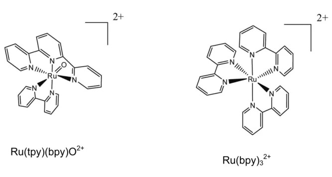
complexes,44,55-57 often target the phosphoribose backbone of RNA.56 The following section will focus on two metallonucleases, Ru(tpy)(bpy)O2+ and Ru(bpy)32+ (Figure 1.2), that were
used for chemical mapping in this work. Both compounds interact with RNA by oxidizing
the guanine nucleobase.
abstraction, or at the nucleobases via radical reactions or electron transfer.58,59 Sugar oxidation occurs nearly five times slower in RNA than in DNA due to the stabilizing effect
of the 2’-OH present in the ribose sugar,60,61 and RNA is much more susceptible to hydrolytic
cleavage of the phosphodiester bond.23,62,63 Base oxidation pathways are similar in both DNA and RNA.
The reactivity of the nucleobases toward oxidation is of the order G > A > C ≈ T / U,
corresponding to the oxidation potentials of the bases.64 Since guanine has the lowest oxidation potential, this is where base damage usually occurs in vivo, occurring either
directly at the guanine position or through migration to a region containing guanine.
Nucleobase cleavage requires a second chemical step, often alkali treatment, to cause
complete strand scission.59 In DNA, this alkali treatment causes hydrolysis of the glycosidic
bond, leaving an abasic site, which can undergo β-elimination of the 3’ phosphate.59 In
RNA, all phosphodiester linkages are subject to alkaline hydrolysis due to the presence of the
2’-hydroxyl. Hydrolysis of the phosphodiester linkages occurs faster in RNA than the
creation of an abasic site and β-elimination.59 In practice, aniline is used to facilitate
complete strand scission in RNA, while piperidine is used for DNA.
One electron oxidation of RNA bases leads to the production of a guanine radical
cation59 that is a relatively strong acid (pKa = 3.9).65 In the mechanism of oxidation determined in DNA (Figure 1.3), the guanine radical undergoes rapid deprotonation to
generate the neutral radical. This is then followed by O2 addition and the eventual
production of the products imidazolone and oxazolone. Alternatively, the guanine radical
cation can become hydrated followed by one electron oxidation to form 8-oxo-guanine.59
DNA damage.66 The preferential degradation pathway of the guanine radical cation
(hydration vs. deprotonation) depends on the context of the guanine. Nucleosides and
single-strands of nucleic acid are expected to favor the imidazolone and oxazolone pathway,
whereas base paired guanines are expected to favor the 8-oxo-guanine pathway due to
stabilization of the radical cation.59
1.3.2.2 Metallonucleases as RNA Mapping Reagents
The ability for metallonucleases to interact with DNA and cause chemical
modification has been known for some time, and their mechanism of action and resulting
cleavage products have been studied in great detail.57,59,61,67 Many of these same compounds have also been used to map the structure of RNA, although the mechanism has not been
studied as in depth, and has in many cases been assumed to occur in the same manner as that
of DNA. Some metallonucleases are able to interact directly with the nucleobase due to their
high oxidation states that are generated either chemically or electrochemically, while other
cleavage agents are activated photochemically. An appealing characteristic of photochemical
reagents is that all the components of a system to be studied can be mixed together without
initiating the chemical reaction until the sample is irradiated.67 Provided that the
chromophore of the photocleavage agent is sensitive to light greater than 300 nm in
wavelength, select excitation of the cleavage agent will occur. The ability to “tune” the
nuclease by changing the ligands allows complexes to be synthesized to match the desired
nucleic acid target. Various metal ligands can be added to the metallonuclease to change
among others,57,73-76 have explored many DNA and RNA structures using transition metal oxidation chemistry. The following discussion focuses on two metallonucleases,
Ru(tpy)(bpy)O2+ (RuO2+, bpy = 2,2’-bipyridine, tpy = 2,2’,2’’-terpyridine) and Ru(bpy)32+,
and their use as probes of RNA structure.
1.3.2.3 Ru(tpy)(bpy)O2+ as an RNA Chemical Mapping Agent and Screen for Small
Molecule Binding
Meyer and coworkers first reported the synthesis of Ru(tpy)(bpy)O2+ in 198177 and later reported that the complex could oxidize organic hydrocarbons and alcohols.78,79 The
complex is made as the aqua complex and is converted via bulk electrolysis to the oxidized
oxo complex through a coupled two proton, two electron process. Since the discovery that
RuO2+ could oxidize DNA under anaerobic conditions,80 RuO2+-nucleic acid oxidation chemistry has been studied extensively.81-85 Due to its size (6 Ǻ) and charge (2+), the RuO2+ compound shows preferential binding to solvent accessible guanines that are prone to cation
binding.42,81,86 It is postulated that oxidation occurs through an inner-sphere Ru(III)-O-G intermediate. Hairpin structures such as the transactivation response (TAR) RNA sequence,
tRNAPhe, and the iron responsive element (IRE) sequence found in human and bullfrog
ferritin have been oxidized with RuO2+, and show the greatest extent of damage at loop guanines (Figures 1.5 and 1.6).55,81,87
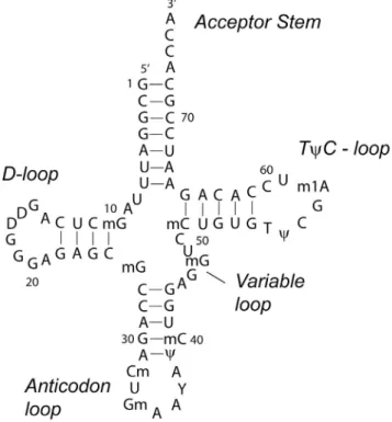
tRNAPhe (Figure 1.6) was studied using RuO2+ chemistry due to the available X-ray crystal structure and the ability to control the RNA conformation via salt concentration.55 In the folded state, tRNAPhe was cleaved mainly at guanines and adenines in the D- and
also studied using RuO2+ chemistry,55 and it was observed that guanine nucleobases found in the loop (G17, G18, and G19) were oxidized more efficiently than guanines found in the stem.
The iron responsive element has also been studied using RuO2+ oxidation
chemistry.42,81,88 In the first study,42 the IRE in full-length bullfrog ferritin mRNA (Figure 1.5A) was probed with RuO2+. Cleavage was seen only at position G16 (the IRE has been
renumbered since the original publication, and was originally termed G14), and not at any
other unpaired guanine residues indicating that this residue was highly solvent exposed. It
was concluded that guanine residues other than G16 were spatially inaccessible to the reagent.
Lack of cleavage at other unpaired guanine residues was later explained by NMR structural
data that suggested that the guanines predicted to be unpaired in the secondary structure were
involved in tertiary interactions with other parts of the RNA molecule.89 Further studies confirmed that the G16 residue is flipped out of the RNA structure and is solvent exposed,
allowing it to make important contacts with the iron regulatory protein (IRP). Ciftan et al.
studied oligonucleotides corresponding to the 30 base IRE hairpin structure and 55 base IRE
hairpin with flanking region.81 RuO2+ cleavage revealed more cleavage sites than in the previous study although G16 was still oxidized to the greatest extent. Other unpaired guanine
residues were also oxidized, which was consistent with the expectations from the predicted
secondary structure.
in the IRE RNA was probed using RuO2+. Three molecules that bound to this pocket were identified, and the effect of one small molecule, yohimbine, on ferritin gene expression was
examined. This small molecule was able to up-regulate ferritin protein synthesis invitro,
validating the use of RuO2+ as a small molecule screen.
1.3.2.4 Ru(bpy)32+ as a Chemical Mapping Agent: The Flash-Quench Technique
Photolysis of a ruthenium chemical mediator to cause oxidative damage in DNA has
been studied extensively by Barton and coworkers.91,92 This oxidative damage occurs primarily through base-labile guanine oxidation. The metal complexes have been used to
study events such as mismatch recognition93,94 and electron transfer from peptides to DNA.95,96 Transition metal complexes such as ruthenium and rhodium can act as direct
photooxidizers in the flash-quench experiment,91 the reaction scheme for which is shown in Figure 1.7. The flash-quench cycle is first initiated by visible light which excites the Ru2+ complex. The excited ruthenium(II) complex *Ru2+ is then quenched by a nonintercalating
electron acceptor (Q), to form Ru3+. The Ru(III) species can be reduced back to the Ru(II) form either through recombination with the reduced quencher (Q-), or through electron transfer with a nearby guanine base resulting in an oxidized guanine radical. The oxidized
guanine radical can return to the resting state by reaction with the reduced quencher or
undergo further reaction to form the guanine oxidation products shown in Figure 1.3. The
flash-quench technique was first used to characterize electron transport in proteins,97 and was later modified for DNA oxidation.91 Recently, Dana Holcomb in our lab has adapted this technique for its use in the oxidation of RNA. (unpublished results)
This flash-quench reaction for oxidation of guanine nucleobases in RNA uses the
common example of an external RNA binder, and its binding is governed by both solvent
binding and electrostatic contributions.74,98 The electron transfer between guanine and Ru(bpy)33+ occurs in less than 200 ns, and once formed, nearly all the guanine radicals decay
within 100 µs.91,92,95 The irreversible reaction of the guanine radical with either oxygen or
water yields oxidative lesions that can be analyzed by gel electrophoresis.95 The flash quench-method of RNA mapping showed oxidation of the guanine residues in the human
ferritin IRE in a similar pattern to those identified using the RuO2+ compound. Preferential oxidation was seen at those residues which were readily solvent accessible and not residues
whose flexibility allowed them to become solvent exposed over time. The use of Ru(bpy)32+
as a footprinting technique continues to be examined in this work.
1.4 Molecular RNA Targets
Promising results have been obtained using a number of small molecules to target
RNA, offering a complimentary approach to the targeting of proteins. Small molecule
ligands have been identified with three major classes of RNA targets in mind: antibacterial,
antiviral, and mRNA. Within each of these classes, various avenues have been pursued to
achieve gene expression regulation, including inhibiting RNA-protein interactions and
preventing protein production by binding to a functionally relevant RNA. To date, all
clinically approved RNA-targeting drugs exert their effect by binding to ribosomal RNA.26
1.4.1 Antiviral Targets: Transactivation Response RNA (TAR RNA)
RNA-protein complex. One example of this interaction is between the TAR RNA and the
Tat protein involved in the regulation of HIV-1.
The HIV-1 genome is made up of a ~9 kb genomic RNA encoding for 15 proteins.99
After integration into the host cell genome, the initial phase of the viral life cycle begins with
transcription from the 5’ end of the RNA. Although transcription is critical to viral
proliferation, RNA polymerase II transcribes poorly from the viral promoters. The HIV-1
protein Tat helps facilitate efficient transcription of the viral genome. In the presence of the
Tat protein, the rate of transcription increases nearly 100-fold to produce full length genomic
RNA.99 Inhibition of the Tat-TAR interaction by binding to TAR, in order to slow or halt production of the viral transcript, has been sought as a potential anti-HIV strategy.100,101 Tat
has been shown to bind to a specific bulged region in the TAR RNA hairpin loop at the
beginning of the viral transcripts.102,103
A variety of techniques have been used to identify compounds able to inhibit the
Tat-TAR interaction, including gel mobility shift assays, absorption and fluorescence
spectroscopy, mass spectrometry, footprinting experiments, scintillation proximity assays,
NMR, and computational screening.52,104-107 Initially, a collection of aminoglycosides was
screened for their ability to disrupt the Tat-TAR interactions.108 Neomycin was found to be
the most potent aminoglycoside with an IC50 of 0.92 µM. In subsequent experiments it was
demonstrated that the binding site for neomycin was located below the bulged region in TAR
RNA. It has been suggested that binding of neomycin induces a conformational change in
the TAR RNA, increasing the off-rate of the Tat-TAR complex.109 Later, Mei and coworkers
developed a high-throughput in vitro screening method and were able to screen
From this screen, two promising compounds were identified, quinoxaline and
tetraaminoquinozaline. The James group has used computer-aided drug design to identify
compounds that are candidates for blocking the Tat-TAR interaction.101,110 Acetopromazine
was identified through the use of a docking program and was found to completely inhibit
formation of the Tat-TAR complex at a concentration of 100 nM.101 An NMR structure was
obtained, showing that acetopromazine bound to TAR RNA in an area containing a
trinucleotide bulge.110 It was later shown that acetopromazine was able to bind internal bulges and terminal loops, but not double stranded RNA or tetraloops.111
1.4.2 mRNA Targets: Inhibiting Ribosome Scanning
Translation in eukaryotes proceeds first with the binding of the small ribosomal
subunit to the eIF-2·GTP·Met·tRNAi complex, followed by binding to the 5’ cap of
mRNA.112 The 5’ end of the mRNA molecule is recognized by the presence of its 5’ cap and two bound initiation factors, eIF4e and eIF4G. The small ribosomal subunit complex then
begins scanning the transcript, searching the 5’UTR for the first AUG codon. After
recognition of the codon by Met-tRNAi, the complex pauses and waits for the large
ribosomal subunit to associate before translation begins. Secondary structure in the 5’UTR is
known to pause or inhibit the scanning process, resulting in translation inhibition. By
binding of a small molecule within the 5’UTR of a specific mRNA, inhibition of translation
and modulation of gene expression can occur.
The in vitro selection process known as SELEX (systematic evolution of ligands by
decreases the in vivo expression of β-galactosidase activity by greater than 90 %. Internal
controls determined that the effects of Hoescht 33258 were not due to general translation
inhibition. In the same manner, Grate and Wilson113 have placed an aptamer to malachite green in the 5’UTR of the CLB2 gene, which encodes for a cyclin that directs the cell cycle
transition from G2 to mitosis in budding S. cerevisiae. They have shown that ligand-induced
folding of the 5’UTR limits binding by the 40S ribosomal subunit and results in a reduction
in the rate of translational initiation.
1.5 Principles of RNA Binding
Recent work has focused on determining the biochemical and biophysical rules
governing small molecule-RNA binding.16,21,107,114 Compounds that interact with RNA show
marked differences from those typically identified as being “drug like” by Lipinski’s rules
and can make targeting a specific RNA difficult.1,14-16,21,115 The small molecules often exceed the established molecular weight limits and are more hydrophobic or polar in nature
than common drugs, due to the need to interact with the negatively charged RNA. Both of
these features could cause a reduction in the bioavailability of the compound. Most small
molecules also have only modest selectivity and affinity for their RNA target when compared
to their protein counterparts.26 The binding constants for many RNA-small molecule interactions are relatively weak, falling in the low micromolar range.14 These drugs can
however be sufficiently potent if binding causes or inhibits a conformational change in the
RNA.
The general affinity of the aminoglycosides for many different RNAs leads to their
promiscuity, the compounds are often useful for in vitro studies, but less useful for cell
culture or in vivo work.26 Aminoglycosides can also be very toxic at high concentrations. Still, a large amount of data has been collected on the aminoglycoside-RNA interaction, and
certain basic principles have emerged that will aid in designing future RNA ligands.
Aminoglycosides have been found to bind pockets created by bases in and around internal
loops and bulged regions or those created by noncanonical interactions. They have shown a
strong dependence for electrostatic interactions, as well as nonionic interactions. They also
have shown that shape complementarity (how well the ligand and RNA fit together) as well
as conformational adaptation (binding induced changes in the ligand and RNA) are a major
contributor of the specificity of small molecules for RNA.
Using the knowledge of binding principles provided by the aminoglycosides, the
Hergenrother group have shown that subsets of deoxystreptamine dimers are able to
selectively bind RNA tetraloops and octaloops through the use of a combinatorial library of
105 deoxystreptamine dimers.1,39 Ligand binding was assessed using fluorescein labeled RNA to obtain an estimate of the dissociation constant and the ligand binding site was
determined using RNase footprinting. Two compounds with nanomolar affinity for hairpin
loops were identified using this screen, one showing preferential binding to a tetraloop, and
the other to an octaloop. These were the first compounds reported to show discrimination
between RNA hairpin loops of various sizes. More work needs to be done to identify the
RNA motifs that small molecules prefer so that compounds can be efficiently designed. In
The rational discovery of small molecule ligands for RNA is still in its infancy
compared to the use of these small molecules to bind proteins and DNA. The complex
structure of RNA increases the difficulty in the identification of potential ligands. To date,
the small molecules identified have little in common as far as their structures and modes of
interaction, making it is hard to generalize what qualifies as a good RNA binder. Detailed
characterization of small-molecule RNA interactions, along with methods for high
throughput screening are necessary to progress toward the goal of selectively targeting RNA
with small molecules.
1.6 Dissertation Focus
This dissertation focuses on the use of secondary structures in mRNA as a small
molecule drug target for regulation of gene expression. Two model systems are investigated,
the iron responsive element in the human ferritin mRNA, which is studied in Chapter 2, and
the preproinsulin glucose regulatory element found in the human preproinsulin mRNA
studied in Chapter 3. Chapter 2 focuses on the ability of a compound identified using
oxidation chemistry to regulate ferritin gene expression. In Chapter 3 the secondary structure
of the preproinsulin glucose regulatory element is first mapped with transition metal
complexes and RNases, then the mapping agents are used to screen for small molecules that
bind to the preproinsulin mRNA. The effect of these compounds on gene regulation is
1.7 Figures
Figure 1.1. Schematic representation of four general classes of RNA secondary structure. A) Base paired RNA forming a helix or duplex structure. B) In an internal loop secondary structure, sequences on both strands cannot be paired. C) A bulged region in RNA occurs when bases on one strand have no partner on the other strand to base pair with. D) Hairpin loop, or stem loop region consisting of a base paired “stem” region with an unpaired terminal loop. The bold line represents a canonical base pair, whereas the circle represents a
A
C
B
D
Duplex / Helix
Internal Loop
Figure 1.2. Structures of the two metal complexes used as metallonucleases in this work
Ru(tpy)(bpy)O
2+Figure 1.3. Proposed mechanism of guanine oxidation in DNA (adapted from Burrows59).
8-oxo-guanine
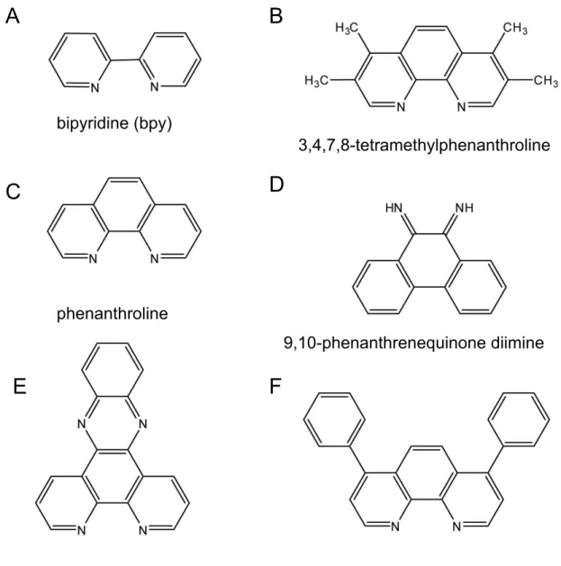
Figure 1.4. Common aromatic ligands used to change the binding properties of
metallonucleases. Bipyridine (bpy) ligands (A) are an example of an electrostatic ligand and also known as external binders. 3,4,7,8-tetramethylphenanthroline (B) and phenanthroline (C) are known groove binders in DNA. 9,10-phenanthrenequinone diimine (D),
dipyridophenazine (E), and 4,7-diphenyl-1,10-phenanthroline (F) are known intercalators.
bipyridine (bpy)
4,7-diphenyl-1,10-phenanthroline dipyridophenazine
9,10-phenanthrenequinone diimine phenanthroline
3,4,7,8-tetramethylphenanthroline
A B
C
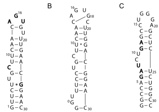
Figure 1.5. Schematic representations of the secondary structure of RNAs mapped with metallonucleases. A) Proposed bullfrog ferritin IRE secondary structure.89 Bold residues represent bases that make direct contacts with the IRP protein.116 B) Proposed structure of the human ferritin IRE.88 There is a base pair across the hairpin loop between C14 and G18.
C) Proposed secondary structure of the TAR element found in HIV-1.101 Bold residues represent bases critical for binding of the Tat protein.
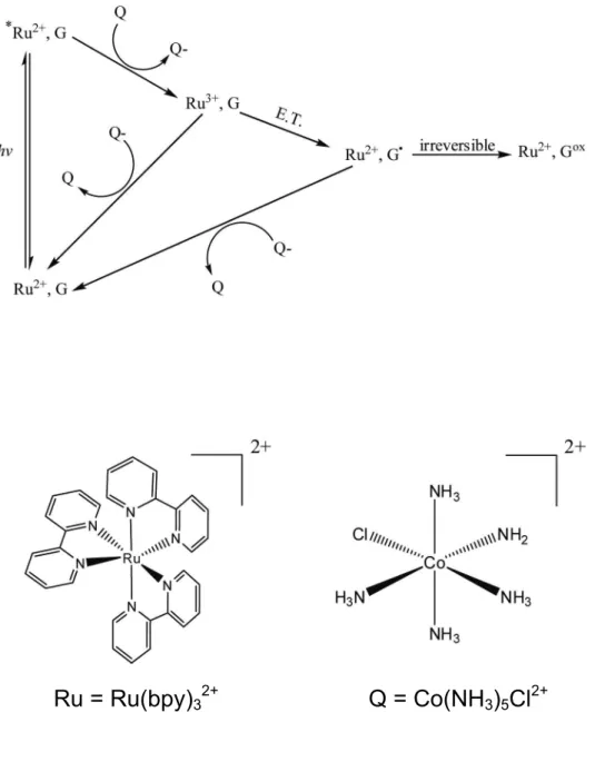
Figure 1.7. Flash-quench reaction scheme and structures of Ru(bpy)32+ and Co(NH3)5Cl2+.
In this scheme, G = Guanine, Q = Quencher. Visible light excites the Ru(bpy)32+ species to
form *Ru(bpy)32+. This excited complex is then quenched by the nonintercalating
Co(NH3)5Cl2+ electron acceptor to form Ru(bpy)33+. The Ru(bpy)33+ can then be reduced
back to Ru(bpy)32+ either through recombination with the reduced quencher or through
electron transfer with a nearby guanine base resulting in a guanine radical. The guanine radical can return to its resting state by reacting with the reduced quencher, or can undergo further reaction to form 8-oxo-guanine or one of its reaction products.
Q = Co(NH
3)
5Cl
2+1. 8 References
(1) Thomas, J. R.; Liu, X.; Hergenrother, P. J. Biochemistry 2006, 45, (36), 10928-10938.
(2) Disney, M. D.; Childs-Disney, J. L. ChemBioChem 2007, 8, 649-656.
(3) Pearson, N. D.; Prescott, C. D. Chem. Biol. 1997, 4, 490-414.
(4) Harvey, I.; Garneau, P.; Pelletier, J. RNA 2002, 8, 452-463.
(5) Xia, T.; Mathews, D. H.; Turner, D. H., Thermodynamics of RNA Secondary Structure Formation. In RNA, 1st ed.; Soll, D.; Nishimura, S.; Moore, P. B., Pergamon: New York, NY, 2001, 21-44.
(6) Faber, C.; Siticht, H.; Schweimer, K.; Rosch, P. J. Biol. Chem. 2000, 275, (27), 20660-20666.
(7) Hsu, M.-C.; Schutt, A. D.; Holly, M.; Slice, L. W.; Sherman, M. I. Science 1991, 254, (5039), 1799.
(8) Barker, J. J. Drug Discov. Today 2006, 11, (9/10), 391-404.
(9) Isaacs, F. J.; Dwyer, D. J.; Collins, J. J. Nat. Biotechnol. 2006, 24, (5), 545-554.
(10) Blount, K. F.; Breaker, R. R. Nat. Biotechnol. 2006, 24, (12), 1558-1564.
(11) DeJong, E. S.; Luy, B.; Marino, J. P. Biopolymers 2002, 2, 289-302.
(12) Ecker, D. J.; Griffey, R. H. Drug Discov. Today 1999, 4, (9), 420-429.
(13) Xavier, K. A.; Eder, P. S.; Giordano, T. Trends Biotechnol. 2000, 18, 349-356.
(14) Foloppe, N.; Matassova, N.; Aboul-ela, F. Drug Discov. Today 2006, 11, (21/22), 1019-1027.
(16) Hermann, T. Biochimie 2002, 84, 869-875.
(17) Tor, Y. ChemBioChem 2003, 4, 998-1007.
(18) Suscheck, S.; Wong, C.-H. Curr. Opin. Chem. Biol. 2000, 4, 678-686.
(19) Mayer, M.; Lang, P. T.; Gerber, S.; Madrid, P. B.; Pinto, I. G.; Guy, R. K.; James, T. L. Chem. Biol. 2006, 13, 993-1000.
(20) Wang, Y.; Rando, R. R. Chem. Biol. 1995, 2, (5), 281-290.
(21) Hermann, T. Angew. Chem. Int. Ed 2000, 39, 1890-1905.
(22) Moore, P. B., A Spectroscopist's View of RNA Conformation: RNA Structural Motifs. In RNA, 1st ed.; Soll, D.; Nishimura, S.; Moore, P. B., Pergamon: New York, 2001, 1-20.
(23) Blackburn, G. M.; Gait, M. J., Nucleic Acids in Chemistry and Biology. 2nd ed.; Oxford University Press: New York, NY, 1996; p.
(24) Weeks, K. M.; Crothers, D. M. Science 1993, 261, (5128), p1574(4).
(25) Carlson, C. B.; Stephens, O. M.; Beal, P. A. Biopolymers 2003, 70, 86-102.
(26) Thomas, J. R.; Hergenrother, P. J. Chem. Rev. 2008, 108, (4), 1171-1224.
(27) Borer, P. N.; Lin, Y.; Wang, S.; Roggenbuck, M. W.; Gott, J. M.; Uhlenbeck, O. C.; Pelczer, I. Biochemistry 1995, 34, (19), 6488-6503.
(28) van den Hoogen, Y. T.; van Beuzekom, A. A.; de Vroom, E.; van der Marel, G. A.; van Boom, J. H.; Altona, C. Nucl. Acids Res. 1988, 16, (11), 5013-5030.
(31) Moore, P. B. Annu. Rev. Biochem. 1999, 68, 287-300.
(32) Mathews, D. H.; Sabina, J.; Zucker, M.; Turner, D. H. J. Mol. Biol. 1999, 288, 911-940.
(33) Mathews, D. H.; Disney, M. D.; Childs, J. L.; Schroder, S. J.; Zucker, M.; Turner, D. H. Proc. Natl. Acad. Sci. U. S. A. 2004, 101, 7287-7292.
(34) Werstuck, G.; Green, M. R. Science 1998, 282, 296-298.
(35) DeNap, J. C. B.; Thomas, J. R.; Musk, D. J.; Hergenrother, P. J. J. Am. Chem. Soc.
2004, 126, (47), 15402-15404.
(36) Dervan, P. B.; Edelson, B. S. Curr. Opin. Struct. Biol. 2003, 13, (3), 284-299.
(37) Wemmer, D. E.; Dervan, P. B. Curr. Opin. Struct. Biol. 1997, 7, (3), 355-361.
(38) Dervan, P. B.; Doss, R. M.; Marques, M. A. Current Medicinal Chemistry - Anti-Cancer Agents 2005, 5, (4), 373-387.
(39) Thomas, J. R.; Liu, X.; Hergenrother, P. J. J. Am. Chem. Soc. 2005, 127, (36), 12434-12435.
(40) Liu, X.; Thomas, J. R.; Hergenrother, P. J. J. Am. Chem. Soc. 2004, 126, (30), 9196-9197.
(41) Yan, Z.; Sikri, S.; Beveridge, D. L.; Baranger, A. M. J. Med. Chem. 2007.
(42) Thorp, H. H.; McKenzie, A. R.; Lin, P. N.; Walden, W. E.; Theil, E. C. Inorg. Chem.
1996, 35, (10), 2773-2779.
(43) Knapp, G., Enzymatic approaches to probing of RNA secondary and tertiary
structure. In RNA Processing Part A: General Methods, Volume 180 ed.; Dahlberg, J. E.; Abelson, J. N., Academic Press: 1989, 192-212.
(45) McPike, M. P.; Sullivan, J. M.; Goodisman, J.; Dabrowiak, J. C. Nucleic Acids Res.
2002, 30, (13), 2825-2831.
(46) McPike, M. P.; Goodisman, J.; Dabrowiak, J. C. Bioorg. Med. Chem. 2002, 10, 3663-3672.
(47) McPike, M. P.; Goodisman, J.; Dabrowiak, J. C., Drug-RNA footprinting. In Drug-Nucleic Acid Interactions, Volume 340 ed.; Chaires, J. B.; Waring, M. J., Academic Press: 2001, 431-449.
(48) Yan, Z.; Baranger, A. M. Bioorg. Med. Chem. Lett. 2004, 14, (23), 5889-5893.
(49) Gayle, A. Y.; Baranger, A. M. Bioorg. Med. Chem. Lett. 2002, 12, (20), 2839-2842.
(50) Kikuta, E.; Aoki, S.; Kimura, E. J. Am. Chem. Soc. 2001, 123, (32), 7911-7912.
(51) Litovchick, A.; Evdokimov, A. G.; Lapidot, A. Biochemistry 2000, 39, (11), 2838-2852.
(52) Dassonneville, L.; Hamy, F.; Colson, P.; Houssier, C.; Bailly, C. Nucl. Acids Res.
1997, 25, (22), 4487-4492.
(53) Peattie, D. A.; Gilbert, W. Proc. Natl. Acad. Sci. U. S. A. 1980, 77, (8), 4679-4682.
(54) Behlen, L. S.; Sampson, J. R.; DiRenzo, A. B.; Uhlenbeck, O. C. Biochemistry 1990, 29, (10), 2515-2523.
(55) Carter, P. J.; Cheng, C.-C.; Thorp, H. H. J. Am. Chem. Soc. 1998, 120, (4), 632-642.
(56) Sigman, D. S.; Mazumder, A.; Perrin, D. M. Chem. Rev. 1993, 93, (6), 2295-2316.
(60) Neyhart, G. A.; Cheng, C.-C.; Thorp, H. H. J. Am. Chem. Soc. 1995, 117, (5), 1463-1471.
(61) Pogozelski, W. K.; Tullius, T. D. Chem. Rev. 1998, 98, (3), 1089-1108.
(62) Pyle, A. M. Science 1993, 261, (5122), p709.
(63) Thorp, H. H. Chem. Biol. 2000, 7, (2), R33-R36.
(64) Steenken, S.; Telo, J. P.; Novais, H. M.; Candeias, L. P. J. Am. Chem. Soc. 1992, 114, (12), 4701-4709.
(65) Candeias, L. P.; Steenken, S. J. Am. Chem. Soc. 1989, 111, (3), 1094-1099.
(66) Shigenaga, M. K.; Park, J.-W.; Cundy, K. C.; Gimeno, C. J.; Ames, B. N., In Vivo Oxidative DNA damage: Measurement of 8-Hydroxy-2'-deoxyguanosine in DNA and urine by high-performance liquid chromatography with electrochemical detection. In
Oxygen Radicals in Biological Systems Part B: Oxygen Radicals and Antioxidants, Volume 186 ed.; Packer, L.; Glazer, A. N., Academic Press: 1990, 521-530.
(67) Armitage, B. Chem. Rev. 1998, 98, (3), 1171-1200.
(68) Neyhart, G. A.; Grover, N.; Smith, S. R.; Kalsbeck, W. A.; Fairley, T. A.; Cory, M.; Thorp, H. H. J. Am. Chem. Soc. 1993, 115, (11), 4423-4428.
(69) Johnston, D. H.; Glasgow, K. C.; Thorp, H. H. J. Am. Chem. Soc. 1995, 117, (35), 8933-8938.
(70) Szalai, V. A.; Thorp, H. H. J. Am. Chem. Soc. 2000, 122, (18), 4524-4525.
(71) Szalai, V. A.; Singer, M. J.; Thorp, H. H. J. Am. Chem. Soc. 2002, 124, (8), 1625-1631.
(72) Holmberg, R. C.; Thorp, H. H. Inorg. Chem. 2004, 43, (16), 5080-5085.
(74) Pyle, A. M.; Rehmann, J. P.; Meshoyrer, R.; Kumar, C. V.; Turro, N. J.; Barton, J. K.
J. Am. Chem. Soc. 1989, 111, (8), 3051-3058.
(75) Chow, C. S.; Behlen, L. S.; Uhlenbeck, O. C.; Barton, J. K. Biochemistry 1992, 31, (4), 972-982.
(76) Chow, C. S.; Barton, J. K. J. Am. Chem. Soc. 1990, 112, (7), 2839-2841.
(77) Moyer, B. A.; Meyer, T. J. Inorg. Chem. 1981, 20, (2), 436-444.
(78) Thompson, M. S.; Meyer, T. J. J. Am. Chem. Soc. 1982, 104, (15), 4106-4115.
(79) Meyer, T. J. J. Electrochem. Soc. 1984, 131, (7), 221C-228C.
(80) Grover, N.; Thorp, H. H. J. Am. Chem. Soc. 1991, 113, (18), 7030-7031.
(81) Ciftan, S. A.; Theil, E. C.; Thorp, H. H. Chem. Biol. 1998, 5, (12), 679-687.
(82) Cheng, C.-C.; Goll, J. G.; Neyhart, G. A.; Welch, T. W.; Singh, P.; Thorp, H. H. J. Am. Chem. Soc. 1995, 117, 2970-2980.
(83) Ciftan, S. A.; Hondros, D. P.; Thorp, H. H. Inorg. Chem. 1998, 37, (7), 1598-1601.
(84) Farrer, B. T.; Thorp, H. H. Inorg. Chem. 2000, 39, (1), 44-49.
(85) Farrer, B. T.; Pickett, J. S.; Thorp, H. H. J. Am. Chem. Soc. 2000, 122, (4), 549-553.
(86) Kalsbeck, W. A.; Thorp, H. H. J. Am. Chem. Soc. 1993, 115, (16), 7146-7151.
(87) Carter, P. J.; Cheng, C.-C.; Thorp, H. H. Inorg. Chem. 1996, 35, (11), 3348-3354.
(89) Gdaniec, Z.; Sierzputowska-Gracz, H.; Theil, E. C. Biochemistry 1998, 37, (6), 1505-1512.
(90) Tibodeau, J. D. Applications of Metal-Mediated Guanine Oxidation in the
Examination of Functionally Relevant Nucleic Acid Structures. Ph.D dissertation, University of North Carolina at Chapel Hill, Chapel Hill, 2005.
(91) Stemp, E. D. A.; Arkin, M. R.; Barton, J. K. J. Am. Chem. Soc. 1997, 119, (12), 2921-2925.
(92) Stemp, E. D. A.; Barton, J. K. Inorg. Chem. 2000, 39, (17), 3868-3874.
(93) Kelley, S.; Boon, E.; Barton, J.; Jackson, N.; Hill, M. Nucl. Acids Res. 1999, 27, (24), 4830-4837.
(94) Yavin, E.; Boal, A. K.; Stemp, E. D. A.; Boon, E. M.; Livingston, A. L.; O'Shea, V. L.; David, S. S.; Barton, J. K. Proc. Natl. Acad. Sci. U. S. A. 2005, 102, (10), 3546-3551.
(95) Wagenknecht, H.-A.; Stemp, E. D. A.; Barton, J. K. J. Am. Chem. Soc. 2000, 122, (1), 1-7.
(96) Wagenknecht, H.-A.; Stemp, E. D. A.; Barton, J. K. Biochemistry 2000, 39, (18), 5483-5491.
(97) Dunn, D. A.; Lin, V. H.; Kochevar, I. E. Biochemistry 1992, 31, (46), 11620-11625.
(98) Kelly, J. M.; Tossi, A. B.; McConnell, D. J.; OhUigin, C. Nucleic Acids Res. 1985, 13, (17), 6017-6134.
(99) Frankel, A. D.; Young, J. A. T. Annu. Rev. Biochem. 1998, 67, (1), 1-25.
(100) Krebs, A.; Ludwig, V.; Boden, O.; Gobel, M. W. ChemBioChem 2003, 4, 972-978.
(102) Selby, M. J.; Bain, E. S.; Luciw, P. A.; Peterlin, B. M. Genes Dev. 1989, 3, (4), 547-558.
(103) Jakobovits, A.; Smith, D. H.; Jakobovits, E. B.; Capon, D. J. Mol. Cell. Biol. 1988, 8, (6), 2555-2561.
(104) Edwards, T. E.; Sigurdsson, S. T. Biochemistry 2002, 41, (50), 14843-14847.
(105) Puglisi, J. D.; Tan, R.; Calnan, B. J.; Frankel, A. D.; Williamson, J. R. Science 1992, 257, p76.
(106) Filikov, A. V.; Mohan, V.; Vickers, T. A.; Griffey, R. H.; Cook, P. D.; Abagyan, R. A.; James, T. L. J. Comput. Aided Mol. Des. 2000, 14, 593-610.
(107) Mei, H. Y.; Mack, D. P.; Galan, A. A.; Halim, N. S.; Heldsinger, A.; Loo, J. A.; Moreland, D. W.; SannesLowery, K. A.; Sharmeen, L.; Truong, H. N.; Czarnik, A. W. Bioorg. Med. Chem. 1997, 5, (6), 1173-1184.
(108) Mei, H.-Y.; Galan, A., A.; Halim, N., S.; Mack, D. P.; Moreland, D. W.; Sanders, K.; Truong, H., N.; Czarnik, A. W. Bioorg. Med. Chem. Lett. 1995, 5, (22), 2755-2760.
(109) Wang, S.; Huber, P. W.; Cui, M.; Czarnik, A. W.; Mei, H.-Y. Biochemistry 1998, 37, (16), 5549-5557.
(110) Du, Z.; Lind, K. E.; James, T. L. Chem. Biol. 2002, 9, 707-712.
(111) Mayer, M.; James, T. L. J. Am. Chem. Soc. 2004, 126, 4453-4460.
(112) Alberts, B.; Johnson, A.; Lewis, J.; Raff, M.; Roberts, K.; Walter, P., Molecular Biology of the Cell. Garland Science: New York, NY, 2002.
(116) Walden, W. E.; Selezneva, A. I.; Dupuy, J.; Volbeda, A.; Fontecilla-Camps, J. C.; Theil, E. C.; Volz, K. Science 2006, 314, 1903-1908.
Chapter 2
In Vitro
Up-regulation of Ferritin Protein Synthesis by Small Molecules
that Target the Iron Responsive Element in Ferritin mRNA
2.1 Abstract
RNA is an attractive therapeutic target because of its many functions in the cell and
its lack of a cellular repair mechanism. Previous work with a Ru(tpy)(bpy)O2+ (RuO2+, bpy
= 2,2’-bipyridine, tpy = 2,2’,2’’-terpeyridine) screen has identified four small molecules that
are able to specifically and selectively bind to the iron responsive element (IRE) structure in
human ferritin mRNA. One small molecule, promazine, has been identified that binds to an
internal bulge region in the IRE sequence that makes important contacts with the iron
regulatory protein (IRP). An RNase I footprinting assay has confirmed that promazine binds
in the bulge region of the IRE. Using an Electrophoretic Mobility Shift Assay (EMSA), we
have shown that this compound can disrupt the binding of IRP1 and IRP2 to the IRE
sequence. Finally, binding of promazine significantly increases the production of ferritin in
an in vitro translation system, and is also able to up-regulate synthesis of the luciferase
2.2 Introduction
2.2.1 Cellular Iron Homeostasis
Iron (Fe) is both an essential nutrient and a potential toxin that plays a role in the
metabolism of all cells.1 It is critical to many diverse functions of the cell, such as DNA
replication, electron transfer, and oxidative stress control. Paradoxically, the presence of
high levels of iron can be harmful, causing inflammation, tissue damage, and toxicity. At
physiological pH, ferric iron (Fe3+) is extremely insoluble and its ability to react with oxygen
to produce dangerous free radicals leads to extremely low free iron concentrations in the cell,
with much of the Fe3+ complexed with proteins.2 It is the balance between the beneficial
aspects of cellular iron and its toxicity that is a challenge. This leads to tight regulation of
the acquisition, storage and utilization of cellular iron.
An intricate system has been developed to handle the iron needs of mammalian cells.
This system involves iron transport and storage proteins (transferrin and ferritin,
respectively), RNA-binding proteins (iron regulatory proteins 1 and 2; IRPs) that regulate the
transport and storage proteins, and specific RNA sequences/structures (iron responsive
elements; IREs) that are the recognition sites for the RNA-binding proteins. Much work has
been done by Theil,3-6 Munro,7-9 Rouault,10-12 Leibold,13,14 and others to elucidate the details
and mechanisms of this system. While there are still some unanswered questions, this system
is one of the most well understood regulatory systems to date.
2.2.1.1 Iron Absorption and Transport
The human body contains ~3-5 g iron, with only 0.5-2 mg entering and leaving the
body daily.15 The vast majority of iron in the body is used in erythroid cells for heme and