Binary Coding, mRNA Information
and Protein Structure
Nikola
Stambuk
ˇ
1, Pa
ˇ
sko Konjevoda
1and Nikola Gotovac
21Ruder Boskovi´c Institute, Zagreb, Croatiaˇ
2Department of Radiology, General Hospital Pozega, Croatiaˇ
We describe new binary algorithm for the prediction ofα
andβ protein folding types from RNA, DNA and amino acid sequences. The method enables quick, simple and accurate prediction ofα and β protein folds on a personal computer by means of a few binary patterns of coded amino acid and nucleotide physicochemical pro-perties. The algorithm was tested with machine learning SMO(sequential minimal optimization)classifier for the
support vector machines and classification trees, on a dataset of 140 dissimilar protein folds. Depending on the method of testing, the overall classification accuracy was 91.43% – 100% and the tenfold cross-validation result of the procedure was 83.57% –>90%.
Genetic code randomization analysis based on 100,000 different codes tested for the protein fold prediction quality indicated that: a)there is a very low chance of p=2:7 10
;4 that a better code than the natural one
specified by the binary coding algorithm is randomly produced, b) dipeptides represent basic protein units
with respect to the natural genetic code defining of the secondary protein structure.
Keywords: RNA, DNA, amino acid, genetic code, pro-tein, structure, prediction, selection, evolution.
1. Introduction
Protein folding prediction from the nucleotide strings is important because the Genome Project has resulted in a large number of gene sequences that code for different, often unknown, protein sequences. Although a large body of literature relates physical and chemical properties of the amino acids and protein folding, relatively little is known about the relationships of the codon composition and secondary protein structure. The genetic code defines how base triplets are assigned to the amino acids in order to code protein molecules 1-5]. As the protein chain is
constructed on the ribosome, it folds up in the way that the free energy is minimized, i.e. the most comfortable configuration is achieved 2, 3]. Recent results indicated that simple binary
coding patterns of amino acid and nucleotide physicochemical properties might be used for design, modeling and prediction of the basic protein folding types 6-11].
The aim of the paper is to present simple, quick and accurate algorithm for the prediction of sec-ondary protein structure from the nucleotide se-quences of newly sequenced exon regions 8-10]. The precision of the model is within the
range of experimental error of the secondary protein structure determination 9]. The method
enables data compression, digitalization of the RNA/DNA sequences and provides a basis for
new heuristic algorithms. The procedure may be applied to database search and data structure analyses(e.g. of GenBank or PDB). Some other
popular and often used methods, e.g. artificial neural networks, do not provide information on the data structure 12].
We investigated the algorithm with respect to mRNA coding ofα (helix)andβ (strand)
pro-tein fold structures. The prediction efficacy of the algorithm was also compared to a large num-ber of randomly produced codes, in order to obtain better insight into the processes of code selection and optimization 3, 13].
2. Results and Discussion
2.1. Nucleotide and Amino Acid Coding
acid and protein synthesis 1-5, 8-14]. Each
triplet consists of 3 bases, selected out of 4 pos-sible ones: uracil(U)or thymine(T), cytosine
(C), adenine (A) and guanine(G) 8-10].
Bi-nary algorithm investigated in this study is based on the representation of 4 nucleotide bases, ac-cording to the notation U or T =00, C = 01,
G = 10 and A = 11 5, 8-10]. It enables the
construction of a linear block code array that re-constructs the genetic code table and accurately predictsα andβ protein folds 5, 8-10].
The first digit of the binary algorithm defines type of the base ring (pyrimidine is coded by
0 and purine by 1), while the second digit
de-fines keto group(0)or amino group(1)of the
ring. Complementarity is achieved by 0$1
digit changes. Partition of complementary base pairs into the weak(A, U or T)and strong(G,
C)hydrogen bonding groups is also defined 5,
8-10].
Quantum chemical donor and electron-acceptor base properties measured by Pullman are also in agreement with the presented binary notation 9]. If the bases that are bad donors and
acceptors are denoted by 0 and good donors and acceptors by 1, the notation corresponds to Fig. 2 and Fig. 3 of the classic Pullman results pub-lished in 1965 (H¨uckel-type calculations) 9,
15].
2.2. Binary Algorithms of the Code
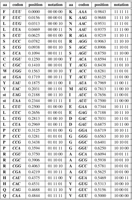
Binary notation of 64 codons and 20 amino acids is presented in Table 1. The information content of each codon sequence is “weighted” according to the classic method of a toss of a fair coin, i.e. by calculating the probabilitiespof each element on 0 1]intervalP 9, 10, 16-20].
The coin is tossedntimes to define the position of each binary address of length nover the al-phabet A= f0, 1g, as follows: p =
P jn=2
n,
jn = 0 for coin tossing outcome 0 and jn = 1
for the outcome 1. This binary algorithm is often applied in information theory for similar purposes 9, 10, 16, 20].
According to the Grantham’s scale of molec-ular polarity, we performed binary defining of amino acid physicochemical properties by par-titioning amino acids into nonpolar group 0(Y,
M, V, L, F, I, W, C)and polar group 1(H, R, Q,
K, N, E, D, P, A, T, S, G) 9, 21]. The groups
were extracted by means of partitioning around
medoids procedure of clustering, with S-Plus software 9, 22, 23].
This method enabled extraction of efficient bi-nary algorithm for the protein fold prediction 9], which confirms experimental results of
Kam-tekar et al. 6]. Binary algorithm of amino acid
polarity reduces the number of analyzed ele-ments within the protein motif(or sliding block)
of lengthnby the factor of 10n(from 20
nto 2n
)
9].
2.3. Amino Acid-Nucleotide Relationships
Groups that share information content with re-spect to both nucleotide and amino acid binary coding of physicochemical properties were ex-tracted by means of the classification tree 8, 9, 23]. Due to the fact that messenger RNA
consists of codons, and serves as a template for amino acid based protein synthesis on the ribosome, codons were treated as independent and related amino acids as dependent variables
(Table 1, Fig. 1).
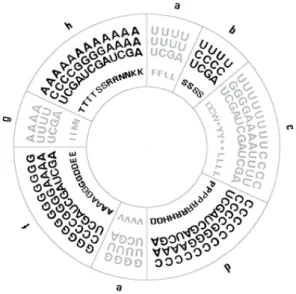
Classification tree extracted, with 100% accu-racy, 8 groups of codons that exhibit close statis-tical relationship based on the physicochemical coding of nucleotide properties and molecular polarity coding of amino acids(Table 1, Fig. 1).
The classification of 8 groups of codons in Fig. 1 is the result of the series of classification rules obtained by means of a procedure of recursive
Fig. 1. Coding patterns of 64 nucleotide triplets and 20 amino acids define codon ring identical to the genetic code table. Codon groups a, c, e, g represent nonpolar amino acids and codon groups b, d, f, h polar amino
aa codon position notation aa codon position notation F UUU 0.0000 00 00 00 K AAA 0.9843 11 11 11 F UUC 0.0156 00 00 01 K AAG 0.9688 11 11 10 L UUG 0.0313 00 00 10 N AAC 0.9531 11 11 01 L UUA 0.0469 00 00 11 N AAU 0.9375 11 11 00 S UCU 0.0625 00 01 00 R AGA 0.9219 11 10 11 S UCC 0.0782 00 01 01 R AGG 0.9063 11 10 10 S UCG 0.0938 00 01 10 S AGC 0.8906 11 10 01 S UCA 0.1094 00 01 11 S AGU 0.8750 11 10 00 C UGU 0.1250 00 10 00 T ACA 0.8594 11 01 11 C UGC 0.1410 00 10 01 T ACG 0.8438 11 01 10 W UGG 0.1563 00 10 10 T ACC 0.8281 11 01 01 st UGA 0.1719 00 10 11 T ACU 0.8125 11 01 00 Y UAU 0.1875 00 11 00 I AUA 0.7969 11 00 11 Y UAC 0.2031 00 11 01 M AUG 0.7813 11 00 10 st UAG 0.2188 00 11 10 I AUC 0.7656 11 00 01 st UAA 0.2344 00 11 11 I AUU 0.7500 11 00 00 L CUU 0.2500 01 00 00 E GAA 0.7344 10 11 11 L CUC 0.2656 01 00 01 E GAG 0.7188 10 11 10 L CUG 0.2813 01 00 10 D GAC 0.7031 10 11 01 L CUA 0.2969 01 00 11 D GAU 0.6875 10 11 00 P CCU 0.3125 01 01 00 G GGA 0.6719 10 10 11 P CCC 0.3281 01 01 01 G GGG 0.6563 10 10 10 P CCG 0.3438 01 01 10 G GGC 0.6401 10 10 01 P CCA 0.3594 01 01 11 G GGU 0.6250 10 10 00 R CGU 0.3750 01 10 00 A GCA 0.6094 10 01 11 R CGC 0.3906 01 10 01 A GCG 0.5938 10 01 10 R CGG 0.4063 01 10 10 A GCC 0.5781 10 01 01 R CGA 0.4219 01 10 11 A GCU 0.5625 10 01 00 H CAU 0.4375 01 11 00 V GUA 0.5469 10 00 11 H CAC 0.4531 01 11 01 V GUG 0.5313 10 00 10 Q CAG 0.4688 01 11 10 V GUC 0.5156 10 00 01 Q CAA 0.4844 01 11 11 V GUU 0.5000 10 00 00 aa=amino acids, st=stop codon
Table 1.Binary coding of nucleotide triplets(codons)and related amino acids.
partitioning 23, 24]. Since this exploratory
sta-tistical technique uncovers structure in data, it is not surprising that codon ring in Fig. 1, ob-tained by means of the algorithm, reconstructs standard genetic code arrangement presented in Table 1.
Classification rules for 8 groups of codons are defined by breakpoints between different po-lar and nonpopo-lar amino acid groups according to the codon positions on the unit interval, as shown in Table 1. Seven breakpoints that sep-arate 8 codon groups into the ones that code for nonpolar and polar amino acids are: 0.055, 0.117, 0.305, 0.492, 0.555, 0.742, and 0.805.
More details on the procedure can be found in ˇ
Stambuk and Konjevoda 9].
2.4. Protein Fold Prediction
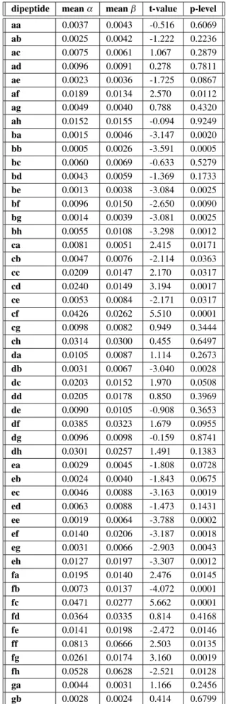
The prediction method is based on the analyses of relative frequencies of 64dipeptidepatterns, i.e. 88 groups, of coded nucleotide and amino
acid physicochemical properties.
The relative frequencies of 64 coding patterns were first analyzed (counted) within the
dipeptides 2, 8, 9]. Then, 64 relative frequency
patterns ofαandβ protein folds were compared by means of SMO machine learning algorithm and classification tree, in order to provide two different and accurate learning algorithms for the protein fold prediction 8, 9, 23-25].
Data were analyzed by means of two software packages. Classification tree was obtained by means of S-Plus 2000 software 8, 9, 22-24].
Weka (Waikato Environment for Knowledge
Analysis)software, version 3.1.7, was used for
the classifications with machine learning Se-quential Minimal Optimization (SMO)
algo-rithm for the Support Vector Machines 8, 9, 25].
Eight groups of codons, specified by means of the nucleotide and amino acid physicochemi-cal properties, differ significantly in α and β
protein folds(Table 2)and enable the
construc-tion of 64dipeptides. Based on 8 letter alphabet permutation, those 64 elementarydipeptide pat-ternscarry over the information on thesymbolic
coding characteristics of basic protein units that define peptide bonds within protein and exon sliding block 2, 8, 9].
The termdipeptideis given in italics since it is not a standard dipeptide consisting of 2 amino acid elements 8, 9].Dipeptiderepresents a
cod-ing pattern for 2 amino acids and their related nucleotide groups based on the physicochemi-cal characteristics defined by means of the cod-ing algorithm presented in Fig. 1 9]. Relative
frequencies of 28 out of 64 possibledipeptides
differ significantly in α and β protein folds, and exhibit distinct patterns relevant for the
pro-group meanα meanβ t-value p-level
a 0.0941 0.1133 -3.123 0.0022 b 0.0654 0.0684 -0.523 0.6019 c 0.1205 0.0945 3.603 0.0004 d 0.1022 0.0796 3.310 0.0012 e 0.1263 0.1526 -4.087 0.0001 f 0.1588 0.1574 0.177 0.8594 g 0.1324 0.1468 -1.956 0.0525 h 0.2002 0.1875 1.887 0.0612
p<0:05(Hotelling’s T=53.4,p<0:000001)
Table 2.Relative frequencies of amino acid and codon groups a-h inαandβ protein folds.
dipeptide meanα meanβ t-value p-level
aa 0.0037 0.0043 -0.516 0.6069 ab 0.0025 0.0042 -1.222 0.2236
ac 0.0075 0.0061 1.067 0.2879
ad 0.0096 0.0091 0.278 0.7811
ae 0.0023 0.0036 -1.725 0.0867
af 0.0189 0.0134 2.570 0.0112
ag 0.0049 0.0040 0.788 0.4320
ah 0.0152 0.0155 -0.094 0.9249 ba 0.0015 0.0046 -3.147 0.0020 bb 0.0005 0.0026 -3.591 0.0005 bc 0.0060 0.0069 -0.633 0.5279 bd 0.0043 0.0059 -1.369 0.1733 be 0.0013 0.0038 -3.084 0.0025 bf 0.0096 0.0150 -2.650 0.0090 bg 0.0014 0.0039 -3.081 0.0025 bh 0.0055 0.0108 -3.298 0.0012
ca 0.0081 0.0051 2.415 0.0171
cb 0.0047 0.0076 -2.114 0.0363
cc 0.0209 0.0147 2.170 0.0317
cd 0.0240 0.0149 3.194 0.0017
ce 0.0053 0.0084 -2.171 0.0317
cf 0.0426 0.0262 5.510 0.0001
cg 0.0098 0.0082 0.949 0.3444
ch 0.0314 0.0300 0.455 0.6497
da 0.0105 0.0087 1.114 0.2673
db 0.0031 0.0067 -3.040 0.0028
dc 0.0203 0.0152 1.970 0.0508
dd 0.0205 0.0178 0.850 0.3969
de 0.0090 0.0105 -0.908 0.3653
df 0.0385 0.0323 1.679 0.0955
dg 0.0096 0.0098 -0.159 0.8741
dh 0.0301 0.0257 1.491 0.1383
ea 0.0029 0.0045 -1.808 0.0728 eb 0.0024 0.0040 -1.843 0.0675 ec 0.0046 0.0088 -3.163 0.0019 ed 0.0063 0.0088 -1.473 0.1431 ee 0.0019 0.0064 -3.788 0.0002 ef 0.0140 0.0206 -3.187 0.0018 eg 0.0031 0.0066 -2.903 0.0043 eh 0.0127 0.0197 -3.307 0.0012
fa 0.0195 0.0140 2.476 0.0145
fb 0.0073 0.0137 -4.072 0.0001
fc 0.0471 0.0277 5.662 0.0001
fd 0.0364 0.0335 0.814 0.4168
fe 0.0141 0.0198 -2.472 0.0146
ff 0.0813 0.0666 2.503 0.0135
fg 0.0261 0.0174 3.160 0.0019
fh 0.0528 0.0628 -2.521 0.0128
ga 0.0044 0.0031 1.166 0.2456
gc 0.0098 0.0075 1.531 0.1280 gd 0.0118 0.0108 0.518 0.6050 ge 0.0047 0.0060 -0.741 0.4602 gf 0.0201 0.0176 1.067 0.2877 gg 0.0051 0.0047 0.390 0.6970 gh 0.0178 0.0208 -1.093 0.2762 ha 0.0138 0.0160 -1.079 0.2823 hb 0.0061 0.0109 -3.293 0.0013 hc 0.0318 0.0290 0.962 0.3376 hd 0.0286 0.0268 0.626 0.5320 he 0.0094 0.0205 -5.060 0.0001 hf 0.0591 0.0640 -1.095 0.2752 hg 0.0164 0.0173 -0.450 0.6532 hh 0.0432 0.0522 -1.894 0.0603
Table 3.Relative frequencies of dipeptides.
tein fold prediction(Hotelling’s T test = 253, p<0:0008; Table 3).
Classification of 140 dissimilar α and β pro-tein folds 8, 9]obtained by means of relative
frequencies of 64 dipeptides, with sequential minimal optimization(SMO)machine learning
algorithm 8, 9, 25], results in 91.43% overall
prediction accuracy and 83.57% correct
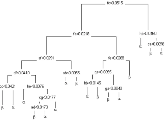
predic-tion under tenfold cross-validapredic-tion of the proce-dure. Classification tree confirms the result of SMO by 100% accurate classification ofα and
β protein folds(Fig. 2).
At the level of a single amino acid element, overall protein fold prediction from the primary sequence is 90%(83.57% under tenfold
cross-validation). However, at the level of dipeptide,
it is not possible to analyze 400 patterns arising from the 2020 dipeptide patterns, since it is
well known that direct inclusion of such number of input parameters requires data reduction or filtration 8, 26]. Consequently, presented
algo-rithm ofdipeptide-baseddata reductionavoids such problems and enables accurate, quick and simple protein fold analysis and/or prediction at
the level of basic protein units(dipeptides)that
define peptide bond between two neighboring amino acids.
The test set of 140 nucleotide sequences used for the computation 8, 9]is, to our knowledge,
the largest available mRNA dataset of 70 α
and 70 β dissimilar (nonhomologous) protein
folds with known three-dimensional structure, i.e., with NMR and those X-ray crystal struc-ture at the resolutions of< 2:5 Angstroms 8,
9, 27-29]. To ensure the best possible accuracy,
we analyzed the prediction results based on the tenfold cross-validation tests 25, 27].
Presented results may reflect the situation when physicochemical information of the codons be-comes a functional component of the secondary protein structure following the translation pro-cess of ribosome mRNA. During the translation process nucleotide information stored within DNA strings, and subsequently transcribed into RNA message, is converted into the polypeptide structure through a series of peptide bonds of the globular proteins 2]. Therefore, it is not
surpris-ing that SMO algorithm prediction ofα andβ
protein folds based on 8 symbolic elements a-h does not contain enough information(and
ele-ments)to accurately predict secondary protein
structure. Consequently, 8-element protein fold prediction based on the coding of monomers is significantly lower(70.17% overall and 68.57%
under tenfold cross-validation) than the fold
prediction based ondipeptides.
2.5. Alphabet Reduction and Fold Prediction
The main influence on the protein folding comes from the side chain properties 2]. Polarity
pa-rameter of the Grantham scale is a typical amino acid side chain property that is correlated to the relative substitution frequency between protein residues 21, 30]. In globular proteins,
polypep-tides of specific stable shapes are folded up due to the interactions of the residual side chains and the interactions with molecules of the medium
2, 31].
The model we described defines protein fold prediction from the nucleotide sequence. When the fold prediction from the amino acid se-quence is needed, the number of groups in Fig. 1 may be reduced until codon-amino acid bias is corrected. The sum of codon groups a, c, e and g defines the group of nonpolar amino acids(0), while the sum of codon groups b, d,
f and h defines a group of polar amino acids
(1). We also evaluated binary classification of
α and β protein folds by means of SMO ma-chine learning algorithm. The prediction results were obtained from the relative frequencies of the binary patterns of polar and nonpolar amino acids within protein sliding blocks of different lengths 32].
The best protein structure prediction result of 100%(tenfold cross-validation: 85%)was
ob-tained with 128 binary heptapeptide patterns 32]. Tree model for heptapeptide-based
classi-fication confirmed the results of SMO machine learning procedure and extracted 12 patterns of amino acid polarity relevant for 100% accurate
αandβ protein fold prediction 32]. This result
is in agreement withdipeptide-based tree clas-sification presented in Fig. 2. It shows that a total number of basic protein units relevant for the secondary structure description is well be-low the finite set of basic folding types recently estimated to be between 500 and 1000 9, 32, 33]. Similar results with respect to fold
predic-tion and procedure cross-validapredic-tions were also obtained for hexapeptides and octapeptides 9, 32].
2.6. Comparison of Prediction Methods
It is worth mentioning that alphabet reduction, e.g. from 8 letters algorithm of amino acid relationships into binary nucleotide-amino acid alphabet, reduces the information content of the sliding block during the protein fold prediction.
α andβ protein fold prediction with 64 dipep-tidesof the 8 letter alphabet is very close to the prediction of hexapeptide sliding block based on binary patterns of amino acid polarity, i.e. the 2 letter alphabet 9]. Both classification
procedures were done on the same dataset 9]
and with identical machine learning classifier. Described nucleotide-amino acid alphabet re-ductions, with respect to physicochemical pa-rameter coding, may be useful in situations when specific length of the protein or nucleotide sliding block is requested for the fold prediction and modeling purposes 9, 32].
2.7. Genetic Code Randomization Analysis
Presented model of nucleotide and amino acid coding of physicochemical parameters was also tested on α and β protein fold dataset 9] by
means of the permutation distribution of ran-domly produced models, i.e. codes 34].
Table 1 codons within 8 groups in Fig. 1. The eight groups a-h consisted of 4, 4, 12, 12, 4, 12, 4 and 12 elements, respectively. Those groups were, as previously discussed, extracted by means of the binary algorithm for physico-chemical coding of nucleotide-amino acid rela-tionships(Table 1, Fig. 1). Another 99,999
arti-ficial models were constructed by a random al-location(assignment)of codons within all
pos-sible 64 dipeptidesthat arise from the 8 Fig. 1 groups. In this way, the codes could be observed with respect to a protein fold prediction quality of: a) amino acid monomers, b) peptide bond
possessing units, i.e. dipeptides consisting of 2 amino acids.
The 99,999 different random permutations of 8 and 64 groups of nucleotide triplets(codons)
were chosen in accordance with the Algorithm P of Knuth 35-37]. The pseudorandom generator
required as input to the algorithm was that pro-vided by Delphi 5 of Borland Inter Inc 36-38].
The result of the models based on the binary patterns of nucleotide and amino acid coding of physicochemical properties(Fig. 1, Table 1)
was added to the results of 99,999 randomly pro-duced codes to obtain a total of 100,000 codes of both groups.
Prediction quality of the codes belonging to both distributions was determined with SMO classi-fier under tenfold cross-validation, from relative frequency patterns 8, 9, 25].
Distribution of the results in Fig. 3 reflects the permutation distribution of random allocation of 64 codons within 8 groups presented in Fig. 1. Eight group(letter)-based protein fold
predic-tion of 68.57% is identical to the random dis-tribution median (68.57%) and almost
identi-cal to the random distribution arithmetic mean
(68.14%).
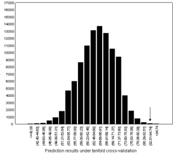
Completely different result is obtained for a random distribution of 64dipeptidesin Fig. 4. This distribution has a mean at 64.95% (
me-dian 65%)with a standard deviation of 6.19%.
Out of 100,000 codes tested for the secondary protein structure prediction, only 27 randomly generated codes gave better structure predic-tion than the binary one based on 64 dipep-tides (83.57% prediction under tenfold
cross-validation, Table 3). The binary code
posi-tion within a cumulative distribuposi-tion(99.973%,
Z=3.008)implies that, with respect to the
sec-ondary protein structure, natural genetic code
defining of codon and amino acid physicochem-ical properties is far away from the random orga-nization. Whendipeptides are observed, there is only 2:710
;4chance to produce better code
than the natural one.
Fig. 3.Protein fold prediction quality of the natural code and 99,999 randomly produced codes. The procedure is
based on 8 codon-amino acid groups(a-h). Arrow
indicates the position of the natural code.
Fig. 4.Protein fold prediction quality of the natural code and 99,999 randomly produced codes. The procedure is based on 64dipeptides, obtained by the permutation of 8 codon-amino acid groups(a-h). Arrow indicates the
The result of our computation experiment is close to the prediction of Freeland et al. 13]that
a code as good as, or better than that chosen by nature, would evolve without selection within the probability interval 210
;4
< p< 10 ;6.
Consequently, dipeptide-based organization of the nucleotide patterns could be essential ele-ment of the secondary protein structure, buried within the genetic code.
3. Conclusion
Binary algorithms presented in this study enable simple, quick and accurate prediction of the sec-ondary protein structure from the primary RNA, DNA and amino acid sequences.
Algorithmic information theory, and related cryp-tographic methods will provide useful tools for further analysis and modeling of the protein structure and genetic code patterns.
References
1] CRICKF. H. C., The Origin of the Genetic Code, Journal of Molecular Biology1968; 38:367–379.
2] DOOLITTLE R. F., Proteins, Scientific American
1985; 253(4):74–83.
3] KNIGHTR. D., FREELANDS. J., LANDWEBERL. F.,
Selection, history and chemistry: the three faces of the genetic code, Trends in Biochemical Sciences
1999; 24:241–247.
4] SWANSONR., A Unifying Concept for the Amino
Acid Code,Bulletin of Mathematical Biology1984; 46(2):187–203.
5] SˇTAMBUKN., On the Genetic Origin of
Comple-mentary Protein Coding, Croatica Chemica Acta
1998; 71(3):573–589.
6] KAMTEKARS., SCHIFFER J. M., XIONGH., BABIK
J. M., HECHT M. H., Protein Design by Binary
Patterning of Polar and Nonpolar Amino Acids,
Science1993; 262:1680–1685.
7] LIH., HELLINGR., TANGC., WINGREENN.,
Emer-gence of Preferred Structures in a Simple Model of Protein Folding,Science1996; 273:666–669.
8] SˇTAMBUKN., KONJEVODAP., New Computational
Algorithm for the Prediction of Protein Folding Types,International Journal of Quantum Chemis-try2001; 84(1):13–22.
9] SˇTAMBUKN., KONJEVODAP., Prediction of
Sec-ondary Protein Structure with Binary Coding Pat-terns of Amino Acid and Nucleotide Physicochem-ical Properties, International Journal of Quantum Chemistry2003; 92(2):123–134.
10] SˇTAMBUKN., Universal Metric Properties of the
Genetic Code, Croatica Chemica Acta 2000; 73(4):1123–1139.
11] JIMENEZ-MONTANOM. A.,DE LAMORA-BASANEZ´
C. R., P¨OSCHELT., The Hypercube Structure of
the Genetic Code Explains Conservative and Non-conservative Aminoacid Substitutions In Vivo and In Vitro,BioSystems1996; 39:117–125.
12] BERGERONB.,Bioinformatics Computing, New
Jer-sey: Prentice Hall, 2003.
13] FREELAND S. J., KNIGHT R. D., LANDWEBER L.
F., Measuring adaptation within the genetic code,
Trends in Biochemical Sciences2000; 25:44–45.
14] WOESEC. R., Evolution of the Genetic Code, Natur-wissenschaften1973; 60:447–459.
15] PULLMAN B., Some Recent Developments in the
Quantum-Mechanical Studies on the Electronic Structure of the Nucleic Acids,Journal of Chemical Physics1965; 43(10):S233–S243.
16] CALUDE C., Information and Randomness, New
York: Springer; 1994.
17] CHAITING. J., A Theory of Program Size Formally
Identical to Information Theory,Journal of the ACM
1975; 22:329–340.
18] CHAITING. J., Information-Theoretic
Incomplete-ness,Applied Mathematics and Computation1992; 52:83–101.
19] CHAITIN G. J., On the Length of Programs for
Computing Finite Binary Sequences, Journal of the Association for Computing Machinery 1966; 13(4):547–569.
20] CHAITING. J.,The Unknowable, Singapore:
Sprin-ger; 1999.
21] GRANTHAMR., Amino Acid Difference Formula
to Help Explain Protein Evolution, Science1974; 185:862–864.
22] Data Analysis Products Division, MathSoft, S-PLUS 2000 Guide to Statistics, vol 2. Seattle: MathSoft; 1999.
23] VENABLESV. N., RIPLEYB. D., Modern Applied Statistics With S-PLUS, New York: Springer; 1997.
24] EVERITTB. S., DUNNG.,Applied Multivariate Data Analysis, London: Arnold; 2001.
25] WITTENI. H., FRANKE.,Data Mining, San
Fran-cisco: Morgan Kaufmann; 2000.
26] EDLERL., GRASSMANNJ., SUHAIS., Role and
Re-sults of Statistical Methods in Protein Fold Class Prediction,Mathematical and Computer Modeling
27] CUFFJ A., BARTONG. J., Evaluation and
Improve-ment of Multiple Sequence Methods for Protein Secondary Structure Prediction,PROTEINS: Struc-ture, Function and Genetics1999; 34:508–519.
28] CUFFJ. A., CLAMPM., SIDDIQUIA. S., FINLAYM.,
BARTON G. J., JPred: A Consensus Secondary Structure Prediction Server, Bioinformatics 1998; 14:892–893.
29] CHOU K. C., MAGGIORA G. M., Domain
Struc-tural Class Prediction, Protein Engineering 1998; 11(7):523–538.
30] MCLACHLANA. D., Repeating Sequences and Gene
Duplication in Proteins,Journal of Molecular Bio-logy1972; 64:417–437.
31] ROSEG. D., GESELOWITZA. R., LESSERG. J., LEE
R. H., ZEHFUS M. H., Hydrophobicity of Amino
Acid Residues in Globular Proteins,Science1985; 229:834–838.
32] SˇTAMBUK N., KONJEVODA P., GOTOVAC N.,
Nu-cleotide Coding of Amino Acid Polarity and Pro-tein Structure,Acta Physica et Chimica Debrecina
2002; 34–35:171–188.
33] DENTON M., MARSHALL C., Laws of the Form
Revisited,Nature2001; 410:417.
34] RAMSEY F. L., SCHAFER D. W., The Statistical Sleuth. A Course in Methods of Data Analysis, Belmont CA: Duxbury Press; 1997.
35] KNUTHD. E.,The Art of Computer Programming,
vol 2. Reading: Addison-Wesley; 1998.
36] WITZTUMD., RIPS E., ROSENBERGY., Equidistant
Letter Sequences in the Book of Genesis,Statistical Science1994; 9(3):429–438.
37] DROSNINM.,The Bible CodeLondon: Orion; 2001.
38] LISCHNER R., Delphi in a Nutshell – A Desktop Quick Reference, Sebastopol CA: O’Reilly & Asso-ciates; 2000.
Received:June, 2004
Accepted:June, 2004
Contact address:
NikolaStambukˇ Paˇsko Konjevoda Ruder Boˇskovi´c Institute Bijenicka cesta 54ˇ
HR-10002 Zagreb Croatia e-mail:[email protected] [email protected]
Nikola Gotovac Department of Radiology General Hospital Pozegaˇ
Osjecka bbˇ
HR-34000 Pozegaˇ
Croatia e-mail:[email protected]
NIKOLASˇTAMBUKwas born in 1959, in Varazdin, Croatia. He receivedˇ
his MD from the Zagreb University School of Medicine in 1984 and completed his internship at Sisters of Mercy Clinical Hospital in Zagreb
(1985–87). He spent the period between 1985 and 1987 as resident at the Railway Health Center in Zagreb and got his MS degree from the Zagreb University School of Medicine in 1987. In 1991 he completed doctoral studies and received his PhD from the Institute for Medical Re-search and Occupational Health, University of Zagreb, where he stayed as a research associate for another year. From 1994 to 2003 he worked at Ruder Boˇskovi´c Institute as senior scientific associate. NikolaStam-ˇ buk has published 59 scientific and professional papers. His scientific interests include immunology and immunochemistry, mathematical and artificial intelligence-based modeling in biomedicine.
PASKOˇ KONJEVODAwas born in 1965, inSibenik, Croatia. He receivedˇ his MD from the Zagreb University School of Medicine in 1993 and his MS degree in pharmacology and toxicology from the same faculty in 2003. Since 2000 he has been a statistical editor of the Croatian Medical Journal. Paˇsko Konjevoda has published 34 scientific and professional papers. His scientific interests are focused on computer modeling of the protein structure and function, and on statistical analysis of medical diagnostic and prognostic tests.
NIKOLAGOTOVACwas born in 1973, in Zagreb, Croatia. He got his MD from the Zagreb University School of Medicine in 1999 and com-pleted his internship at General Hospital Pozega between 1999 andˇ
2000. Since 2000 he has been a resident at Radiology Department, General Hospital Pozega. Nikola Gotovac has published 6 scientificˇ