Dr J.E. Evangalin et al JMSCR Volume 06 Issue 09 September 2018 Page 448
Original Research Article
Histomorphological Spectrum of Prostatic Lesions and Its Correlation with
Prostate Specific Antigen Level
Authors
Dr J.E. Evangalin
1, Dr P. Aruthra
2*, Dr V. Eswari
31
Final year Post Graduate, 2Assistant Professor, 3Head of the Department of Pathology
Meenakshi Medical College, Hospital and Research Institute, Enathur, Kanchipuram, Tamilnadu, India *Corresponding author
Dr P. Aruthra
Email: [email protected]
Abstract
Background: Prostatic lesions are important health problem among Indian men. Currently Prostate specific antigen (PSA) test is used widely in the diagnosis of prostatic lesions
Aim: To evaluate the prevalence and distribution of prostatic lesions in kanchipuram and the diagnostic use
of PSA levels in screening malignant lesions of prostate.
Material & Methods:Prospective study of 60 cases including both benign and malignant lesions of prostate are taken as study sample, received in the department of pathology at Meenakshi medical college, Kanchipuram from January to December 2017. Sections are H&E stained and studied under microscope.
PSA values are collected for all the 60 cases.
Results: Out of 60 cases, 20 are benign prostatic Hyperplasia (BPH), 25 are BPH with prostatitis, 7 are prostatic intra-epithelial neoplasia and 8 are prostatic adenocarcinoma. PSA values are classified as : 0-4 ng/dl as normal , 4.1-10 ng/dl mild, 10.1- 20 ng/dl moderate and >20 ng/dl severe .There is mild PSA value elevation in 40% cases, moderate elevation in 8.9% cases and severe elevation in 2.2% cases of non-neoplastic prostatic lesions. Similarly there is mild elevation of PSA value in 6.7% cases, moderate elevation in 6.7% cases and severe elevation in 60% cases in neoplastic lesions of prostate. Elevated PSA value is significantly associated with the malignant lesions with p value < 0.0001.
Conclusion: Prostatic lesions are more common seen in the elderly. BPH are more common than prostatic
carcinoma. Prostate specific antigen level is raised in both the neoplastic and non-neoplastic lesions of prostate. But the values are significantly raised in the neoplastic lesions prostatic carcinoma. Hence the estimation of PSA level in serum can be used as a diagnostic tool for screening malignant lesions of prostate.
Keywords:Prostatic lesions, PSA, screening tool.
Introduction
Prostatic carcinoma is an important growing health problem in India and is at present the second leading site cancer among males in the urban Indian population(1). It is also the most common malignant
tumour in men over the age of 65 years. The prevalence of prostate cancer rises with age, and can reach up to 60% in men at the of age of 80.(2). Currently prostate specific antigen (PSA) levels are being used widely for the screening and the
www.jmscr.igmpublication.org Impact Factor (SJIF): 6.379
Dr J.E. Evangalin et al JMSCR Volume 06 Issue 09 September 2018 Page 449 diagnosis of carcinoma prostrate. Its level may be
elevated in patients with prostate cancer due to the leakage of PSA into the blood which are widely produced by the prostatic cancer cells and they precede clinical disease by 10 years or longer(3). PSA has been
Used extensively over the past five decades as a marker in the diagnosis of prostate cancer along with Digital rectal examination (DRE). DRE and PSA has been recommended test in guidelines of the American cancer society since 1993 for annual check-up of men aged 50 years or above and has resulted in early diagnosis and treatment(4).
PSA is normally formed as a proenzyme, secreted primarily by the secretory acinar cells that line the prostate and are secreted into the lumen(5). Elevated PSA is also found in pathological conditions of the prostate such as hyperplasia, inflammation and tumours where the destruction of cell membrane leads to the release of PSA into the circulation(6). PSA has also been noted to be detected in trace amounts in the peri-urethral glands, adrenal neoplasm, renal cell carcinoma, endometrium, normal breast tissue, breast tumour, breast milk(6). Since PSA is normally found in low concentration in serum, elevated PSA in serum makes it an important marker of cancer(7). Investigations reveal that every gram of cancer prostate tissue increases the value of serum PSA for 2.3 ng/ml in average, while every gram of hyperplasic tissue increases the same parameter 10 times less compared to cancer tissue(8). The upper limit of normal for PSA values is generally considered to be 4.0ng/mL, between 4 and 20ng/mL is considered borderline and more than 20ng/mL is considered high. Patients with a PSA value greater than 4 ng/mL, regardless of DRE results, generally undergo biopsy. The cut-off value of 4.0ng/mL represents the level at which the highest sensitivity and highest specificity are present. At present there is no value of PSA for the definitive diagnosis of prostatic malignancy and a positive finding on DRE is also not always 100% specific, hence biopsy of the prostate is still required for the diagnosis of prostate malignancy(9).
Materials and Methods
This is a prospective study done in 60 prostatic specimens including Trans-Urethral resection of prostate (TURP) and prostatic biopsies received in Meenakshi medical college hospital and research institute, Enathur, Kanchipuram from December 2016 to 2017 done on patients who had the clinical symptoms of prostatism on digital rectal examination (DRE). Specimens were fixed in 10% neutral buffered formalin, sections taken at 4 micron thickness, stained in Haematoxylin & Eosin and studied under microscope. Blood samples were collected and serum prostate specific antigen levels were evaluated. The standard protocol diagnostic methods applied for prostatic lesions were digital rectal examination (DRE), Trans abdominal ultrasonography of prostate, determination of serum PSA and biopsy of prostate. The test used for statistical analysis is the calculation of Mean, standard deviation and chi square test by using graph iPad software.
Observations and Results
Dr J.E. Evangalin et al JMSCR Volume 06 Issue 09 September 2018 Page 450
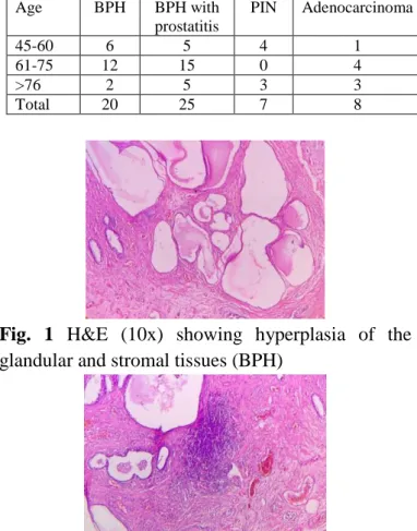
Table 1 showing age-wise distribution of prostatic lesions
Age BPH BPH with
prostatitis
PIN Adenocarcinoma
45-60 6 5 4 1
61-75 12 15 0 4
>76 2 5 3 3
Total 20 25 7 8
Fig. 1 H&E (10x) showing hyperplasia of the glandular and stromal tissues (BPH)
Fig. 2 H&E (10x) showing features of BPH along with infiltration by lymphocytes and plasma cells in the stroma (BPH with prostatitis
Fig.3 H&E (10x) showing prostatic glands with features of crowding , nuclear stratification and pleomorphism (PIN)
Fig.4 H&E (40x) - showing fused glands without any intervening stroma, gleason scoring of 8 (4+4) (prostatic adenocarcinoma)
Table 2 shows the most common clinical symptom encountered in prostatic lesions is increased frequency of micturition. Second most common symptom being acute retention of urine, and the other symptoms observed are painful micturition and Hematuria.
Table 2- showing various clinical symptoms seen in prostatic lesions
Symptoms No.
of cases
BPH BPH
with prosta -titis
PIN Adenocar
cinoma
Increased frequency of urination
34 17 10 3 4
AUR 20 1 14 2 3
Painful micturition
3 0 1 2 0
Haematuria 3 2 0 0 1
Total 60 20 25 7 8
PSA values were normal in 10 cases of BPH, 12 cases of BPH with prostatitis, 3 cases of PIN (2 low grade & 1 High grade) and 1 case of prostatic adenocarcinoma also were noted to have normal PSA levels. Mild elevation of PSA was noted in 7 cases of BPH, 11 cases of BPH with prostatitis and in 1 case of PIN( High grade). Marked elevation in of PSA was noted in 1 case of BPH, 3 cases of PIN (High grade)and 6 cases of prostatic adenocarcinoma. Mean PSA value in BPH was 4.47+/- 4.21, in BPH with prostatitis is 4.79 +/- 4.22, in PIN 19.95 +/- 28.83, and in prostatic adenocarcinoma 109.35+/-142.05was noted as shown in Table 3
Table 3- showing PSA levels with various prostatic lesions
PSA Level(ng /dl)
BPH BPH with
prostatitis
PIN Adenoc
arcinom a
0-4 10 12 3 1
4.1-10 7 11 1 0
10.1-20 2 2 0 1
>20 1 0 3 6
Mean PSA Value
4.47 +/- 4.21
4.79+/- 4.22
19.95 +/- 28.83
Dr J.E. Evangalin et al JMSCR Volume 06 Issue 09 September 2018 Page 451
Discussion
Carcinoma of prostate is the most common cancer in India among males due to increasing life expectancy and relatively better diagnostic methods. The gold standard triad for diagnosing prostate cancer comprised of Digital rectal examination (DRE), Prostate specific antigen ( PSA) level and Transrectal ultrasonography(10). The Digital rectal examination (DRE) has always been the primary method for evaluating the prostate.. Digital rectal examination (DRE) is neither specific nor sensitive enough to detect prostate cancer(11). To improve the detection rate of the prostate cancer, the Digital rectal examination (DRE) should be followed by a diagnostic test with high sensitivity. Prostate specific antigen (PSA) testing provides such a method, being very sensitive. The frequency of the diagnosis of prostatic carcinoma has beensubstantially increased after the introduction of Prostate specific antigen (PSA) screening test. In our study the incidence of BPH is 80% which is close with studies done by Wadgaonkar et al(12) (83.7%). The incidence of prostatic adenocarcinoma is 13.3% which is also close with studies by Wadgaonkar et al(12) (13.75%) and PIN is 11.7% which is close with Jasani et al(13) study (7.2%). The common presenting symptom is increased frequency of micturition followed by acute retention of urine. Prostatic lesions are more common in the elderly age group . Mean age is 67+/-8.69 in BPH, 68.84+/-11.56 in BPH with prostatitis, 68.28+/-10.98 in PIN and 75.25+/-10.60 in prostatic adenocarcinoma Table 4 .
Table 4 Histopathological diagnosis in various studies
Histopathological Diagnosis
Wadgaonkar et al(12).
Jasani et al(13)
Present stud-y
BPH +/-
prostatitis
83.75% 56% 80%
Prostatic Adenocarcinoma
13.75% 32% 13.3%
PIN 1.25% 7.22% 11.7%
Transitional cell carcinoma
1.25% 11%
In our study, Benign prostate hyperplasia is seen in maximum number 45 (75%) of cases and in that
Dr J.E. Evangalin et al JMSCR Volume 06 Issue 09 September 2018 Page 452
Table 5- PSA value in non-neoplastic prostatic lesions
PSA value
Jasani et al(13)
Alpesh puri p.et all(14)
Sanjaykum ar c. Chauhan et
al(15).
Present study
Mild 63.7% 23.9% 21.6% 40%
Moderate 27.4% 69% 11.3% 8.9%
Severe 8.8% 7% 7.2% 2.2%
Normal 59.8% 48.9%
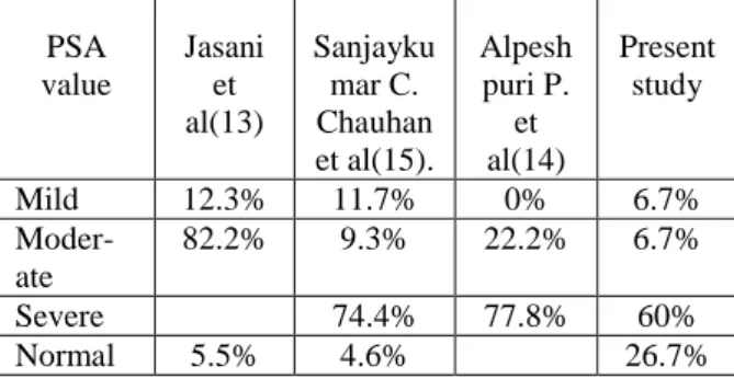
Table 6- PSA value in neoplastic prostatic lesions
PSA value
Jasani et al(13)
Sanjayku mar C. Chauhan et al(15).
Alpesh puri P.
et al(14)
Present study
Mild 12.3% 11.7% 0% 6.7%
Moder-ate
82.2% 9.3% 22.2% 6.7%
Severe 74.4% 77.8% 60%
Normal 5.5% 4.6% 26.7%
Conclusion
Prostatic lesions are more common in the elderly age group, BPH more common than carcinoma of prostate. Prostate specific antigen level is raised in both the neoplastic and non-neoplastic lesions of prostate. But the values are significantly raised in neoplastic lesions (prostatic carcinoma). Hence the estimation of PSA level in serum can be used as a diagnostic tool for screening malignant lesions of prostate.
Acknowledgements: Nil
Conflict of Interest: Nil
References
1. Jain S, Saxena S, Kumar A. Epidemiology of prostate cancer in India. Meta gene [Internet]. 2014 Dec [cited2018 May 5]; 2:
596–605. Available from
http://www.ncbi.nlm.nih.gov/pubmed/25606 442
2. Prostate Cancer - Cancer Stat Facts [Inter-net]. [cited 2018 May 5]. Available from: https://seer.cancer.gov/statfacts/html/prost.ht ml
3. Akdas A, Tarcan T, Türkeri L, Cevik I, Biren T, Gürmen N. The diagnostic accuracy of digital rectal examination, transrectal ultrasonography, prostate-specific antigen (PSA) and PSA density in prostate carcinoma. Br J Urol [Internet]. 1995 Jul [cited 2018 May 5];76(1):54–6. Available from:
http://www.ncbi.nlm.nih.gov/pubmed/75442 05
4. De La Rosette J, Perachino M, Thomas D, Madersbacher S, Desgrandchamps F, Alivizatos G, et al. Guidelines on benign prostatic hyperplasia. [cited 2018 May 6]; Available from: https://uroweb.org/wp- content/uploads/EAU-Guidelines-Bening-Prostatic-Hyperplasia-2001.pdf
5. Hoffman A, Half EE. Update on Screening for Urological Malignancies. Rambam Maimonides Med J [Internet]. 2017 [cited 2018 May 5];8(4). Available from: http://www.ncbi.nlm.nih.gov/pubmed/29059 045
6. Banerjee B, Iqbal B, Kumar H, Kambale T, Bavikar R. Correlation between prostate specific antigen levels and various prostatic pathologies. J Med Soc [Internet]. 2016 [cited 2018 May 6];30(3):172. Available from: http://www.jmedsoc.org/text.asp 2016/30/3/172/191184
7. Atan A, Guzel O. How should prostate specific antigen be interpreted Turkish J Urol [Internet]. 2013 Sep [cited 2018 May 6];39(3):188–93. Available from: http://www.ncbi.nlm.nih.gov/pubmed/26328 106
8. Zivkoviae S. Correlation between prostate-specific antigen and histopathological difference of prostate carcinoma. Arch Oncol [Internet]. 2004 [cited 2018 May 6];12(3):148–51. Available from: http://www.doiserbia.nb.rs/img/doi/0354-7310/2004/0354-73100403148Z.pdf
Dr J.E. Evangalin et al JMSCR Volume 06 Issue 09 September 2018 Page 453 antigen testing intervals for the detection of
curable prostate cancer. JAMA [Internet]. 1997 May 14 [cited 2018 May 6];277 (18):1456–60. Available from: http://www.ncbi.nlm.nih.gov/pubmed/91457 18
10.Franco OE, Arima K, Yanagawa M, Kawamura J. The usefulness of power Doppler ultrasonography for diagnosing prostate cancer: Histological correlation of each biopsy site. BJU Int. 2000;85(9):1049– 52.
11.Unal D, Sedelaar JP, Aarnink RG, van Leenders GJ, Wijkstra H, Debruyne FM, et al. Three-dimensional contrast-enhanced power Doppler ultrasonography and conventional examination methods: the value of diagnostic predictors of prostate cancer. BJU Int. 2000;86:58–64.
12.Correlation of serum prostate specific antigen ( PSA ). 2013;3(2):274–81.
13.Utility D, Prostate OF, Antigen S, Of D, Lesions P. International Journal of Biomed-ical and Advance Research 268. :2–6. 14.Goswami AP, Rupala G, Goswami NN.
Serum PSA level in Prostatic lesions with Histopathological correlation in Gujarat and histologic findings in biopsy specimens of men with prostatic disease . Material and methods : This. :33–8.