Original Research Article
Red cell distribution width as a prognostic marker in mechanically
ventilated children admitted in pediatric critical care unit of tertiary
care centre, India
Swati M. Gadappa*, Manas Kumar Behera
INTRODUCTION
The practice of paediatric critical care is dynamic and evolving. Paediatric population is a vulnerable group necessitating standard care for medically and surgically ill children. Red cell distribution width (RDW) is a simple and low-cost measure that denotes the variability in red blood cell size.1 It is widely available measure and
is routinely reported as part of a complete blood count
(CBC). Any process that releases reticulocytes in the circulation will result in an increase in RDW.2 Recently
many studies suggest that RDW may also be useful as a biomarker of disease severity and clinical outcomes in critically ill patients. An increased RDW is an independent predictor of all-cause mortality in sepsis, congestive heart failure, and adult critical illness, and has been shown to improve acute physiology scoring for risk prediction in critically ill adults.3 The resulting acute rise
ABSTRACT
Background: Paediatric population is a vulnerable group necessitating standard care for medically and surgically ill children. Red cell distribution width (RDW) is a simple and low-cost measure that denotes the variability in red blood cell size. Any process that releases reticulocytes in the circulation will result in an increase in RDW. RDW may also be useful as a biomarker of disease severity and clinical outcomes in critically ill patients.
Methods: Retrospective cohort study of all patients between 1month-12yrs of age, mechanically ventilated in Paediatric intensive care unit. Those patients with RDW on admission and complete data for PIM3 (Paediatric Index of Mortality 3) were included. Analyses included correlation, logistic regression analysis, and receiver operating characteristic (ROC) curves.
Results: Retrospective analysis of data on 93 consecutive critically ill children admitted in PICU was done between Jan 2015- June 2016. We noted statistically significant correlation between mortality and anemia (10.24 g/dL, SD 2.26; 8.78 g/dL, SD 2.60.p = 0.009), LOS on MV (p = 0.008), RDW (p = 0.002), shock (p = 0.004) and ventilator associated Pneumonia (p = 0.024). Mortality increased as length of stay on mechanical ventilation increased (4.13 days, SD 2.125 versus 6.94 days, SD 7.603 p = 0.008). The cut-off of 18.10 was chosen as Mean RDW. Based on AUROC, RDW is independently associated with high risk of mortality.
Conclusions: RDW measured within 24 hours of PICU admission was independently associated with length of stay on mechanical ventilation and mortality in a general PICU population. We recommend the need for multicentric, prospective longitudinal studies to determine the optimum utility of RDW to enhance decision making in PICU.
Keywords: Critical care outcome, Mechanical ventilation, RDW
Department of Pediatrics, Smt. Kashibai Navale Medical College and General Hospital, Pune, Maharashtra, India
Received:13 August2018 Accepted:18 August 2018
*Correspondence: Dr. Swati M. Gadappa
E-mail: [email protected]
Copyright: © the author(s), publisher and licensee Medip Academy. This is an open-access article distributed under the terms of the Creative Commons Attribution Non-Commercial License, which permits unrestricted non-commercial use, distribution, and reproduction in any medium, provided the original work is properly cited.
in RDW may therefore reflect the degree of the underlying inflammatory state and provide useful prognostic information about intensity of resource utilization and risk of mortality.4-7 Some recent studies
have found associations of an increased RDW with mortality, irrespective of mean cellular volume (MCV) and hemoglobin levels. Associations were reported for patients with heart failure, acute myocardial infarction, community-acquired pneumonia, pulmonary hypertension and in the general population.8-16
Data on the utility of RDW as a biomarker of clinical outcomes in the paediatric population are more limited. In critically ill children admitted to ICU, RDW is associated with risk of death and is suggested as an independent prognostic marker.3,17-19 There are very few
studies examining RDW as a biomarker in a general paediatric intensive care unit (PICU) population, esp. critical children requiring mechanical ventilation.20 The
characterization of such a readily available biomarker may provide a simple, pragmatic tool to stratify patients by severity of illness and identify those at risk for increased resource utilization and poor outcomes to facilitate early interventions and triage decisions without additional costs or the need for a complex assay. We therefore studied the association of RDW at PICU admission with length of stay (LOS) and mortality in mechanically ventilated children to determine its potential application as a simple and significant biomarker for prognostication in the critically ill paediatric population.
METHODS
Study design
Retrospective analysis was carried out in all patients, age 1month to 12years, who were consecutively admitted to the 8-bedded PICU between January 2015 to June 2016 in Tertiary care Hospital and Medical College in a Semi-urban region of Pune city, Maharashtra.
We serve low to middle income population as an economical and tertiary referral unit for pediatric medical and surgical cases, however this excludes pediatric patients who are post-cardiac surgery or those who need extracorporeal membrane oxygenation.
Present study was a retrospective analytic-descriptive study, carried out in Pediatric Intensive Care Unit of a tertiary care hospital.
Inclusion criteria
All critically ill children, 1mth to 12yrs age, admitted in Pediatric Intensive Care Unit and requiring mechanical ventilation, were included in the study. Deaths occurring in the Operation theatre, if any, were included only if the operation occurred during the PICU stay and was a therapy for the illness requiring PICU care.
Exclusion criteria
Patients who had PICU stay of less than 24 hrs, those who were admitted in CPR and remained unstable for >2 hrs, preterm infants, those who received blood transfusion prior to admission in PICU and those with death within 24 hrs of PICU admission were excluded. Patients requiring temporary (<24 hrs) postoperative mechanical ventilation for respiratory support were not included in the study. Those patients with incomplete data for PIM-3 score were excluded. This study was approved by the Institutional Ethics Committee and a waiver of consent was granted to perform this retrospective chart review of existing data.
Patient data, outcomes and covariates
Patient file records were analysed through which baseline characteristics, demographical data, laboratory and clinical data, possible risk factors and outcome were studied. We calculated Pediatric Index of Mortality-III scores as marker for disease severity for each patient using the available data. Detailed complete blood count report which routinely includes RDW (done on Sysmax machine) was noted. The normal range in our laboratory was 11-16%.
PIM3 will be calculated automatically through data entered in Anzics CORE - Severity Score a Calculation of PIM3 (and PIM3 risk of death%)21
PIM3val = (3.8233 * Pupils) – (0.5378 * Elective) + (0.9763 * MechVent) + (0.0671 * (absolute Base Excess)) – (0.0431*SBP) + (0.1716*(SBP*SBP/1000)) +
(0.4214 (100*FiO2/PaO2)) –
(1.2246*Recov_CardBypPr) –
(0.8762*Recov_CardNonBypPr) –
(1.5164*Recov_NonCardPr) + (1.6225* VHRdiag) + (1.0725*HRdiag) – (2.1766*LRdiag) – 1.7928
PIM3 risk of death = ePIM3val / (1 + ePIM3val ). Categorisation of diagnosis will be done based on PIM3 guidelines.
We defined invasive ventilation as being ventilated mechanically through an endotracheal tube or a tracheostomy. When a patient was re-intubated within 24 hours after initial extubation, both episodes were considered as one (same case). Length of stay (LOS) in PICU is a commonly used clinical endpoint in lung disease studies. However, as many factors have an impact on the LOS in PICU that may be easily confounded in a retrospective analysis, hence LOS on mechanical ventilation was analysed as a secondary parameter.18 In
the subgroup analysis of ventilated patients, we evaluated the association between RDW and PIM3 score, length of stay on ventilation and mortality.
standard definitions using consensus guidelines, International Classification of Diseases, 9th Revision, Clinical Modification (ICD-9) codes.22,23 Patients
presenting with “shock” included all those with septic shock, cardiac arrest, hypovolemic shock, cardiogenic shock, anaphylactic shock, and shock not otherwise specified who had documentation of hemodynamic compromise or organ dysfunction requiring fluid resuscitation or vasopressor support. Any patient with a new or pre-existing hematologic or oncologic diagnosis was included in the “hematology/oncology” subgroup. Anemia was defined as Hemoglobin of less than 10g/dL. Acute kidney injury was diagnosed and staged as per RIFLE criteria. Hepatic Dysfunction was defined as a combination of total bilirubin of >2mg/dL and either an alkaline phosphatise or serum aminotransferase of greater than twice normal.24 Ventilator associated pneumonia
was defined as per Johanson’s criteria (Presence of new/persistent infiltrates on Chest radiograph and any 2 of: Febrile >38.3C; Leucocytosis or leucopenia; Purulent tracheobronchial secretions).
Lab analysis
RDW was measured as part of the routine haematological tests according to the formula:
RDW = (Coefficient of Variability of RBC ÷ mean MCV) × 100.
Periodic quality checks are routinely performed as part of the clinical laboratory accreditation requirements.
Statistical methods
To test the association between RDW and mortality in critically ill children, the RDW values were split into quartiles. The RDW values for three quartiles [25th, 50th
and 75th centile] are 16.6, 19.6 and 22.7. These cut-offs
are taken for dividing the total frequency in to 4 equal parts. Multivariable logistic regression was used to test the association of RDW on the first day of pediatric intensive care unit (PICU) admission with prolonged PICU length of stay (LOS) >72 hours and mortality.
The variables used included age, haemoglobin, WBC count, LOS on mechanical ventilation (LOS MV), time of MV, RDW, PIM3 score.
A paired Student’s ‘t’ test was used for comparisons. For all tests a p value less than 0.05 was considered statistically significant. All analyses were performed with SPSS v24 for Mac. The area under the receiver operating characteristic curve (AUROC) was utilised to predict sensitivity and specificity of RDW with and without anemia; as an independent predictor of mortality.
RESULTS
We had about 950 admissions in the 8 bedded PICU during January 2015 to June 2016, of which 119 were mechanically ventilated. Based on the inclusion criteria and availability of PIM3 parameters for scoring, 93 patients were included in the study. Overall PICU mortality was 10.1%. Patients included 52 males (55.9%) and 41 females (44.1%).
Age wise distribution (min 0.16 yr - max 12 yr) was as follows: <1yr = 34 (36.6%), 1-5 yr = 52 (55.9%) and 6-12 yr = 07 (7.5%). The chief primary medical diagnosis were respiratory disease (n = 30, 32.25%), cardiovascular (n = 18, 19.35%), neurological (n = 18, 19.35%), gastrointestinal and hepatic (n = 16, 17.20%) and Hemato-Oncology (n = 08, 8.6%). Other descriptive statistic details for RDW, PIM3 and MV-LOS (Length of stay on mechanical ventilation) are given in Table 1.
Table 1: Descriptive statistics.
N Min Max Mean SD
Age 93 0.16 12.00 2.1208 2.52949
Hb 93 4.50 17.70 9.2677 2.58041
WBC 93 2.80 47.99 13.6613 7.69023
MV LOS Days 93 1 52 6.00 6.447
RDW 93 11.60 36.50 20.2882 4.92116
PIM3 Score 93 -6.9083 118.0800 11.132377 33.3966209
Min: Minimum; Max: Maximum; SD: Standard Deviation
We noted Severe acute malnutrition (SAM) in 33(35.5%) and MAM in 16(17.2%). 47.3% (n = 44) had no malnutrition. Time of initiating mechanical ventilation in patients was <24 hr = 48 (51.6%), 24-72 hrs = 26 (28%) and >72 hrs = 19 (20.4%). Time of ventilation had statistically significant correlation with mortality (p =
pneumonia. 61.3% (n = 57) had sepsis. Leucocytosis was seen in 62 (66.7%) and Leucopenia was present in 06 (6.5%) cases. Anemia was noted in 53 (57%) patients. Hepatic dysfunction was seen in 28 (30.1%) patients.
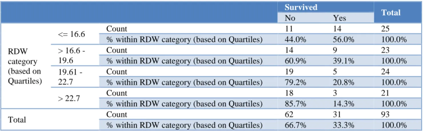
Acute kidney injury was noted in 19 (20.4%) patients. Amongst these critically ill children, mortality was 66.7% (n = 62). Frequency of patients based on RDW quartiles were: ≤16.6 (n = 25, 26.9%); 16.6-19.6 (n = 23, 24.7%), 19.61-22.7 (n = 24, 25.8%) and >22.7 (n = 21, 22.6%). Frequency of mortality based on these RDW Quartiles are mentioned in Table 2.
We noted statistically significant correlation between mortality and anemia (10.24 g/dL, SD 2.26; 8.78g/dL, SD2.60. p = 0.009), LOS on MV (4.13 days, SD 2.125 vs 6.94 days, SD 7.603 p = 0.008), RDW (Mortality was 44%, n = 11/25 in RDW <16.6; RDW >22.7 mortality was 84.7%, n = 18/21. P = 0.002), shock (p = 0.004) and Ventilator associated Pneumonia (p = 0.024). Chi square test analysis of various parameters are provided in Table 3.
Table 2: Survival data in RDW category (based on Quartiles).
Survived
Total
No Yes
RDW category (based on Quartiles)
<= 16.6 Count 11 14 25
% within RDW category (based on Quartiles) 44.0% 56.0% 100.0% > 16.6 -
19.6
Count 14 9 23
% within RDW category (based on Quartiles) 60.9% 39.1% 100.0% 19.61 -
22.7
Count 19 5 24
% within RDW category (based on Quartiles) 79.2% 20.8% 100.0%
> 22.7 Count 18 3 21
% within RDW category (based on Quartiles) 85.7% 14.3% 100.0%
Total Count 62 31 93
% within RDW category (based on Quartiles) 66.7% 33.3% 100.0%
Table 3: Correlations of mortality.
Outcome measures p OR
Anemia 0.009 2.517 (1.04-6.09) RDW 0.002 4.255 (1.64-11.02) Shock 0.004 5.429(1.66-17.74) VAP 0.024 2.629 (1.101-6.577) Sepsis 0.591 1.000 (0.413-2.423) AKI 0.057 3.246 (0.868-12.15) Hepatic dysfunction 0.085 2.292 (0.817-6.430)
Figure 1: Overall RDW.
Multivariable analyses were performed using imputed data, and adjusted odds ratios (ORs) with 95% confidence intervals are presented. The area under the receive operating characteristic curve (AUROC) was used to determine the scope of RDW as an independent predictor for mortality and to define optimal cut-points for sensitivity and specificity. To further confirm the benefit of RDW as prognostic marker we controlled RDW for anemia and utilised AUROC to determine sensitivity and specificity. As per AUROC with Overall RDW, the best cut-off of RDW is 18.45 with Sensitivity = 71.0% and Specificity = 67.8% (Figure 1).
After controlling for anemia, the cut-off was 18.10 with sensitivity = 72.7% and specificity = 61.1% (Figure 2). PIM3 score had sensitivity = 72.7% and specificity = 62%. Thus as per the present study, RDW of more than 18.10 is independently associated with high risk of mortality
DISCUSSION
It was found that RDW measured within 24 hours of PICU admission was independently associated with mortality, with or without anemia. We also found significant correlation of presence of anemia, shock, ventilator associated pneumonia and length of stay on mechanical ventilation with mortality. Being a tertiary referral centre for many critical and end stage patients due to mainly economical and other reasons, we endeavoured to evaluate the significance of a routine test like Red cell distribution width as an independent risk factor for mortality in critically ill children. Although RDW demonstrated high sensitivity but low specificity as an independent predictor of mortality, it corresponded fairly to sensitivity and specificity of PIM 3 score in the present study. This is an important observation as a low cost test of RDW performed nearly at par with more complex index of mortality. Analysing the Odd’s ratio for the Cohort with mortality, it demonstrated that there is a 1.579 chance that with high RDW the patient will not survive.
Recent studies have found associations of an increased RDW with mortality, irrespective of mean cellular volume (MCV) and haemoglobin levels. Associations were reported for patients with heart failure, acute myocardial infarction, and community-acquired pneumonia pulmonary hypertension and in the general population.8-16 Data on the utility of RDW as a biomarker
of clinical outcomes in the paediatric population are more limited. In critically ill children admitted to ICU, RDW is associated with risk of death and is suggested as an independent prognostic marker.3,17-19 There are very few
studies examining RDW as a biomarker in a general paediatric intensive care unit (PICU) population, esp. critical children requiring mechanical ventilation.20 Our
data demonstrate that RDW at the time of PICU admission may help to alert PICU clinicians to a subgroup of critically ill pediatric population who are at risk for adverse outcomes. Early identification will be a boon to provide early intervention to these patients and improve the outcome of intensive care unit.
The most attractive properties of RDW as a pragmatic clinical biomarker are its relative low cost and near universal availability compared to other proposed biomarkers in this population. Though AUROC for RDW indicates its moderate utility in mortality prediction, the more objective clinical assessment, laboratory data and complex scores like PIM3, PRISM 24 (Paediatric risk of mortality), PELOD (Paediatric Logistic Organ Dysfunction) scores may help in specific triaging and
specific management decisions. Moreover many experts have cautioned on the usage of these complex indices to predict outcomes for individual patient.25
RDW is known to be elevated in states of ineffective red cell production and increased red cell destruction, which are a common feature in a variety of infectious and inflammatory conditions.1 An association between
increasing RDW and elevated levels of acute phase reactants including erythrocyte sedimentation rate, high sensitivity C-reactive protein, and interleukin-6 has been demonstrated in adults, suggesting that RDW may be elevated in the setting of acute inflammatory states secondary to rapid red blood cell destruction or blunted erythropoiesis.3-5 However, many prior studies have
emphasized that raised RDW is predictive of outcome even after controlling inflammatory markers. Thus, indicating that inflammation alone cannot entirely explain the pathophysiologic processes leading to RDW elevation in critical illness.3,4,26
In the present study, patients with the highest RDW were more likely to present with infection, sepsis, and shock. Although this implies a causative role for inflammation to increase RDW, we lacked specific measures to determine the extent to which inflammation modified the association of RDW with LOS or mortality in our patients. A key point, however, is that RDW is most likely to be a marker of an underlying patho-physiological process (i.e., inflammation, impaired erythropoiesis, or bone marrow dysfunction) rather than itself being a cause of adverse clinical outcomes. An increase in RDW can be linked to hypoxemia.2 Transient
decreases in oxygen partial pressures (PaO2) leads to a ‘pulsatile’ erythropoietin (EPO) release through hypoxia-inducible transcription factors. These will in its turn trigger the release of immature reticulocytes into the circulation leading to anisocytosis and a higher RDW in the affected patient. Supporting this hypoxemia - anisocytosis pathway and the value of RDW in lung pathology, elevated RDW values have been found in diverse respiratory disease processes reflecting differences in disease severity.2
Adult studies have demonstrated that RDW remains an independent predictor of mortality after controlling for recent blood transfusion.3-5,27 In tropical country like
India where 45% children are malnourished, underlying nutritional anemia cannot be ruled out. Also, due to financial constraints, iron studies could not be conducted. However, this also contributed in determining utility of RDW in predicting outcome, irrespective of anemia and transfusion status; thus making it more practically relevant. Demonstration on AUROC of almost similar predictability of mortality by RDW after controlling for anemia suggests that anemia cannot account for our findings.
confounding factors like anemia, inflammatory biomarkers, markers of hypoxia could not be included in detail. Many critical patients could not be included due to lack of data for PIM3 score or lack of RDW report, which may contribute to selection bias. Finally, being a retrospective study active evaluation of clinical progression and of biomarkers of inflammation would have improved the outcome of the study.
CONCLUSION
RDW measured within 24 hours of PICU admission was independently associated with length of stay on mechanical ventilation and mortality in a general PICU population. RDW being low cost and universally available, can be utilized in predicting mortality in critically ill children. We recommend the need for multicentric, prospective longitudinal studies to determine the optimum utility of RDW to enhance decision making in PICU.
ACKNOWLEDGEMENTS
Authors would like to thank Dean, Smt. Kashibai Navale Medical College and General Hospital, Narhe, Pune.
Funding: No funding sources Conflictof interest: None declared
Ethicalapproval: The study was approved by the Institutional Ethics Committee
REFERENCES
1. Qurtom HA, Al-Saleh QA, Lubani MM, Hassanein A, Kaddoorah N, Qurtom MA, et al. The value of red cell distribution width in the diagnosis of anaemia in children. Eur J Pediatr. 1998;148:745-8. 2. Yčas JW, Horrow JC, Horne BD. Persistent increase
in red cell size distribution width after acute diseases: A biomarker of hypoxemia? Clin Chim Acta. 2015;448:107-17.
3. Bazick HS, Chang D, Mahadevappa K, Gibbons FK, Christopher KB. Red cell distribution width and allcause mortality in critically ill patients. Crit Care Med. 2011;39:1913-21.
4. Forhecz Z, Gombos T, Borgulya G, Pozsonyi Z, Prohászka Z, Jánoskuti L. Red cell distribution width in heart failure: Prediction of clinical events and relationship with markers of ineffective erythropoiesis, inflammation, renal function, and nutritional state. Am Heart J. 2009;158:659-66. 5. Lippi G, Targher G, Montagnana M, Salvagno GL,
Zoppini G, Guidi GC. Relation between red cell distribution width and inflammatory biomarkers in a large cohort of unselected outpatients. Arch Pathol Lab Med. 2009;133:628-32.
6. Sipahi T, Koksal T, Tavil B, Akar N. The effects of acute infection on hematologic parameters. Ped Hemato Oncol. 2004;21:513-20.
7. Scharte M, Fink MP. Red blood cell physiology in critical illness. Crit Care Med. 2003;31:S651-7. 8. Dabbah S, Hammerman H, Markiewicz W. Relation
between red cell distribution width and clinical outcomes after acute myocardial infarction. Am J Cardiol. 2010;105:312-7.
9. Pascual-Figal DA, Bonaque JC, Redondo B. Red blood cell distribution width predicts long-term outcome regardless of anaemia status in acute heart failure patients. Eur J Heart Fail. 2009;11:840-6. 10. van Kimmenade RR, Mohammed AA,
Uthamalingam S. Red blood cell distribution width and 1-year mortality in acute heart failure. Eur J Heart Fail. 2010;12:129-36.
11. Tonelli M, Sacks F, Arnold M. Relation between red blood cell distribution width and cardiovascular event rate in people with coronary disease. Circ. 2008;117:163-8.
12. Braun E, Domany E, Kenig Y. Elevated red cell distribution width predicts poor outcome in young patients with community acquired pneumonia. Crit Care. 2011;15:R194.
13. Hampole CV, Mehrotra AK, Thenappan T. Usefulness of red cell distribution width as a prognostic marker in pulmonary hypertension. Am J Cardiol. 2009;104:868-72.
14. Patel KV, Ferrucci L, Ershler WB. Red blood cell distribution width and the risk of death in middle-aged and older adults. Arch Intern Med. 2009;169:515-23.
15. Patel KV, Semba RD, Ferrucci L, Newman AB, Fried LP, Wallace RB, et al. Red cell distribution width and mortality in older adults: a meta-analysis. J Gerontol Series A Biomed Sci Med Sci. 2009;65(3):258-65.
16. Perlstein TS, Weuve J, Pfeffer MA. Red blood cell distribution width and mortality risk in a community-based prospective cohort. Arch Intern Med. 2009;169:588-94.
17. Büyükkoçak U, Gencay I, Ates G, Çağlayan O. Red blood cell distribution width and mortality in icu patients; a cross sectional retrospective analysis red blood cell distribution width and mortality in icu patients. Enliven: J Anesthesiol Crit Care Med. 2014;1(4):1-4.
18. Wang F, Pan W, Shuming P, Ge Junbo, Wang S, Chen M. Red cell distribution width as a novel predictor of mortality in ICU patients. Ann Med. 2011;43(1):40-6.
19. Hunziker S, Celi LA, Lee J, Howell MD. Red cell distribution width improves the simplified acute physiology score for risk prediction in unselected critically ill patients. Crit Care. 2012;16(3):R89. 20. Ramby AL, Goodman DM, Wald EL, Scott L.
Weiss red blood cell distribution width as a pragmatic marker for outcome in paediatric critical illness. PLos One. 2015;10(6):e0129258.
nloads/PIM2%20%26%20PIM3%20Information%2 0Booklet%20Mar16.pdf
22. Weiss SL, Parker B, Bullock ME, Swartz S, Price C, Wainwright MS, et al. Defining pediatric sepsis by different criteria: Discrepancies in populations and implications for clinical practice. Pediatr Crit Care Med. 2012;13:e210-8.
23. Goldstein B, Giroir B, Randolph A. International pediatric sepsis consensus conference: definitions for sepsis and organ dysfunction in pediatrics. Pediatr Crit Care Med. 2005;6:2-8.
24. Sands KE, Bates DW, Lanken PN, Graman PS, Hibberd PL, Kahn KL, et al. Epidemiology of sepsis syndrome in 8 academic medical centers. JAMA. 1997;16:234-40.
25. Straney L, Clements A, Parslow RC, Pearson G, Shann F, Alexander J, et al. Paediatric index of mortality 3: an updated model for predicting
mortality in pediatric intensive care. Pediatr Critical Care Med. 2013;14(7):673-81.
26. Ku NS, Kim H, Oh HJ, Kim YC, Kim MH, Song JE, et al. Red cell distribution width is an independent predictor of mortality in patients with Gram-negative bacteremia. Shock. 2012;38:123-7. 27. Lacroix J, Cotting J. Pediatric Acute Lung Injury
and Sepsis Investigators (PALISI) Network. Severity of illness and organ dysfunction scoring in children. Ped Crit Care Med. 2005;6:S126-34.