Non-Surgical Therapy Reduces Presence of JP2 Clone in
Localized Aggressive Periodontitis
Danielle K Burgess, MS*, Hong Huang, BS†, Peter Harrison, MS†,‡, Theodora Kompotiati,
DDS, MS, Ikramuddin Aukhil, MS†, and Luciana M Shaddox, DDS, MS, PhD
*University of North Carolina School of Dentistry, Chapel Hill, North Carolina, United States
†Department of Periodontology, University of Florida College of Dentistry, Gainesville, Florida,
Unites States ‡Division of Periodontology, School of Dental Science, Trinity College Dublin,
Ireland
Abstract
Background—Previous studies have provided substantial evidence on the association of Aggregatibacter actinomycetemcomitans (Aa), and its highly leukotoxic JP2 genotype, with localized aggressive periodontitis (LAP). The present study aims to evaluate the presence of JP2 in LAP individuals following periodontal treatment.
Methods—Sixty African American LAP patients between ages 5 and 25 years were examined. At baseline, pocket depth (PD), clinical attachment level (CAL), bleeding on probing (BoP) and plaque index were measured and subgingival plaque was collected from both LAP diseased sites and healthy sites for each participant. Patients received whole-mouth ultrasonic debridement, scaling and root planning and a 7-day prescription of amoxicillin and metronidazole. Participants were re-evaluated, re-sampled and received regular maintenance therapy at 3, 6 and 12-months post-treatment. PCR technique was used detect presence of the JP2 genotype before and after treatment.
Results—At baseline, the JP2 sequence was identified in 75% of LAP diseased sites and in 56.67% of healthy sites. At 3, 6 and 12-months post-treatment, patient compliance was 40, 31 and 31, respectively, and JP2 detection decreased to 17.5%, 6.45% and 3.23% respectively, in diseased sites (P<0.001) and to 2.5%, 3.23% and 0% respectively, in healthy sites (P<0.001). Clinical parameters of disease were also significantly reduced after therapy (p<0001). Additionally, significant correlations were observed between JP2 presence and mean PD (p < 0.002) and CAL (p < 0.001), post-therapy.
Conclusion—Periodontal therapy was successful in reducing clinical parameters of LAP and the subgingival presence of JP2 in diseased and healthy sites.
Corresponding Author: Luciana M. Shaddox, DDS, MS, Ph.D., Department of Periodontology and Oral Biology, University of Florida College of Dentistry, P.O. Box 100434, Gainesville, FL 32610-0434, Phone: 352-273-8368, Fax: 352-273-6192,
DISCLAIMERS:
HHS Public Access
Author manuscript
J Periodontol
. Author manuscript; available in PMC 2017 December 01.Published in final edited form as:
J Periodontol. 2017 December ; 88(12): 1263–1270. doi:10.1902/jop.2017.170285.
A
uthor Man
uscr
ipt
A
uthor Man
uscr
ipt
A
uthor Man
uscr
ipt
A
uthor Man
uscr
Keywords
Aggregatibacter actinomycetemcomitans; periodontitis; aggressive; juvenile; prepubertal; therapy; pathogenicity; inflammation
Characterized by severe anatomical and physiological destruction of the periodontal structures supporting the first molars and/or incisors, localized aggressive periodontitis (LAP) is an inflammatory disease that typically presents in adolescent individuals that are otherwise systemically healthy. 1, 2 LAP prevalence varies substantially between age groups and ethnicities, however research evidence describes a more pronounced frequency of LAP occurrence within adolescent populations of African ancestry, significantly more so than Hispanic and Caucasian populations. 3–5 While the rationale behind this endemic remains to be elucidated, clinical investigations of Aggregatibacter actinomycetemcomitans (Aa), a resident bacterium of the oral microbiota may help provide possible explanations.6–8 One such explanation examines the concept of two separate etiologies for LAP; the first considers minimally leukotoxic genotypes of A. actinomycetemcomitans as opportunistic pathogens across several ethnic groups worldwide. The second, recognizes the highly leukotoxic JP2 genotype of A. actinomycetemcomitans as an exogenous pathogen that exhibits racial tropism towards persons of African descent and is presumed to employ interfamilial dissemination, 9 the latter also found for Aa. 10, 11 for Research has shown that individuals infected with this highly leukotoxic JP2 Aa have a higher risk of developing attachment loss than those infected with non-JP2 genotype. 8 A similar study reports statistical significance between the presence of the JP2 genotype and the conversion from a healthy periodontal state to a LAP diseased state with LAP susceptible African American families. 12 Furthermore, a number of studies have associated the JP2 clone with more severe forms of LAP. 13–15
The polymorphonuclear (PMN) and monocyte cell-specific leukotoxin assembled by all A. actinomycetemcomitans is produced in 10–20 times greater amounts in the JP2 genotypes than in the minimally pathogenic genotypes.16 Additionally, Brogan and coworkers describes this enhanced leukotoxicity as a consequence of a unique 530 base-pair deletion within the promoter region of the leukotoxin operon. This deletion subsequently allows the inclusion and participation of a second promoter in transcription of the leukotoxin gene thereby leading to greater production of the toxin. 16 Thus, this organism appears to be of great toxicity and strong association with this disease.
Although previous periodontitis treatment studies have shown significant correlations between the suppression of A. actinomycetemcomitans presence and reductions in the clinical parameters of aggressive periodontitis 17–20, the JP2 and non-JP2 genotype persistence after non-surgical treatment of LAP patients remains unclear. One study looked into JP2 genotype specifically after treatment of periodontitis patients. Cortelli et al.16 conducted a study in which a group of Brazilian periodontitis participants harboring the JP2 and the non-JP2 sequences were evaluated at baseline and re-evaluated at 12 months post scaling/root planning, systemic antibiotics and periodontal surgery. At 12 months, clinical measurements were significantly reduced in both groups, it was noted however, that the
A
uthor Man
uscr
ipt
A
uthor Man
uscr
ipt
A
uthor Man
uscr
ipt
A
uthor Man
uscr
presence of the JP2 genotype negatively affected the response to treatment. 21 Due to geographical changes on bacterial colonization of aggressive periodontitis, in specifically Aa JP2 genotype, it is important to evaluate the potential reduction of this virulent clone in different populations/continents after no-surgical therapy and the maintenance of such results long-term post-therapy.
We have recently reported our cross-sectional findings on the prevalence of the JP2 and non-JP2 sequences of A. actinomycetemcomitans within a cohort of 60 African American children diagnosed with LAP, of whom (50) 83% carried the JP2 genotype and (17) 28.33% carried the non-JP2 genotype.22 Understanding host and bacterial response to periodontal treatment and disease progression/resolution during maintenance is crucial to the
development of improved treatment protocols. Therefore, the objective of this longitudinal study is to investigate, within this population of LAP patients, the impact of mechanical and a single dose of systemic antibiotic therapy on the detection of JP2 and non-JP2 genotypes of A. actinomycetemcomitans, as well as possible concomitant impacts on the clinical manifestation of the disease over a 12-month period.
MATERIALS AND METHODS
Participant Population
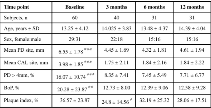
The data reported herein was gathered from a subject group of 60 African American LAP patients with a baseline mean age of 13.25 ± 4.12 years; 42 with LAP affecting permanent dentition and 18 primary dentition (see Table 1 for demographic and clinical parameters). Participants were recruited from the Leon County Health Department (Tallahassee, FL) between April 2007 and November 2015, from the Dental Clinical Research Unit (Gainesville, FL) between February 2014 to March 2014, from the Duval County Health Department (Jacksonville, FL) between February 2014 and March 2016 and from Jackson County Health Department (Marianna, FL) between April 2015 and April 2016. All participants and legal guardians of participants under 18 received verbal briefing of the investigation, and written consent/assent was obtained before proceeding with the study. The study protocol was reviewed and approved by the Institutional Review Board at the
University of Florida (IRB# 201400349) and is registered in Clinicaltrials.gov (#NCT01330719).
Study inclusion criteria were as follows: ages of 5 and 25 years old, systemically in good health as evidenced by medical history and a diagnosis of LAP according to criteria detailed in Armitage 1999 and Albandar 2014. 1,2 More specifically, patients should present at least two sites with pocket depths (PD) greater than or equal to 5mm, with bleeding on probing (BoP), clinical attachment levels (CAL) of 2mm or greater and bone loss detected by radiographs, on first molars and/or incisors, but in no more than two teeth other than first molars and incisors. Exclusion criteria: patient diagnosed with any systemic diseases or conditions that could influence the characteristics or clinical presentation of LAP (ex: diabetes, blood disorders etc.), patient who reported the use of antibiotics within 3 months prior to first visit, or use of any medications that could influence periodontal characteristics or interfere with response to periodontal treatment (ex: cyclosporine, phenytoin), pregnant and/or lactating women, and smokers.
A
uthor Man
uscr
ipt
A
uthor Man
uscr
ipt
A
uthor Man
uscr
ipt
A
uthor Man
uscr
Clinical Examination & Sampling
At baseline, 3, 6 and 12 months the following clinical parameters were measure and six sites per tooth by previously calibrated examiners (LMS and PH): pocket depth (PD), clinical attachment level (CAL), bleeding on probing (BoP) and plaque index (PI) using a UNC-15mm§ probe and stored using a periodontal software**. Bitewing and periapical radiographs were taken to assess bone loss and further confirm diagnosis. Per patient, subgingival plaque was obtained from two LAP diseased sites (PD≥5mm, BoP, CAL≥2mm and radiographic bone loss) of which the deepest pockets were targeted and from two healthy similar sites (interproximal site, posterior or anterior, depending on diseased site sampled, PD≤3mm, no BoP, no CAL, no bone loss). Plaque samples were harvested using sterile absorbent endodontic paper points†† and stored at −80° C until processed.
Bacterial DNA extraction and JP2 sequence detection proceeded as previously described. 22 Briefly, bacterial DNA was isolated from the subgingival plaque samples using the DNA Purification Kit, following the manufacturer instructions‡‡. Target sequences were amplified under standard polymerase chain reaction (PCR) conditions using a PCR Master Mix with HF buffer§§ and primers previously mentioned by Poulsen and coworkers: ltx3 (5′– GCCGACACCAAAGACAAAGTCT-3′) and ltx4 (5′–
GCCCATAACCAAGCCACATAC-3′).23
Periodontal Treatment
The periodontal treatment methods employed within this study were performed by two calibrated examiners (LMS and PH) and are previously described in detail. 24 Treatment was administered subsequent to recording baseline periodontal parameters/sample collection and consisted of a full-mouth supra-subgingival debridement using an ultrasonic device***, scaling and root planning of diseased sites under local anesthesia, as needed. Additionally, patients were prescribed one single regimen of systemic antibiotics: a combination of 500mg amoxicillin and 250mg metronidazole25–27 (dosage adjusted for children <40 kg), t.i.d for 7 days. Antibiotic compliance and side effects were verified at next visit with patient/parent, returned medication bottles and medication log. All patients received oral hygiene instructions and an accompanying oral care kit consisting of an electronic toothbrush†††, toothpaste, dental floss, and interproximal brushes as needed. Participants were requested to be present at 3, 6, and 12 month appointments after baseline for re-examination, at which they received additional full mouth supra-subgingival debridement with ultrasonic device, scaling and root planning of remaining diseased sites, as needed, and oral hygiene instructions. Patients did not receive additional antibiotics at follow up visits.
§Hu-Friedy, Chicago, IL
**Florida Probe Corporation Gainesville, FL
††Paperpoints, medium size, Kerr Endodontics, Orange, CA ‡‡MasterPure EPICENTRE Biotechnologies, Madison, WI, USA §§New England BioLabs Inc., Ipswich, MA, USA
***Cavitron Jet Plus, Dentsply, York PA
†††Oral B Vitality TM Dual Clean, Proctor & Gamble, Cincinnati, OH, USA
A
uthor Man
uscr
ipt
A
uthor Man
uscr
ipt
A
uthor Man
uscr
ipt
A
uthor Man
uscr
Statistical Analysis
Power analysis for this study was determined by baseline differences between LAP and controls as described previously.22 Post-Hoc power analysis run for the chi-square test for the main outcome variable (presence of JP2 post treatment in diseased sites) showed a power of 89.6% for the lowest N (31), at 12 months. Statistical analyses on the presence of JP2 and non-JP2 strains and correlations with clinical parameters were conducted using a statistical software‡‡‡. Chi-square and Fisher exact tests were used to compare the prevalence of positive strains (JP2 and non-JP2) at each timepoint, before and after treatment, out of a total of available sites (no data imputing performed). Pearson/Spearman correlations were used to correlate clinical parameters/reductions with the presence of JP2 strains and ANOVA/ Kruskal Wallis with Dunn’s multiple comparisons were used to compare clinical parameters before and after treatment among timepoints. Significance was determined at p value <0.05. All analysis were conducted using a statistical software§§§.
RESULTS
Sixty patients were included in this study. Our baseline analysis of JP2 genotype prevalence within this group has been recently published. 22 An impressive 83% of this study
population carried the JP2 genotype in either a diseased site (75%) or in a healthy site (56.67%). The non-JP2 sequence proved to be significantly less pronounced within this group, presenting in only 18.33% of both diseased and heathy sites. Co-infection with both JP2 and non-JP2 sequences was observed in 15% of both diseased and healthy sites. 22 Among the 60 participants enrolled at baseline, 40 (66.67%) returned at 3 months for post-treatment evaluation and 31 (51.67%) returned at both 6 and 12 months (see supplementary Fig. 1 in online Journal of Periodontology).
A significant reduction in the prevalence of A. actinomycetemcomitans was observed following periodontal treatment. At 3, 6 and 12-months post-therapy, detection of the JP2 genotype in LAP diseased sites had reduced to 17.5% (7/40), 6.45% (2/31) and 3.23% (1/31), respectively, compared to 75% at baseline (p < 0.0001, Fig. 1a). In LAP healthy sites, the post-treatment JP2 detection rate was 2.5%, 3.23% and 0% at 3, 6 and 12 months respectively, compared to 56.67% at baseline (P<0.001) (Fig 1b). In diseased sites, non-JP2 sequence prevalence was also reduced from 18.33% at baseline to 0% at 3 months and to 3.23% at both 6 and 12 months (p<0.01, Fig 2a), and in healthy sites from 18.33% at baseline to 2.5% at 3 months and 3.23% at 6 and 12 months respectively (p<0.01, Fig 2b). Interestingly, if we look at only those 31 patients that were evaluated at 12 months, their baseline proportion of diseased sites with JP2 clone is 68% (21 total) and only 1 patient of those 31 (3%) presented this species at 12 months post-therapy (P<0.001).
Similarly, significant improvements were also observed in PD and CAL post-treatment readings (Figure 3a). At baseline, PD and CAL means for LAP diseased sites measured 6.55mm and 3.98mm respectively. Post-therapy evaluations at 3, 6 and 12 months reported reductions to the following mean PDs: 4.45mm, 4.32mm and 4.61mm and corresponding
‡‡‡GraphPad Prism, version 5.00, San Diego California USA §§§GraphPad Prism, GraphPad Software, La Jolla, CA.
A
uthor Man
uscr
ipt
A
uthor Man
uscr
ipt
A
uthor Man
uscr
ipt
A
uthor Man
uscr
CAL means: 1.75mm, 1.84mm and 1.84mm, respectively (p < 0.001, Fig 3a). Reductions in BoP were also noted across post-treatment time points, however they were non-significant (Table 1). The pre-therapy baseline data showed no significant difference between the mean PD and mean CAL of LAP diseased sites that were positive for the JP2 sequence and those that were negative for JP2 sequence. 22 However, after treatment, significant positive correlations were observed between JP2 presence and PD (p=0.002 and r2=0.2889) and between JP2 presence and CAL (p<0.001 and r2=0.3619). A significant difference was noted between the PD and CAL of sites with and without the JP2 sequence at baseline and the PD and CAL of the same sites at the subsequent visits (p < 0.001), markedly, as the number of JP2 positive sites decreased due to treatment, so did the PD and CAL readings (Fig. 3b). It is important to note that there were no reported side effects with the use of this regimen by any LAP participant.
DISCUSSION
Research describes the microbiological environments associated with periodontal health, as a homeostatic balance in which potentially pathogenic species are presumed to naturally exist within the indigenous microflora. Colonization by these pathogens is kept to a
minimum due to competition from commensal species.28 However, slight disturbances in the subgingival environment may be disruptive to this homeostasis thereby enabling antagonistic species to thrive and outcompete commensal ones. Such shifts in the microbial ecosystems are believed to be the driving forces behind the development of inflammatory diseases of the periodontium. 28–30
Microbial analyses of the subgingival microflora of LAP subjects indicates that these individuals maintain a collection of organisms that is significantly different from that of heathy individuals. 31 Specifically, within African American adolescent populations, the presence of the highly virulent JP2 genotype of A. actinomycetemcomitans has garnered much attention; this species, as evidenced by a number of studies appears to be strongly implicated in etiology of localized aggressive periodontitis in this group. 9, 31–34 Findings from our preliminary investigation concerning the prevalence of the JP2 genotype within a group of 60 African American LAP patients further substantiates this claim, as 83% of this study population harbored the highly leukotoxic JP2 genotype. 22 The purpose of this study was to evaluate the impact of periodontal treatment on the stability of the JP2 genotype of Aa as it related to the effectiveness of treatment on the clinical presentation of LAP within this group.
The results of the present study are consistent with previous treatment studies that have reported a positive correlation between the reduction of A. actinomycetemcomitans and the resolution of LAP. 17, 35 Considering both LAP diseased and healthy sites within the present study, we observed significant reductions in the detectability of both the JP2 and non-JP2 sequences following mechanical debridement and antibiotic therapy (Figs. 1 and 2). A correlation between diseased sites harboring or not harboring the JP2 sequence at baseline alone, was not significant with the mean PD and CAL in those sites; this could be due to too few LAP sites negative for JP2 (15) as compared to JP2 positive sites (45), and the disease being clinically very uniform among those sites (all sites deeper than 5mm). However,
A
uthor Man
uscr
ipt
A
uthor Man
uscr
ipt
A
uthor Man
uscr
ipt
A
uthor Man
uscr
following treatment, a correlation was found between the JP2 and both PD and CAL, i.e. the sites with persistent JP2 sequence after treatment presented significantly deeper PD and higher CAL levels that the JP2 negative sites (figure 3). Furthermore, we noted that the elimination of the JP2 genotype (the conversion of a site from JP2 positive to JP2 negative) in the long term after treatment corresponded to improvements and maintenance in the clinical presentation of LAP. Specifically, both PD and CAL measurements were noted to be significantly reduced after treatment, and remained reduced, for up to 12-months post-therapy. These results corroborate with previous findings, where the elimination of JP2 was correlated with positive clinical presentation. 21 These correlations presented here provide further evidence for the implication of the JP2 genotype as an associated pathogen in LAP disease and attests for the first time the effectiveness of this non-surgical treatment protocol for this population in 12 months post-therapy. In fact, the long-term clinical benefits and 4-year maintenance of this treatment protocol has been previously reported for all LAP patients in this trial. 36 Noteworthy, this is a one-time regimen of antibiotics for the treatment of this population at baseline and they are followed by regular maintenance following this treatment. Thus, the significant reduction of this pathogen long-term with one time antibiotic use is of extreme clinical benefit here for this disease.
The aforementioned treatment study by Cortelli et al. 2009 21 presents results comparable to the present study, furthermore the authors report that those patients infected with the non-JP2 genotype appeared to respond more favorably to treatment than those infected with the JP2 genotype. Reductions in the detectability of non-JP2 clone of A.a were also observed in the present study 12-months post-treatment, however, the non-JP2 genotype was only present in 28.33% of sites in the present study, majority of which occur as a co-infection with the JP2 sequence. Thus, we were not able to statistically compare the effects of non-JP2 alone in the response to treatment in the present investigation. However, our results
regarding JP2 do corroborate with this previous investigation as previously mentioned.
The JP2 genotype was also detected in 56.67% of healthy sites examined at baseline, and after treatment, significant reduction in the presence of JP2 genotype was observed in these sites as well. The notable presence of this bacterium within healthy sites of LAP patients and within the heathy control group (40%) analyzed previously 22 indicates that simply detecting the JP2 sequence may not be sufficient to predict disease onset. Nonetheless, the presence of this bacterial genotype may increase host susceptibility for commensal-pathogen imbalance. This, along with genetic predisposition, and a heightened host responsiveness, 37 may lead to disease onset. Interesting to also note here that our previous study looking at a slightly smaller sample size of this cohort (51 LAP patients) and under a 16S-rRNA-microarray based assay (HOMIM) have shown >50% of diseased sites harboring Aa (OT531) in this population, 31 although the specific JP2 genotype evaluated here was more prevalent, as detected with regular PCR (75% of diseased sites). It is also important to note that the detection rate of this species after treatment could have been higher if more sites were collected at baseline and post-treatment. 38 However, given the localization of this specific disease (only a few sites in the mouth effected), the attrition data (missed appointments following treatment, which is a common occurrence for this population) and the very low prevalence of this disease, a higher number of post-treatment sample collection was not possible in the present investigation. Additionally, the fact that some patients missed
A
uthor Man
uscr
ipt
A
uthor Man
uscr
ipt
A
uthor Man
uscr
ipt
A
uthor Man
uscr
timepoints and therefore proper maintenance could lead to recurrence of the JP2
colonization. However, despite these limitations, it is important to note that the present study still showed a high prevalence of JP2 at baseline, followed by a very low detection rate of this species post-treatment, with a power of >80%.
Another limitation in this study is the lack of quantification of the actual bacterial load within the sites analyzed. This study only evaluated the presence/absence of this species. The actual amount of this species load in the sites could potentially influence the onset and the severity of LAP manifestation and consequently strongly influence response to treatment. However, we were able to show that this treatment protocol was able to significantly reduce the detection of JP2 in LAP and maintain low detection rates, along with clinical stability, over 12 months post-therapy.
CONCLUSIONS
This study showed that mechanical periodontal therapy with a single regimen of amoxicillin and metronidazole was successful in reducing and maintaining a low rate of detection of JP2 in diseased and healthy sites of African Americans diagnosed with LAP, along with a significant reduction and stability of clinical parameters of the disease, 12 months post-therapy.
Acknowledgments
The authors would like to thank the NIH/NIDCR R01DE019456 for the financial support awarded to this research, as well as the doctors and staff at the Leon, Duval, and Jackson County Health Department dental centers for their assistance in coordinating our visits to their clinics, patient referrals and dental care. This study is registered in Clinicaltrials.gov (#NCT01330719).
References
1. Albandar JM. Aggressive periodontitis: case definition and diagnostic criteria. Periodontol 2000. 2014; 65:13–26. [PubMed: 24738584]
2. Armitage GC. Development of a classification system for periodontal diseases and conditions. Ann Periodontol. 1999; 4:1–6. [PubMed: 10863370]
3. Albandar JM, Tinoco EM. Global epidemiology of periodontal diseases in children and young persons. Periodontol 2000. 2002; 29:153–176. [PubMed: 12102707]
4. Loe H, Brown LJ. Early onset periodontitis in the United States of America. J Periodontol. 1991; 62:608–616. [PubMed: 1770420]
5. Susin C, Haas AN, Albandar JM. Epidemiology and demographics of aggressive periodontitis. Periodontol 2000. 2014; 65:27–45. [PubMed: 24738585]
6. Darby I, Curtis M. Microbiology of periodontal disease in children and young adults. Periodontol 2000. 2001; 26:33–53. [PubMed: 11452905]
7. Fine DH, Markowitz K, Furgang D, et al. Aggregatibacter actinomycetemcomitans and its relationship to initiation of localized aggressive periodontitis: longitudinal cohort study of initially healthy adolescents. J Clin Microbiol. 2007; 45:3859–3869. [PubMed: 17942658]
8. Haubek D, Ennibi OK, Poulsen K, Vaeth M, Poulsen S, Kilian M. Risk of aggressive periodontitis in adolescent carriers of the JP2 clone of Aggregatibacter (Actinobacillus) actinomycetemcomitans in Morocco: a prospective longitudinal cohort study. Lancet. 2008; 371:237–242. [PubMed:
18207019]
A
uthor Man
uscr
ipt
A
uthor Man
uscr
ipt
A
uthor Man
uscr
ipt
A
uthor Man
uscr
9. Haubek D, Poulsen K, Westergaard J, Dahlen G, Kilian M. Highly toxic clone of Actinobacillus actinomycetemcomitans in geographically widespread cases of juvenile periodontitis in adolescents of African origin. J Clin Microbiol. 1996; 34:1576–1578. [PubMed: 8735124]
10. de Monteiro MF, Casati MZ, Taiete T, et al. Periodontal clinical and microbiological characteristics in healthy versus generalized aggressive periodontitis families. J Clin Periodontol. 2015; 42:914– 921. [PubMed: 26392039]
11. Gunsolley JC, Ranney RR, Zambon JJ, Burmeister JA, Schenkein HA. Actinobacillus
actinomycetemcomitans in families afflicted with periodontitis. J Periodontol. 1990; 61:643–648. [PubMed: 2231231]
12. Bueno LC, Mayer MP, DiRienzo JM. Relationship between conversion of localized juvenile periodontitis-susceptible children from health to disease and Actinobacillus
actinomycetemcomitans leukotoxin promoter structure. J Periodontol. 1998; 69:998–1007. [PubMed: 9776028]
13. Ennibi OK, Benrachadi L, Bouziane A, Haubek D, Poulsen K. The highly leukotoxic JP2 clone of
Aggregatibacter actinomycetemcomitans in localized and generalized forms of aggressive periodontitis. Acta Odontol Scand. 2012; 70:318–322. [PubMed: 22251014]
14. Haubek D, Ennibi OK, Abdellaoui L, Benzarti N, Poulsen S. Attachment loss in Moroccan early onset periodontitis patients and infection with the JP2-type of Actinobacillus
actinomycetemcomitans. J Clin Periodontol. 2002; 29:657–660. [PubMed: 12354092]
15. Zambon JJ, Haraszthy VI, Hariharan G, Lally ET, Demuth DR. The microbiology of early-onset periodontitis: Association of highly toxic Actinobacillus actinomycetemcomitans strains with localized juvenile periodontitis. J Periodontol. 1996; 67:282–290.
16. Brogan JM, Lally ET, Poulsen K, Kilian M, Demuth DR. Regulation of Actinobacillus
actinomycetemcomitans leukotoxin expression: analysis of the promoter regions of leukotoxic and minimally leukotoxic strains. Infect Immun. 1994; 62:501–508. [PubMed: 8300209]
17. van Winkelhoff AJ, Rodenburg JP, Goene RJ, Abbas F, Winkel EG, de Graaff J. Metronidazole plus amoxycillin in the treatment of Actinobacillus actinomycetemcomitans associated periodontitis. J Clin Periodontol. 1989; 16:128–131. [PubMed: 2921374]
18. Akincibay H, Orsal SO, Sengun D, Tozum TF. Systemic administration of doxycycline versus metronidazole plus amoxicillin in the treatment of localized aggressive periodontitis: a clinical and microbiologic study. Quintessence Int. 2008; 39:e33–39. [PubMed: 18567166]
19. van Winkelhoff AJ, Tijhof CJ, de Graaff J. Microbiological and clinical results of metronidazole plus amoxicillin therapy in Actinobacillus actinomycetemcomitans-associated periodontitis. J Periodontol. 1992; 63:52–57. [PubMed: 1313103]
20. Xajigeorgiou C, Sakellari D, Slini T, Baka A, Konstantinidis A. Clinical and microbiological effects of different antimicrobials on generalized aggressive periodontitis. J Clin Periodontol. 2006; 33:254–264. [PubMed: 16553634]
21. Cortelli SC, Costa FO, Kawai T, et al. Diminished treatment response of periodontally diseased patients infected with the JP2 clone of Aggregatibacter (Actinobacillus) actinomycetemcomitans. J Clin Microbiol. 2009; 47:2018–2025. [PubMed: 19458180]
22. Burgess D, Huang H, Harrison P, Aukhil I, LMS. Aggregatibacter actinomycetemcomitans in African Americans with Localized Aggressive Periodontitis. J Den Res Clin Transl Sci. 2017; 2:249–257.
23. Poulsen K, Ennibi OK, Haubek D. Improved PCR for detection of the highly leukotoxic JP2 clone of Actinobacillus actinomycetemcomitans in subgingival plaque samples. J Clin Microbiol. 2003; 41:4829–4832. [PubMed: 14532234]
24. Shaddox LM, Goncalves PF, Vovk A, et al. LPS-induced inflammatory response after therapy of aggressive periodontitis. Journal of dental research. 2013; 92:702–708. [PubMed: 23788609] 25. Buchmann R, Nunn ME, Van Dyke TE, Lange DE. Aggressive periodontitis: 5-year follow-up of
treatment. J Periodontol. 2002; 73:675–683. [PubMed: 12083543]
26. Heller D, Varela VM, Silva-Senem MX, Torres MC, Feres-Filho EJ, Colombo AP. Impact of systemic antimicrobials combined with anti-infective mechanical debridement on the microbiota of generalized aggressive periodontitis: a 6-month RCT. J Clin Periodontol. 2011; 38:355–364. [PubMed: 21303403]
A
uthor Man
uscr
ipt
A
uthor Man
uscr
ipt
A
uthor Man
uscr
ipt
A
uthor Man
uscr
27. Tinoco EM, Beldi MI, Campedelli F, et al. Clinical and microbiological effects of adjunctive antibiotics in treatment of localized juvenile periodontitis. A controlled clinical trial. J Periodontol. 1998; 69:1355–1363. [PubMed: 9926765]
28. Bartold PM, Van Dyke TE. Periodontitis: a host-mediated disruption of microbial homeostasis. Unlearning learned concepts. Periodontol 2000. 2013; 62:203–217. [PubMed: 23574467] 29. Berezow AB, Darveau RP. Microbial shift and periodontitis. Periodontol 2000. 2011; 55:36–47.
[PubMed: 21134227]
30. Marsh PD. Dental plaque as a biofilm and a microbial community - implications for health and disease. BMC Oral Health. 2006; 6(Suppl 1):S14. [PubMed: 16934115]
31. Shaddox LM, Huang H, Lin T, et al. Microbiological characterization in children with aggressive periodontitis. Journal of dental research. 2012; 91:927–933. [PubMed: 22863892]
32. Cortelli JR, Cortelli SC, Jordan S, Haraszthy VI, Zambon JJ. Prevalence of periodontal pathogens in Brazilians with aggressive or chronic periodontitis. J Clin Periodontol. 2005; 32:860–866. [PubMed: 15998269]
33. Haraszthy VI, Hariharan G, Tinoco EM, et al. Evidence for the role of highly leukotoxic
Actinobacillus actinomycetemcomitans in the pathogenesis of localized juvenile and other forms of early-onset periodontitis. J Periodontol. 2000; 71:912–922. [PubMed: 10914794]
34. Haubek D, Dirienzo JM, Tinoco EM, et al. Racial tropism of a highly toxic clone of Actinobacillus actinomycetemcomitans associated with juvenile periodontitis. J Clin Microbiol. 1997; 35:3037– 3042. [PubMed: 9399490]
35. Slots J, Rosling BG. Suppression of the periodontopathic microflora in localized juvenile periodontitis by systemic tetracycline. J Clin Periodontol. 1983; 10:465–486. [PubMed: 6579058] 36. Miller KA, Branco-de-Almeida LS, Wolf S, et al. Long-term clinical response to treatment and
maintenance of localized aggressive periodontitis: a cohort study. J Clin Periodontol. 2017; 44:158–168. [PubMed: 27767222]
37. Shaddox L, Wiedey J, Bimstein E, et al. Hyper-responsive phenotype in localized aggressive periodontitis. Journal of dental research. 2010; 89:143–148. [PubMed: 20042739]
38. Socransky SS, Haffajee AD. The bacterial etiology of destructive periodontal disease: current concepts. J Periodontol. 1992; 63:322–331. [PubMed: 1573546]
A
uthor Man
uscr
ipt
A
uthor Man
uscr
ipt
A
uthor Man
uscr
ipt
A
uthor Man
uscr
Figure 1.
Figure 1A. Chi-square and Fisher exact tests show difference between baseline (BL) and all timepoints after treatment on the frequencies of JP2 positive diseased sites in LAP patients (****P<0.0001).
Figure 1B. Chi-square and Fisher exact tests show difference between baseline (BL) and all timepoints after treatment on the frequencies of JP2 positive healthy sites in LAP patients (****P<0.0001).
A
uthor Man
uscr
ipt
A
uthor Man
uscr
ipt
A
uthor Man
uscr
ipt
A
uthor Man
uscr
Figure 2.
Figure 2A. Chi-square and Fisher exact tests show difference between baseline (BL) and all timepoints after treatment on the frequencies of non-JP2 positive diseased sites in LAP patients (**P<0.01).
Figure 2B. Chi-square and Fisher exact tests show difference between baseline (BL) and all timepoints after treatment on the frequencies of non-JP2 positive healthy sites in LAP patients (**P<0.01).
A
uthor Man
uscr
ipt
A
uthor Man
uscr
ipt
A
uthor Man
uscr
ipt
A
uthor Man
uscr
Figure 3.
Figure 3A. Anova (Kruskal-wallis with Dunn’s multiple comparisons) show significant differences between baseline (BL) mean pocket depth (PD) and mean clinical attachment levels (CAL) of the sampled diseased site and all other timepoints, up to 12 months (****p<0.001).
Figure 3B. Spearman correlation show significant positive correlation between JP2 (JP2+) presence and PD (r=0.2889, p=0.002) and CAL (r=0.3619, p<0.0001) and Mann-Whitney tests also showed a significant difference between JP2+ and JP2 negative (JP2-) sites in terms of PD (***p=0.0002) and CAL (****p<0.001).
A
uthor Man
uscr
ipt
A
uthor Man
uscr
ipt
A
uthor Man
uscr
ipt
A
uthor Man
uscr
A
uthor Man
uscr
ipt
A
uthor Man
uscr
ipt
A
uthor Man
uscr
ipt
A
uthor Man
uscr
ipt
Table 1
Demographics and Clinical Parameters before and after treatment.
Time point Baseline 3 months 6 months 12 months
Subjects, n 60 40 31 31
Age, years ± SD 13.25 ± 4.12 14.025 ± 3.83 13.48 ± 4.37 14.39 ± 4.04
Sex, female:male 29:31 22:18 15:16 15:16
Mean PD site, mm 6.55 ± 1.78*** 4.45 ± 1.69 4.32 ± 1.81 4.61 ± 1.94
Mean CAL site, mm 3.98 ± 1.85*** 1.75 ± 2.11 1.84 ± 2.16 1.84 ± 2.22
PD > 4mm, % 16.07 ± 10.74*** 8.35 ± 7.41 7.45 ± 5.49 7.71 ± 6.77
BoP, % 20.28 ± 23.87** 12.73 ± 8.00 12.39 ± 9.06 12.58 ± 9.28
Plaque index, % 36.57 ± 23.87 24.8 ± 14.56* 32.19 ± 25.32 28.06 ± 17.51
SD=standard deviation,
*** p<0.0001,
**
P<0.01 all timepoints compared to baseline.
*