Genome-wide maps of alkylation damage, repair,
and mutagenesis in yeast reveal mechanisms
of mutational heterogeneity
Peng Mao,
1,4Alexander J. Brown,
1,4Ewa P. Malc,
2Piotr A. Mieczkowski,
2Michael J. Smerdon,
1Steven A. Roberts,
1,3and John J. Wyrick
1,31
School of Molecular Biosciences, Washington State University, Pullman, Washington 99164, USA;
2Department of Genetics,
Lineberger Comprehensive Cancer Center, University of North Carolina, Chapel Hill, North Carolina 27599, USA;
3Center
for Reproductive Biology, Washington State University, Pullman, Washington 99164, USA
DNA base damage is an important contributor to genome instability, but how the formation and repair of these lesions is
affected by the genomic landscape and contributes to mutagenesis is unknown. Here, we describe genome-wide maps of
DNA base damage, repair, and mutagenesis at single nucleotide resolution in yeast treated with the alkylating agent methyl
methanesulfonate (MMS). Analysis of these maps revealed that base excision repair (BER) of alkylation damage is
signifi-cantly modulated by chromatin, with faster repair in nucleosome-depleted regions, and slower repair and higher mutation
density within strongly positioned nucleosomes. Both the translational and rotational settings of lesions within nucleosomes
significantly influence BER efficiency; moreover, this effect is asymmetric relative to the nucleosome dyad axis and is
reg-ulated by histone modifications. Our data also indicate that MMS-induced mutations at adenine nucleotides are significantly
enriched on the nontranscribed strand (NTS) of yeast genes, particularly in BER-deficient strains, due to higher damage
formation on the NTS and transcription-coupled repair of the transcribed strand (TS). These findings reveal the influence
of chromatin on repair and mutagenesis of base lesions on a genome-wide scale and suggest a novel mechanism for
tran-scription-associated mutation asymmetry, which is frequently observed in human cancers.
[Supplemental material is available for this article.]
DNA base lesions are the most frequent class of DNA damage, aris-ing from reactive oxygen species, spontaneous base hydrolysis, deamination, or endogenous and exogenous alkylating agents. Base lesions must be efficiently repaired by the base excision repair (BER) pathway to avoid mutagenesis or cell death, outcomes which can contribute to carcinogenesis, neurodegenerative diseases, and aging (Wallace et al. 2012; Akbari et al. 2015). BER is initiated by lesion-specific DNA glycosylases that cleave the N-glycosidic bond of damaged bases to generate an abasic site. For example, alkyladenine glycosylase (AAG) and 3-methyladenine DNA glyco-sylase (Mag1) are responsible for recognizing and cleaving the ma-jor type of alkylation damage,N-methylpurine (NMP) lesions (e.g., 3-methyladenine [3meA] and 7-methylguanine [7meG]), in hu-man and yeast cells, respectively (Bauer et al. 2015). The resulting abasic site is cleaved by apurinic/apyrimidinic endonuclease (APE1 in humans), followed by repair synthesis and DNA ligation (Bauer et al. 2015).
While it is well established that nucleotide excision repair (NER), which repairs helix-distorting DNA lesions such as UV pho-toproducts, is significantly modulated by cellular chromatin (Nag and Smerdon 2009), the effects of chromatin on BER in vivo are poorly understood. In vitro studies using purified BER enzymes have shown that each step in the BER pathway is significantly in-hibited within a strongly positioned nucleosome, particularly
when the lesion has an inward rotational setting or translational position near the nucleosome dyad (i.e., the position of intersec-tion of DNA with the central dyad axis of the nucleosome) (Rodriguez et al. 2015). However, it is not clear to what extent nu-cleosomes affect BER in vivo, since nucleosome positioning in eu-karyotic genomes is generally weaker than the positioning sequences used for in vitro studies (Mao et al. 2017) and the pres-ence of cellular chromatin-remodeling enzymes can facilitate BER in nucleosomes (Hinz and Czaja 2015; Rodriguez et al. 2015). Moreover, nucleosomes in vivo are marked by different histone post-translational modifications, which could potentially alter re-pair efficiency (Rodriguez et al. 2016).
Our understanding of how chromatin and other genomic fea-tures influence the formation and repair of DNA base damage, such as DNA alkylation, has been limited by the lack of genome-wide methods for mapping this class of DNA lesions. Unlike NER, which has been extensively studied using novel genome-wide methods (Hu et al. 2015; Mao et al. 2016; Yu et al. 2016), there are currently no published studies measuring BER of DNA base lesions on a genomic scale. One recent study mapped the rep-licative incorporation of uracil (a form of base damage) across the yeast genome using a method called Excision-seq (Bryan et al. 2014). However, this technique required very high, nonphysiolog-ical levels of uracil incorporation (Wyrick and Roberts 2015) and did not measure subsequent repair of uracil lesions. Alternative
4Co-first authors
Corresponding authors: [email protected], jwyrick@vetmed .wsu.edu
Article published online before print. Article, supplemental material, and publi-cation date are at http://www.genome.org/cgi/doi/10.1101/gr.225771.117.
methods have been proposed for map-ping DNA base damage (Li et al. 2015; Riedl et al. 2016), but these have yet to be applied on a genome-wide scale. Moreover, little is known about how chromatin or other genomic features in-fluence mutagenesis associated with DNA base damage across the genome, due to a lack of relevant genome-wide mutation data.
In this study, we developed a novel method known as N -methylpurine-se-quencing (NMP-seq) to map methyl methanesulfonate (MMS)-induced alkyl-ation damage across the yeast genome at single nucleotide resolution. We focused on DNA alkylation damage, as it is an im-portant class of base lesions in eukaryotic cells that arises from endogenous (e.g., S-adenosyl methionine) and exogenous damaging agents, including chemother-apeutics such as temozolomide (Fu et al. 2012), and because it is commonly used as a model lesion for BER studies (e.g., Li and Smerdon 2002a; Li et al. 2015). We also analyzed the genome-wide dis-tribution of mutations arising from MMS treatment in wild-type and repair-deficient strains and compared the pat-terns of mutagenesis with the observed variations in lesion formation and repair across the genome.
Results
A map of alkylation lesions at single
nucleotide resolution across the yeast
genome
To precisely map alkylation NMP lesions throughout the genome, we developed the seq method (Fig. 1A). NMP-seq is adapted from methods previously used for mapping ribonucleotide lesions (Ding et al. 2015) and UV-induced cyclo-butane pyrimidine dimers (Mao et al. 2016). The major difference between
these methods is that NMP-seq employs the AAG and APE1 BER enzymes to generate single-strand breaks containing free 3′OH im-mediately upstream of the NMP lesions 3-methyladenine (3meA) and 7-methylguanine (7meG) (see Fig. 1A), two DNA lesions which comprise >90% of all alkylation DNA damage induced by the model alkylating agent MMS (Friedberg et al. 2006). Free 3′OH ends generated by AAG and APE1 cleavage are then ligated to a biotinylated A adaptor, which is used to purify, PCR-amplify, and sequence the resulting DNA fragments. By mapping the se-quencing reads back to the genome sequence, the location of the NMP lesion can be precisely determined.
We used the NMP-seq method to map NMP lesions through-out the yeast genome following treatment with MMS. Wild-type yeast cells were treated with 0.2% MMS for 10 min, and then geno-mic DNA was isolated from yeast immediately after MMS
associated with 3meA lesions (relative to T-associated reads) was more apparent when yeast cells were treated with a higher dose of MMS (0.4% for 10 min) (Supplemental Fig. S1). Consequently, we used this dose (0.4% MMS) for all subsequent NMP-seq exper-iments. NMP-seq reads associated with other nucleotides (i.e., C or T) could represent rare alkylation lesions on pyrimidine bases (Friedberg et al. 2006), background due to low levels of AAG cleav-age at undamcleav-aged DNA, or incomplete blocking of 3′OH groups by terminal transferase (Fig. 1A). The number of 7meG and 3meA NMP-seq reads decreased in the WT 30-min time point (Fig. 1B), likely reflecting ongoing BER of these NMP lesions.
Repair of NMP lesions is significantly modulated by the
stereotypic organization of nucleosomes within yeast genes
To characterize the effects of genomic and chromatin contexts on repair of NMP lesions, we used NMP-seq to map NMP lesions fol-lowing 1 or 2 h of repair in WT cells treated with 0.4% MMS. We focused on repair of 7meG lesions because this is the predominant class of lesion induced by MMS and thus could be more readily an-alyzed during the NMP-seq repair time course. We anan-alyzed the av-erage distribution of 7meG lesions adjacent to the transcription start sites (TSSs) of 5762 yeast genes. To account for potential se-quence biases across yeast genes, the frequency of 7meG reads was normalized by the number of G nucleotides at each position relative to the TSS (i.e., from−500 bp upstream of, to +650 bp downstream from the TSS) on both DNA strands. The distribution of normalized 7meG reads adjacent to the TSS showed a striking distribution following 2-h repair, with low levels of 7meG lesions immediately upstream of the TSS, and periodic peaks of 7meG le-sions downstream from the TSS (Fig. 1C, top panel). The distribu-tion of 7meG lesions after 2-h repair was very similar on the transcribed strand (TS) and nontranscribed strand (NTS) (Fig. 1C, top panel;Supplemental Fig. S2), indicating that, unlike UV-in-duced DNA photoproducts (Hanawalt and Spivak 2008; Hu et al. 2015; Mao et al. 2016), MMS-induced 7meG lesions in WT yeast are not more rapidly repaired on the TS.
To determine whether the pattern of 7meG lesions represents differences in repair efficiency, as opposed to differences in initial damage levels, we calculated the fraction of 7meG lesions remain-ing (i.e., unrepaired) followremain-ing 2-h repair relative to a 0-h control. Because repair is ongoing even during the 10-min MMS exposure in the WT strain, we used a matchedmag1deletion strain treated with the same MMS dose for the 0-h time point. Since Mag1 is the sole DNA glycosylase that recognizes NMP lesions in yeast (Bauer et al. 2015), themag1Δmutant should eliminate BER during MMS exposure and thus represent the initial distribution of NMP lesions. The fraction of unrepaired 7meG lesions showed the same trend as before, with low levels of unrepaired 7meG lesions imme-diately upstream of the TSS and periodic peaks of unrepaired 7meG lesions downstream from the TSS (Fig. 1C, middle panel). A simi-lar, although less pronounced, trend of unrepaired 7meGs was ap-parent following 1-h repair (Fig. 1C, middle panel).
Downstream from the TSS, peaks of unrepaired 7meG lesions had a periodicity of∼164 bp (1-h repair) and∼160 bp (2-h repair), which matches the estimated 160- to 165-bp nucleosome repeat length in yeast (Cui et al. 2012). This suggests that the heteroge-neous distribution of unrepaired 7meG reads following 2-h repair may reflect the inhibitory effect of nucleosomes on BER. To test this hypothesis, we analyzed a published map of nucleosome dyad positions in yeast (Brogaard et al. 2012). This analysis re-vealed that the peaks of unrepaired 7meG lesions downstream
from the TSS coincided with the principal dyad locations of the +1, +2, +3, and +4 nucleosomes (cf. bottom panel with top and middle panels of Fig. 1C), while low levels of unrepaired 7meG up-stream of the TSS largely coincided with nucleosome-depleted re-gions (NDR) present in many yeast promoter rere-gions (Yuan et al. 2005). We also analyzed a published yeast DNase-seq data set (Zhong et al. 2016), which is an alternative measure of chromatin structure and accessibility. Comparison with the DNase-seq data revealed that peaks of unrepaired 7meG lesions corresponded to regions with a low density of DNase-seq reads (i.e., low chromatin accessibility due to positioned nucleosomes), while low levels of unrepaired 7meG in the NDR upstream of the TSS coincided with high levels of DNase-seq reads (Supplemental Fig. S3). This analysis indicates that differences in chromatin accessibility due to the stereotypic organization of nucleosomes across yeast genes significantly modulate the repair of 7meG lesions. Similar patterns of repair were also detected at individual genes (Fig. 1D,E).
To confirm that the observed patterns of unrepaired 7meG le-sions were due to nucleosome-dependent variations in BER effi-ciency, we repeated the repair time course in a mag1Δ strain. Analysis of themag1ΔNMP-seq data revealed that the levels of 7meG lesions showed only a marginal decrease following 1- or 2-h incubation (Supplemental Fig. S4A). Presumably, this reflects on-going DNA replication and trans-lesion synthesis in themag1Δ
strain (Johnson et al. 2007), which would cause an apparent decrease in the levels of NMP lesions. Importantly, analysis of the 1- and 2-hmag1Δdata indicated that there was no correlation between the fraction of 7meG lesions remaining and nucleosome positions in themag1Δmutant (Supplemental Fig. S4B,C), indicat-ing that Mag1 is required for the patterns of 7meG repair observed in WT cells. In summary, this analysis indicates that the stereo-typic nucleosome organization within genes (e.g.,−1 nucleosome, NDR, +1 nucleosome, etc. [Jiang and Pugh 2009; Rando and Winston 2012]) significantly modulates the efficiency of BER of 7meG lesions, with slower repair associated with the central dyad locations of nucleosomes and faster repair in NDRs.
BER efficiency is regulated by the translational and rotational
settings of the lesions within strongly positioned nucleosomes
in vivo
To better understand how the positioning of an NMP lesion within a nucleosome affects its repair in vivo, we analyzed the repair of 7meG lesions among a set of∼10,000 strongly positioned nucleo-somes (nucleosome score > 5) in the yeast genome (Brogaard et al. 2012; Mao et al. 2016). Analysis of 7meG lesions remaining (i.e., unrepaired) following 2-h repair revealed a greater fraction of unpaired lesions near the nucleosome dyad relative to more distal re-gions of nucleosomal DNA (Fig. 2A) or the adjacent linker DNA
(Supplemental Fig. S5A), indicating that the translational
position-ing of DNA lesions relative to the nucleosome dyad affects repair efficiency. A similar trend was observed following 1-h repair
(Supplemental Fig. S5B), although the effect was less pronounced.
the patterns present in WT cells represent differences in BER efficiency.
Analysis of ∼7500 weakly positioned nucleosomes in the yeast genome (nucleosome positioning score < 1) revealed a much less pronounced effect of translational positioning on re-pair, as there was only a slightly higher fraction of lesions remain-ing near the nucleosome dyad (Fig. 2C). Similarly, there was only a relatively small difference in 7meG lesions remaining at outward versus inward rotational settings within weakly positioned nucle-osomes. These data indicate that the impact of nucleosomes on BER efficiency in vivo is dependent upon nucleosome positioning strength.
Analysis of 7meG removal for each individual DNA strand within the high-resolution Brogaard et al. nucleosome map (Brogaard et al. 2012) also revealed a striking asymmetry relative to the nucleosomal dyad (Fig. 2D). The fraction of unrepaired
7meG lesions following 2-h repair among strongly positioned nu-cleosomes in the yeast genome (Brogaard et al. 2012) was analyzed for each individual DNA strand aligned in a 5′to 3′orientation. For both the plus and minus strands, positions upstream (or 5′) of the nucleosome dyad showed a significant effect of rotational setting on the fraction of unrepaired 7meG lesions, while this effect was less pronounced at positions downstream (or 3′) from the nucleo-some dyad (Fig. 2D). Quantification of the data confirmed that there was a higher relative difference in unrepaired 7meG lesions at“In”relative to“Out”rotational settings for positions 5′of the nucleosome dyad relative to positions 3′from the dyad (P< 0.05)
(Supplemental Table S1). In parallel, we analyzed chromatin
relative to positions 3′ from the dyad (P< 0.05) (Fig. 2E;
Supplemental Table S1), consistent with our 7meG repair data
(and a recent study [Zhong et al. 2016]).
Closer examination of the data indicates that the rotational asymmetry across the dyad for both the repair and DNase I data sets was largely driven by differences at“Out”rotational settings. There was a significant difference in the fraction of unrepaired 7meG lesions for“Out”rotational settings 5′of the dyad relative to the equivalent settings 3′from the dyad (P< 0.05); in contrast, there was no significant difference in the fraction of 7meG lesions remaining at“In”rotational settings across the dyad (P> 0.05). A similar trend was observed for the DNase-seq data, as“Out” rota-tional settings 5′of the nucleosome dyad were more accessible to DNase I cleavage than“Out”rotational settings 3′from the dyad (P< 0.05), whereas there was no significant difference in accessibil-ity at“In”rotational settings (P> 0.05). It has previously been sug-gested that this asymmetry in DNase I chromatin accessibility may be due to the orientation of the DNA strands relative to the other DNA gyre (Zhong et al. 2016). Inspection of the nucleosome struc-ture supports this model, as DNA strands 5′of the dyad consistent-ly orient toward the solvent at“Out”rotational settings, while at the same locations DNA strands 3′from the dyad consistently or-ient toward the other DNA gyre (Fig. 2F) and thus are less accessible to DNase I and BER factors such as Mag1.
The effect of translational positioning on BER efficiency
is regulated by histone modifications
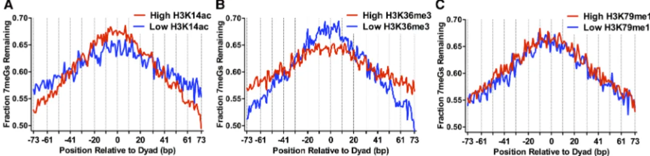
Histone post-translational modifications (PTMs) such as histone acetylation alter nucleosome dynamics (Neumann et al. 2009), and particular histone acetylation sites are associated with efficient NER (Yu et al. 2016); however, it is not known how histone acety-lation or other histone PTMs affect BER. To address this question, we analyzed our NMP-seq data using a nucleosome-resolution map of steady-state histone modifications in yeast (Weiner et al. 2015). This study showed that yeast histone modifications can be clustered based on their genome-wide distribution into two ma-jor groups: (1) histone PTMs associated with the 5′coding regions of yeast genes (e.g., H3K4me3, H3K9ac, H3K14ac, etc.), and (2) his-tone PTMs associated with the 3′ coding regions of yeast genes (H3K36me3, H3K79me3, etc.). Analysis of representative histone PTMs from each of these groups (i.e., H3K14ac and H3K36me3) re-vealed significant differences in BER of 7meG lesions among nu-cleosomes with high or low levels of these histone PTMs. There
was a smaller fraction of unrepaired 7meG lesions following 2-h re-pair at distal translational positions in nucleosomes with high lev-els of H3K14ac relative to nucleosomes with low levlev-els of H3K14ac, while there was a relatively higher fraction of unrepaired 7meG lesions near the nucleosome dyad (Fig. 3A). A similar trend was observed for a number of other histone PTMs associated with group 1 (e.g., H3K4me3, H3K23ac, H4K5ac) (Supplemental Fig. S6A–C). These results suggest that high levels of pre-existing his-tone acetylation are associated with more rapid BER at distal trans-lational locations within nucleosomes and paradoxically slower repair at translational positions near the nucleosome dyad.
The opposite trend was observed for nucleosomes with high levels of group 2 histone PTMs (i.e., H3K36me3). Nucleosomes with high levels of H3K36me3 had a higher fraction of 7meG le-sions remaining at distal translational locations relative to the low H3K36me3 controls following 2-h repair but a lower fraction of unrepaired 7meG lesions near the nucleosome dyad (Fig. 3B). Roughly similar trends were observed for a number of other group 2 histone PTMs (e.g., H3K36me2, H3K79me3) (Supplemental Fig.
S6D,E). These results may in part be due to the negative correlation
between H3K36me3 and histone acetylation (Weiner et al. 2015), since H3K36 methylation functions to recruit histone deacetylase complexes to the 3′ coding region of yeast genes (Lee and Shilatifard 2007).
However, not all histone modifications were associated with distinct patterns of repair. For example, there was no apparent dif-ference in the pattern of unrepaired 7meG lesions between nucle-osomes with high and low levels of H3K79 monomethylation (H3K79me1) (Fig. 3C). While pre-existing histone PTMs were asso-ciated with distinct patterns of repair assoasso-ciated with translational positioning in nucleosomes, the effect of rotational setting on re-pair was not apparent in this nucleosome map. This is likely due to the relatively low precision of the MNase-ChIP-seq method in de-fining nucleosome dyad positions (Weiner et al. 2015).
MMS-induced mutations are enriched near the dyad of strongly
positioned nucleosomes in yeast
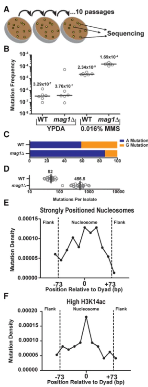
Since nucleosome structure causes such striking effects on the re-pair of NMPs in vivo, we next sought to determine whether the de-creased repair efficiency within strongly positioned nucleosomes ultimately resulted in an increase in MMS-induced mutation den-sity in these regions. We therefore subjected WT andmag1Δyeast to 10 repeated 2-d exposures of 0.016% MMS to accumulate
MMS-induced mutations and subsequently sequenced the ge-nomes of 24 independent clonal isolates of each genotype (Fig. 4A) to determine the location and spectra of the acquired muta-tions. Treatment of WT andmag1Δyeast with a single 2-d exposure to 0.016% MMS increased CanRmutation frequencies∼71- and ∼450-fold over untreated yeast, respectively (P= 0.0022 for both by Mann-WhitneyUtest) (Fig. 4B), indicating that greater than 98.6% and 99.8%, respectively, of accumulated mutations in the MMS-treated strains are the result of MMS-induced DNA lesions. Whole-genome sequencing of MMS-treated WT yeast revealed that, in accordance with previously published MMS-induced tion spectra (Roberts et al. 2012), nearly equal numbers of muta-tions occurred at“A”nucleotides compared to“G”nucleotides (∼59% and∼41%, respectively) (see Fig. 4C), despite 7meG lesions being formed approximately eightfold more frequently than 3meA. This result is consistent with prior studies (Roberts et al. 2012) and highlights the greater mutagenicity of 3meA lesion rela-tive to 7meG (Shrivastav et al. 2010). Compared to WT yeast,mag1Δ yeast displayed a much higher overall number of mutations per ge-nome (Fig. 4D) and a greater number of A-mutations relative to G-mutations (∼86% and∼14%, respectively,P< 0.0001) (Fig. 4C), in-dicating that MMS-treatment is inducing mutagenic NMP lesions that are normally repaired by Mag1 and highlighting the greater ef-ficiency of Mag1 in removing 3meA lesions (Bjoras et al. 1995).
Analysis of the trinucleotide sequence context in which MMS-induced mutations and lesions occurred (Supplemental
Figs. S7, S8) indicated that the relative abundances of G- and
A-mu-tations, as well as 7meG and 3meA lesions, strongly correlated with the trinucleotide composition of the yeast genome (P≤
0.002; Pearson coefficient > 0.75 for independent comparisons of G-mutation to G bases in WT yeast and A-mutations to A bases inmag1Δby Pearson correlation tests;P= 0.042; Pearson coeffi-cient = 0.51 for A-mutations compared to A bases in WT yeast), in-dicating that MMS-induced lesions and mutations occur largely independent of sequence specificity. Additionally, both 7meG-and 3meA-induced mutations produce biased base substitutions (favoring G to A or T and A to G) and occur with little to no se-quence specificities (Supplemental Fig. S8).
We next determined the frequency of mutations occurring within∼10,000 strongly positioned nucleosomes and their sur-rounding linker DNA, normalizing the number of mutations ob-served across the nucleosome by the relevant sequence content of A and G bases. We found that, similar to the patterns observed with NMP-seq data (Supplemental Fig. S5A), strongly positioned nucleosomes had significantly higher mutation densities com-pared to the flanking linker DNA (P= 0.0149), with mutation den-sity peaking near the dyad axis (Fig. 4E;Supplemental Fig. S9B). In contrast, mutation densities in weakly positioned nucleosomes (∼7500 nucleosomes) displayed no significant difference (P= 0.2935) between positions near the nucleosome dyad and those in the flanking linker DNA (Supplemental Fig. S9A,B). These find-ings indicate that reduced BER efficiency in strongly positioned nucleosomes, particularly near the dyad axis, is a key determinant of MMS-induced mutation density in vivo.
We also examined mutation density among nucleosomes with differing levels of histone PTMs, since our NMP-seq data indi-cated that histone PTMs such as H3K14ac and H3K36me3 signifi-cantly altered the pattern of repair of NMP lesions within nucleosomes. Consistent with the repair data (Fig. 3B), nucleo-somes with low levels of H3K36me3 showed a peak of mutation density near the dyad axis (Supplemental Fig. S9C). In contrast, the effect of translational positioning on mutation density was
Figure 4. Analysis of MMS-induced mutations across the genome of WT andmag1Δyeast. (A) Individual isolates (n= 24) of WT andmag1Δyeast strains were grown on 0.016% MMS-supplemented yeast extract peptone dextrose adenine (YPDA) media at 30°C for 48 h. Separate lines were then streaked for singles (not explicitly shown) on additional MMS plates, for a total of 10 passages. At the end of the passaging, unique isolates were pick-ed for whole-genome sequencing. (B) CanRfrequencies were measured for
six independent isolates of WT andmag1Δstrains of yeast (open circles), which were either untreated (grown on YPDA media) or treated for 2 d with 0.016% MMS. Black horizontal bars and numbers indicate the medi-an frequency of Cmedi-anRfor each genotype and treatment. Data were
ana-lyzed using a two-tailed Mann-WhitneyUtest. (C) Percentage of A- and G-mutations in whole-genome sequencing of MMS-treatedmag1Δand WT strains. (D) Number of mutations per isolate from whole-genome se-quencing of MMS-treated WT andmag1Δstrains. (E) Strongly positioned nucleosomes have elevated mutation density relative to flanking DNA re-gions in WT yeast. Mutations from MMS-treated WT isolates were mapped to strongly positioned nucleosomes (score > 5) and normalized by se-quence context. An area of∼180 bp was examined, which consisted of the main nucleosome (147 bp) and an additional flank on either side (∼30 bp total). Each DNA sequence was then divided into 11 bins, each of∼16 bp. Comparisons of the number of mutations observed between nucleosome-bound and flanking DNA was performed usingχ2analysis.
less apparent for nucleosomes with high levels of H3K36me3
(Supplemental Fig. S9D). A similar trend was observed for
nucleo-somes with high levels of H3K14ac (Fig. 4F), again consistent with our repair data (Fig. 3A), although the difference between nucleo-somes with high and low H3K14ac was less clear (cf. Fig. 4F and
Supplemental Fig. S9E). These findings suggest that variations in
repair efficiency in nucleosomes due to histone PTMs can influ-ence mutation rates.
MMS-induced A-mutations are enriched on the
NTS of yeast genes and are associated with higher
levels of 3meA lesions
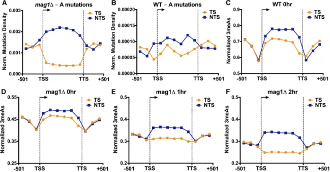
Analysis of MMS-induced mutations at adenine nucleotides (i.e., A to G, A to C, or A to T mutations) revealed a novel asymmetry in mutation density between the TS and NTS of yeast genes. This ef-fect was particularly apparent in the MMS-treatedmag1Δstrain, as there were fivefold more A-mutations on the NTS relative to the TS in yeast coding sequences, even after normalizing for the frequen-cy of“A”nucleotides in the TS and NTS (P< 0.0001 byχ2test; Fig.
5A). In MMS-treated WT yeast, we also observed a significant en-richment of A-mutations occurring on the NTS relative to the TS (P= 0.0023) (Fig. 5B), although the magnitude of the enrichment was smaller than in themag1Δstrain. In contrast, flanking promot-er and downstream DNA lacked a significant strand asymmetry in themag1Δ(P= 0.9014) (Fig. 5A) and wild-type strains (P= 0.5473). Surprisingly, the densities of A-mutations in both the TS and NTS in themag1Δstrain significantly differed from that of the promoter and downstream flanking regions, with A-mutations being elevat-ed in the NTS and relevat-educelevat-ed in the TS relative to flanking DNA (Fig. 5A). This suggests that two independent processes occurring on
the NTS and TS may contribute to the transcriptional asymmetry of MMS-induced mutations.
To investigate the molecular mechanisms underlying the strand asymmetry in MMS-induced A-mutations, we first analyzed the initial formation of 3meA lesions in our NMP-seq data sets. While the levels of 3meA lesions were much lower than 7meG sions overall, we consistently observed higher levels of 3meA le-sions on the NTS of yeast genes relative to the TS immediately following MMS treatment (i.e., WT 0-h andmag1Δ0-h samples) (see Fig. 5C,D). The magnitude of this difference was relatively small, ranging from 6%–11% more 3meA lesions on the NTS rela-tive to the TS after normalizing for the frequency of A nucleotides on the NTS and TS, but was consistent across multiple experiments
(Supplemental Fig. S10A–C). There were also consistently higher
3meA lesions on the NTS relative to flanking DNA (∼11%–18% more 3meA lesions) (see Fig. 5C,D). These differences were magni-fied in amag1Δstrain treated with a chronic dose of 0.02% MMS for 3 h (∼24% more 3meA lesions on the NTS relative to the TS and∼17% more 3meA lesions on the NTS relative to flanking DNA) (see Supplemental Fig. S10D), experimental conditions that more closely mimic themag1ΔMMS mutation experiments. These results indicate that higher 3meA lesion formation on the NTS of yeast genes likely contributes to transcriptional asymmetry in MMS-induced A-mutations.
3meA lesions are preferentially repaired on the TS
in a BER-deficient yeast strain
While strand-specific differences in 3meA lesion formation can explain the elevated levels of A-mutations on the NTS, this mech-anism cannot explain the significantly lower levels of A-mutations
on the TS relative to flanking DNA, since initial levels of 3meA le-sions on the TS were generally higher than flanking DNA (Fig. 5C, D). However, analysis of 3meA lesions in subsequent repair time points (i.e., 1- and 2-h) in themag1Δstrain revealed a progressive loss of 3meA lesions on the TS, both relative to the NTS and flank-ing DNA (Fig. 5E,F). To compare repair efficiency between TS and NTS, we analyzed the fraction of 3meA lesions remaining at the 1-and 2-h repair time points in themag1Δstrain relative to initial damage levels (i.e.,mag1Δ0-h). While overall 3meA lesions de-creased in themag1Δstrain following a 1- or 2-h repair incubation, presumably reflecting ongoing DNA replication and trans-lesion synthesis in themag1Δstrain (Johnson et al. 2007), the levels of 3meA lesions decreased more rapidly on the TS relative to the NTS or flanking DNA (Fig. 6A,B). While the transcribed region showed a clear strand-specific difference in 3meA repair, there was no difference in 3meA removal in the flanking promoter and downstream sequences (Fig. 6A,B). Moreover, there was no strand-specific difference in 7meG repair in themag1Δstrain at the 1- and 2-h repair time points (Fig. 6C; Supplemental Fig. S11), indicating the observed repair activity is specific for 3meA le-sions. These results indicate that in a BER-deficient strain there is preferential repair of 3meA lesions on the TS of yeast genes, provid-ing a potential explanation for the significantly lower levels of A-mutations on the TS in the MMS-treatedmag1Δstrain.
Based on these results, we hypothesized that transcription-coupled nucleotide excision repair (TC-NER) functions as a backup repair pathway to preferentially remove 3meA lesions from the TS of yeast genes to prevent MMS-induced mutagenesis and cytoxic-ity in BER-deficient cells. To test this hypothesis, we analyzed the
MMS sensitivity of yeast mutants lacking both BER (mag1Δ) and TC-NER (rad26Δ) pathways. While themag1Δmutant was sensitive to MMS, as expected, none of the NER-deficient mutants (i.e.,
rad14Δ,rad16Δ, andrad26Δ) were sensitive to low doses of MMS when BER was functional (Fig. 6D). However, themag1Δrad26Δ
double mutant showed enhanced MMS sensitivity (Fig. 6D), con-sistent with a previous study (Lee et al. 2002), indicating that TC-NER may function in NMP removal in a BER-deficient strain. In contrast, deletion ofRAD16,which causes a specific defect in the global genome nucleotide excision repair (GG-NER) pathway and is highly sensitive to UV irradiation (Supplemental Fig. S12) but does not affect TC-NER, did not show elevated MMS sensitivity in amag1Δbackground (Fig. 6D). Deletion ofRAD14, which elim-inates both TC-NER and GG-NER, also significantly enhanced the MMS sensitivity of amag1Δstrain to an even greater extent than themag1Δrad26Δdouble mutant (Fig. 6D). Previous studies have shown that deletion ofRAD26 does not completely eliminate TC-NER in yeast (Li and Smerdon 2002b), which may explain the higher MMS sensitivity in themag1Δrad14Δdouble mutant. These data are consistent with the model that the Rad26-mediated TC-NER pathway specifically repairs cytotoxic and mutagenic 3meA lesions on the TS of yeast genes.
Discussion
While efficient repair of DNA base lesions is critical to maintaining genome integrity and preventing mutations, the impact of chro-matin and other genomic features on BER and mutagenesis in vivo is not well understood. Here, we utilized a novel method
known as NMP-seq to map the initial formation and repair of DNA alkylation damage across the yeast genome at single nucleotide res-olution. Our results indicate that BER of MMS-induced alkylation damage is significantly modulated by cellular chromatin, with slower repair of lesions at translational positions near the nucleo-some dyad and at inward rotational settings. Furthermore, we show that the effect of translational and rotational positioning on repair is asymmetric relative to the nucleosome dyad and signif-icantly modulated by histone PTMs. Importantly, slower repair within strongly positioned nucleosomes correlated with an in-creased frequency of MMS-induced mutations in yeast, indicating that chromatin-associated variations in BER efficiency can impact mutation rates in vivo. Finally, we find that MMS-induced muta-tions at adenine nucleotides show a striking DNA strand asymme-try among yeast genes, particularly in the absence of BER, with a significantly higher mutation frequency on the NTS relative to the TS. We show that this is due to higher initial 3meA formation on the NTS of yeast genes and preferential removal of 3meA lesions from the TS. In summary, our data reveal that chromatin structure, backup DNA repair activities, and transcription are prominent sculptors of mutation distributions associated with alkylation DNA base damage.
We find that repair of 7meG lesions in cellular chromatin is significantly modulated by the translational and rotational setting of the lesion, with faster repair of lesions at“Out”rotational set-tings and translational positions distal from the nucleosome dyad, including linker DNA, and slower repair at“In”rotational settings and translational positions near the dyad. While these findings are generally consistent with in vitro studies of BER in
“designed”nucleosomes (Hinz and Czaja 2015; Rodriguez et al. 2015), the effects of translational and rotational setting on BER ef-ficiency have not been previously demonstrated in vivo (Li and Smerdon 2002a; Li et al. 2015). These nucleosome-dependent dif-ferences in BER efficiency translate to a striking pattern of BER sur-rounding yeast genes. Peaks of unrepaired 7meG lesions correlate with the stereotypic positioning of nucleosomes downstream from the TSS (e.g., the +1 nucleosome, +2 nucleosome, etc.), while repair of 7meG lesions occurs much more rapidly in NDRs up-stream of the TSS. Furthermore, we show that the effects of rota-tional setting on BER efficiency are primarily confined to the 5′ half of each nucleosomal DNA strand due to strand-specific differ-ences in chromatin accessibility at“Out”rotational settings. This asymmetry has not previously been detected in biochemical stud-ies of BER in nucleosomes but could be tested using defined nucle-osome substrates in vitro.
Histone PTMs have been shown to regulate other repair path-ways (e.g., NER and DNA double-strand break repair), but their role in BER is unclear (Mao and Wyrick 2016). Our analysis indicates that pre-existing histone PTMs cause subtle but significant differ-ences in BER efficiency among nucleosomes in vivo. For example, high levels of histone acetylation, such as H3K14ac, are associated with faster repair at distal translational positions near the DNA exit/entry sites but paradoxically slower repair near the nucleo-some dyad. Histone acetylation is often associated with increased nucleosomal DNA unwrapping, particularly at distal translational positions (Neumann et al. 2009), which could explain the faster re-pair of 7meG lesions at these positions in highly acetylated nucle-osomes. While slower repair near the nucleosome dyad among highly acetylated nucleosomes seems counterintuitive, a recent re-port suggests that H3K14ac and other histone acetylations (i.e., H3K56ac) inhibit DNA polymeraseβactivity during repair of le-sions located near the dyad of nucleosome substrates in vitro
(Rodriguez et al. 2016). The opposite trend was observed for his-tone PTMs associated with the 3′ coding regions of yeast genes (e.g., H3K36me3), presumably because these histone PTMs are as-sociated with histone deacetylation (Lee and Shilatifard 2007; Weiner et al. 2015). While our data indicate that MMS-induced mutations in wild-type yeast are significantly elevated near the nu-cleosome dyad relative to flanking regions among strongly posi-tioned nucleosomes, this effect appears to be magnified among nucleosomes with high levels of H3K14ac or correspondingly low levels of H3K36me3. Notably, in wild-type yeast, MMS-in-duced mutagenesis mirrors the effect of translational positioning on BER efficiency within these nucleosomes, suggesting that dif-ferences in repair due to histone PTMs may be an important con-tributor to mutation rates. It would be interesting to determine to what extent this mechanism regulates mutation rate in human cancers, since it is known that certain histone PTMs correlate with mutation density in sequenced cancer genomes (Schuster-Bockler and Lehner 2012).
In the course of analyzing MMS-induced mutations, we dis-covered a significantly higher frequency of mutations at adenine nucleotides on the NTS relative to the TS of yeast genes. This DNA strand asymmetry was magnified in the BER-deficient
mag1Δstrain, suggesting that rapid removal of 3meA lesions by Mag1 suppresses this effect. Our data suggest that two distinct mechanisms contribute to this novel transcriptional asymmetry.
First, there is higher initial 3meA formation on the NTS of yeast genes, even after normalizing for differences in DNA se-quence context. Previous studies have suggested that ongoing transcription can render the NTS more susceptible to chemical modifications, as the NTS becomes transiently single-stranded dur-ing transcription-associated R-loop formation (Kim and Jinks-Robertson 2012); it is possible that a similar mechanism contrib-utes to the higher frequency of 3meA formation and A-mutations on the NTS of yeast genes. However, single-stranded DNA also transiently accumulates on the lagging strand during DNA replica-tion and A-mutareplica-tions were not enriched on the lagging strand in
mag1Δyeast, instead showing a slight (but significant) enrichment on the leading strand (Supplemental Fig. S13). Thus, either muta-genesis associated with increased formation of 3meA on single-stranded DNA occurs in a transcription-specific context or other mutagenic processes occurring during leading strand synthesis (possibly strand bias in the usage of error-free polymerases) ob-scure this mutation signature at the replication fork.
however, it is not known to what extent this occurs with eukaryot-ic RNA polymerases. We hypothesize that persistent 3meA lesions may trigger TC-NER in yeast by stalling RNA polymerase II, imply-ing that TC-NER may play a broader role in repairimply-ing nonbulky DNA lesions than previously appreciated. While our data suggest that TC-NER repairs certain classes of alkylation damage in yeast, it is not clear if TC-NER plays a similar role in mammalian cells. A previous study reported that there was no strand bias in the re-pair of 3meA lesions at theDHFRgene in a BER-deficient murine cell line lacking AAG (Plosky et al. 2002), even though NER con-tributes to NMP lesion removal at this locus. It will be important to analyze the genome-wide repair of alkylation damage in human cells, since these findings may have important implications for un-derstanding the molecular basis of transcriptional asymmetries in mutations found in many human cancers (Alexandrov et al. 2013; Roberts and Gordenin 2014).
Methods
MMS treatment, NMP-seq library preparation, and sequencing
Yeast cells (BY4741) were grown in YPD (yeast extract, peptone, dextrose) medium to OD600≈0.8. MMS (Sigma-Aldrich) was
add-ed to the yeast culture to a final concentration of 0.2% (v/v) or 0.4%, and incubated at 30°C for 10 min with shaking to induce al-kylation damage. Cell pellets were spun down and washed twice with sterile deionized water to remove remaining MMS. Aliquots were taken before adding MMS and immediately after MMS incu-bation for“No MMS”and“MMS 0-h”samples, respectively. The remainder of the cells was resuspended in prewarmed YPD medi-um and incubated in a 30°C shaker, and samples were collected at different repair times as indicated. For chronic MMS treatment, yeast cells (OD600≈0.6) were grown in YPD containing 0.02%
MMS for 3 h. MMS was then removed, and cells were collected for DNA isolation.
Yeast genomic DNA was isolated as previously described (Mao et al. 2016). The procedure of NMP-seq library preparation is sim-ilar to our published CPD-seq method (Mao et al. 2016), except that damaged DNA was incubated with recombinant human AAG glycosylase (AAG 1-79Δ, a gift from Dr. Leona Samson at MIT) to cleave NMPs. The resulting abasic sites were further pro-cessed by incubating DNA with recombinant AP endonuclease APE1 (NEB) to generate new 3′OHs at NMP sites. The resulting li-brary was amplified for approximately five cycles by PCR, using primers complementary to trP1 and A, size-selected with AMpure XP beads, and sequenced on an Ion Torrent Proton platform (Life Technologies). Validation of the NMP-seq library, including trP1 ligation, free 3′OH blocking, and A adaptor ligation, was con-ducted as previously described for CPD-seq damage mapping (Mao et al. 2016). DNA adaptor and primer sequences are provided in the
Supplemental Methods.
NMP-seq data analysis
NMP-seq data analysis was performed using a modified version of the published protocol for analyzing CPD-seq data (Mao et al. 2016). A detailed description of the data analysis methods is pro-vided in theSupplemental Methods.
Accumulation of MMS-induced mutations in yeast
Diploid wild-type or mag1Δ yeast (BY4743) accumulated muta-tions by being repeatedly passaged on YPDA media containing 0.016% MMS. Briefly, individual colonies were streaked for singles on YPDA supplemented with 0.016% MMS, then allowed to grow
for 48 h. Then, a single colony unique to each streak was picked and struck out onto another MMS-containing YPDA plate. This was repeated for 10 passages. Total genomic DNA was purified from 24 independent WT andmag1Δisolates, sequenced, and mu-tations called similar to Sakofsky et al. (2014). A detailed descrip-tion of these methods and the analysis of the mutadescrip-tion distribution are provided in theSupplemental Methods. All muta-tions identified are listed inSupplemental Table S2.
Data access
The NMP-seq data from this study have been submitted to the NCBI Gene Expression Omnibus (GEO; http://www.ncbi.nlm. nih.gov/geo/) under accession number GSE98031. The MMS-in-duced mutation sequencing data from this study have been sub-mitted to the NCBI Sequence Read Archive (SRA; https://www. ncbi.nlm.nih.gov/sra) under accession number SRP105152.
Acknowledgments
We thank Mark Wildung and Wei Wei Du for technical assistance with Ion Proton sequencing and Amelia Hodges for helpful com-ments and suggestions. This research was supported by grants from the National Institute of Environmental Health Sciences (NIEHS) (R01ES002614 to J.J.W. and M.J.S., R03ES027945 to P.M., and R00ES022633 to S.A.R.), Department of Defense Congressionally Directed Medical Research Programs (BC141727 to S.A.R.), and an internal grant from the Washington State University College of Veterinary Medicine (to J.J.W.).
References
Akbari M, Morevati M, Croteau D, Bohr VA. 2015. The role of DNA base ex-cision repair in brain homeostasis and disease.DNA Repair32:172–179. Alexandrov LB, Nik-Zainal S, Wedge DC, Aparicio SA, Behjati S, Biankin AV, Bignell GR, Bolli N, Borg A, Borresen-Dale AL, et al. 2013. Signatures of mutational processes in human cancer.Nature500:415–421. Bauer NC, Corbett AH, Doetsch PW. 2015. The current state of eukaryotic
DNA base damage and repair.Nucleic Acids Res43:10083–10101. Bjoras M, Klungland A, Johansen RF, Seeberg E. 1995. Purification and
prop-erties of the alkylation repair DNA glycosylase encoded the MAG gene fromSaccharomyces cerevisiae.Biochemistry34:4577–4582.
Brogaard K, Xi L, Wang JP, Widom J. 2012. A map of nucleosome positions in yeast at base-pair resolution.Nature486:496–501.
Bryan DS, Ransom M, Adane B, York K, Hesselberth JR. 2014. High resolu-tion mapping of modified DNA nucleobases using excision repair en-zymes.Genome Res24:1534–1542.
Cui F, Cole HA, Clark DJ, Zhurkin VB. 2012. Transcriptional activation of yeast genes disrupts intragenic nucleosome phasing.Nucleic Acids Res
40:10753–10764.
Ding J, Taylor MS, Jackson AP, Reijns MA. 2015. Genome-wide mapping of embedded ribonucleotides and other noncanonical nucleotides using emRiboSeq and EndoSeq.Nat Protoc10:1433–1444.
Friedberg EC, Walker GC, Siede W, Wood RD, Schultz RA, Ellenberger T. 2006.DNA repair and mutagenesis. ASM Press, Washington, DC. Fu D, Calvo JA, Samson LD. 2012. Balancing repair and tolerance of DNA
damage caused by alkylating agents.Nat Rev Cancer12:104–120. Hanawalt PC, Spivak G. 2008. Transcription-coupled DNA repair: two
de-cades of progress and surprises.Nat Rev Mol Cell Biol9:958–970. Hinz JM, Czaja W. 2015. Facilitation of base excision repair by chromatin
remodeling.DNA Repair36:91–97.
Hu J, Adar S, Selby CP, Lieb JD, Sancar A. 2015. Genome-wide analysis of hu-man global and transcription-coupled excision repair of UV damage at single-nucleotide resolution.Genes Dev29:948–960.
Jiang C, Pugh BF. 2009. A compiled and systematic reference map of nucle-osome positions across theSaccharomyces cerevisiaegenome.Genome Biol
10:R109.
Johnson RE, Yu SL, Prakash S, Prakash L. 2007. A role for yeast and human translesion synthesis DNA polymerases in promoting replication through 3-methyl adenine.Mol Cell Biol27:7198–7205.
Lee JS, Shilatifard A. 2007. A site to remember: H3K36 methylation a mark for histone deacetylation.Mutat Res618:130–134.
Lee SK, Yu SL, Prakash L, Prakash S. 2002. YeastRAD26, a homolog of the humanCSBgene, functions independently of nucleotide excision repair and base excision repair in promoting transcription through damaged bases.Mol Cell Biol22:4383–4389.
Li S, Smerdon MJ. 2002a. Nucleosome structure and repair of N-methylpur-ines in theGAL1-10genes ofSaccharomyces cerevisiae.J Biol Chem277: 44651–44659.
Li S, Smerdon MJ. 2002b. Rpb4 and Rpb9 mediate subpathways of transcrip-tion-coupled DNA repair in Saccharomyces cerevisiae. EMBO J 21: 5921–5929.
Li M, Ko T, Li S. 2015. High-resolution digital mapping ofN-methylpurines in human cells reveals modulation of their induction and repair by near-est-neighbor nucleotides.J Biol Chem290:23148–23161.
Mao P, Wyrick JJ. 2016. Emerging roles for histone modifications in DNA excision repair.FEMS Yeast Res16:fow090.
Mao P, Smerdon MJ, Roberts SA, Wyrick JJ. 2016. Chromosomal landscape of UV damage formation and repair at single-nucleotide resolution.Proc Natl Acad Sci113:9057–9062.
Mao P, Wyrick JJ, Roberts SA, Smerdon MJ. 2017. UV-induced DNA damage and mutagenesis in chromatin.Photochem Photobiol93:216–228. Nag R, Smerdon MJ. 2009. Altering the chromatin landscape for nucleotide
excision repair.Mutat Res682:13–20.
Neumann H, Hancock SM, Buning R, Routh A, Chapman L, Somers J, Owen-Hughes T, van Noort J, Rhodes D, Chin JW. 2009. A method for genetically installing site-specific acetylation in recombinant his-tones defines the effects of H3 K56 acetylation.Mol Cell36:153–163. Plosky B, Samson L, Engelward BP, Gold B, Schlaen B, Millas T, Magnotti M,
Schor J, Scicchitano DA. 2002. Base excision repair and nucleotide exci-sion repair contribute to the removal ofN-methylpurines from active genes.DNA Repair1:683–696.
Plosky BS, Frank EG, Berry DA, Vennall GP, McDonald JP, Woodgate R. 2008. Eukaryotic Y-family polymerases bypass a 3-methyl-2′ -deoxyade-nosine analogin vitro and methyl methanesulfonate-induced DNA damagein vivo.Nucleic Acids Res36:2152–2162.
Racine JF, Zhu Y, Mamet-Bratley MD. 1993. Mechanism of toxicity of 3-methyladenine for bacteriophage T7.Mutat Res294:285–298. Rando OJ, Winston F. 2012. Chromatin and transcription in yeast.Genetics
190:351–387.
Riedl J, Fleming AM, Burrows CJ. 2016. Sequencing of DNA lesions facilitat-ed by site-specific excision via base excision repair DNA glycosylases yielding ligatable gaps.J Am Chem Soc138:491–494.
Roberts SA, Gordenin DA. 2014. Hypermutation in human cancer genomes: footprints and mechanisms.Nat Rev Cancer14:786–800.
Roberts SA, Sterling J, Thompson C, Harris S, Mav D, Shah R, Klimczak LJ, Kryukov GV, Malc E, Mieczkowski PA, et al. 2012. Clustered mutations in yeast and in human cancers can arise from damaged long single-strand DNA regions.Mol Cell46:424–435.
Rodriguez Y, Hinz JM, Smerdon MJ. 2015. Accessing DNA damage in chro-matin: preparing the chromatin landscape for base excision repair.DNA Repairdoi: 10.1016/j.dnarep.2015.04.021.
Rodriguez Y, Hinz JM, Laughery MF, Wyrick JJ, Smerdon MJ. 2016. Site-spe-cific acetylation of histone H3 decreases polymeraseβactivity on nucle-osome core particles in vitro.J Biol Chem291:11434–11445. Sakofsky CJ, Roberts SA, Malc E, Mieczkowski PA, Resnick MA, Gordenin
DA, Malkova A. 2014. Break-induced replication is a source of mutation clusters underlying kataegis.Cell Rep7:1640–1648.
Schuster-Bockler B, Lehner B. 2012. Chromatin organization is a major in-fluence on regional mutation rates in human cancer cells.Nature488:
504–507.
Shrivastav N, Li D, Essigmann JM. 2010. Chemical biology of mutagenesis and DNA repair: cellular responses to DNA alkylation.Carcinogenesis
31:59–70.
Wallace SS, Murphy DL, Sweasy JB. 2012. Base excision repair and cancer. Cancer Lett327:73–89.
Weiner A, Hsieh TH, Appleboim A, Chen HV, Rahat A, Amit I, Rando OJ, Friedman N. 2015. High-resolution chromatin dynamics during a yeast stress response.Mol Cell58:371–386.
Wyrick JJ, Roberts SA. 2015. Genomic approaches to DNA repair and muta-genesis.DNA Repair36:146–155.
Yu S, Evans KE, van Eijk P, Bennett M, Webster RM, Leadbitter M, Teng Y, Waters R, Jackson SP, Reed SH. 2016. Global genome nucleotide exci-sion repair is organized into domains that promote efficient DNA repair in chromatin.Genome Res26:1376–1387.
Yuan GC, Liu YJ, Dion MF, Slack MD, Wu LF, Altschuler SJ, Rando OJ. 2005. Genome-scale identification of nucleosome positions inS. cerevisiae. Science309:626–630.
Zhong J, Luo K, Winter PS, Crawford GE, Iversen ES, Hartemink AJ. 2016. Mapping nucleosome positions using DNase-seq. Genome Res 26:
351–364.