B.-J. van Rossum
Structure refinement
of photosynthetic components
with multidimensional MAS NMR
dipolar correlation spectroscopy
B.-J
.
v
an
Rossum
Structur
e
re
finement
of
photosynthetic
components
with
MAS
NMR
spectr
Structure refinement of photosynthetic components
with multidimensional MAS NMR dipolar correlation
Structure refinement of photosynthetic components
with multidimensional MAS NMR dipolar correlation
spectroscopy
PROEFSCHRIFT
ter verkrijging van
de graad van Doctor aan de Universiteit Leiden,
op gezag van de Rector Magnificus Dr. W.A. Wagenaar,
hoogleraar in de faculteit der Sociale Wetenschappen,
volgens besluit van het College voor Promoties
te verdedigen op donderdag 11 mei 2000
te klokke 15.15 uur
door
Barth-Jan van Rossum
P
ROMOTIECOMMISSIEPROMOTORES : prof. dr. H.J.M. de Groot prof. dr. J. Lugtenburg REFERENT : prof. dr. S. Vega OVERIGE LEDEN : prof. dr. J. Reedijk
prof. dr. A.J. Hoff prof. dr. D. Bedeaux
Contents
Chapter 1 General introduction
1.1 Introduction ... 8
1.2 Photosynthesis ... 9
1.3 MAS NMR spectroscopy and photosynthesis ... 11
1.4 Heteronuclear (1H-13C) dipolar correlation spectroscopy ... 12
References ... 15
Chapter 2 Theoretical background: frequency-switched Lee-Goldburg irradiation and Lee-Goldburg cross-polarization 2.1 Introduction ... 17
2.2 Lee-Goldburg irradiation ... 19
2.3 Lee-Goldburg cross-polarization ... 22
References ... 25
Chapter 3 High magnetic field and high-speed MAS for enhanced proton resolution in heteronuclear (1H-13C) dipolar correlation spectroscopy 3.1 Introduction ... 27
3.2 Experimental ... 28
3.3 Results and discussion ... 30
3.4 Conclusions ... 34
References ... 34
Chapter 4 Heteronuclear dipolar correlation spectroscopy with frequency-switched Lee-Goldburg homonuclear decoupling 4.1 Introduction ... 36
4.2 Frequency-switched Lee-Goldburg irradiation ... 37
4.3 Heteronuclear (1H-13C) dipolar correlation spectroscopy ... 38
4.3.1 Preparation and evolution ... 38
4.3.2 The mixing period ... 39
4.3.3 Short CP contact times ... 40
4.3.4 Lee-Goldburg CP ... 43
4.3.5 Multidimensional correlation spectroscopy ... 43
Contents 6
4.4 The Lee-Goldburg scaling factor ... 46
4.5 Frequency-switched LG and sample rotation ... 48
4.6 Experimental details ... 49
References ... 50
Chapter 5 A method for measuring heteronuclear (1H-13C) distances in high-speed MAS NMR 5.1 Introduction ... 51
5.2 Experimental ... 52
5.3 Results ... 54
5.4 Discussion ... 63
5.5 Conclusions ... 66
References ... 66
Chapter 6 The 3-D structure of self-assembled chlorophyll a / H2O from multispin labeling and MAS NMR 2-D dipolar correlation spectroscopy in high magnetic fields 6.1 Introduction ... 68
6.2 Experimental ... 70
6.3 Results ... 72
6.3.1 13C-13C homonuclear dipolar correlation spectroscopy ... 72
6.3.2 1H-13C heteronuclear dipolar correlation spectroscopy ... 76
6.4 Discussion ... 78
6.4.1 Chemical shift and distance constraints on the structural model of the Chl a / H2O stacks ... 78
6.4.2 Chemical shift and distance constraints on the structural model of the Chl a / H2O layered sheets ... 82
6.4.3 The bilayer structure of the aggregate ... 85
6.4.4 The suprastructure of the aggregate ... 87
6.5 Conclusions ... 90
References ... 90
Chapter 7 Proton shifts from high-field 2-D and 3-D high-speed CP/MAS 13C dipolar correlation spectroscopy of aggregated BChl c in uniformly 13C-enriched chlorosomal antennae of Chlorobium tepidum 7.1 Introduction ... 92
7.2 Experimental ... 94
7.3 Results ... 96
7.4 Discussion ... 102
References ... 114
Chapter 8 Binding of QA and QB in Rhodobactersphaeroides R26 reaction centers 8.1 Introduction ... 116
8.2 Experimental ... 119
8.2.1 Preparation of [1-13C] QA Rb. sphaeroides R26 RCs ... 119
8.2.2 Preparation of [1-13C] QB Rb. sphaeroides R26 RCs ... 121
8.2.3 NMR spectroscopy... 122
8.3 Results ... 123
8.4 Discussion ... 128
8.5 Conclusions ... 132
References ... 133
Chapter 9 13C MAS NMR evidence for structural similarity of L162YL mutant and Rhodobacter sphaeroides R26 RC, despite widely different cytochrome c2 -mediated re-reduction kinetics of the oxidized primary donor Abstract ... 136
1. Introduction ... 136
2. Experimental ... 137
3. Results ... 139
4. Discussion ... 141
Acknowledgements ... 143
References ... 143
Chapter 10 General discussion and outlook... 144
Summary ... 146
Samenvatting ... 150
Publications ... 154
Curriculum vitae ... 156
1
Chapter
1.1 Introduction
Magic-angle spinning nuclear magnetic resonance (MAS NMR) dipolar correlation
spectroscopy is rapidly forthcoming as a versatile technique for de novo structure
determination of microscopically ordered systems without long-range translation symmetry
[1]. It has been shown that de novo structure determination is possible with 13C homonuclear
MAS NMR dipolar correlation spectroscopy when multiple labeling is used [1-4]. While 13C
homonuclear dipolar correlation spectroscopy is now being used routinely in assignment and
structure refinement studies of organic solids, 1H MAS NMR has not yet found a widespread
application as a tool for structure determination. Due to the combination of the strong
homonuclear dipolar interactions between the abundant protons in the solid state and the
small proton chemical shift dispersion, the proton resolution is often very limited which
makes 1H NMR in solids difficult.
The scope of the research described in this thesis is to demonstrate that protons can be
used in structure refinement studies of biological systems in the solid state using
cross-polarization (CP) MAS NMR dipolar correlation spectroscopy. To this end, first a set of
NMR techniques should be developed to suppress the 1H homonuclear dipolar interactions
under MAS conditions. In a second step, the versatility of protons for structure
determination with solid-state NMR should be demonstrated by exploiting the novel NMR
techniques in a structural investigation of various biological systems.
The contents in the thesis are divided into two main parts. Chapters 2-5 focus on the
development of the solid-state NMR. Chapter 2 provides a short theoretical background,
while Chapters 3-5 are concerned with the development of the novel solid-state NMR
spectroscopy techniques that enable the use of protons in multidimensional heteronuclear
dipolar correlation spectroscopy. The second part of the thesis, Chapters 6-9, is dedicated to
the application of these and other solid-state NMR techniques to study systems in the field of
photosynthetic research, like native chlorosome antennae, synthetic antenna model systems
and photosynthetic bacterial reaction centers.
1.2 Photosynthesis
Photosynthesis is the collection of life-sustaining processes that convert (solar) light-energy
into chemical energy, which is stored in energy-rich organic material, collectively called
biomass. Photosynthetic organisms can be divided into two groups. Plants, algae and
cyanobacteria belong to the first group that is capable of oxygenic photosynthesis, in which
the light-energy is used in a reductive fixation of carbon dioxide into carbohydrates under
oxidization of water. In this process oxygen is produced. The second group of
photosynthetic organisms comprises the anoxygenic photosynthetic bacteria, that use
molecules other than water as an electron donor, for instance H2S or organic acids. In the
anoxygenic photosynthetic energy conversion no oxygen is produced. The photosynthetic
organelles that are investigated in this thesis are extracted from bacteria of this second
group.
Photosynthesis is a multistep process with a high degree of compartimentalization. It
takes place in a set of complex molecules located in or attached to the photosynthetic
membrane. The two major steps that can be distinguished in the primary processes of the
photosynthetic energy conversion are:
I: The harvesting of light and transfer of the light-energy to the reaction center (RC)
II: Charge separation and subsequent electron transport in the RC
The absorption of light, the conversion of the light-energy into electronic excitation-energy
and the subsequent migration of the excitation-energy to the RC in step I, take place in the
photosynthetic antenna system. In higher plants, green algae, heliobacteria and some purple
bacteria, the antenna system is embedded within the photosynthetic membrane. These
antennae consists of chlorophyll-carotenoid-protein complexes. In other photosynthetic
bacteria the antenna is situated in extra-membranous organelles, connected to the
photosynthetic membrane. For instance, in green sulfur bacteria (Chlorobiaceae) and green
filamentous bacteria (Chloroflexaceae), the major light-harvesting apparatus is formed by
chlorosomes, which are oblong bodies that are attached to the cytoplasmic side of the cell
membrane [5-7]. In contrast to plant antenna systems, the chlorosomes are nearly
protein-free and their structure and function rely on the self-aggregation of the antenna pigments not
mediated by a protein [8,9]. In this respect the chlorosomes form a unique class of
photosynthetic antennae. Chlorosomes can be separated from the photosynthetic apparatus
and have been the subject of many studies (for a recent review of the experimental work, see
Chapter 1 10
The RC is a transmembrane protein complex. It plays a key-role in the photosynthetic
energy conversion, since here the actual charge separation and photochemistry take place.
RCs of purple photosynthetic bacteria, for instance Rhodobacter (Rb.) sphaeroides, have
been extensively investigated in the past few years. Nowadays efficient procedures exist for
growing strains of this species, for the isolation of the RC complexes in the native
membrane, and for the preparation and manipulation of samples for a wide variety of
investigations [11]. For the purple bacteria Rhodopseudomonas viridis and Rb. sphaeroides,
RCs have been crystallized and studied with X-ray diffraction, from which a detailed
knowledge about their structure was obtained [12-16]. The RCs of the purple bacteria Rb.
sphaeroides R26 consist of three polypeptide subunits supporting nine cofactors: four
bacteriochlorophylls (B), two bacteriopheophytins (Φ), two quinones (Q) and one non-heme Fe2+. Two bacteriochlorophyll molecules form the special pair P. The cofactors are arranged
in two branches, designated A and B, with a nearly two-fold symmetry (Fig. 1.1).
Upon illumination, the special pair is photo-oxidized and an electron is transferred
along the A-branch across the membrane, first to the primary quinone QA. QA is tightly and
permanently bound to the protein and serves as a one-electron gate. It temporarily accepts a
single electron, which is subsequently transferred to the secondary quinone QB. Following a
second excitation, QB is doubly reduced and doubly protonated and leaves the RC as a
diquinol. Subsequently, the empty QB binding pocket is occupied by a new ubiquinone-10
(UQ10) molecule which is taken up from the quinone-pool (for a review, see e.g. [17]).
regenerates the oxidized special pair P+ to P. After replacement with a fresh UQ10 molecule,
the RC has returned to its original state and the reaction is cyclic.
Under physiological conditions, electrons have a strong preference to travel along the
A-branch. Thus, despite the apparent two-fold symmetry of the A- and B-branch (cf. Fig.
1.1), the molecular mechanisms of the function of the RC are highly asymmetric [18]. The
question why nature has chosen for this functional symmetry-breaking has been an important
issue in the photosynthesis research field during the past decade.
Unraveling the molecular mechanisms of photosynthesis and gaining fundamental
knowledge about one of the most important processes in living nature already provide
important motives for studying the concepts of photosynthesis. Second, a thorough
understanding of the molecular mechanisms of photosynthetic energy conversion will
potentially be of help for the development and improvement of artificial photosynthesis
devices, which can become an important source of renewable energy in the foreseeable
future [19].
1.3 MAS NMR spectroscopy and photosynthesis
In recent years, progress has been made in forging pathways for obtaining MAS NMR
access to membrane proteins in general and to photosynthetic components in particular [20].
These studies rely on the use of labeling schemes and 13C CP/MAS NMR. The natural
abundance of 13C is low, ~ 1 %, and in order to enhance the sensitivity, the use of 13C
enrichment is a prerequisite. For instance, using labeled spheroidene obtained by total
synthesis, the configuration of the 15-15’ bond of the carotenoid reconstituted into R26 RCs
was established [21]. The electronic ground-state of the one-electron gate QA in the RC has
been probed with MAS NMR and isotope labeling [22]. An advantage of selective labeling
is a direct and straightforward chemical shift assignment of the response from the nucleus of
interest. However, selective labeling is most often difficult to realize. In particular for
chlorophyll, to arrive at a complete set of specifically labeled molecules at every individual
position will take many years of organic synthesis work.
Recently, different routes for obtaining information from multiply enriched samples
were explored. The details of the electronic ground-state structure of the RC protein complex
of Rb. sphaeroides have been investigated for tyrosine side chains labeled at the 4’-position,
with particular focus on M210 and L162, that are of importance for the efficiency of the
charge separation and re-reduction processes in the RC [23-25]. In these studies, all
tyrosines in the RC were selectively labeled, and the assignment of the response from the
Chapter 1 12
another approach, a novel example of photochemically-induced dynamic nuclear
polarization (photo-CIDNP) was discovered, yielding strong emissive signals for QA
depleted, or pre-reduced uniformly 15N-labeled RCs [26]. Finally, it was found that the large
chemical shift dispersion of the 13C response of ~ 200 ppm can be exploited for
high-resolution dipolar correlation spectroscopy of 13C nuclei in multiply enriched samples, due
to a truncation of the homonuclear dipolar interactions by the chemical shift dispersion in
high magnetic fields. This yields remarkably narrow lines in the 2-dimensional (2-D) MAS
NMR 13C homonuclear dipolar correlation spectra of uniformly 13C-enriched ([U-13C])
chlorophylls [2]. It was used recently to refine the structure at the molecular level of an
uniformly labeled intact chlorosome photosynthetic antenna system that is inaccessible to
X-ray or solution NMR approaches [2-4,27].
1.4 Heteronuclear (
1H-
13C) dipolar correlation spectroscopy
An important aim of this thesis is to demonstrate that protons can be utilized in MAS NMR
for assignment strategies, structure determination and structure-function studies of
microscopically ordered systems without long-range translation symmetry in general, and
photosynthetic components in particular. Thus far, the dipolar correlation spectroscopy has
not found a widespread application to study hydrogens in large biological preparations like
membrane proteins in the solid state. Protons play a very important role in the structure and
function of proteins, since they are involved in the formation of hydrogen bonds that
determine the secondary structure of a protein. In addition, protons take part in the binding
of cofactors to a protein and stabilize the self-assembly of pigments in e.g. chlorosome
antenna systems. However, the dipolar line-broadening in the solid state generally results in
a proton resolution that is insufficient for structural research.
In the current opinion an improved resolution can only be obtained by taking
advantage of the large 13C chemical shift dispersion in heteronuclear (1H-13C) correlation
spectroscopy. First, in Chapter 2 a simple theoretic framework is provided to describe the
effect of off-resonance radio-frequency irradiation of the protons during 1H evolution and
CP. Next, in Chapter 3 it is shown that increasingly high magnetic fields are essential to
improve the spectral resolution in multidimensional spectroscopy with MAS. It is
demonstrated that straightforward high-speed MAS heteronuclear (1H-13C) CP wide-line
separation (CP/WISE) spectroscopy [28] performed at a high magnetic field and without any
homonuclear decoupling scheme during the proton evolution, already yields resolved 1H-13C
correlations. Proton chemical shifts can be obtained directly from such spectra [29].
dipolar correlation spectra of multispin 13C clusters can be acquired at high spinning speeds
when frequency-switched Lee-Goldburg irradiation is applied during the 1H evolution
[30-33]. The assignment of solid-state proton chemical shifts from the heteronuclear correlation
spectra can provide information about the electronic structure of a densely packed solid.
Non-bonding interactions can be quite strong in the solid state and even small shifts can be
significant, if the shift effects are correlated in the sense that they follow a pattern or that
they are extended over a region of the molecule [3,4]. In addition, the range of the coherent
spin-diffusion in the solid state is intrinsically much larger than in solution [34]. It is shown
in Chapter 4 that Lee-Goldburg CP [35,36] in combination with heteronuclear dipolar
correlation spectroscopy can be exploited to detect 1H-13C heteronuclear intermolecular
correlations and to provide unambiguous structural restraints [37].
In Chapter 5, a method is presented that can be applied in uniformly 13C-enriched
compounds to extract 1H-13C heteronuclear distances with good precision from CP build-up
curves, which are recorded at high MAS rates under simultaneous suppression of the 1H
homonuclear dipolar interactions. The Fourier transform of the time-oscillatory
magnetization build-up curves provides direct access to heteronuclear (1H-13C) dipolar
coupling strengths. An empirical relation between the heteronuclear distance and the dipolar
coupling strength is constructed from a series of simulations. It is demonstrated for a [U-13C]
tyrosine·HCl model compound that this relationship can be useful in the translation of
experimental coupling strengths into distances between the coupled spins. The
experimentally determined internuclear distances compare very well with the distance
information extracted from the neutron diffraction structure of tyrosine·HCl [38].
A concept for structure determination using 13C homonuclear and 1H-13C heteronuclear
dipolar correlation spectroscopy is presented in Chapter 6 [1,4]. The concept is applied in a
3-dimensional (3-D) structure determination study of aggregates of [U-13C] chlorophyll a /
H2O. Chlorophyll a (Chl a) is the green pigment involved in photosynthetic harvesting of
light and subsequent conversion of light-energy into chemical energy by higher plants and
related species, like algae and cyanobacteria. It forms aggregates when exposed to water
[39]. The aggregated Chl a is thought to represent a paradigm for a system that is potentially
important for artificial-photosynthesis research [39]. It is shown in Chapter 6 that knowledge
about the electronic structure deduced from the solid-state proton assignment from
intramolecular heteronuclear (1H-13C) correlations, as well as distance constraints obtained
from the observation of several intermolecular heteronuclear correlations, provide
information that can be interpreted consistently into a 3-D structural model of the
self-assembled Chl a / H2O.
In Chapter 7, the arrangement of [U-13C] bacteriochlorophyll (BChl) c in intact
Chapter 1 14
heteronuclear dipolar correlation spectroscopy. Since 3-D crystals of the chlorosome antenna
were not yet obtained, the system is not amenable to high-resolution diffraction techniques.
BChl c is the major chromophore of the chlorosomes of the bacterium Chlorobium tepidum,
and is known to form aggregates [40-42]. BChl c is related to Chl a in the sense that both
pigments have three unsaturated pyrrole rings, unlike e.g. BChl a and BChl b, that only have
two unsaturated rings. There is growing evidence that the internal structure in the
chlorosomes is based on the self-organization of BChl cnot directly mediated by proteins
[8,9]. In particular, from previous NMR studies using 13C homonuclear dipolar correlation
spectroscopy it was concluded that the stacking of BChl c in the chlorosomes and in
artificial aggregates is highly similar, which provides convincing evidence that indeed the
self-organization of the chromophore is the main mechanism to support the structure of the
chlorosomes [3]. In Chapter 7 the proton assignment from the heteronuclear correlation
spectroscopy is used to refine the model for the arrangement of the BChl c in the
chlorosomes.
In Chapter 8, the binding of ground-state QA and QB in the RC protein complex of Rb.
sphaeroides and the formation of hydrogen bonds to the surrounding protein is investigated.
Knowledge about the hydrogen-bonding interactions of the quinones to the protein can help
to understand the different electrochemical function and binding properties of QA and QB in
the RC in the ground state (Fig. 1.1). To this end, RCs reconstituted with [1-13C] UQ10 [43]
for QA or QB are studied with heteronuclear dipolar correlation spectroscopy and CP
build-up curves of the label signal are recorded. In this way, the proton(s) that interact with the 1-13
C=O of QA or QB are characterized in terms of their chemical shift and the distance to the
1-13C of the quinones. Strong evidence is provided by the NMR for a strong
hydrogen-bonding interaction in ground-state RCs of both 1-13C=O QA and QB with the surrounding
protein. This contrasts with Fourier transform infrared spectroscopy, which suggested an
essentially free or weakly bound 1-13C=O QA [44-46].
Finally, in Chapter 9, the protein environment of the tyrosine residue L162 that is of
importance for the efficiency of the charge separation and re-reduction processes in the RC
of Rb. sphaeroides is investigated. To this end, details of the electronic ground-state
structure are studied for L162YL mutant RCs, i.e., with leucine substituted for tyrosine
L162, in which all tyrosines were selectively labeled at the 4’-position [25]. In this study an
unambiguous assignment of the solid-state NMR response from L162 is achieved by
comparing the data of [4’-13C] Tyr enriched L162YL mutant RCswith [4’-13C] Tyr enriched
RCs of Rhodobacter sphaeroides R26 and of the [4’-13C] Tyr enriched mutant M210YW
[23-25]. M210YW and L162YL are the first two mutant RCs that have been studied to
References
[1] van Rossum, B.-J.; Boender, G.J.; Mulder, F.M.; Raap, J.; Balaban, T.S.; Holzwarth, A.; Schaffner, K.;
Prytulla, S.; Oschkinat, H.; de Groot, H.J.M. (1998) Spectrochim. Acta A 54, 1167.
[2] Boender, G.J.; Raap, J.; Prytulla, S.; Oschkinat, H.; de Groot, H.J.M. (1995) Chem. Phys. Lett. 237, 502.
[3] Balaban, T.S.; Holzwarth, A.R.; Schaffner, K.; Boender, G.-J.; de Groot,H.J.M. (1995) Biochemistry
34, 15259.
[4] Boender, G.J. (1996) Ph.D. Thesis, Leiden University, the Netherlands.
[5] Staehelin, L.A.; Golecki, J.R.; Fuller, R.C.; Drews, G. (1978) Arch. Mikrobiol. 119, 269. [6] Staehelin, L.A.; Golecki, J.R.; Drews, G. (1980) Biochim. Biophys. Acta 589, 30. [7] Olson, J.M. (1980) Biochim. Biophys. Acta594, 33.
[8] Holzwarth, A.R.; Griebenow, K.; Schaffner, K. (1990) Z. Naturforsch. 45C, 203.
[9] Holzwarth, A.R.; Griebenow, K.; Schaffner, K. (1992) J. Photochem. Photobiol. A 65, 61. [10] Olson, J.M. (1998) Photohem. Photobiol.67, 61.
[11] Feher, G.; Okamura, M.Y. (1978) in: the Photosynthetic Bacteria (R.K. Clayton, W.R. Sistrom, Eds.),
p. 349, Plenum Press, New York.
[12] Deisenhofer, J.; Epp, O.; Miki, K.; Huber, R.; Michel, H. (1985) Nature318, 618.
[13] Allen, J.P.; Feher, G.; Yeates, T.O.; Komiya, H.; Rees, C.D. (1988) Proc. Natl. Acad. Sci. USA 85, 8487.
[14] Chang, C.H.; El-Kabbani, O.; Tiede, D.; Norris, J.; Schiffer, M. (1991) Biochemistry30, 5352.
[15] Chirino, A.J.; Lous, E.J.; Huber, M.; Allen, J.P.; Schenck, C.C.; Paddock, M.L.; Feher, G.; Rees, D.
(1994) Biochemistry33, 4584.
[16] Ermler, U.; Fritzsch, G.; Buchanan, S.K.; Michel, H. (1994) Structure2, 925
[17] Bixon, M.; Fajer, J.; Feher, G.; Fied, J.H.; Gamliel, G.; Hoff, A.J.; Levanon, H.; Möbius, K.; Nechustai,
R.; Norris, J.R.; Schertz, A.; Sessler, J.L.; Stehlik, D. (1991) Isr. J. Chem. 32-4, 369. [18] Kirmaier, C.; Holten, D. (1990) Biochemistry30, 609.
[19] van Rossum, B.; Soede, C.; Steensgaard, D.; Holzwarth, A.; Schaffner, K.; Raap, J.; Lugtenburg, J.;
Gast, P.; Hoff, A.; de Groot, H. (1999) submitted for the proceedings ofthe 8th European Conference
on the Spectroscopy on Biological Molecules’, August 29 - September 2, 1999, Twente, The
Netherlands.
[20] de Groot, H.J.M. (1996) in: Biophysical Techniques in Photosynthesis (Advances in Photosynthesis) (J.
Amesz, A.J. Hoff, Eds.), Vol. 3, p. 299, Kluwer academic publishers.
[21] de Groot, H.J.M.; Gebhard, G.; v.d. Hoef, I.; Hoff, A.J.; Lugtenburg, J.; Violette, C.A.; Frank, H.A.
(1992) Biochemistry31, 12446.
[22] van Liemt, W.B.S.; Boender, G.J.; Gast, P.; Hoff, A.J.; Lugtenburg, J.; de Groot, H.J.M. (1995)
Biochemistry34, 10229.
[23] Fischer, M.R.; de Groot, H.J.M.; Raap, J.; Winkel, C.; Hoff, A.J.; Lugtenburg, J. (1992) Biochemistry
Chapter 1 16
[24] Shochat, S.; Gast, P.; Hoff, A.J.; Boender, G.J.; van Leeuwen, S.; van Liemt, W.B.S.; Vijgenboom, E.;
Raap, J.; Lugtenburg, J.; de Groot, H.J.M. (1995) Spectrochim. ActaA 51, 135.
[25] van Rossum, B.-J.; Wachtveitl, J.; Raap, J.; v.d. Hoeff, K.; Gast, P.; Lugtenburg, J.; Oesterhelt, D.; de
Groot, H.J.M. (1997) Spectrochim. ActaA53, 2201.
[26] Zysmilich, M.; McDermott, A.E. (1994) J. Am. Chem. Soc.116, 8362.
[27] Boender, G.J.; Balaban, T.S.; Holzwarth, A.R.; Schaffner, K.; Raap, J.; Prytulla, S.; Oschkinat, H.; de
Groot, H.J.M. (1995) in: Photosynthesis: From Light to Biosphere (P. Mathis, Ed.), Vol. 1, p. 347,
Kluwer Academic Publishers, Boston, Dordrecht, London.
[28] Schmidt-Rohr, K.; Clauss, J.; Spiess, H.W. (1992) Macromolecules25, 3273.
[29] van Rossum, B.-J.; Boender, G.J.; de Groot, H.J.M. (1996) J. Magn. Reson. A120, 274. [30] Lee, M.; Goldburg, W.I. (1965) Phys. Rev. A 140, 1261.
[31] Bielecki, A.; Kolbert, A.C.; Levitt, M.H. (1989)Chem. Phys. Lett.155, 341.
[32] Bielecki, A.; Kolbert, A.C.; de Groot, H.J.M.; Griffin, R.G.; Levitt, M.H. (1990) Advances in Magnetic
Resonance 14, 111.
[33] van Rossum, B.-J.; Förster, H.; de Groot, H.J.M. (1997) J. Magn. Reson. 124, 516.
[34] Mulder, F.M.; Heinen, W.; van Duin, M.; Lugtenburg, J.; de Groot, H.J.M. (1998) J. Am. Chem. Soc.
120, 12891.
[35] Caravatti, P.; Bodenhausen, G.; Ernst, R.R. (1982) Chem. Phys. Lett.89, 363. [36] Wu, C.H.; Ramamoorthy, A.; Opella, S.J. (1994) J. Magn. Reson.A 109, 270.
[37] van Rossum, B.-J.; Prytulla, S.; Oschkinat, H.; de Groot, H.J.M. (1998) in: Magnetic Resonance and
Related Phenomena (D. Ziessow, W. Lubitz, F. Lendzian, Eds.), Vol. 1, p. 38, Technische Universität,
Berlin.
[38] Frey, M.N.; Koetzle, T.F.; Lehmann, M.S.; Hamilton, W.C. (1973) J. Chem. Phys.58, 2547. [39] Worcester, D.L.; Michalski, T.J.; Katz, J.J. (1986) Proc. Natl. Acad. Sci. U.S.A. 83, 3791. [40] Bystrova, M.I.; Mal’gosheva, I.N.; Krasnovskii, A.A. (1979) Mol. Biol. Engl. Trans. 13, 440. [41] Smith, K.M.; Kehres, L.A.; Fajer, J. (1983) J. Am. Chem. Soc. 105, 1387.
[42] Miller, M.; Gillbro, T.; Olson, J.M. (1993) Photochem. Photobiol. 57, 98.
[43] van Liemt, W.B.S.; Steggerda, W.F.; Esmeijer, R.; Lugtenburg, J. (1994) Rec. Trav. Chim. Pays-Bas
113, 153.
[44] Brudler, R.; de Groot, H.J.M.; van Liemt, W.B.S.; Steggerda, W.F.; Esmeijer, R.; Gast, P.; Hoff, A.J.;
Lugtenburg, J.; Gerwert, K. (1994) EMBO J. 13, 5523.
[45] Breton, J.; Boullais, C.; Burie, J.-R.; Nabedryk, E.; Mioskowski, C. (1994) Biochemistry33, 14378. [46] Brudler, R.; de Groot, H.J.M.; van Liemt, W.B.S.; Gast, P.; Hoff, A.J.; Lugtenburg, J.; Gerwert, K.
(1996) in: Reaction Centers of Photosynthetic Bacteria, Structure and Dynamics (M.E. Michel-Beyerle,
2
2.1 Introduction
Many solid-state NMR experiments rely on the application of radio frequency (RF) pulses to
manipulate the nuclear spin Hamiltonian. In most experiments, the RF field
) cos(
2 )
( 1 RF RF
RF t = H
ω
t+ψ
H is applied on or nearly on-resonance. In this case
ω
RF≅γ
H0, which is positive by definition [1]. Hereγ
is the gyromagnetic ratio of the spin and H0 is the applied static magnetic field (Fig 2.1A). Alternatively, the design of an NMR techniquecan be based on the application of an off-resonance RF field. In that case an effective field
Heff arises, inclined at an angle
θ
≠π
2 with respect to the rotating frame z-axis (Fig 2.1B). In this thesis, the focus will be on techniques that enable the use of protons insolid-state NMR. In solids, the protons, or I spins, are mutually coupled via strong homonuclear dipolar interactions. For a static sample, the homonuclear dipolar interaction between the I
spins in the spin-pair approximation can be expanded into five terms HIIM, with M = -2, …, +2 [2]. Only HII0 is secular, which means that it commutes with the static-field Zeeman interaction. In a high magnetic field, this is the term that remains and the truncated
Hamiltonian for the homonuclear dipolar interaction has the form [2]
) 3
(
0 II
∑
<
⋅ − =
j i
j i zj zi ij I I
a I I
H ,
frequency-switched Lee-Goldburg irradiation
and Lee-Goldburg cross-polarization
Chapter 2 18 with 3 2 2 2 I
0 (3cos 1)
2
4 ij
ij ij
r
a −
−
=
γ
θ
π
µ
h,
where rij is the size of the distance vector rij between the spins i and j, and
θ
ij is the angle between rij and the z-axis. For off-resonance RF irradiation, HII0 can be transformed to a tilted frame, with the new ~z -axis along the effective field Heff =H1ex +[H0 −(ω
RF/γ
I)]ez(Fig. 2.1B). This yields the tilted Hamiltonian [3]
secular non 2 2 1 II ) ~ ~ ~ ~ 3 ( ) 1 cos 3 ( ~ H
H = − ⋅
∑
− ⋅ +<j i j i zj zi ij I I
a I I
θ
.The non-secular part of this Hamiltonian contains terms that do not commute with
∑
iIzi
~ .
In Section 2.2, Lee-Goldburg (LG) irradiation is discussed, which can be used to
suppress the strong homonuclear dipolar coupling between the I spins [3]. During LG
irradiation, the RF frequency is chosen off-resonance in such a way that the effective field
eff
H is inclined at the magic angle
θ
m = tan−1( 2) = 54.7° with respect to the static magnetic field H0 along the z-axis. As a result, the secular part of the homonuclear dipolar interaction vanishes due to the 21(3cos2θ
−1) dependence in Eq. (2.3) [3]. With0 I RF
LG=
ω
−γ
H∆ and |
ω
1I|=γ
IH1I, a LG condition can be defined according tom I
1
1(| |/ LG)
tan−
ω
∆ =θ
.In Section 2.3, cross-polarization (CP) between an 1H spin I and a 13C spin S with
suppression of the 1H homonuclear dipolar interactions using LG irradiation is discussed.
During CP, the two spin species are irradiated simultaneously on-resonance and
θ
=π
2 forboth (Fig 2.1A), yielding 2 21
2
1(3cos θ −1)=− in Eq. (2.3). In the doubly-rotating frame the
spin Hamiltonian for a single spin S coupled to a set of interacting spins I during CP has the
form [4]
∑
∑
∑
+ − − ⋅ + = < i z zi i j i j i xj xi ij x ixi S a I I bI S
I 21 (3 )
S 1 I
1
ω
I Iω
Hwith ω1I =−γIH1I and ω1S =−γSH1S, which are negative for positive γ in a right-handed coordinate system [1]. This Hamiltonian is again truncated and contains only the part of the
homonuclear dipolar interaction that commutes with the RF irradiation term along the x-axis.
The last term represents the heteronuclear dipolar interaction HIS between the S spin and the I spins, with
3 2 2
S I
0 (3cos 1)
2
4 i
i i
r
b −
−
= γ γ θ
where ri is the distance between S and Ii, and θi is the angle between the distance vector ri
and the z-axis. If the CP experiment is performed in such a way that the I spins are irradiated
off-resonance at a Lee-Goldburg condition, the third term in Eq. (2.4) due to the
homonuclear dipolar interaction between the I spins vanishes.
During magic-angle spinning (MAS) NMR, the sample is rotated with a spinning
speed ωr 2π around an axis that is inclined at the magic angle θm with respect to the z-axis. With MAS, the form of the spin operators in the Hamiltonians in Eqs. (2.3) and (2.4)
remains the same, while the coefficients of the homonuclear and heteronuclear dipolar
interaction aij(t) and bi(t) contain terms that vary periodically in time with frequencies kωr,
with k = −2, −1, +1, +2 [5].
In the terminology of Maricq and Waugh, the homonuclear dipolar interactions
between like spins in a rotating sample are homogeneous, which means that the
corresponding Hamiltonian does not commute with itself at different times [6]. In practice,
the spinning speed ωr 2π is often in the slow MAS range 0 2 II 2
r 2 ) | |
(ω π << H , and the homonuclear dipolar couplings lead to a homogeneous broadening of the NMR response. In
contrast, the dipolar interactions between unlike spins are inhomogeneous, since the
interaction terms commute with themselves at all times. In absence of homonuclear dipolar
couplings, the total spin Hamiltonian comprising chemical shift and heteronuclear dipolar
coupling terms is also inhomogeneous. During MAS, the spectrum associated with such a
inhomogeneous Hamiltonian breaks up in a pattern of spinning side-bands, with the relative
intensities of the spinning side-bands determined by both the dipolar interaction and the
chemical shift [6,7]. At high MAS rates >10 kHz the heteronuclear dipolar interaction and
the chemical shift anisotropy are substantially reduced by the sample spinning. During CP,
the RF irradiation of the two spin species renders the total spin Hamiltonian homogeneous,
even in absence of homonuclear dipolar interactions. This effectively leads to a recoupling
of the IS interactions that are otherwise averaged by the MAS and the relevant dynamics will
involve the homogeneous part of the spin Hamiltonian.
2.2 Lee-Goldburg irradiation
In Chapter 4 it will be demonstrated that the use of frequency- and phase-switched
Lee-Goldburg irradiation (FSLG) during the 1H evolution of 2-D heteronuclear (1H-13C) dipolar
correlation spectroscopy strongly enhances the proton resolution. In this section the effect of
off-resonance RF irradiation on the spin Hamiltonian of strongly coupled I spins will be
Chapter 2 20
For a static sample in high magnetic field, the secular term HII0 in Eq. (2.1) comprises the major source of line-broadening [8] and it needs to be attenuated for high-resolution
spectroscopy. The application of an off-resonance RF field HRF(t)=2H1cos(ωRFt+ψRF) to the I spins in the off-resonance rotating frame, rotating with angular frequency ωframe =−ωRF along the z-axis of the laboratory frame, yields a truncated Hamiltonian
{
}
0II 1 I I RF 0
I( ) H
H =
∑
− − − +i
xi zi
zi
iI γ H ω γ I γ H I
δ ,
with the chemical shift dispersion
∑
iδiIzi included explicitly. When the RF power is high, 2
0 II 2
eff) | |
(
γ
H >> H , H can be transformed to a tilted rotating frame, defined by the transformation∏
iexp{
−iθmIyi}
[9], with the tilted ~z -axis along the direction of theeffective field Heff =H1ex +(H0 −
ω
RFγ
I)ez . In this tilted frame, HII0 transforms to five terms H~IIM, which yields, withω
eff =−γ
IHeff{
+ −}
+ ∑( )
= − =∑
2 2 II eff ~ ~ ) sin ( ~ ) cos ( ~ M M M i xi i zii I I H
H
δ
θ
ω
δ
θ
λ
θ
,where = − = − = ± ±
θ
θ
λ
θ
θ
θ
λ
θ
θ
λ
2 4 3 2 2 3 1 2 2 1 0 sin ) ( cos sin ) ( ) 1 cos 3 ( ) (( )
( )
= = + = = ⋅ − =∑
∑
∑
< + + −
< + +
− < j i j i ij j i j zi zj i ij j i j i zj zi ij I I a I I I I a I I a ~ ~ ~ ~ ) ~ ~ ~ ~ ( ~ ~ ) ~ ~ ~ ~ 3 ( ~ † 2 II 2 II † 1 II 1 II 0 II H H H H
H I I
From Eq. (2.9) the following commutation relations are obtained
[
M]
MiIzi II M II
~ ~ , ~ H H =
∑
,hence H~II0 is the only secular term in Eq. (2.7) [3].
During FSLG irradiation, the RF field HRF(t) is frequency switched between LG
0 I
LG = ±∆ ∆
±
γ
Hω
with ∆LG= 21 2ω
1I , and phase switched between LG ∆ ±ψ
, withπ
ψ
ψ
+∆LG− −∆LG = , after successive periodsτ
LG = 2 3⋅(2π
ω
1I) [10,11]. As a result,ω
efftoggles between ∆LG cos
θ
and −∆LG cosθ
. Subsequent transformation to the frame rotating withω
eff along the z~-axis is performed using the propagator∏
− = i zi I t i tUeff( ) exp(
ω
eff ~ ) ,and leads to the two time-dependent Hamiltonians
∑
−= −
∆ ±
i ziI t
U t U
t) ( ) ~ ( ) ~
( ~ eff 1 eff eff LG
ω
H Hfor
ω
eff =±∆LG cosθ
during the successive periodsτ
LG and{
}
( )
+ ∑ ±( )
+ − = ≠− = ∆ ±∑
2 02 eff II
0 II 0 eff eff LG ~ ) exp( ~ ) sin ~ cos ~ ( sin ~ ) cos ( ) ( ~ M M M M i yi xi i zi i t M t I t I I t H H H
θ
λ
ω
θ
λ
ω
ω
θ
δ
θ
δ
m .When the off-resonance frequency is set to a LG condition, the H~II0 term vanishes due to the
( )
12(
3cos2 1)
0
θ
=θ
−λ
dependence [3]. The remaining linewidth originates from the non-secular terms H~II±1 and H~II±2 [3]. These non-secular terms will be truncated when the tilted effective field is large, i.e., (γ
Heff)2 >>| H~IIM |2. The truncation will be most effective for the2 II
~±
H terms which have a 2
ω
eff time-dependence.It is important to realize that all five terms H~IIM in the tilted rotating frame originate from the single term HII0 in the rotating frame. This implies an additional advantage when
0 II
H is attenuated by a high static field, since in that case the terms H~IIM should also be reduced. An increased proton chemical shift dispersion in high fields effectively attenuates
the truncated homonuclear dipolar couplings and high magnetic fields have a line-narrowing
effect on the MAS proton response. An experimental verification of this phenomenon is
presented in Chapter 3.
The factor cos
θ
=1 3 in Eq. (2.13) scales the chemical shift dispersion. The time-dependent transverse part will be refocused due to the sign reversal ofω
eff between the two successive periods. By settingω
effτ
LG =2π
, the two frames rotating with ±ω
eff along thez
~-axis will coincide with the tilted rotating frame at the beginning and at the end of each
period
τ
LG. Hence the evolution of the spin system can be monitored from the ±ω
eff rotating frames, in which it evolves under the two Hamiltonians according to Eq. (2.13), andthe proton magnetization will effectively evolve in the plane perpendicular to the tilted ~z -axis.
For a spinning sample, the coefficient aij of the homonuclear dipolar interaction in Eq.
(2.1) is time-dependent. Since we assumed above that the RF power is high in the sense
2 0 II 2
eff) | |
(
γ
H >> H , the secular part of the Hamiltonian (2.13) will be suppressed due to the( )
21(
3cos2 1)
0
θ
=θ
−λ
dependence. On the other hand, the non-secular terms with M ≠ 0, in Eq. (2.13) will be suppressed over a full FSLG cycle 2⋅τ
LG, provided that the cycle time is short compared to the period of the sample rotation. In theory, this implies that the RF power (2.11)(2.12)
Chapter 2 22
should be sufficiently high, since this will both lead to a short 2⋅
τ
LG and to a more complete truncation of the non-secular part of the Hamiltonian. In practice, however, it turns out thatin high magnetic fields a moderately high RF power corresponding with an 1H nutation
frequency of ~ 60 kHz is already sufficient to achieve good resolution with FSLG irradiation
at MAS rates up to 15 kHz, as will be shown in Chapter 4.
2.3 Lee-Goldburg cross-polarization
In Chapter 5 the time-evolution of the signal intensity of a 13C spin S during CP/MAS with
Lee-Goldburg irradiation applied to a small finite number of I spins (1H) is analyzed
numerically. In this section, we present a first-order analytical theoretical description of a
two-spin system that consists of a spin S coupled to a single spin I, rotating with a MAS rate
π
ω
r 2 .The spin Hamiltonian in the doubly rotating frame for the two-spin system during the
mixing time of the Lee-Goldburg CP (LG-CP) experiment can be represented as
z z z
x
x S I b t I S
I 1S LG ( )
I
1 + −∆ +
=
ω
ω
H ,
where ∆LG=
ω
RF−ω
0I , withω
0I =−γ
IH0I, the frequency offset for the RF irradiation applied to the I spin [3,12,13]. The last term in Eq. (2.14) represents the heteronucleardipolar interaction HIS between the two spins, with [14]
[
cos( ) cos(2 2 )]
2 )
(t =
ω
d G1ω
rt+φ
+G2ω
rt+φ
b ,
3 IS 2 S I 0 d 4 r h
γ
γ
π
µ
ω
=− .In the tilted frame, defined by the transformation exp
{
−iθ
mIy} {
exp −i(π
2)Sy}
, the tilted spin Hamiltonian can be written as H~= H~0+ H~1(t), wherez z S I ~ ~ ~ S 1 I eff, 0 =
ω
+ω
H ,
(
IxSx IzSx)
t b
t) ( )sin( )~~ cos( )~~ (
~
m m
1 =
θ
−θ
H ,
with the effective field
ω
eff,I =−(ω
12I+∆LG2)12. The time-independent part H~0 can be removed from the Hamiltonian by transformation to the interaction frame, according to{ }
i 0t 1{
i 0t}
* 1 ~ exp ~ ~ exp ~ H H H
H = − . For the n = ±1 Hartmann-Hahn (HH) matching condition, (2.14)
(2.15)
r S 1 I
eff,
ω
ω
ω
− =± , the flip-flop part representing the heteronuclear dipolar interaction has the form{
}
[
{
exp(( ) ) exp( ( ) )}
~ ~ ) ) ( exp( ) ) ( exp( ~ ~ sin 4 ~ S 1 I eff, r S 1 I eff, r S 1 I eff, r S 1 I eff, r m 1 d * 1
φ
ω
ω
ω
φ
ω
ω
ω
φ
ω
ω
ω
φ
ω
ω
ω
θ
ω
i t i i t i S I i t i i t i S I G − + − − + + − + + − − + − + + + − ⋅ = + − − + Hwhich leads to a time-independent Hamiltonian
[
]
[
]
− = − + − ⋅ + = − − + ⋅ = + − − + + − − + r 1S I eff, m 1 d r 1S I eff, m 1 d * 1 for ) exp( ~ ~ ) exp( ~ ~ sin 4 for ) exp( ~ ~ ) exp( ~ ~ sin 4 ~ω
ω
ω
φ
φ
θ
ω
ω
ω
ω
φ
φ
θ
ω
i S I i S I G i S I i S I GH .
Likewise, the n = ±2 HH matching conditions,
ω
eff,I −ω
1S =±2ω
r , yield a Hamiltonian of theform
{
}
[
{
exp((2 ) 2 ) exp( (2 ) 2 )}
~ ~ ) 2 ) 2 ( exp( ) 2 ) 2 ( exp( ~ ~ sin 4 ~ S 1 I eff, r S 1 I eff, r S 1 I eff, r S 1 I eff, r m 2 d * 1
φ
ω
ω
ω
φ
ω
ω
ω
φ
ω
ω
ω
φ
ω
ω
ω
θ
ω
i t i i t i S I i t i i t i S I G − + − − + + − + + − − + − + + + − ⋅ = + − − + Hwith time-independent terms
[
]
[
]
− = − + − ⋅ + = − − + ⋅ = + − − + + − − + r 1S I eff, m 2 d r 1S I eff, m 2 d * 1 2 for ) 2 exp( ~ ~ ) 2 exp( ~ ~ sin 4 2 for ) 2 exp( ~ ~ ) 2 exp( ~ ~ sin 4 ~ω
ω
ω
φ
φ
θ
ω
ω
ω
ω
φ
φ
θ
ω
i S I i S I G i S I i S I GH .
The factor sin
θ
m in these expressions for H~1* scales the heteronuclear dipolar interaction. Only the flip-flop term is relevant for polarization transfer. The single quantum termx zS I t
b( )cos(
θ
m)~~− in Eq. (2.16) does not lead to polarization transfer and has been discarded. In addition, double quantum terms of the form I~+S~+ and I~−S~− have been left out in Eqs. (2.17)-(2.20), since for the n = ±1 and ±2 HH matching conditions, these terms connect
diagonal elements in the product basis mImS that have a large difference in energy and can be discarded according to perturbation theory. Finally, rapidly oscillating terms of the form
,
cos
ω
rt cos2ω
rt, etc., are neglected, since they should be small on average compared to the time-independent term. This is justified as long 41G|n| d m|r |
ω
sinθ
ω
>> .As an example, we will now study the time-dependent S spin magnetization build-up
during LG-CP, adjusted for the
ω
eff,I −ω
1S =+ω
r HH matching condition. The derivation of the expressions for the other matching conditions proceeds in a fully analogous way. Wewill write the Hamiltonian for the heteronuclear dipolar coupling as
(2.17)
(2.18)
(2.19)
Chapter 2 24
[
~ ~ exp( ) ~ ~ exp( )]
4 ~*
1
φ
φ
δ
I S i +I S −i= + − − +
H ,
with
δ
≡G1ω
dsinθ
m. Following excitation of the I spins, the density operator at the beginning of the LG-CP period in the interaction frame can be written asρ
0 =−Z−1β
Lω
0I~Iz, withβ
L =1kBT. The time-evolution ofρ
under H~1* can be evaluated using) ~ exp( ) ~ exp( )
(t iH1*t
ρ
0 iH1*tρ
= − , which leads to the following expression, in matrix-notation in the product basis mImS ,+ − − + − − + + − − − − ⋅ − = − t t i t i t Z t i i
δ
δ
φ
δ
φ
δ
ω
β
ρ
2 1 2 1 2 1 2 2 1 2 2 1 2 1 2 1 2 1 I 0 L 1 4 1 cos sin ) exp( 0 0 sin ) exp( cos 0 0 0 0 0 0 0 0 ) (The S-spin signal S(t) can be calculated by evaluation of the expectation value of S~z, according to <S~z(t)>=Tr(
ρ
(t)S~z), which leads to(
1 cos)
(1 exp( ) exp( )))
( 21 2
2 2 1 I 0 L 1 4 1 2 1 I 0 L 1 4
1Z t Z t t
t
S =− −
β
ω
−δ
=− −β
ω
− +iδ
− −iδ
. .
Hence, during the LG-CP, the S-spin signal oscillates with angular frequency 21
δ
around theaverage value −14Z−1
β
Lω
0I, and Fourier transformation ofI 0 L 1 4 1 )
(t + Z−
β
ω
S results in a
spectrum with two singularities at frequencies 21 1 d m 2
1
δ
ω
sinθ
ω
=± =± G .The expectation value for <S~z(t)> has been evaluated for a single crystallite. For a powder sample, it should be integrated over all crystallites. With 43sin(2 m)sin(2 )
1 ij
G =
θ
θ
[14], and
θ
ij the angle that the internuclear vector connecting spins i and j makes with the (2.21)(2.22)
rotor axis, we can write ( ) 12 0sin(2 ),
ij
ij
δ
θ
θ
ω
= where we defined 43 dsin( m)sin(2 m)0
ω
θ
θ
δ
≡ )cos( m
d
θ
ω
= . The powder average can be evaluated using S(
ω
)=P(θ
ij) dθ
ij(ω
)/dω
, with P(θ
ij) the angular distribution function [15]. Since the interaction is axially symmetric, we simply have P(θ
ij)=sinθ
ij [15], which leads to2 0 4 1 2 0 4 1 2 1 2 0 4 1 2 1 ) ( 2 ) ( ) ( ) (
δ
ω
δ
ω
δ
ω
ω
− − + + − − =S ,
with −
δ
0 2≤ω
≤δ
0 2. This powder spectrum is plotted in Fig. 2.2. It represents the LG-CP S-spin spectrum for the IS spin-pair, which is obtained after Fourier transformation of thetime-oscillating S-spin signal build-up [16]. The shape resembles a static Pake-pattern [17],
although the characteristic high-frequency ‘ears’ are missing. The frequency splitting
between the two maxima equals
δ
0, which is related to the heteronuclear distance rIS viaω
d in Eq. (2.15).References
[1] Levitt, M.H. (1997) J. Magn. Reson.126, 164.
[2] Ernst, R.R.; Bodenhausen, G.; Wokaun, A. (1987) in: Principles of Nuclear Magnetic Resonance in
One and Two Dimensions; Vol. 14 of International Series of Monographs on Chemistry; Clarendon
Press, Oxford.
[3] Lee, M.; Goldburg, W.I. (1965) Phys. Rev.A 140, 1261.
[4] Pines, A.; Gibby, M.G.; Waugh, J. (1973) J. Chem. Phys.59, 569. [5] Stejskal, E.O.; Schaefer, J.; Waugh, J.S. (1977) J. Magn. Reson.28, 105. [6] Maricq, M.M.; Waugh, J.S. (1979) J. Chem. Phys.70, 3300.
[7] Roberts, J.E.; Harbison, G.S.; Munowitz, M.G.; Herzfeld, J.; Griffin, R.G. (1987) J. Am. Chem. Soc.
109, 4163.
[8] Abragam, A. (1961) in: The principles of Nuclear Magnetism; Oxford University Press, London.
[9] Redfield, A.G. (1955) Phys. Rev. 98, 1787.
[10] Bielecki, A.; Kolbert, A.C.; Levitt, M.H. (1989) Chem. Phys. Lett.155, 341.
[11] Bielecki, A.; Kolbert, A.C.; de Groot, H.J.M.; Griffin, R.G.; Levitt, M.H. (1990) Adv. Magn. Reson. 14, 111.
[12] Caravatti, P.; Bodenhausen, G.; Ernst, R.R. (1982) Chem. Phys. Lett.89, 363.
[13] van Rossum, B.-J.; Prytulla, S.; Oschkinat, H.; de Groot, H.J.M. (1998) in: MagneticResonance and
Related Phenomena (D. Ziessow, W. Lubitz, F. Lendzian, Eds.), Vol. 1, p. 38, Technische Universität,
Berlin.
[14] Bennet, A.E.; Griffin, R.G.; Vega, S. (1994) in: NMR-Basic Principles and Progress; Vol. 33; p. 1,
Springer-Verlag, Berlin.
Chapter 2 26
[15] Schmidt-Rohr, K.; Spiess, H. (1994) in: Multidimensional Solid-state NMR and Polymers; Academic
Press, London.
[16] Bertani, P.; Raya, J.; Reinheimer, P.; Goneon, R.; Delmotte, L.; Hirschinger, (1999)J.SolidState NMR
13, 219.
3
Parts of this chapter were published in J. Magn. Reson.A120 (1996) 274-277
3.1 Introduction
It is generally accepted that 1H-13C heteronuclear dipolar correlation spectroscopy in solids requires the application of multiple-pulse techniques to suppress the strong homonuclear dipolar interactions between the abundant protons in order to achieve a sufficient line-narrowing in the 1H dimension. In this chapter, it is shown that a high magnetic field strength, in combination with fast magic-angle spinning (MAS), can already improve the proton resolution to such an extent that 1H-13C correlations and proton chemical shifts can be directly obtained from 2-dimensional (2-D) spectra, collected without the application of 1H homonuclear decoupling schemes during the proton evolution. In addition, it will be shown that in favourable cases, heteronuclear correlations between molecules, e.g. intermolecular hydrogen bonds, can be obtained with the simplest possible pulse schemes.
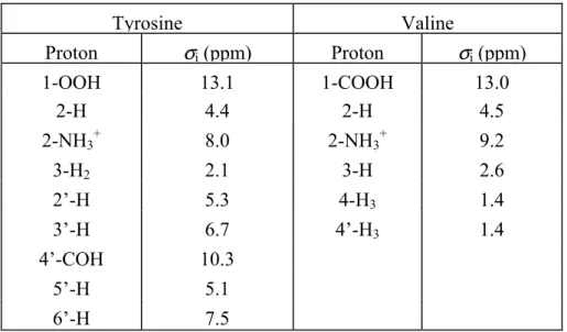
To demonstrate this, the most straightforward and very simple ‘cross-polarization wideline-separation’ (CP/WISE) technique is used [1] (Fig. 3.1A) to study a small model compound, labeled tyrosine (Fig. 3.1B). The CP/WISE pulse scheme does not apply any proton line-narrowing method. Following a
π
2 pulse on the protons, a time increment t1before the CP allows for the observation of the proton evolution with detection via the
carbons. Since the protons are allowed to evolve freely during t1, the technique enables a
comparison of the 1H homonuclear dipolar line-broadening under various experimental conditions. The purpose of this chapter is to demonstrate the effectiveness of increasingly
higher fields in combination with rapid spinning for enhancing the proton resolution in
heteronuclear correlation spectroscopy. We recognize that the 1H resolution in the correlation spectra presented below is still limited and that additional improvement can be
achieved by implementation of multiple-pulse line-narrowing techniques to suppress the
strong 1H homonuclear dipolar couplings. However, the point that we would like to make here is that almost every pulse scheme involving direct or indirect detection of protons can,
and in fact should, benefit from the additional resolution enhancement provided by a strong
Chapter 3 28
magnetic field and a high MAS rate. Hence, we feel that in the design of new pulse schemes,
their applicability at high magnetic field strengths and at high MAS speeds should be an
important consideration. In Chapter 4, a more sophisticated technique exploiting
frequency-switched Lee-Goldburg irradiation during the 1H evolution will be presented, providing excellent proton resolution in 2-D correlation spectra recorded at high fields and with fast
MAS [2].
3.2 Experimental
The CP/WISE correlation spectra have been recorded using MSL-400 (9.4 T) and DMX-600
(14.1 T) spectrometers, equipped with 4mm triple- and double-resonance CP/MAS probes,
respectively (Bruker, Karlsruhe, Germany). A home-built spinning-speed controller was
used to keep the spinning speed
ω
r 2π
constant to within a few Hz [3]. A ramped-amplitude CP sequence (RAMP-CP) was implemented to restore a broader Hartmann-Hahnmatching profile at high MAS frequencies [4]. To avoid homonuclear coherence transfer
processes in both proton and uniformly 13C-enriched carbon spin reservoirs during CP and to guarantee that each carbon will effectively receive its magnetization only from the
neighbouring protons, the RAMP-CP mixing times were kept short, typically <500 µs. In
Chapters 4 and 5
a different route will be followed, where heteronuclear polarization transfer is established
under simultaneous 1H homonuclear decoupling. This allows for the use of longer CP transfer times without loss of selectivity in the correlation experiment. At the highest field
strength, the protons are decoupled from the carbons during the acquisition time t2 by use of
the two-pulse phase-modulation (TPPM) decoupling scheme [5]. The phase-modulation
angle and pulse length for the TPPM decoupling are 20 degrees and 8 µs, respectively. Finally, phase-sensitive detection in the 1H dimension has been simulated by varying the proton preparation pulse in a TPPI scheme [6].
Chapter 3 30
The t1 acquisition time for both 2-D spectra is 1.066 ms. Prior to Fourier
transformation, a sine-squared apodization in the proton dimension was applied in the t1
domain, phase-shifted by
π
5. A Lorentz-Gauss window with the maximum at 0.1 of the acquisition time and a broadening of 50 Hz was applied in the t2 dimension.3.3 Results and discussion
Fig. 3.2 shows 2-D heteronuclear (1H-13C) dipolar correlation spectra from a preparation of uniformly 13C-enriched ([U-13C]) L-tyrosine⋅HCl (Cambridge Isotopes), recorded at higher
magnetic field strengths of 9.4 T (Fig. 3.2A) and 14.1 T (Fig. 3.2B). The spinning speed for
the measurement at 9.4 T is 14.5 kHz, while the 14.1 T dataset has been obtained with a
MAS rate
ω
r 2π
= 15.0 kHz. The RAMP-CP mixing time for the experiments was fixed at 100 µs. This short CP mixing time ensures that predominantly the protons in the immediate vicinity of a particular carbon contribute to the signal build-up. It is clear from the twodatasets shown in Fig. 3.2 that the resolution in both dimensions, which is already quite good
at the moderately high field of 9.4 T, is considerably improved when the field strength is
increased to 14.1 T. In particular the downfield 1H signals correlated with 1-13C at 172.2 ppm and with 4’-13C at 151.6 ppm are much better resolved at the highest field strength. The improved resolution in the 13C dimension in Fig. 3.2B is due to a combination of the higher field and the good performance of the TPPM decoupling. From Fig. 3.2B a
complete assignment of the proton chemical shifts is readily obtained. This assignment is
listed in Table 3.1.
In order to compare the resolution in the proton dimension for the two different field
strengths in more detail, vertical slices representing the proton signals correlated with
separate carbons were extracted from the two 2-D spectra (Fig. 3.3). For an objective
comparison of the linewidths, the data have been processed without apodization in t1 and
were plotted on a Hz scale. Fig. 3.3A shows the proton responses correlated with the 1-13C at 172.2 ppm, which mainly represent the 1-OO1H signals, Fig. 3.3B the 2-1H resonances correlated with 2-13C at 56.3 ppm, and Fig. 3.3C the signals from the 5’-1H observed via the aromatic 5’-13C at 118.0 ppm. The 1H slices obtained from the spectrum recorded at 9.4 T are represented with dashed lines, while the solid lines label the data at a field of 14.1 T. As
can be verified from the slices, the proton resolution is enhanced for the 2-D correlation
spectrum recorded at the strongest field of 14.1 T. In particular, the broad foot in the spectra
recorded at a field of 9.4 T appears not to be present in the high-field data.
The linewidths for the proton resonances at the two fields have been determined and
at 9.4 T. However, the effective gain in proton resolution is different for the various protons.
For instance, the resolution enhancement expressed in terms of the linewidth in ppm for the
Chapter 3 32
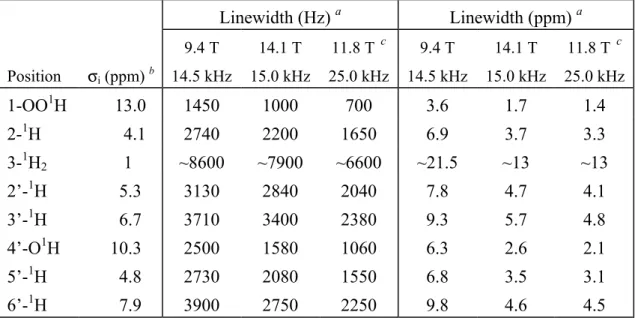
Table 3.1: Solid-state NMR proton shifts
σi
and linewidths of L-tyrosine·HClLinewidth (Hz) a Linewidth (ppm) a
Position σi (ppm) b
9.4 T 14.5 kHz
14.1 T 15.0 kHz
11.8 T c 25.0 kHz
9.4 T 14.5 kHz
14.1 T 15.0 kHz
11.8 T c 25.0 kHz
1-OO1H 13.0 1450 1000 700 3.6 1.7 1.4
2-1H 4.1 2740 2200 1650 6.9 3.7 3.3
3-1H2 1 ~8600 ~7900 ~6600 ~21.5 ~13 ~13
2’-1H 5.3 3130 2840 2040 7.8 4.7 4.1
3’-1H 6.7 3710 3400 2380 9.3 5.7 4.8
4’-O1H 10.3 2500 1580 1060 6.3 2.6 2.1
5’-1H 4.8 2730 2080 1550 6.8 3.5 3.1
6’-1H 7.9 3900 2750 2250 9.8 4.6 4.5
aFull width at half height (FWHH), from spectra processed without apodization in t
1. bAt a magnetic field of 14.1 T. cData supplied by and used with kind permission of Bruker [8].
14.1 T compares well with the typical widths achieved with more elaborate line-narrowing
techniques. The improvement for the other protons is within the range 1.7-1.9, with
exception of the 3’-1H resonance for which a factor 1.6 is found. The enhancement of the overall proton resolution is obviously more than the factor 1.5 due to the increase in the
chemical shift dispersion alone. Since the field strength is the only parameter that was
varied, the reduction of the linewidth can readily be related to an effect of the increased
magnetic field. It can thus be concluded that the high-field resolution enhancement is
non-linear, since apart from the increased proton chemical shift dispersion, an additional
narrowing of the proton resonances is observed, induced by the increased magnetic field
strength itself.
The 1H shift of 13 ppm for the 1-OOH correlated with 1-13C is in the characteristic downfield range of 8-15 ppm for hydrogen-bonded protons. From the neutron diffraction
structure of tyrosine⋅HCl, it is known that the carboxylic proton forms a hydrogen bond to the phenolic oxygen of the 4’ hydroxyl group [7]. The importance of the hydrogen-bonded
proton in the initial stages of the CP transfer is evident from the observation of a weak
intermolecular correlation of the 4’-13C with the carboxylic proton around 13 ppm in the dataset obtained at 14.1 T, indicated with an arrow in Fig. 3.2B. Since the 2-D dataset was
![Table 6.1. 13 C chemical shifts in solution ( σ liq ) of monomeric Chl a in [U- 2 H] tetrahydrofuran [24,25]](https://thumb-us.123doks.com/thumbv2/123dok_us/8310150.2200818/80.892.177.688.278.1072/table-chemical-shifts-solution-liq-monomeric-chl-tetrahydrofuran.webp)


