Journal of Athletic Training 2016;51(12):1053–1070 doi: 10.4085/1062-6050-51.7.01
Óby the National Athletic Trainers’ Association, Inc
www.natajournals.org position statement
National Athletic Trainers’
Association Position Statement:
Management of Acute Skin
Trauma
Joel W. Beam, EdD, LAT, ATC*;
Bernadette Buckley, PhD, LAT, ATC*;
William R. Holcomb, PhD, ATC, FNATA, FNSCA
†
;
Mario Ciocca, MD
‡
*Clinical and Applied Movement Sciences, Brooks College of Health, University of North Florida, Jacksonville; †School of Kinesiology, University of Southern Mississippi, Hattiesburg; ‡Department of Sports Medicine, University of North Carolina at Chapel Hill
Objective: To present recommendations for the cleansing, debridement, dressing, and monitoring of acute skin trauma in patients.
Background: Acute skin trauma is common during partic-ipation in athletic and recreational activities. Clinical decisions and intervention protocols after injury vary among athletic trainers and are often based on ritualistic practices. An understanding of cleansing, debridement, and dressing tech-niques; clinical features of infection and adverse reactions; and monitoring of acute skin trauma is critical for certified athletic trainers and other allied health and medical professionals to
create a local wound environment that promotes healing and lessens the risk of complications.
Recommendations: These guidelines are intended to provide the certified athletic trainer and others participating in athletic health care with specific knowledge about and recom-mendations for the management of acute skin trauma.
Key Words: abrasions, avulsions, blisters, incisions, lacer-ations, punctures, cleansing, debridement, nonocclusive dress-ings, occlusive dressdress-ings, infection, adverse reactions
T
raumatic injury to the skin is common among athletes participating in all sports.1 The exact frequencies of abrasions, avulsions, blisters, inci-sions, lacerations, and punctures are difficult to calculate because many patients do not seek medical attention after injury; for others, their activity level is initially unaffected, and the injury is not recorded on surveillance reports. Unreported skin trauma and inappropriate wound manage-ment can result in delayed healing, cross-contamination, bacterial colonization, and infection, adversely affecting the overall health and playing status of the patient. Managing acute skin trauma through appropriate cleansing, debride-ment, and dressing techniques can create an environment conducive to healing and lessen the risk of complications.2,3 Wound-management techniques have undergone drastic changes over the last 50 years, and other allied health care professions, organizations, and facilities have developed guidelines that serve as standards of care.4–6 However, guidelines for the management of acute skin trauma by athletic trainers (ATs) are limited in the literature.1,7 The development and implementation of cleansing, debride-ment, and dressing techniques for acute skin trauma arecritical for ATs to successfully deliver health care services to patients. The following review and recommendations provide information on the management of acute skin trauma and guidelines for ATs and other allied health and medical professionals who care for patients.
RECOMMENDATIONS
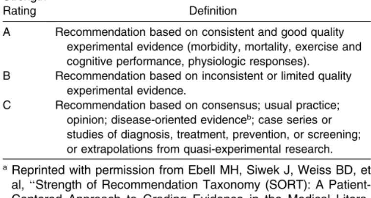
This position statement is based on current research and literature with regard to the cleansing, debridement, dressing, and monitoring of acute skin trauma. We independently categorized the studies and literature using the Strength of Recommendation Taxonomy (SORT) developed by the American Academy of Family Physi-cians.8 The taxonomy grades the quality of the data from the literature (level of evidence) and provides strength ratings for the suggested recommendations with the letter
A,B, orC (Table 1).
follow-up, and supplies for athletic training facilities and kits.
Cleansing
1. The wound bed and adjacent periwound tissues should be thoroughly cleansed as soon as possible after acute skin trauma.9–11Strength of recommendation: C
2. After initial cleansing, the wound should be cleansed only when clinically necessary (eg, visibly contaminated, clinically infected).9,12–18Strength of recommendation: C
3. Irrigation can be used to cleanse superficial- to full-thickness abrasions, incisions, and lacerations.19,20
Strength of recommendation: B
a. To effectively remove wound debris, irrigation pressure should range between 4 and 15 pounds per square inch (psi; 27.58–103.42 kPa).11,19,21–23
Strength of recommendation: B
b. A range of 7 to 11 psi (48.26–75.84 kPa) can be achieved with a 35-mL syringe and an 18- to 20-gauge needle, needle hub, or plastic cannula.11,19,23,24
Strength of recommendation: B
4. Showering can be used to cleanse postoperative inci-sions.19,20,25Strength of recommendation: A
5. Hydrotherapy (eg, whirlpool baths and soaks) may be used during cleansing to hydrate a wound.11,24,26Strength
of recommendation: C
6. Scrubbing or swabbing of the wound bed should be avoided because it may be ineffective in reducing bacterial counts and can further contaminate and damage the granulation tissue.9,13,24,26–29 Strength of
recommen-dation: C
7. Normal saline and potable tap water should be used as cleansing agents with superficial- to full-thickness abrasions, incisions, and lacerations.19,20,25,30–33
Strength of recommendation: A
8. Tap water should be avoided in the presence of exposed bone or tendon, although the basis for this guideline is unclear.34
Strength of recommendation: C
9. Antiseptics (eg, povidone-iodine, hydrogen peroxide) should be used with caution as cleansing agents because some may be toxic to tissues.19,35–43 Strength of
recommendation: B
10. Irrigation with 1% povidone-iodine can be used to cleanse acute, traumatic, contaminated wounds.19
Strength of recommendation: B
11. The cleansing solution should be delivered to the wound at a temperature between 98.68F and 107.68F (378C and 428C).13,14,44,45
Strength of recommendation: C
Debridement
12. The area of acute skin trauma should be thoroughly cleansed and debrided before dressings are applied.4,46–53
Strength of recommendation: B
13. Debridement should continue until well-vascularized, healthy granulation or epithelial tissue is exposed.54
Strength of recommendation: C
14. The method of debridement should be based on the type of wound, amount and type of debris, training and expertise of the health care provider, and cost-effective-ness and time-effectivecost-effective-ness of the technique.55,56
Strength of recommendation: C
15. Irrigation can serve as an extension of cleansing for debridement of superficial- to full-thickness abrasions, avulsions, blisters, incisions, lacerations, and punc-tures.3,57–59Strength of recommendation: C
a. Irrigation pressure should range between 4 and 15 psi (27.58–103.4 kPa).3,57,59–61 Strength of
recommenda-tion: C
b. To avoid driving debris and contaminants deeper into puncture wounds, an irrigation pressure of 2 to 4 psi (13.79–27.58 kPa) should be used.3,57,59–61Strength of
recommendation: C
16. Hydrotherapy debridement (eg, whirlpool baths and soaks) should be avoided because it presents a risk of cross-contamination and is neither cost-effective nor time-effective.62,63
Strength of recommendation: C
17. Wet-to-dry debridement should be avoided because tissue removal is nonselective and painful.54,55,57,59,64–70
Strength of recommendation: C
18. Wet-to-moist debridement may be used for superficial- to full-thickness abrasions, avulsions, blisters, incisions, and lacerations and with the formation of eschar. However, care should be taken to protect healthy granulation tissue.59Strength of recommendation: C
19. Scrubbing should be used only for superficial- to partial-thickness abrasions, avulsions, blisters, incisions, and lacerations contaminated with large quantities of small debris (eg, sand, grass, clay, asphalt).66
Strength of recommendation: C
20. Conservative sharp debridement can be used after cleansing to remove loosely adhering, devitalized tissue from superficial- to full-thickness abrasions, avulsions, blisters, incisions, and lacerations.71,72 Strength of
rec-ommendation: C
21. Chemical debridement (eg, sodium hypochlorite, hydro-gen peroxide, silver) should be avoided because some elements and compounds may damage viable tis-sue.71,73,74
Strength of recommendation: C
Table 1. Strength of Recommendation Taxonomy8a Strength
Rating Definition
A Recommendation based on consistent and good quality experimental evidence (morbidity, mortality, exercise and cognitive performance, physiologic responses).
B Recommendation based on inconsistent or limited quality experimental evidence.
C Recommendation based on consensus; usual practice; opinion; disease-oriented evidenceb; case series or
studies of diagnosis, treatment, prevention, or screening; or extrapolations from quasi-experimental research.
aReprinted with permission from Ebell MH, Siwek J, Weiss BD, et
al,‘‘Strength of Recommendation Taxonomy (SORT): A Patient-Centered Approach to Grading Evidence in the Medical Litera-ture,’’February 1, 2004, Vol 69, No 3, American Family Physician Copyright Ó2004 American Academy of Family Physicians. All Rights Reserved.
bPatient-oriented evidence measures outcomes that matter to
22. Autolytic debridement should be used for selective proteolytic digestion of necrotic tissue.51,55,57,58,64,71,75–85
Strength of recommendation: B
a. Autolytic debridement can be used with the following wounds:
i. Postoperative incisions.79 Strength of
recommen-dation: B
ii. Superficial- to full-thickness abrasions, avulsions, and lacerations. Strength of recommendation: C
iii. Superficial- to full-thickness blisters after removal of the necrotic roof with conservative sharp debridement.71Strength of recommendation: C
iv. Superficial- to partial-thickness punctures.
Strength of recommendation: C
b. Autolytic debridement should be avoided with infect-ed wounds.57,66,75 Strength of recommendation: C
Dressings
23. After thorough cleansing and debridement, an area of acute skin trauma should be covered with an appropriate dressing until fully healed (Table 2).2,86–90
Strength of recommendation: B
24. Nonocclusive dressings can be used as primary dressings with the following wounds (Table 3):
a. Woven and nonwoven gauze for clinically infected abrasions, avulsions, blisters, incisions, lacerations, or punctures.91,92Strength of recommendation: C
b. Woven, nonwoven, and impregnated gauze for puncture wounds that have cavities.93–96 Strength of
recommendation: C
c. Wound-closure strips with superficial, linear lacera-tions and postoperative incisions under minimal static and dynamic tension.3,97,98 Strength of
recommenda-tion: A
d. Woven gauze with superficial- to full-thickness abrasions, avulsions, blisters, incisions, and
lacera-tions to achieve wet-to-moist debridement.55,68,91,94
Strength of recommendation: C
e. Woven and nonwoven gauze, nonadherent pads, and adhesive strips or patches for superficial- to partial-thickness abrasions, avulsions, and blisters and superficial-thickness incisions, lacerations, and punc-tures as a temporary dressing and on irregular body surfaces.91,95,99
Strength of recommendation: C
25. Woven or nonwoven gauze, nonadherent pads, and adhesive strips or patches can be used as secondary dressings to absorb moderate-to-heavy exudate and provide additional protection for superficial- to full-thickness abrasions, avulsions, blisters, lacerations, punc-tures, and traumatic and postoperative incisions.91,94,100,101
Strength of recommendation: C
26. Occlusive dressings should be used as primary dressings with the following wounds (Table 4):
a. Alginates, films, foams, hydrocolloids, and hydrogels for superficial- to partial-thickness abrasions, avul-sions, blisters, inciavul-sions, lacerations, and punc-tures.2,90,91,95,102–112
Strength of recommendation: A
b. Alginates, foams, and hydrocolloids for partial- to full-thickness abrasions, avulsions, blisters, lacerations, and traumatic and postoperative incisions.2,90,91,102,106,109–117
Strength of recommendation: A
c. Dermal adhesives for partial- to full-thickness lacer-ations and traumatic and postoperative incisions in areas of low skin tension.97,98,118,119 Strength of
recommendation: A
d. Alginates and foams (nonsilver or silver impregnated) or silver-impregnated films and hydrocolloids for contaminated and clinically infected abrasions, avul-sions, blisters, inciavul-sions, and lacerations.91,94,120,121
Strength of recommendation: C
27. Films and hydrocolloids can be used as secondary dressings to provide impermeability to microorganisms, improve adherence, and promote a moist wound envi-ronment for superficial- to full-thickness abrasions, avulsions, blisters, lacerations, punctures, and traumatic and postoperative incisions.* Strength of recommenda-tion: C
Identification of Infection and Adverse Reactions
28. The athletic training staff should monitor the patient and wound area for clinical features of wound infection, including fever, pain, edema, erythema, warmth, wound dehiscence, and delayed wound healing.124–128Strength of
recommendation: C
29. For treatment of bacterial infections, refer to the National Athletic Trainers’ Association position statement on skin diseases.129
Strength of recommendation: B
30. Infection rates in patients with acute skin trauma can be reduced with appropriate cleansing, debridement, and dressing techniques.39,125,130–132
Strength of recommenda-tion: B
31. Skin antisepsis is effective in reducing the incidence of surgical-site infections in incisions and lacerations.133
Strength of recommendation: A
Table 2. Sample Dressings
Brand Name Type Manufacturer
Allevyn Foam Smith & Nephew Pty, Limited, Mount Waverley, Victoria, Australia
Aquaflo Hydrogel Covidien, Mansfield, MA Aquaheal Hydrogel Spenco Medical
Corporation, Waco, TX Bioclusive Film Johnson & Johnson,
Medical LTD, Ascot, United Kingdom Dermabond Dermal adhesive Ethicon, Somerville, NJ Duoderm Hydrocolloid ConvaTec, Princeton, NJ Histoacryl Dermal adhesive Aesculap AG, Tuttlingen,
Germany Leukostrips Wound-closure strip Smith & Nephew Pty PolyMem Foam Ferris Mfg, Burr Ridge, IL Polyskin II Film Covidien
SteriStrips Wound-closure strip 3M Company, St Paul, MN Tegaderm Alginate, film, foam,
hydrocolloid, and hydrogel
3M Company
Ultec Pro Alginate/ hydrocolloid
Covidien
32. Oral antibiotics should not be used prophylactically unless the abrasion, avulsion, blister, incision, laceration, or puncture is heavily contaminated.134,135
Strength of recommendation: A
33. Topical antimicrobial agents can reduce infection rates in acute skin trauma but should be used judiciously given the emergence of resistant bacterial strains.135–142
Strength of recommendation: A
34. The patient should be monitored for the development of adverse reactions stemming from the use of some cleansing solutions, topical antimicrobial agents, and
nonocclusive and occlusive dressings.143–147
Strength of recommendation: C
35. The patient’s health status (eg, malnutrition, smoking, diabetes) may contribute to the development of adverse reactions and should be considered.130,148–161
Strength of recommendation: B
36. The athletic training staff should monitor the patient and wound area for clinical features of adverse reactions, including erythematous rash, eczematous reaction, vesi-cles, white discoloration, tenderness, nodularity, burning, pruritus, or systemic reactions such as urticaria and anaphylaxis.143–146,162–165
Strength of recommendation: C
Table 3. Nonocclusive Dressing Indications
Indications Primary Dressing Secondary Dressing Other Considerations
Clinically infected abrasions, avulsions, blisters, incisions, lacerations, and punctures
Woven and nonwoven gauze Woven or nonwoven gauze; nonadherent pads; adhesive strips or patches; adhesive gauze; nonadherent, self-adherent, and adherent tapes and wraps
Daily dressing changes required
Punctures with cavities Woven, nonwoven, and impregnated gauze
Woven or nonwoven gauze; nonadherent pads; adhesive strips or patches; adhesive gauze; nonadherent, self-adherent, and adherent tapes and wraps
Daily dressing changes required
Superficial, linear lacerations and postoperative incisions
Wound-closure strips Woven or nonwoven gauze; nonadherent pads; adhesive strips or patches; nonadherent, self-adherent, and adherent tapes and wraps
Use only on wounds with minimal static and dynamic tension
Superficial- to full-thickness abrasions, avulsions, blisters, incisions, and lacerations for wet-to-moist debridement
Woven gauze Woven or nonwoven gauze; nonadherent pads; nonadherent, self-adherent, and adherent tapes and wraps
Use care to protect healthy granulation tissues
Superficial- to partial-thickness abrasions, avulsions, blisters, superficial-thickness incisions, lacerations, and punctures as temporary dressings
Premoistened woven and nonwoven gauze; nonadherent pads; adhesive strips or patches
Woven or nonwoven gauze; nonadherent pads; adhesive strips or patches; adhesive gauze; nonadherent, self-adherent, and adherent tapes and wraps
Follow-up in athletic training facility for comprehensive management
Superficial- to partial-thickness abrasions, avulsions, blisters, superficial-thickness incisions, lacerations, and punctures on irregular body areas
Woven and nonwoven gauze; nonadherent pads; adhesive strips or patches
Woven or nonwoven gauze; nonadherent pads; adhesive strips or patches; adhesive gauze; nonadherent, self-adherent, and adherent tapes and wraps
Daily dressing changes required
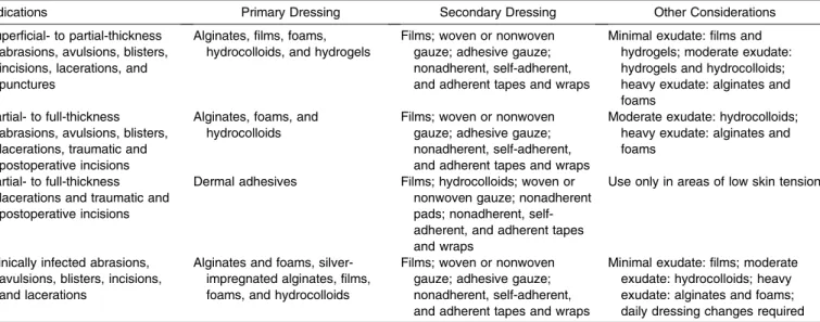
Table 4. Occlusive Dressing Indications
Indications Primary Dressing Secondary Dressing Other Considerations
Superficial- to partial-thickness abrasions, avulsions, blisters, incisions, lacerations, and punctures
Alginates, films, foams, hydrocolloids, and hydrogels
Films; woven or nonwoven gauze; adhesive gauze; nonadherent, self-adherent, and adherent tapes and wraps
Minimal exudate: films and hydrogels; moderate exudate: hydrogels and hydrocolloids; heavy exudate: alginates and foams
Partial- to full-thickness abrasions, avulsions, blisters, lacerations, traumatic and postoperative incisions
Alginates, foams, and hydrocolloids
Films; woven or nonwoven gauze; adhesive gauze; nonadherent, self-adherent, and adherent tapes and wraps
Moderate exudate: hydrocolloids; heavy exudate: alginates and foams
Partial- to full-thickness lacerations and traumatic and postoperative incisions
Dermal adhesives Films; hydrocolloids; woven or nonwoven gauze; nonadherent pads; nonadherent, self-adherent, and adherent tapes and wraps
Use only in areas of low skin tension
Clinically infected abrasions, avulsions, blisters, incisions, and lacerations
Alginates and foams, silver-impregnated alginates, films, foams, and hydrocolloids
Films; woven or nonwoven gauze; adhesive gauze; nonadherent, self-adherent, and adherent tapes and wraps
37. Treatment of the adverse reaction should consist of identifying the reaction, removing the causative agent, and directing appropriate measures at the reaction.143–146,162–165
Strength of recommendation: C
38. Criteria for physician referral include
a. Any evidence of deeper injury that may require repair, evaluation for nerve or tendon damage, or concern for heavy contamination.Strength of recommendation: C
b. Wounds that develop erythema, warmth, edema, drainage, pain, or rash or demonstrate delayed healing.
Strength of recommendation: C
Follow-Up
39. The athletic training staff should visually inspect the patient, wound area, and dressing daily throughout the healing process.
a. The visual inspection should include the patient, wound bed, and periwound tissues for the presence of adverse reactions and infection. If any signs or symptoms are present, the patient should be referred to a physician.Strength of recommendation: C
b. Frequency of dressing changes varies based on the type of dressing (Table 5). A dressing change is warranted with evidence of dressing channel forma-tion, separation from periwound tissues, significant exudate accumulation, strike-through, leakage, or wound desiccation.89,91,94,166,167 Strength of
recom-mendation: C
c. Dressing transitions are required as reductions in exudate and new tissue growth occur during healing.†
Strength of recommendation: C
40. Patients should be educated on dressing guidelines to increase compliance.
a. Patients should follow instructions from the athletic training staff on dressing changes. Occlusive dressings can stay in place over wounds for longer periods than nonocclusive dressings.91,94,105,122,170–172
Strength of recommendation: C
b. Patients will notice changes in the dressing over time. An accumulation of moisture beneath occlusive dressings may be visually detected or demonstrated by expansion of the dressing over the wound bed and
should not be confused with infection.91,103,105,173
Strength of recommendation: C
c. Patients must immediately report dressing channel formation, separation from periwound tissues, seal breach, strike-through, or leakage or signs or symp-toms of adverse reactions and infection.89,91,94,166,167
Strength of recommendation: C
Supplies for Athletic Training Facilities and Kits
41. Supplies to manage acute skin trauma should be available in the athletic training facility.
a. Cleansing: normal saline, potable tap water, 35-mL syringe, 18- to 20-gauge needle hub or plastic cannula, antiseptic skin cleanser, and clean basin or cup. b. Debridement: normal saline, potable tap water, 35-mL
syringe, 18- to 20-gauge needle hub or plastic cannula, woven gauze, high-porosity sponge or surgical scrub brush, and sterile scissors and tweezers.
c. Dressings: nonocclusive dressings (woven, nonwoven, and impregnated sterile gauze, nonadherent pads, adhesive strips and patches, and wound-closure strips), occlusive dressings (alginates, films, foams, hydrogels, hydrocolloids, and dermal adhesives), adhesive gauze, and nonadherent, self-adherent, and adherent tapes and wraps.
d. Miscellaneous: sterile or clean (or both) drapes or towels, biohazard container, boxed gloves, face or eye shields (or both), and topical antibiotics.
42. Supplies to manage acute skin trauma should be available in athletic training kits.
a. Cleansing: normal saline, potable tap water, 35-mL syringe, 18- to 20-gauge needle hub or plastic cannula, and clean basin or cup.
b. Debridement: same as cleansing.
c. Dressing: nonocclusive dressings (woven and nonwo-ven sterile gauze, nonadherent pads, and adhesive strips and patches), adhesive gauze, and nonadherent, self-adherent, and adherent tapes and wraps.
d. Miscellaneous: boxed gloves, face or eye shields (or both), and biohazard container.
BACKGROUND
Acute skin trauma is a disruption of the integrity of the epidermis, dermis, or subcutaneous tissues (or a combina-tion of these). Acute wounds are characterized based on the mechanism of injury and resultant tissue damage and include traumatic abrasions, avulsions, blisters, lacerations, punctures, and traumatic and postoperative incisions. The mechanisms of injury of acute skin trauma are shear and tensile forces and tensile loads.174 Acute wounds can also be characterized by the amount of tissue damage.175
Superficial-thickness wounds involve damage to the superficial epidermis. Partial-thickness wounds extend through the epidermis and into the superficial dermis.
Full-thickness wounds extend through the epidermis and dermis and into the subcutaneous adipose tissue. Acute wounds proceed through an orderly and timely reparative process of inflammation, proliferation, and remodeling that results in restoration of anatomic and functional integrity and wound appearance.176 Some have characterized acute
Table 5. Recommended Duration of Wound Dressing Usagea
Dressing Type Duration, d
Woven, nonwoven, and impregnated sterile gauze; nonadherent pads; and adhesive strips and patches87,91,93,94,105,113,168
1
Wound-closure strips174 5–10
Alginates105 7
Films and foams91,94,105,122,170 3–7
Hydrogels94,105,122 1–7
Hydrocolloids94,105,122,170 5–7
Dermal adhesives171,172 5–10
aWounds without dressing-integrity problems (eg, wrinkling or
bunching, channel formation, separation from periwound tissues, strike-through, leakage of exudate) or clinical features of adverse reactions or infection.89,91,94
wounds as those that heal themselves within 4 to 6 weeks.124
THE EVIDENCE AND LITERATURE REVIEW
Cleansing
Wound cleansing, considered a critical part of the management of acute skin trauma, is the process of applying a nontoxic solution to aid in the removal of exudate, bacteria, foreign debris, and dressing residue to create an environment conducive for healing.39,177 Acute wounds are initially considered to be contaminated; cleansing is necessary to remove any debris and facilitate healing.9,11,17 After the initial cleansing, it may not be necessary to cleanse wounds at every dressing change but rather to rehydrate a wound to create a moist environment, visualize and assess the wound, minimize trauma during adherent dressing removal, or promote patient com-fort.9,12–18,178 If signs of clinical infection are present, then continued wound cleansing is necessary.
Technique. The selection of an appropriate cleansing technique is necessary to create an optimal environment for wound healing (ie, a moist, clean, warm environment). The more commonly used techniques include irrigation, showering, hydrotherapy (eg, whirlpool baths and soaks), and scrubbing and swabbing.
Irrigation. Irrigation, the steady flow of solution across the wound surface, is the preferred method of cleans-ing.11,39,179 The purpose of irrigation is to remove loose debris and excess wound secretions to create an optimal healing environment. This method allows for newly granulating tissue to be preserved, bacteria and debris to be effectively removed, and the comfort and convenience of the patient to be ensured. The potential risks associated with this technique include splash back, additional trauma, and bacteria driven into deeper tissues if the pressure is excessive.11
Although many irrigation recommendations are based on trials involving chronic wounds, investigators11,19–25,29,39 agree that high-pressure irrigation (eg, 4–15 psi [27.58– 103.42 kPa]) is the best practice for cleansing superficial- to full-thickness abrasions, incisions, and lacerations. Au-thors19of an evidence-based medicine review found that a pressure of 13 psi (89.63 kPa) was effective in reducing infection and inflammation in lacerations and chronic wounds among children and adults, although no consensus exists as to the optimal pressure that should be used. Researchers24agree that low-pressure irrigation (eg,,4 psi [27.58 kPa]) is not as effective in cleansing and serves only to moisten the wound bed. Pressures greater than 25 psi (172.4 kPa) may be necessary to debride a wound but are not recommended for routine cleansing, as they may damage healthy granulation tissue.13,24
The equipment commonly used for irrigation includes bulb syringes, pressurized canisters, and syringes with an attached needle or catheter. A 35-mL syringe with an 18- to 20-gauge needle, needle hub, or plastic cannula exerts pressures in the range of 7 to 11 psi (48.26–75.84 kPa), delivering a solution without damage to the tissue.11,19,23,24
Showering. Showeringis effective for larger traumatic wounds, although the pressure is rarely controlled. Authors of a Cochrane review20 examining the effects of
postop-erative showering versus no showering found strong evidence of no difference in the infection and healing rates of incisions. However, investigators19,20reported that patients derived a sense of health and well-being from showering.
Hydrotherapy. Whirlpool baths can be used as an aggressive form of cleansing; the turbulence of the water dislodges debris from the wound bed. More often used for chronic wounds, bathing in lukewarm tap water has become an increasingly common practice, although its role may be considered more for debridement than cleansing.11,24,26 Researchers19suggested that whirlpool therapy may reduce pain and inflammation in surgical incisions during the first 72 postoperative hours. Clinically, this method of cleansing may be used to hydrate a wound, even though concerns and potential risks include disrupting the moisture balance of the wound bed, macerating periwound tissues, and impairing healing by introducing microorganisms from the immersion fluid.11
Scrubbing and Swabbing. Scrubbing and swabbing
involve the use of a material (eg, gauze, foam) to wipe the surface of the wound in a systematic manner. This method lacks the evidence necessary to show that it creates the optimal wound environment: scrubbing and swabbing redistribute bacteria over the wound as opposed to removing them.9,13,24,26–29 Cotton wool fiber remnants from woven gauze often contaminate the wound and have resulted in nonwoven swabs being the material of choice for swab-bing.9,13,29 Swabbing can be used to cleanse the periwound tissues or wounds with loose necrotic tissue or slough (moist necrotic tissue).26,27 However, others9,13,24,26–29 caution against using the swabbing technique on the wound bed because of residual damage to the granulating tissue. Overall, no evidence supports or refutes scrubbing and swabbing to cleanse wounds.19
Solutions. The selection of an appropriate nontoxic solution to remove debris and create an environment to promote healing is a critical component of wound cleansing. Various cleansing solutions are recommended for their therapeutic value.180The characteristics of an ideal solution are nontoxicity to human tissue, effectiveness in the presence of organic material, ability to reduce the number of microorganisms, low likelihood of causing sensitivity reactions, cost-effectiveness, availability, and shelf-life stability.41,178,181 Based on these characteristics, many authors7,12,20,182 consider normal saline the most appropriate cleansing solution for acute skin trauma, although the use of potable tap water has become more widely accepted.
contamination and infection.178 Tap water has the advan-tages of being efficient, cost-effective, and accessible.20Yet tap water should not be used to cleanse wounds in which tendon or bone is exposed. Normal saline is the recommended solution for these wounds.34
Antiseptics. Topical antiseptics are antimicrobial agents that kill or reduce the number of microorganisms that may impede wound healing. Commonly used solutions include povidone-iodine (eg, Betadine; Purdue Products LP, Stamford, CT) and hydrogen peroxide.36,38–41,183,184 However, the use of antiseptics as prophylactic antimicro-bial agents for acute skin trauma has been controversial for many years.35–37,40,184
Several investigators19,37 have examined the effects of povidone-iodine and the product Betadine on rates of infection and healing. In 1 evidence-based medicine review,19 infection rates were compared between acute wounds that were cleansed with 1% povidone-iodine or with normal saline. Among contaminated postoperative incisions and traumatic lacerations, 1% povidone-iodine was favored over normal saline. An individual trial in this review19examining lacerations demonstrated no differenc-es in infection ratdifferenc-es between 1% povidone-iodine and normal saline. Authors37 of a more recent evidence-based medicine review examined the use of iodine and other local wound care methods (eg, honey, silver-impregnated dressings) on postoperative incisions and various acute wounds and noted no differences in rates of infection or healing. An additional narrative review35 examining the effects of povidone-iodine on healing in animal and human wound models has raised questions about the benefits of the solution; delayed wound healing, reduced wound strength, and increased rates of infection were noted when acute skin trauma was treated with the antiseptic. In a small investigation, researchers41 demonstrated the safe use of antimicrobials in diluted concentrations. A 1 : 10 diluted solution of Betadine was effective against bacteria and not harmful to human fibroblasts.41 A 1 : 5 diluted solution of Betadine was toxic to fibroblasts and, therefore, one may conclude that the commercially produced solution of Betadine would be toxic as well.41
Other investigations have examined the efficacy of hydrogen peroxide as an antiseptic in animal and human wound models. In a narrative review,38hydrogen peroxide demonstrated minimal effects in reducing the bacterial bioburden (quantity of microorganisms), but the findings were inconclusive regarding cytotoxic effects on tissues and rates of healing. Small experimental investigations41,42 support the finding that hydrogen peroxide is ineffective in reducing microorganisms in the wound bed. However, these trials demonstrated a greater cytotoxic effect of hydrogen peroxide on tissues,42,43which perhaps delayed healing.
Temperature. Athletic trainers must also consider the temperature of the solution to facilitate healing. The recommended temperature of the cleansing solution is between 98.68F and 107.68F (378C and 428C).13,14,44,45 Mitotic activity (reproduction of cells essential to healing) decreases as the wound temperature drops after cleansing or dressing changes. It can take up to 40 minutes for the wound bed to return to its original temperature and up to 3 hours for mitotic activity to return to normal.45Therefore, using a cool cleansing solution may delay the healing process.14Although research directly examining the effects
of various solution temperatures on wound healing is lacking, some evidence suggests that lower temperatures may delay wound healing.45
Debridement
Debridement is the removal of necrotic or devitalized tissue, microorganisms, contaminated tissue, fibrin or foreign bodies, and cellular debris from the wound bed.53,54,75,185After acute skin trauma, debridement should be used to decrease the risk of infection and create an environment suitable for healing. Thorough cleansing of the wound should be performed initially and, if necessary, debridement used to remove remaining debris or devital-ized tissue from the wound bed. The wound should be debrided until only normal vascularized tissue remains.54,56 Debridement can decrease the bacterial concentration within the wound bed, decreasing the bacterial bioburden83; improve the function of leukocytes, reducing the risk of infection; shorten the inflammatory phase, decreasing the energy required for healing; and remove debris and tissue from the wound bed, eliminating the physical barrier to healing.66
Technique.Many debridement methods are available to health care providers. The methods that ATs can consider using include irrigation, hydrotherapy, dry, wet-to-moist, scrubbing, conservative sharp, chemical, and autolytic debridement.
Irrigation. Irrigationis the delivery of normal saline or potable tap water to the wound bed in a constant or pulsed stream.3,57–59 Irrigation can serve as an extension of cleansing to remove loose superficial debris or necrotic tissue from superficial- to full-thickness abrasions, avul-sions, blisters, inciavul-sions, lacerations, and punctures.3,57–59 Recommended pressure ranges from 4 to 15 psi (27.58– 103.4 kPa) for all wounds except punctures, for which 2 to 4 psi (13.79–27.58 kPa) is recommended to avoid driving debris and contaminants deeper into the wound.3,57,59,61
Hydrotherapy. Whirlpool baths and soaks use potable tap water to soften and remove devitalized tissue and toxic debris and dilute the bacterial content of the wound bed. The body part and wound are submerged in water between 95.98F and 102.28F (35.58C and 39.08C) for 20 to 30 minutes.63 Although commonly used, hydrotherapy is not recommended for acute skin trauma. This technique can increase the risk of cross-contamination from the whirlpool tub or other container, and it is neither cost-effective nor time-effective to drain, properly clean and disinfect, and refill the whirlpool tub or container after each patient.63
Wet to Moist. Similar to wet-to-dry debridement, wet-to-moist debridementis the placement of woven gauze with large pores that is premoistened with normal saline or potable tap water over the wound bed. The gauze is allowed to remain on the wound bed for minutes to hours and removed before drying is complete. Wet-to-moist debride-ment may be used for superficial- to full-thickness abrasions, avulsions, blisters, incisions, and lacerations. This technique is a rapid method of debridement and allows for removal of debris, devitalized tissue, and eschar (black necrotic tissue, scab), while protecting healthy granulation tissue and producing minimal pain.59
Scrubbing. Scrubbingis the use of a sponge with high porosity (90 pores/in2 [14 pores/cm2]) or a surgical scrub brush, along with normal saline or potable tap water, to scour the wound bed from the middle toward the wound margins in a circular pattern.66Scrubbing can be used with superficial- to partial-thickness abrasions, avulsions, blis-ters, incisions, and lacerations contaminated with large quantities of small debris (eg, sand, grass, clay, asphalt).66 This technique is nonselective, with potential removal of healthy granulation tissue, and the mechanical pressure of the sponge or brush can produce pain.
Conservative Sharp Debridement. Conservative sharp debridement is the use of sterile scissors and forceps or tweezers to remove loosely adhering devitalized tissue that lies superficial to viable tissue on the wound bed.71 Conservative sharp debridement of acute skin trauma is typically performed in a single visit and may be used on superficial- to full-thickness abrasions, avulsions, blisters, incisions, and lacerations.72 Applicable state practice acts may allow ATs to perform this technique but should be verified.174
Chemical Debridement. Chemical debridement is the application of sodium hypochlorite, hydrogen peroxide, or povidone-iodine in water-based solutions; silver; or honey directly to the wound bed or the use of chemical-impregnated dressings over the wound. Although chemicals may facilitate debridement, their use is controversial and not recommended for acute skin trauma.71,73,74 Some authors have recommended chemicals for debridement of infected wounds71or with wet-to-moist debridement.75The effervescent effect of hydrogen peroxide may aid debride-ment, but hydrogen peroxide and sodium hypochlorite are thought to be cytotoxic to healthy granulation tissue.74 Silver and silver-impregnated dressings have been used for centuries to provide antimicrobial debridement of colonized and clinically infected wounds. In an evidence-based medicine review, Vermeulen et al121 demonstrated insuffi-cient evidence for the use of silver-impregnated dressings to manage contaminated or infected acute and chronic wounds.
Autolytic Debridement. Autolytic debridement is the use of the body’s mechanisms to promote proteolytic digestion of necrotic tissue in a moist environment created by the application of occlusive dressings such as alginates, films, foams, hydrogels, and hydrocolloids.76,79,83,85 The moist environment allows endogenous collagenase en-zymes within the wound to liquefy necrotic tissue, which can then be more easily digested by macrophag-es.51,55,57,58,75,77,80,82 The body selectively digests only nonviable tissue and the moist environment allows for painless debridement.71Autolytic debridement can be used
with superficial- to full-thickness abrasions, avulsions, incisions, and lacerations; superficial- to full-thickness blisters after removal of the necrotic roof with conservative sharp debridement71; and superficial- to partial-thickness punctures. Autolytic debridement is slow, occurring over several days, and is not recommended for infected wounds.57,66,75
In an evidence-based medicine review, Lewis et al79 examined the use of dressings in the autolytic debridement of postoperative incisions. Overall, modern occlusive dressings were favored over gauze dressings for healing. Findings from individual trials in the review79produced no clear evidence of differences in rates of healing among polyurethane foam, silicone foam, and alginate dressings. Several authors78,81,84have investigated the effectiveness of autolytic, wet-to-dry, and enzymatic debridement among various chronic wounds. Autolytic debridement using hydrogel and hydrocolloid dressings and wet-to-dry debridement using normal saline-soaked gauze produced a satisfactory environment to promote healing of pressure ulcers.81In this study, the hydrogel dressing was preferred because the transparent construction allowed for visual assessment of the wound bed and removal of the dressing did not damage healthy tissue. Other authors84have shown hydrogel dressings to be effective in managing chronic and necrotic wounds with slough. For debridement of leg ulcers, no differences were demonstrated between autolytic and enzymatic debridement in rates of healing.78
Dressings
It is well established that acute wounds should be covered with a dressing to support the healing process rather than be left uncovered and exposed to the external environ-ment.2,86–90 Dressings used with acute skin trauma must promote an environment that will facilitate complete healing in the shortest possible amount of time.100,112 A variety of nonocclusive and occlusive dressings are available, but purchasing a wide selection is likely not cost-effective for athletic training facilities. Athletic trainers may choose to select and become proficient in using a few brands of dressings from each category.
Nonocclusive Dressings. Nonocclusive dressings are readily accessible and used in most athletic training facilities for the management of acute skin trauma.181,186 These dressings are available in a variety of forms and include woven, nonwoven, and impregnated sterile gauze, nonadherent pads, adhesive strips and patches, and wound-closure strips.
and postoperative incisions with minimal static and dynamic tension.3,97,98 Woven gauze with an open-weave pattern can be used for mechanical (wet-to-moist) debride-ment of superficial- to full-thickness abrasions, avulsions, blisters, incisions, and lacerations.55,68,91
Woven and nonwoven gauze, nonadherent pads, and adhesive strips or patches premoistened with normal saline95,99 or potable tap water can be used as temporary primary dressings for superficial- to partial-thickness abrasions, avulsions, and blisters and superficial-thickness incisions, lacerations, and punctures.91 This dressing technique is appropriate when an immediate return to physical activity is necessary. Further evaluation and appropriate cleansing, debridement, and dressing should be conducted in the athletic training facility. Woven and nonwoven gauze, nonadherent pads, and adhesive strips or patches can also be applied to wounds sustained on irregular body surfaces when other dressings cannot be held in place.
Secondary Dressings. Secondary dressings are de-signed to be used in combination with primary dressings to provide additional absorption, protection, or occlusion for the wound bed.187Woven or nonwoven gauze, nonadherent pads, and adhesive strips or patches can be used as secondary dressings for superficial- to full-thickness abrasions, avulsions, blisters, lacerations, punctures, and traumatic and postoperative incisions.91,94,100,101Woven and nonwoven gauze can be applied over the primary dressing to absorb moderate-to-heavy amounts of exudate, which can leak from or strike through the primary dressing.100
Strike-through is the leakage of exudate from the wound bed that becomes visible through the dressing. Gauze and nonadherent pads can provide additional padding and protection to the wound. Roll gauze and adhesive strips or patches can also assist in securing the primary dressing to the periwound tissues.
Occlusive Dressings. Semipermeable and impermeable occlusive dressings are designed to interact with the wound to facilitate healing and lessen the risk of infection and adverse reactions. Among the numerous classes of occlusive dressings, alginates, films, foams, hydrogels, hydrocolloids, and dermal adhesives are the most accessible to ATs for managing acute skin trauma.
Primary Dressings. For several wound types, occlusive dressings should be used as primary dressings and applied directly to the wound bed. Alginate, film, foam, hydrocol-loid, and hydrogel dressings should be used for superficial-to partial-thickness abrasions, avulsions, blisters, incisions, lacerations, and punctures.2,90,91,95,102–112 Films and hydro-gels are indicated for superficial-thickness wounds based on their ability to manage low levels of exudate. Films are nonabsorbent; with higher levels of exudate, channels (progression of exudate from the wound bed to the perimeter of the dressing) can form in the dressing and compromise the seal edge and barrier properties with subsequent leakage.91 Hydrogels can absorb minimal-to-moderate amounts of exudate and are unique in that they can also donate moisture to the wound bed,107 thereby lowering the risk of desiccation in minimally draining superficial wounds.108Partial-thickness wounds with mod-erate exudate should be managed with hydrocolloids or hydrogels. Hydrogels should be closely monitored because maceration of the wound and periwound tissues can occur
from the high water content and slow absorption of exudate.103,106 Alginate and foam dressings are indicated for partial-thickness wounds with heavy exudate. High absorbency allows alginates and moderate-to-high moisture vapor transmission (evaporation of fluid from the wound bed through the dressing) and high absorbency enables foam dressings to manage large amounts of exudate.104
Partial- to full-thickness abrasions, avulsions, and blisters should be managed with alginates, foams, and hydrocol-loids; lacerations and incisions can be managed with alginates, foams, hydrocolloids, or dermal adhesives.‡ Hydrocolloids should be applied to partial- to full-thickness abrasions, avulsions, and blisters accompanied by moderate exudate.109,115 Hydrocolloids can also be used for lacera-tions and traumatic and postoperative incisions with moderate exudate and adequate tissue approxima-tion.113,114,116 Alginates and foams should be used to manage the heavy exudate typically produced by partial-to full-thickness abrasions, avulsions, blisters, lacerations, and traumatic and postoperative incisions with adequate tissue approximation.91,109,117 Lacerations and traumatic and postoperative incisions in areas of low skin tension that require tissue approximation can be closed with dermal adhesives.97,98,118,119
Several classes of occlusive dressings can be used for contaminated and clinically infected wounds. Alginates and foams can be applied to clinically infected abrasions, avulsions, blisters, incisions, and lacerations.91,94,120,121The high absorbency of these dressings can effectively manage the significant amounts of drainage associated with infection, but daily changes are required.91,94,120 Antimi-crobial silver dressings can be used for contaminated and clinically infected abrasions, avulsions, blisters, incisions, and lacerations.120,121Alginate, film, foam, and hydrocol-loid dressings are available with different concentrations and release rates of silver.
Secondary Dressings. Occlusive dressings can be used as secondary dressings in combination with primary dressings on superficial- to full-thickness abrasions, avulsions, blisters, lacerations, punctures, and traumatic and postoperative incisions. Although most occlusive dressings are available in sheet form with an adhesive backing, some foams and hydrogels are nonadhesive and require a secondary dressing.91,105,108,109 Films are appro-priate as secondary dressings to secure foams and hydrogels to provide occlusion122and can also be applied in combination with other occlusive dressings in sheet form for additional adherence to the periwound tissues and to prevent leakage of excess exudate from heavily draining wounds. Films and hydrocolloids can serve as secondary dressings for lacerations and traumatic and postoperative incisions that have been closed with sutures,3,101,113,114,116,123 staples,101 or dermal adhe-sives.3,101
Healing. Authors of evidence-based medicine reviews have focused on nonocclusive and occlusive dressing interventions for split-thickness skin graft (STSG) donor sites and traumatic and postoperative wounds in various populations. Among the broad categories of nonocclusive and occlusive dressings, strong evidence indicated that occlusive dressings were favored over nonocclusive
dressings for rates of healing in STSGs.102 This surgical wound is the equivalent of a superficial- to partial-thickness abrasion.175 In the review,102 hydrocolloids and films were favored over nonocclusive dressings and hydrocolloids were also superior to other occlusive dressings for healing. Individual trials in the review102revealed that alginates and foams were superior to nonocclusive dressings for healing. An additional review111 showed no differences among nonocclusive and occlusive dressings in rates of healing among STSGs and postoperative incisions. An individual trial in this review111 demonstrated that foams were superior to nonocclusive dressings in healing of STSGs. Evidence-based reviews examining the effects of nonocclusive and occlusive dressings impregnated with silver on uninfected189and contaminated and infected121,190 acute and chronic wounds provide no clear evidence to support their effectiveness in increasing healing rates.
Authors of other evidence-based medicine reviews have reported on the efficacy of dermal adhesives for traumatic and postoperative wounds. A Cochrane review98examined standard wound closure (sutures, staples, wound-closure strips) and dermal adhesives for postoperative incisions. The findings demonstrated that sutures lesseneddehiscence
(separation of wound edges); wound-closure strips im-proved surgeons’ assessments of cosmetic appearance; and sutures, staples, and wound-closure strips increased sur-geons’ satisfaction regarding ease of use. For time to complete closure, the review revealed inconsistent findings between standard wound-closure techniques (sutures, staples, wound-closure strips) and dermal adhesives. Among dermal adhesives, low-viscosity adhesive was favored over high-viscosity products, and octyl cyanoacry-late was favored over butyl cyanoacrycyanoacry-late for time to complete closure. An additional Cochrane review97 report-ed on dermal adhesives and standard wound closure (sutures, staples, wound-closure strips) for traumatic lacerations. Dermal adhesives lessened pain, the time to complete closure, and the rate of erythema but increased the risk of dehiscence. No differences were found in cosmetic outcomes. Standard wound-closure techniques (sutures, staples, wound-closure strips) were favored as easier to use. A comparison of the dermal adhesives butyl cyanoac-rylate and octyl cyanoaccyanoac-rylate demonstrated no differences in levels of pain, time to complete closure, dehiscence, or cosmetic outcome.
Experimental and clinical investigations present addi-tional evidence for the benefits of occlusive dressings. In several small trials examining rates of healing in standard-ized abrasions, authors found that hydrocolloids and films were superior to nonocclusive dressings,90 other occlusive dressings,2,112 and no dressing.2,90 Hydrocolloids used as secondary dressings for sutured traumatic lacerations and postoperative incisions showed increased patient mobili-ty191,192and greater exudate control192when compared with nonocclusive dressings. For rates of healing, findings were inconsistent when hydrocolloids as secondary dressings for sutured and nonsutured lacerations and incisions were compared with nonocclusive dressings.113,193,194
Pain.Increased levels of pain are associated with the use of nonocclusive dressings. In an evidence-based medicine review, Wiechula102noted that nonocclusive dressings used as primary dressings for STSGs produced greater levels of pain on visual analogue scales at rest and with ambulation
than occlusive dressings. Findings from individual trials in the review102 demonstrated that hydrocolloids, films, and foams resulted in less pain than nonocclusive dressings. Several authors191,194,195 have shown that hydrocolloids used as secondary dressings decreased levels of pain in sutured lacerations and postoperative incisions compared with nonocclusive dressings.
Infection. Rates of infection with various nonocclusive and occlusive dressings have been reported in the literature. In a review110 of 75 studies (3047 wounds) examining infection under occlusive dressings and 36 studies (1085 wounds) examining infection under nonocclusive dressings used as controls for occlusive dressings, researchers demonstrated overall infection rates of 2.6% and 7.1%, respectively. This significant finding in overall rates was supported by further analyses of infection for dressings by wound type (eg, ulcers, burns, STSGs, abrasions, and lacerations), with occlusive dressings favored over nonocclusive dressings.110 In an evidence-based medicine review,102occlusive dressings produced less infection than nonocclusive dressings in the management of STSGs. Among specific dressings, hydrocolloids and films were favored over nonocclusive dressings for decreased rates of infection. In a Cochrane review,97rates of infection did not differ between the dermal adhesives butyl cyanoacrylate and octyl cyanoacrylate in the closure of traumatic lacerations. In contrast, a separate Cochrane review98 revealed no differences in rates of infection between nonocclusive standard wound closure (sutures, staples, wound-closure strips) and occlusive dermal adhesives in closure of postoperative incisions. Authors of several evidence-based medicine reviews examined the effects of nonocclusive and occlusive dressings impregnated with silver on uninfected189and contaminated and infected121,190 acute and chronic wounds. No clear evidence supported their effectiveness to prevent or control infection. An evidence-based review101 examined the efficacy of nonocclusive and occlusive dressings for postoperative incisions healing by primary intention (tissue approximation using sutures, staples, dermal adhesives, or a combination of these). The authors101found no clear evidence for differences in rates of surgical-site infection among nonocclusive dressings, occlusive dressings, and no dressings. Additionally, there was no clear evidence for the most effective dressing to lessen rates of infection.
Identification of Infection and Adverse Reactions
The goal in treating acute skin trauma is to achieve rapid healing while providing optimal function and cosmetic results and minimizing adverse events. Adverse outcomes may occur when a phase of healing is delayed or prolonged; may result from the cleansing, debridement, or dressing technique or material used; or be due to the health status of the patient. Most acute skin trauma heals without consequence, although it is important to manage these wounds appropriately to lessen the number of poor outcomes and allow functional return to activity as quickly as possible.
protective defense mechanisms. Wounds can then become contaminated with bacteria. Although bacteria are present, multiplication of bacteria has not yet taken place.126 Colonization is a normal state, with multiplying microorganisms present in the wound but no host reaction and no clinical indication or evidence of tissue damage.196 Bacterial contamination of the wound will not delay the healing process, and colonization of skin microflora may actually enhance healing.39 Healing becomes impaired when colonization progresses to critical colonization and then to infection.39 Critical colonization is the transition state between colonization and invasive wound infection.196 Critical colonization is reached when the host defenses are unable to maintain the balance of organisms at colonization.126 This transition period is a multifactorial process and is specific to the individual patient and the particular bacteria in the wound.126At critical colonization, the granulation bed of the wound may appear unhealthy, but there is no tissue invasion and the only clinical feature may be delayed healing.196
Infection occurs when the presence of multiplying bacteria overwhelms the host defenses and subsequent host injury occurs.126 The most common causes of uncompli-cated skin and soft tissue infections are group A b -hemolytic Streptococcus and Staphylococcus aureus bac-teria.197–202Data from the SENTRY Antimicrobial Surveil-lance Program monitoring skin and soft tissue infections indicated that S aureus was the most common pathogen among complicated and hospitalized patients, followed by
Pseudomonas aeruginosa, Enterococcus, Escherichia coli,
Enterobacter,Klebsiella, andStreptococcus.203 Streptococ-cal infections may be underrepresented because many cases, in contrast with other organisms, are mild and do not require patients to be hospitalized.204 Additionally, the incidence of streptococcal infections is difficult to assess given the lack of specimens and reliance on studies using unconventional identification methods.197 The progression to infection is multifactorial and depends on the number of bacteria, their virulence and pathogenicity, and the host’s ability to mount an immune response.196 Intact skin often contains microflora at 105organisms per gram of tissue.196 Infection can occur when the level of bacterial growth exceeds 105 organisms per gram of tissue, although it can also occur at lower levels for more virulent bacteria such as
b-hemolytic Streptococcus.125 Depending on the nature of the wound and the organism involved, more than 50% of patients with wounds containing.105organisms per gram of tissue will develop an infection.205 Factors affecting wound infection include history, location, and timing, which can predict the level of bacteria in a wound.130 The longer the time from injury until treatment, the greater the bacterial bioburden of the wound. The mean time for those with.105organisms per gram of tissue is 5.17 hours from the time of injury.130When an infection does occur, wound healing is impaired through multiple mechanisms. Infection decreases oxygen tension; prolongs the inflammatory phase; impairs leukocyte chemotaxis and migration, phagocytosis, intracellular killing, angiogenesis, and epi-thelialization; and produces collagen breakdown.124
Clinical features of infection are due to the excessive inflammatory response surrounding the wound. Cellulitis occurs when the infection spreads through the dermis and subcutaneous tissue.134 Signs and symptoms may include
fever, pain, edema, erythema, warmth, wound dehiscence, and delayed wound healing.124–128 Cellulitis borders are smooth and ill defined. Patients with more severe infections may present with vesicles, bullae, pustules, necrosis, ascending lymphangitis, and regional lymphadenopathy.127 Infection should be recognized and addressed promptly to prevent progression. Antibiotic treatment should be direct-ed toward the most common pathogens or suspectdirect-ed cause of infection.197–202 Systemic antibiotics rather than topical antibiotics are appropriate for cellulitis. For nonpurulent cellulitis, empiric treatment is aimed at b-hemolytic
Streptococcus and methicillin-susceptible S aureus and increasing coverage for community-associated methicillin-resistantS aureus(CA-MRSA) for those who worsen in 48 hours or develop an abscess.204 If a simple abscess is present or the wound is draining purulent material, the patient should be referred to a physician for possible incision and drainage. If the wound is drained and the infection does not appear to be spreading through the subcutaneous tissue, then systemic antibiotics are not needed.130 For those with a purulent cellulitis, empiric antibiotic therapy is given for CA-MRSA.204 More information on the treatment of bacterial skin infections can be found in the National Athletic Trainers’ Association position statement on skin diseases.129
at higher risk of infection.138,206 Location of the wound predicts infection: infection rates are higher in the thigh and arm and lower in the face and scalp.207 Patient comorbid-ities, including extremes of age, diabetes, chronic renal failure, obesity, malnutrition, and immunocompromise due to illness or medications, can increase the risk of infection.138 Wounds that receive delayed treatment are at higher risk, and a delay greater than 10 hours becomes an important risk factor for infection.207 Questions regarding the need for systemic prophylactic antibiotics require the patient to be referred to a physician. Topical antimicrobial agents may effectively reduce rates of infection with acute skin trauma. Authors142 of a small review of clinical investigations examining the efficacy of topical antimicro-bials revealed a decrease in infection rates among superficial- and partial-thickness abrasions, lacerations, punctures, and sutured lacerations compared with topical preparations without an antibiotic. Findings from individual trials in this review142 showed no differences in rates of infection among triple antibiotic (neomycin, bacitracin, polymyxin B), mupirocin, bacitracin zinc ointment, and povidone-iodine cream. However, the period of use should be limited to help prevent emergence of resistant bacterial strains, hypersensitivity reactions, and adverse effects on wound healing.135–141
Adverse Reactions. Adverse reactions may occur with materials used to treat acute skin trauma. Allergic contact dermatitis can be caused by cleansing solutions and dressings but is more common with topical antimicrobial agents and neomycin in particular.145,162 Clinical features vary and may include pruritus, eczematous plaque, edema and erythema with vesicles, or more generalized dermatitis.162 Treatment involves removal of the offending substance and application of topical corticosteroids.162 Patients with more severe cases may require systemic corticosteroids and oral antihistamines.162 Less frequently, immunoglobulin E-mediated allergic reactions and anaphylaxis may occur with topical antimicrobial use. Clinical features occur shortly after administration and can include pruritus, urticaria, dyspnea, throat swelling, nausea, diarrhea, dizziness, and death.143,144 Treatment of anaphylaxis is emergent and includes epinephrine, corticosteroids, and antihistamines.143 Chemical burns have occurred from prolonged contact with povidone-iodine in whirlpool baths and soaks or under an occlusive device or dressing.163Povidone-iodine has also been associated with metabolic acidosis, cardiovascular instability, renal insufficiency, and death when used indiscriminately for large wounds.40,163 Patients with chemical burns may present with pain, well-demarcated erythema, bullae, and vesicles. Treatment is supportive with topical antibiotics and dressings that promote healing.163 Foreign-body reactions can develop from the use of dermal adhesives.146 Clinical features are wound-site tenderness followed by the appearance of a tumor or mass.146 The adhesive should be removed and the wound monitored.146
Nonocclusive and occlusive wound dressings have been implicated in immediate and delayed reactions due to occlusion or excessive adhesion of the dressing to the wound.147 Folliculitis, caused by occlusion of the skin, occurs at the base of hair follicles over the periwound tissues.208 Clinical features may include multiple papules and pustules in areas covered by an occlusive dressing,
commonly areas that were shaved before dressing applica-tion. Treatment consists of dressing removal and referral for possible culture and systemic antibiotic therapy.208 Maceration, which is often associated with chronic wounds, may also occur from acute skin trauma and contribute to delayed healing.164 It is a function of wound exudate and can extend the wound and contribute to pain.164Maceration can be identified clinically by white discoloration of the periwound tissues. Erythematous maceration can occur, causing the skin to appear red and inflamed, and be associated with burning, stinging, or itching.165 Wounds that are developing maceration require dressing removal and reassessment to determine if dressing selection, prolonged dressing wear time, or associated infection is contributing to the production of exudate.164,165Desiccation of nonocclusive dressings and adhesion of occlusive dressings to the wound bed require remoistening before removal to prevent tissue avulsion.57,59,64,67,69,91
Adverse outcomes may occur because of the status or health of the patient. Although the data are inconclusive, malnutrition may contribute to a delay in healing and the development of infection.154 Anti-inflammatory medica-tions such as topical or systemic steroids,155,156nonsteroidal anti-inflammatories,157,158 and COX-2 inhibitors159 may suppress wound healing. Smoking may also impair healing for multifactorial reasons, including a decrease in collagen production. Diabetes may delay tissue repair and wound contraction, reduce incision-breaking strength, and increase susceptibility to infection.160 Wound healing may also be delayed in those with chronic renal insufficiency, acute or chronic liver disease, peripheral vascular disease, or AIDS.130,148–153,161
Criteria for Referral. Most patients with acute skin trauma can be treated by ATs without complication. Indications for physician referral include deep wounds that require tissue approximation with sutures or staples; heavily contaminated wounds that require more extensive cleansing, debridement, or possibly prophylactic antibiotics; and wounds with tendon or nerve injury. Consideration for referral of an acute uncomplicated wound should be given for patients with immunocompromised conditions. Patients with wounds that can be managed in the athletic training facility should be treated and followed closely. A delay in normal healing, development of an allergic reaction, or clinical features of infection or adverse reactions including erythema, warmth, edema, pus, or pain inconsistent with findings require physician referral.
Follow-Up
Appropriate management of acute skin trauma requires the AT to monitor the patient, the wound area, and the dressing until healing is complete. Daily visual inspections of the patient, wound bed, and periwound tissues are performed to identify signs and symptoms associated with the development of adverse reactions and infection. Some dressings (eg, wound-closure strips, films, hydrogels, dermal adhesives) can remain in place while the AT partially visualizes the wound bed.
wound. Nonocclusive and occlusive dressings are designed to remain on the wound bed for varying periods of time in the absence of dressing-integrity problems, adverse reac-tions, or infection (Table 5). However, these recommenda-tions are only guidelines, and without adverse reacrecommenda-tions or infection, alginates, films, foams, hydrogels, and hydrocol-loids may remain on the wound bed longer than 7 days.209 The integrity and physical properties of a dressing will influence performance and healing outcomes. Wrinkling or bunching of the dressing and the formation of channels from the wound bed to the edge, separation of the dressing edges from the periwound tissues, or excessive accumula-tion of exudate leading to strike-through can result in leakage of exudate.89,91,94Leakage of exudate and failure of the dressing to maintain a barrier to the external environment can increase the risk of cross-contamination and infection and may lead to wound desiccation.89,166,167
Dressing transitions are also necessary as the wound progresses through the phases of healing.§No one dressing is appropriate for every wound,210,211and few dressings are suited for treating a single wound throughout the healing process.100Dressing transitions may be required as healing progresses, based on cellular and chemical changes in the wound bed, such as decreasing levels of exudate and new epithelial growth.100 Dressings that become saturated with exudate in the early phases of healing may require additional changes to lessen the risk of leakage and maceration.91,168 Reductions in exudate levels as healing progresses indicate the need for a change to a dressing that is less absorbent or has a lower moisture vapor transmission rate to maintain sufficient moisture over the wound bed.91,94,104Dry wounds with minimal exudate may require a moisture-donating dressing to prevent desiccation of the wound bed.108
Several suggestions are provided to assist with clinical application of wound dressings. Nonocclusive and occlu-sive dressings are available in a variety of sizes to fit the diameter of the wound, and most can be cut to obtain a proper fit. Generally, the dressing should be 1 to 2 cm larger than the wound bed to provide an adequate edge seal on the periwound tissues.91,105,122,170 Most nonocclusive dressings require a secondary dressing, whereas the majority of occlusive dressings do not. Nonocclusive dressings can be secured with adhesive gauze (eg, Cover-Roll; BSN Medical Inc, Charlotte, NC; or Omnifix; Hartmann USA, Rock Hill, SC) or nonadherent, self-adherent, or adherent tapes and wraps. Adhesive gauze applied to the edges of films, foams, and hydrogels and over the entire surface of alginates and hydrocolloids effectively secures these dressings to patients for practices and competitions.2,112 Dressing removal should be approached with caution to avoid trauma to the wound bed. Maintaining moisture over the wound bed prevents occlusive dressings from adhering and protects against damage to the tissue upon removal.105 Dressings that are adhered to the wound bed should be moistened with normal saline or tap water through irrigation before removal.91 With minimal exudate over the wound, some occlusive dressings (eg, films) can adhere to the wound bed and require moistening for removal.105
Education for patients on wound dressings and the recommended guidelines for wear is needed to ensure
compliance. Patients must be encouraged to follow the directions provided by the athletic training staff for daily monitoring and identifying the signs and symptoms of infection and adverse reactions. Guidelines specific to occlusive-dressing wear and length of time over the wound bed should prevent unnecessary dressing changes. As a moist environment is created, the collection of exudate will be visible under transparent film and hydrogel dressings. This brownish fluid should not be confused with infec-tion.103 Alginates and hydrocolloids react with and absorb exudate over the wound bed, producing a gel-like mass.91,105,173 This gel can have a foul odor and should not be mistaken for infection.91,105 Absorption of exudate can cause swelling and expansion of occlusive dressings over the wound bed. Direct forces from contact with equipment, playing surfaces, and other individuals can rupture the dressings or force exudate to the dressing edges, resulting in the formation of channels and subsequent leakage.
Supplies for Athletic Training Facilities and Kits
Athletic training facilities and kits should have supplies for the management of acute skin trauma. Minimal supplies are required for kits used during practices and competitions. Timely cleansing, debridement, and dressing of wounds are often necessary to return patients to activity as soon as possible. Infection-control guidelines must be followed when managing patients with acute skin trauma. The Occupational Safety and Health Administration,212 Centers for Disease Control and Prevention,213 National Athletic Trainers’ Association,129 and National Collegiate Athletic Association214 guidelines provide infection-control mea-sures to implement in the management of acute wounds. The measures include developing a written organizational control plan; educating staff members regarding control measures; promoting the cleansing and disinfecting of tables, supplies, and laundry; ensuring hand-hygiene and personal-hygiene practices among staff members and patients; adhering to sport association recommendations; and verifying documentation of recommended measures. CONCLUSIONS
Certified ATs and other allied health and medical professionals must be able to manage patients with acute skin trauma to promote healing and lessen the risk of complications. This position statement presents recommen-dations to educate clinicians about cleansing, debridement, and dressing techniques; recognition, management, and prevention of infection and adverse reactions; and moni-toring and educating the patient.
DISCLAIMER