The design and synthesis of new synthetic low molecular weight
heparins
K. Chandarajoti1,2, J. Liu3, and R. Pawlinski1
1 Division of Hematology and Oncology, McAllister Heart Institute, Department of Medicine,
University of North Carolina at Chapel Hill, Chapel Hill, NC 27599 USA 2 Department of
Pharmaceutical Chemistry, Faculty of Pharmaceutical Sciences, Prince of Songkla University, Hat
Yai, Songkhla 90112, Thailand 3 Division of Chemical Biology and Medicinal Chemistry, Eshelman
School of Pharmacy, University of North Carolina, Chapel Hill, North Carolina 27599, USA
Abstract
Low molecular weight heparins (LMWH) have remained the most favorable form of heparin in clinics since 1990s’ owing to its predictable pharmacokinetic properties. However, LMWH is mainly eliminated through kidney, thus limits its use in renal-impaired patients. In addition, the anticoagulant activity of LMWH is only partially neutralized by protamine. LMWH is obtained from a full-length, highly sulfated polysaccharide harvested from porcine mucosal tissue. The depolymerization involved in LMWH production generates a broad size distribution of LMWH fragments (6-22 sugar residues). This, combined with the various methods used to produce commercial LMWHs, result in variable pharmacological and pharmacokinetic properties. An alternative, chemoenzymatic approach offers a method for the synthesis of LMWH that has the potential to overcome the limitations of current LMWHs. This review summarizes the application of a chemoenzymatic approach to generate LMWH and the rationale for development of a synthetic LMWH.
Keywords
Anticoagulants; Chemistry Techniques; Synthetic; Enzymes; Heparin; Low-Molecular-Weight; Protamines
History of heparin
Heparan sulfate is a polysaccharide-based natural product, widely expressed on mammalian cell surfaces and in the extracellular matrix. Its contribution to the number of biological processes, including the embryonic development, inflammatory response, bacterial/viral infection and blood coagulation has been previously summarized [1, 2]. Heparin, highly sulfated form of heparan sulfate, is a powerful anticoagulant that has been clinically used for
HHS Public Access
Author manuscript
J Thromb Haemost
. Author manuscript; available in PMC 2017 June 01.Published in final edited form as:
J Thromb Haemost. 2016 June ; 14(6): 1135–1145. doi:10.1111/jth.13312.
A
uthor Man
uscr
ipt
A
uthor Man
uscr
ipt
A
uthor Man
uscr
ipt
A
uthor Man
uscr
almost a century. Despite the development of new anticoagulants in recent years, heparin remains the drug of choice in several medical conditions [3]. Heparin was originally isolated in 1916 from canine liver cells by Jay McLean and William Howell. The name heparin comes from the Greek word “hepar” for liver, the organ from which heparin was first isolated [4]. During the early years (1930-1950) of heparin manufacturing, a number of different tissue sources (e.g. dog liver, bovine liver and lung) were evaluated for large-scale production. Over the last 3 decades, porcine intestinal tissue has become the major source for heparin [5].
The heparin extraction process starts with boiling porcine intestines, to collect mucosal membranes and then drying them to obtain crude heparin extracts. Heparin extracts are then combined and purified before being formulated as an injectable drug [6]. Heparin exists in a heterogeneous form. The molecular weight (MW) for heparin ranges from 3,000-30,000 Daltons [7]. Only one third of heparin polysaccharide chains display anticoagulant properties implying that a specific sequence in the polysaccharide chain is required for this activity [8, 9]. During 1970-1980, a depolymerized form of heparin was introduced, namely, low molecular weight heparin (LMWH) [10]. Thus, the full-length heparin is referred to as unfractionated heparin (UFH). LMWH has a smaller molecular size range resulting in greater bioavailability and longer half-life compared to UFH [10, 11] (Table 1). These properties have made LMWH more attractive in clinical use, primarily in the outpatient setting. LMWH has the added benefit of conferring a lower risk for heparin-induced thrombocytopenia (HIT) than UFH [12].
Heparin prevents clot formation through the pentasaccharide domain
A robust procoagulant response is essential for hemostasis, but excessive activation of coagulation can result in thrombosis. The coagulation cascade can be divided into the extrinsic, intrinsic and common pathways. Small amounts of thrombin generated by the extrinsic pathway are amplified via the intrinsic pathway resulting in the production of large amounts of thrombin. Subsequently, thrombin cleaves fibrinogen to fibrin resulting in clot formation [13]. Antithrombin (AT) is the endogenous inhibitor of coagulation proteases that primarily inactivates thrombin and factor Xa (FXa) [14, 15]. Heparin has a unique
pentasaccharide structure within its polysaccharide chain that allows interaction with AT [16, 17]. Upon binding to AT, heparin induces a conformational change in AT resulting in 1000-fold enhancement of FXa and thrombin inhibition (Figure 1) [17, 18]. The inhibition of FXa depends exclusively on the presence of the specific pentasaccharide unit while the anti-thrombin activity requires the extension of at least 18 sugar residues [15] in addition to the presence of the pentasaccharide unit [19]. The landmark work by Walenga and
colleagues demonstrated that specific inhibition of FXa by a synthetic pentasaccharide results in antithrombotic activity [20]. This finding opened a new avenue of the antithrombotic drug development.
Clinical applications of commercially available heparins
Commercial heparins are categorized into three forms corresponding to their average molecular weight: UFH (MWavg ~14,000), LMWH (MWavg ~ 3,500-6,000), and the
A
uthor Man
uscr
ipt
A
uthor Man
uscr
ipt
A
uthor Man
uscr
ipt
A
uthor Man
uscr
synthetic pentasaccharide, fondaparinux (MW 1508.3) [21]. UFH and LMWH are manufactured from animal sources whereas the pentasaccharide is produced by chemical synthesis [22, 23]. LMWH is derived from UFH by chemical and enzymatic
depolymerization [5], resulting in smaller size polysaccharide fragments [5].
The unique pharmacokinetic profiles of the three forms of heparin have resulted in certain formulations being preferred over others in clinical practice based on the indication [3]. UFH is the oldest form of heparin possessing potent anticoagulation effect which is rapidly and efficiently reversed by an antidote, protamine. Although UFH has several clinical advantages, including the prevention and treatment of venous and arterial thromboembolism, the wide range molecular weight distribution of UFH is associated with differences in bioavailability between individual patients, thus making its anticoagulant activity less predictable [24]. This necessitates routine monitoring in patients receiving UFH to ensure appropriate dosing. Furthermore, 2-3% of patients treated with UFH develop HIT, a potentially life-threatening complication of heparin treatment [25]. Thus, UFH is better suited for use in hospitalized patients under close monitoring. UFH is commonly prescribed for patients undergoing cardiopulmonary bypass or hemodialysis because of its rapid onset of action [26] and short half-life. An added advantage is that clearance of UFH is largely independent of renal function making it safe for use in patients with renal dysfunction [27]. LMWH offers a number of advantages over UFH including predictable bioavailability through subcutaneous administration [28], a longer plasma half-life [29], and lower
incidences of HIT [27]. LMWH has also been associated with a more favorable bleeding risk (UFH = 2.3%, LMWH =1.4%) [30]. These advantages make LMWH a good choice for outpatient treatment and prophylaxis of venous thromboembolism, and the management of patients with acute myocardial infarction and unstable angina [3]. LMWH however does dependent largely on renal function for clearance and should therefore be avoided or used with caution in patients with compromised renal function [27]. Fondaparinux has a similar profile to LMWH but several case reports suggested that it may be safe to use in patients with HIT [3, 31, 32]. In addition, Kang and colleagues reported that fondaparinux has similar effectiveness and safety as argatroban and danaparoid in patients with suspected HIT [33]. Currently, LMWH is the most commonly prescribed form of heparin.
The need for development of a synthetic LMWH
Dangers of dependence on animals for the supply chain
The 2007-2008 worldwide heparin crises indicated that heparin production is vulnerable to contamination and adulteration [34, 35]. Structurally similar to heparin, oversulfated chondroitin sulfate (OSCS) is a semi-synthetic material that was intentionally made to adulterate crude heparin for economic gain by the manufacturers [36, 37]. The
contamination of heparin with OSCS caused allergic reactions and death in over a hundred patients receiving UFH [38]. This incident has raised concerns over the safety and reliability of animal-sourced UFH and LMWH [37, 39]. Although the newly implemented heparin monograph [40] safeguards against the possibility of future OSCS contamination in heparin products, concerns remain regarding risk for contamination due to the source and
methodologies involved in heparin production. Further, unforeseeable events such as
A
uthor Man
uscr
ipt
A
uthor Man
uscr
ipt
A
uthor Man
uscr
ipt
A
uthor Man
uscr
widespread disease outbreaks affecting the animal source (e.g. swine flu outbreak) can significantly affect drug manufacturing and availability. The shortcomings of animal-sourced heparin production have provoked researchers to seek alternative methods to produce heparin.
Variability in pharmaceutical profile between different LMWHs
Commercially available LWMHs differ in size distribution with MW ranging from 3,500-6,000. This is the result of differences in methods used to depolymerize full-length UFH to produce LMWH. For example, enoxaparin (MWavg ~4500) is produced using
benzylation followed by alkaline hydrolysis; dalteparin (MWavg ~ 6000) is derived from
controlled nitrous acid depolymerization; and tinzaparin (MWavg ~6500) is prepared by
controlled heparinase digestion [41].The polydispersity in chain length may affect metabolic clearance because short oligosaccharides require renal clearance while larger
polysaccharides are eliminated by the liver [42]. Furthermore, a structural characterization study on these three LMWHs demonstrated that the depolymerization methods potentially damage the pentasaccharide region of the LMWHs, thus reducing their anticoagulant potency [43].
In addition, depended on the molecular weight, heparins can differentially affect the function of tissue factor pathway inhibitor (TFPI) which is a natural anticoagulant inhibiting tissue factor /FVIIa complex via FXa–dependent manner. It has been shown that longer chain polysaccharide release more TFPI from the surface of endothelial cells compared to the lower molecular weight [44]. Thus, different LWMH brands possessing the heterogeneity of saccharide chain length may variably affect TFPI release.
As mentioned earlier, the requirement of the saccharide chain length to exert the inactivation of FXa and FIIa dictates a ratio between the anti-FXa and anti-FIIa activities (FXa/FIIa ratio). A full length UFH efficiently inhibits both FXa and (FXa/FIIa resulting in the anti-FXa/anti-FIIa ratio of 1:1. LMWH, on the other hand, predominantly inhibits FXa resulting in a larger ratio of anti-FXa/anti-FIIa e.g. enoxaparin (4-16 sugar residues), 3.9:1; dalteparin, 2.5:1 (8-22 sugar residues) and Tinzaparin (4-18 sugar residues), 2:1. A majority of the smaller LMWH fragments presented in LMWH are only capable of inhibiting FXa. Thus, different LMWHs produced from various depolymerization procedures differ in their anti-FXa/antiFIIa ratios due to their polydispersity. Consistent with that, the U.S. FDA indicates that none of the LMWH product brands are interchangeable [45].
The lack of an effective antidote for LMWH
The U.S. FDA approved protamine for use as an antidote for UFH in the event of bleeding complications [46]. Protamine is a highly basic protein predominantly composed of arginine residues, displaying positive charge under physiological pH. The polycationic structure of protamine interacts with highly negatively charged heparin. Protamine efficiently reverses the anticoagulant activity of UFH, however only partially neutralizes LMWHs [47, 48]. The affinity for the interaction between protamine and heparin depends on negative charge density and the length of the polysaccharide chains [49]. Thus, UFH is more efficiently neutralized by protamine than LMWH due to its longer chain size. In addition to chain size,
A
uthor Man
uscr
ipt
A
uthor Man
uscr
ipt
A
uthor Man
uscr
ipt
A
uthor Man
uscr
the polydispersity of LMWH also contributes towards efficacy of neutralization by
protamine. For example, short chain LMWHs (≤ decasaccharides) that demonstrate anti-FXa activity are too short to interact with protamine. As such, larger oligosaccharides in LMWH can be neutralized by protamine while smaller fragments still display anticoagulant activity. This explains why protamine only partially neutralizes LMWHs.
Dependence of LMWH on renal function
UFH is safe in patients with renal impairment because it is metabolized predominantly by the liver whereas fondaparinux is contraindicated as it is cleared primarily through the kidney [50]. The obvious contrast in clearance between UFH and fondaparinux suggests that the size of heparin fragments plays a key role in the elimination process. A scavenger receptor, namely stabilin-2, highly expressed in the sinusoidal endothelial cells of the liver, is primarily responsible for the clearance of heparin [51]. Stabilin-2 was identified as a systemic clearance receptor for UFH and a portion of LMWH [52]. The investigation on stabilin-2 receptor revealed that the scavenger receptor recognizes a minimal length of at least 10 sugar residues and the presence of hydroxyl group at C3 position of N-sulfo glucosamine (GlcNS) residue within the heparin chain [42]. While LMWHs can be used in patients with mild to moderate renal impairment with close monitoring and dose
adjustments, there is considerable risk for decreased clearance and development of bleeding complications associated with supratherapeutic plasma drug levels [53].
Development of an enzyme-based method to synthesize heparin
Biology of heparin synthesis
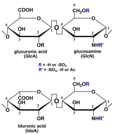
Heparin is found in mast cell granules and interacts with histamine, proteases and other inflammatory mediators [54]. The heparin polysaccharide consists of a disaccharide repeating unit of either glucuronic acid (GlcA) or iduronic acid (IdoA) and glucosamine (GlcN) residues. The disaccharide unit is capable of carrying sulfo groups (Figure 2) [55].
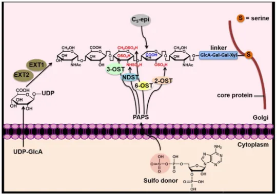
Biosynthesis of heparin occurs in the endoplasmic reticulum and the Golgi and involves a series of specialized enzymes. Generally, the pathway consists of three phases: initiation, polymerization and modification of the chain. The initiation phase involves formation of a linker that tethers to a serine residue of serglycin, a core protein presented in the mast cell (Figure 3). Then, chain polymerization takes place by formation of a copolymer of N-acetylglucosamine (GlcNAc) and GlcA, and this forms the backbone of heparin. This chain elongation process is driven by two polymerases known as exostosin glycosyltransferase 1 (EXT 1) and EXT 2 [56]. The unmodified polysaccharide backbone subsequently undergoes chain modification by a series of sulfations and epimerization. The first modification step is N-sulfation of GlcNAc residues by N-deacetylase/N-sulfotransferase (NDST), which is a critical step for subsequent modification. NDST has two functions: removal of the acetyl group deacetylase activity, NDase) and installation of an N-sulfo group
(N-sulfotransferase activity, NST) [57]. After N-sulfation, C5-epimerase (C5-epi) following by
2-O-sulfation take place. C5-epi converts the D-glucuronic acid (GlcA) to L-iduronic acid
(IdoA) [58]. 2-O-sulfotransferase catalyzes the transfer of a sulfo group to the 2-O-position of either IdoA or GlcA, but preferentially to the IdoA residue [59]. The addition of a sulfo
A
uthor Man
uscr
ipt
A
uthor Man
uscr
ipt
A
uthor Man
uscr
ipt
A
uthor Man
uscr
group at the 6-OH of GlcN is modified by 6-O-sulfotransferases [60]. Lastly, 3-O-sulfotransferases add a sulfo group at the 3-OH of GlcN (39), and this is critical for AT binding and anticoagulant activity (Figure 3) [61, 62].
Creation of synthetic heparin polysaccharides
Synthesis of the pentasaccharide is purely chemical. It is extremely difficult, if not impossible, to synthesize polysacharides in the size range of LMWH using this method. However, the utilization of biosynthetic enzymes offers an alternative strategy to synthesize heparin oligosaccharides. These enzymes have high regioselectivity towards their substrates thereby eliminating the need for protecting groups in the chemical synthesis. This in turn drastically shortens the synthetic route and improves the product yield [21].
Heparin biosynthetic enzymes have been extensively studied and characterized [63]. Several crystal structures of heparin biosynthetic enzymes are available providing information necessary for designing heparin structures with specific biological functions [64-66]. It is now feasible to alter substrate specificity and synthesize certain structures that cannot be obtained from the wild type proteins [67]. Further, the sequence of enzymatic modifications can be altered making it feasible to create the exact structure of a specific, desired target compound. To date, the majority of these biosynthetic enzymes have been expressed in Esherichia Coli (E. coli) [68]. In 2011, the research groups of Liu and Linhardt successfully utilized a chemoenzymatic method to synthesize an ultra low molecular weight heparin with equivalent anticoagulant potency to that of fondaparinux [69].
Strategies for chemoenzymatic synthesis of heparin
The key components for the chemoenzymatic synthesis of heparin are enzymes, sugar substrates and a sulfo donor. Most of the biosynthetic enzymes have been characterized [67]. However, recombinant form of certain enzymes are difficult to obtain. Furthermore, some natural sugar nucleotides are not compatible with the chemoenzymatic synthesis strategy therefore unnatural nucleotide sugars are required. Finally, the commercial sulfo donor is prohibitively expensive for a large scale synthesis. The following strategies have been developed to overcome these problems.
Altering biosynthetic enzymes
Although several mammalian heparin biosynthetic enzymes have been successfully
expressed in the bacterial expression system, certain enzymes e.g. EXT 1 and EXT 2 cannot be expressed in E. coli. To overcome this problem, bacterial glycosyltransferases from the E. Coli K5 strain (N-acetyl glucosaminyl transferase, KfiA) [70] and heparosan synthase 2 from Pasteurella multocida (pmHS2) [71, 72] are utilized in the synthesis of heparin backbone [73]. The bacteria produce extracellular carbohydrate capsule consisting of a disaccharide unit that is identical to the unmodified heparin backbone suggesting the use of bacterial glycosyltransferase counterparts.
NDST is a key enzyme in heparin biosynthesis as previously discussed. However, NDST is not suitable for the chemoenzymatic synthesis because of two reasons: first, NDST is a large protein with more than 800 amino acid residues, and is difficult to express in E. coli in high
A
uthor Man
uscr
ipt
A
uthor Man
uscr
ipt
A
uthor Man
uscr
ipt
A
uthor Man
uscr
levels. Second, the conversion of GlcNAc to GlcNS residue by NDST has a specific pattern, depending on the isoform [74]. Thus, the use of NDST does not allow the freedom to vary the position of the GlcNS residue in a heparin product. As mentioned previously, N-sulfotransferase (NST) is the C-terminal of NDST, consisting of only N-N-sulfotransferase activity. NST is only 260 amino acid residues long, and is expressed in E. coli at a very high level [65]. Therefore, using only the NST domain with the modified glucosamine substrate, allows the completion of the N-sulfation step and overcomes the limitations of NDST.
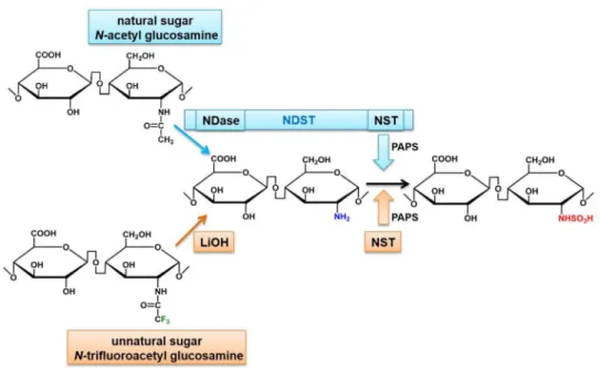
Modifying the sugar substrate
Formation of a glycosidic linkage in the core backbone of heparin requires uridine
diphosphate sugar substrates: UDP-GlcNAc and UDP-GlcA. In the N-sulfo addition step by NDST, NDase removes the N-acetyl group and converts GlcNAc (CH3CONH) to the free
amino group (GlcNH2). NST adds a sulfo group to GlcNH2 resulting in GlcNS
(GlcNHSO3). Because NDST is not available, an alternative method using NST and an
unnatural uridine diphosphate sugar substrate were used to introduce the GlcNS residues. Here, the presence of GlcNH2 substrate is required for the sulflation by NST. The
implementation of an unnatural UDP-N-trifluoroacetyl glucosamine (UDP-NTFA) serves as an analog of UDP-GlcNAc [75, 76] as they are structurally similar [77]. The GlcNTFA residue is introduced into the heparin core backbone by glycosyltransferases both KfiA and pmHS2. The N-trifluoroacetyl group (CF3CONH) from the GlcNTFA residue can be
removed by a basic condition (lithium hydroxide) yielding GlcNH2 residue (Figure 4).
Preparing in-house sulfate donor
In addition to polysaccharide backbone synthesis, sulfate modifications crucially dictate biological activity of heparin. The sulfation steps by multiple sulfotransferases require a sulfate donor known as 3’-phosphoadenosine 5’-phosphosulfate (PAPS). PAPS is expensive and the availability of PAPS is a key determinant of cost-effectiveness when considering large-scale synthesis for clinical use. This limitation has been addressed by the synthesis of PAPS using an enzymatic approach in a one-pot format using ATP sulfurylase (from Kluyveromyces lactis), APS kinase (from Penicillium chrysogenum) and pyrophosphatase (from E.coli). Bacterial expression of all three enzymes yielding crude extracts are efficiently enough to convert ATP and sulfate to PAPS in a gram scale [78].
Alternatives for low molecular weight heparin production
It has been well established that heparin binds to a number of biological proteins resulting in various pharmacological effects. Heparin compound obtained from natural sources is heterogeneous with a number of drawbacks that have already been discussed. Recent technological advances in carbohydrate chemistry (both chemical and enzymatic synthesis) have made development of synthetic LMWH for clinical applications feasible.
Non-enzymatic synthesis of larger heparin molecules remains a challenge. Protecting and de-protecting steps of reactive hydroxyl groups are essential for a chemical synthesis of carbohydrate. In addition, the stereoselectivity is critically important. The Gardiner research group successfully synthesized a lead compound in gram-scale (12 sugar residue
heparin-A
uthor Man
uscr
ipt
A
uthor Man
uscr
ipt
A
uthor Man
uscr
ipt
A
uthor Man
uscr
related compound) by constructing a starting disaccharide that is used as a precursor building block for repeated elongation cycles [79, 80]. The resulting product is useful for pharmacokinetic and metabolic studies in vivo.
M118 is an improved LMWH that was rationally designed to circumvent the polydispersity of commercial LMWHs. M118 has a high potency with reduced polydispersity [81] that results from a stepwise manufacturing process starting from isolation of high potency UFH. The isolated UFH is depolymerized by a patented heparin lyase mutant which shortens UFH into LMWH fragments while the AT-binding domain largely remains intact. The fragments are further selected for a portion of high anti-FXa and anti-thrombin activities. The resulting LMWH displays superior anticoagulant activity than the parent UFH (and other LMWHs) due to the decrease in polydispersity. However, M118 is still derived from animal-source material.
In addition to the development of M118, our group created a simple and rapid enzymatic method to produce a LMWH that has a narrow size distribution [82]. The method involves use of a dual function pmHS2 to simultaneously add GlcN and GlcA to a starting saccharide precursor in a one-pot enzymatic reaction. The one-pot synthetic format limits the size distribution of the resulting sugar backbone in a range of 8-14 sugar residues which is narrower than that of commercial LMWHs (6-22 sugar residues). The sugar backbones produced by the controlled enzymatic reaction were further subjected to sulfo modifications yielding a narrow size distribution LMWH with potent anti-FXa activity. This enzymatic approach eliminates need for animal source material and improves both polydispersity and anticoagulant potency.
A new homogeneous, reversible LMWH
A new synthetic, homogeneous LMWH was designed such that it had the following beneficial properties: potent anticoagulant activity, hepatic clearance, and effective neutralization with protamine. Recently, we synthesized a series of homogeneous LMWHs that were composed of 6, 8, 10, or 12 sugar residues (6mer, 8mer, 10mer and 12mer) (Figure 5A) [83]. All of these synthetic LMWHs were structurally designed to carry the
pentasaccharide motif for AT binding. The initial step in the synthesis was building a sugar backbone. We employed a commercially available monosaccharide, 1-O-(para-nitrophenyl)-glucuronide (GlcA-pNP), as the starting monosaccharide. Two bacterial glycosyltransferases (KfiA and pmHS2) were used in a step-wise elongation process. Placement of sulfo groups at N- and O-positions were driven by the specific sulfotransferases responsible for these positions while altering of the GlcA configuration was catalyzed by C5-epimerase (Figure
5B).
The pharmacological properties of these synthetic LMWHs were investigated both in vitro and in vivo in comparison to UFH and enoxaparin [83]. All of the synthetic LMWHs showed superior potency in the anticoagulant activity in comparison to UFH and enoxaparin. We probed the binding of these LMWHs to the stabilin-2 receptor (systemic clearance receptor for UFH and a portion of LMWH [52]) both in vitro and in vivo. Larger LMWHs (10mer and 12 mer) were internalized by stabilin-2 and retained in the liver, while the
A
uthor Man
uscr
ipt
A
uthor Man
uscr
ipt
A
uthor Man
uscr
ipt
A
uthor Man
uscr
smaller LMWHs (6mer and 8mer) showed low binding to the receptor. We next determined the ability of protamine to neutralize the synthetic LMWHs. The smaller LMWHs (6mer, 8mer and 10 mer) displayed fairly low sensitivity to protamine neutralization. Further, the anticoagulant activity of the larger LMWH (12mer) was still not fully reversible by protamine. Therefore, we further modified the 12mer by adding an extra sulfo group to obtain S12mer (Figure 5A). The S12mer demonstrated the same protamine reversibility as UFH. These finding were confirmed in an ex vivo mouse model. Finally, in vivo studies using a mouse-tail-clip bleeding model showed that protamine reduced the prolonged bleeding time induced by the S12mer (77).
Our data suggested that protamine only neutralized LMWH fragments larger than 10 sugar residues. Commercial LMWH is a mixture of LMWH fragments with a broad size
distribution and this wide range in polymer size is associated with variable affinity towards protamine. This explains the partial protamine neutralization observed with commercial LMWHs. The excellent sensitivity toward protamine neutralization shown by the S12mer suggests both size of the sugar backbone and the sulfation pattern contribute to the sensitivity toward protamine neutralization.
Conclusion
The advancement in understanding the substrate specificity of heparin biosynthetic enzymes is critically important to conduct successful enzymatic synthesis. An enzymatic approach circumvents the difficulty in heparin synthesis and provides an alternative to synthesize the complex, carbohydrate containing molecules that reach the limit of the organic synthesis. The use of biosynthetic enzymes not only provides a high regio- and stereo-selectivity for a formation of glycosidic bond but also solves the problems of protection and de-protection steps required for chemical synthesis.
It has been established that a sulfation pattern as well as the size of heparin fragments are key factors contributing to the interaction between heparin and heparin-binding proteins [84]. Other therapeutic applications of heparin have been extensively studied e.g. anti-inflammatory property. With the well-developed enzymatic approach, custom designed heparin, without anticoagulant activity to avoid the hemorrhagic side effect, can be achieved. The development of synthetic version of heparins not only eliminates the disadvantages and risks of using animal derived-medicine; it also supports further biological studies of heparin and its related compounds.
The production of synthetic heparin in laboratories has already reached to gram scale, a nearly 500,000-fold increase compared with the first synthesis published in 2002 [85]. This improvement is the result of more efficient enzyme expressions and the higher efficiency in the synthesis of enzyme co-factors. Although the cost of synthetic heparins synthesis is still higher than the production cost of natural heparins, further development of the
chemoenzymatic approach should result in cost-effective products, accelerating the modernization of LMWH drugs.
A
uthor Man
uscr
ipt
A
uthor Man
uscr
ipt
A
uthor Man
uscr
ipt
A
uthor Man
uscr
Acknowledgments
We would like to thank Raj Kasthuri MD for his valuable input and critical reading of the manuscript.
This work was supported by grants from the National Institutes of Health: HL096679; HL117659
References
1. Bishop JR, Schuksz M, Esko JD. Heparan sulphate proteoglycans fine-tune mammalian physiology. Nature. 2007; 446:1030–7. [PubMed: 17460664]
2. Perrimon N, Bernfield M. Specificities of heparan sulphate proteoglycans in developmental processes. Nature. 2000; 404:725–8. [PubMed: 10783877]
3. Garcia DA, Baglin TP, Weitz JI, Samama MM. American College of Chest P. Parenteral anticoagulants: Antithrombotic Therapy and Prevention of Thrombosis. Chest (9th). 2012; 141:e24S–43S. American College of Chest Physicians Evidence-Based Clinical Practice Guidelines. [PubMed: 22315264]
4. Mc LJ. The discovery of heparin. Circulation. 1959; 19:75–8. [PubMed: 13619023]
5. Linhardt RJ, Gunay NS. Production and chemical processing of low molecular weight heparins. Seminars in thrombosis and hemostasis. 1999; 25(Suppl 3):5–16. [PubMed: 10549711] 6. Liu CSMaJ. China’s heparin revisited: What went wrong and has anything changed? Journal of
Commercial Biotechnology. 2013; 19:1–13.
7. Johnson EA, Mulloy B. The molecular-weight range of mucosal-heparin preparations. Carbohydrate research. 1976; 51:119–27. [PubMed: 1000525]
8. Lam LH, Silbert JE, Rosenberg RD. The separation of active and inactive forms of heparin. Biochemical and biophysical research communications. 1976; 69:570–7. [PubMed: 1267803] 9. Andersson LO, Barrowcliffe TW, Holmer E, Johnson EA, Sims GE. Anticoagulant properties of
heparin fractionated by affinity chromatography on matrix-bound antithrombin iii and by gel filtration. Thrombosis research. 1976; 9:575–83. [PubMed: 1006626]
10. Johnson EA, Kirkwood TB, Stirling Y, Perez-Requejo JL, Ingram GI, Bangham DR, Brozovic M. Four heparin preparations: anti-Xa potentiating effect of heparin after subcutaneous injection. Thrombosis and haemostasis. 1976; 35:586–91. [PubMed: 989965]
11. Harenberg J. Pharmacology of low molecular weight heparins. Seminars in thrombosis and hemostasis. 1990; 16(Suppl):12–8. [PubMed: 1962899]
12. Martel N, Lee J, Wells PS. Risk for heparin-induced thrombocytopenia with unfractionated and low-molecular-weight heparin thromboprophylaxis: a meta-analysis. Blood. 2005; 106:2710–5. [PubMed: 15985543]
13. Sparkenbaugh E, Pawlinski R. Interplay between coagulation and vascular inflammation in sickle cell disease. Br J Haematol. 2013; 162:3–14. [PubMed: 23593937]
14. Rosenberg RD, Damus PS. The purification and mechanism of action of human antithrombin-heparin cofactor. The Journal of biological chemistry. 1973; 248:6490–505. [PubMed: 4738234] 15. Danielsson A, Raub E, Lindahl U, Bjork I. Role of ternary complexes, in which heparin binds both
antithrombin and proteinase, in the acceleration of the reactions between antithrombin and thrombin or factor Xa. The Journal of biological chemistry. 1986; 261:15467–73. [PubMed: 3782075]
16. Casu B, Oreste P, Torri G, Zoppetti G, Choay J, Lormeau JC, Petitou M, Sinay P. The structure of heparin oligosaccharide fragments with high anti-(factor Xa) activity containing the minimal antithrombin III-binding sequence. Chemical and 13C nuclear-magnetic-resonance studies. The Biochemical journal. 1981; 197:599–609. [PubMed: 7325974]
17. Choay J, Petitou M, Lormeau JC, Sinay P, Casu B, Gatti G. Structure-activity relationship in heparin: a synthetic pentasaccharide with high affinity for antithrombin III and eliciting high anti-factor Xa activity. Biochemical and biophysical research communications. 1983; 116:492–9. [PubMed: 6651824]
18. Olson ST, Bjork I, Sheffer R, Craig PA, Shore JD, Choay J. Role of the antithrombin-binding pentasaccharide in heparin acceleration of antithrombin-proteinase reactions. Resolution of the
A
uthor Man
uscr
ipt
A
uthor Man
uscr
ipt
A
uthor Man
uscr
ipt
A
uthor Man
uscr
antithrombin conformational change contribution to heparin rate enhancement. The Journal of biological chemistry. 1992; 267:12528–38. [PubMed: 1618758]
19. Xu Y, Pempe EH, Liu J. Chemoenzymatic Synthesis of Heparin Oligosaccharides with both Anti-Xa and Anti-IIa Activities. J Biol Chem. 2012 10.1074/jbc.M112.358523.
20. Walenga JM, Fareed J, Petitou M, Samama M, Lormeau JC, Choay J. Intravenous antithrombotic activity of a synthetic heparin pentasaccharide in a human serum induced stasis thrombosis model. Thromb Res. 1986; 43:243–8. [PubMed: 3738864]
21. Linhardt RJ, Liu J. Synthetic heparin. Current Opinion in Pharmacology. 2012; 12:217–9. [PubMed: 22325855]
22. Petitou M, Duchaussoy P, Lederman I, Choay J, Jacquinet JC, Sinay P, Torri G. Synthesis of heparin fragments: a methyl alpha-pentaoside with high affinity for antithrombin III. Carbohydrate research. 1987; 167:67–75. [PubMed: 3690577]
23. Petitou M, van Boeckel CA. A synthetic antithrombin III binding pentasaccharide is now a drug! What comes next? Angewandte Chemie. 2004; 43:3118–33. [PubMed: 15199558]
24. Gray E, Mulloy B, Barrowcliffe TW. Heparin and low-molecular-weight heparin. Thrombosis and haemostasis. 2008; 99:807–18. [PubMed: 18449410]
25. Kelton JG, Warkentin TE. Heparin-induced thrombocytopenia: a historical perspective. Blood. 2008; 112:2607–16. [PubMed: 18809774]
26. Heres EK, Speight K, Benckart D, Marquez J, Gravlee GP. The clinical onset of heparin is rapid. Anesth Analg. 2001; 92:1391–5. [PubMed: 11375810]
27. Hirsh J, Raschke R. Heparin and low-molecular-weight heparin: the Seventh ACCP Conference on Antithrombotic and Thrombolytic Therapy. Chest. 2004; 126:188S–203S. 10.1378/chest.
126.3_suppl.188S. [PubMed: 15383472]
28. Weitz JI. Low-molecular-weight heparins. The New England journal of medicine. 1997; 337:688– 98. [PubMed: 9278467]
29. Bradbrook ID, Magnani HN, Moelker HC, Morrison PJ, Robinson J, Rogers HJ, Spector RG, Van Dinther T, Wijnand H. ORG 10172: a low molecular weight heparinoid anticoagulant with a long half-life in man. British journal of clinical pharmacology. 1987; 23:667–75. [PubMed: 3606928] 30. Quinlan DJ, McQuillan A, Eikelboom JW. Low-molecular-weight heparin compared with
intravenous unfractionated heparin for treatment of pulmonary embolism: a meta-analysis of randomized, controlled trials. Annals of internal medicine. 2004; 140:175–83. [PubMed: 14757615]
31. Kuo KH, Kovacs MJ. Fondaparinux: a potential new therapy for HIT. Hematology. 2005; 10:271– 5. [PubMed: 16085538]
32. Kuo KH, Kovacs MJ. Successful treatment of heparin induced thrombocytopenia (HIT) with fondaparinux. Thrombosis and haemostasis. 2005; 93:999–1000. [PubMed: 15886823] 33. Kang M, Alahmadi M, Sawh S, Kovacs MJ, Lazo-Langner A. Fondaparinux for the treatment of
suspected heparin-induced thrombocytopenia: a propensity score-matched study. Blood. 2015; 125:924–9. [PubMed: 25515959]
34. B. W. In China. The New York Times. The New York Times; 2010. Strong Debut for Supplier of Heparin; p. B6
35. Blossom DB, Kallen AJ, Patel PR, Elward A, Robinson L, Gao G, Langer R, Perkins KM, Jaeger JL, Kurkjian KM, Jones M, Schillie SF, Shehab N, Ketterer D, Venkataraman G, Kishimoto TK, Shriver Z, McMahon AW, Austen KF, Kozlowski S, et al. Outbreak of adverse reactions associated with contaminated heparin. The New England journal of medicine. 2008; 359:2674–84. [PubMed: 19052120]
36. Guerrini M, Beccati D, Shriver Z, Naggi A, Viswanathan K, Bisio A, Capila I, Lansing JC, Guglieri S, Fraser B, Al-Hakim A, Gunay NS, Zhang Z, Robinson L, Buhse L, Nasr M, Woodcock J, Langer R, Venkataraman G, Linhardt RJ, et al. Oversulfated chondroitin sulfate is a contaminant in heparin associated with adverse clinical events. Nature biotechnology. 2008; 26:669–75. 37. Liu H, Zhang Z, Linhardt RJ. Lessons learned from the contamination of heparin. Nat Prod Rep.
2009; 26:313–21. [PubMed: 19240943]
38. Kishimoto TK, Viswanathan K, Ganguly T, Elankumaran S, Smith S, Pelzer K, Lansing JC, Sriranganathan N, Zhao G, Galcheva-Gargova Z, Al-Hakim A, Bailey GS, Fraser B, Roy S,
A
uthor Man
uscr
ipt
A
uthor Man
uscr
ipt
A
uthor Man
uscr
ipt
A
uthor Man
uscr
Rogers-Cotrone T, Buhse L, Whary M, Fox J, Nasr M, Dal Pan GJ, et al. Contaminated heparin associated with adverse clinical events and activation of the contact system. The New England journal of medicine. 2008; 358:2457–67. [PubMed: 18434646]
39. Zhang Z, Weiwer M, Li B, Kemp MM, Daman TH, Linhardt RJ. Oversulfated chondroitin sulfate: impact of a heparin impurity, associated with adverse clinical events, on low-molecular-weight heparin preparation. Journal of medicinal chemistry. 2008; 51:5498–501. [PubMed: 18754653] 40. Convention USP. U.S. Pharmacopeia 38 and National Formulary 33. Slp. United States
Pharmacopeial: May. 2015 2015
41. Merli GJ, Groce JB. Pharmacological and clinical differences between low-molecular-weight heparins: implications for prescribing practice and therapeutic interchange. P T. 2010; 35:95–105. [PubMed: 20221326]
42. Pempe EH, Xu Y, Gopalakrishnan S, Liu J, Harris EN. Probing structural selectivity of synthetic heparin binding to stabilin protein receptors. The Journal of biological chemistry. 2012; 287:20774–83. [PubMed: 22547069]
43. Bisio A, Vecchietti D, Citterio L, Guerrini M, Raman R, Bertini S, Eisele G, Naggi A, Sasisekharan R, Torri G. Structural features of low-molecular-weight heparins affecting their affinity to antithrombin. Thrombosis and haemostasis. 2009; 102:865–73. [PubMed: 19888521] 44. Ma Q, Tobu M, Schultz C, Jeske W, Hoppensteadt D, Walenga J, Cornelli U, Lee J, Linhardt R,
Hanin I, Fareed J. Molecular weight dependent tissue factor pathway inhibitor release by heparin and heparin oligosaccharides. Thrombosis research. 2007; 119:653–61. [PubMed: 16824584] 45. Nightingale SL. Appropriate Use of Low-Molecular-Weight Heparins (LMWHs). Journal of the
American Medical Association. 1993; 270:1672. [PubMed: 8411485]
46. Crowther MPaMA. Neutralization of heparin activity. In: Rebecca Lever BMaCPP. , editor. Handbook of experimental pharmacology. Springer; 2012. p. 266
47. Holst J, Lindblad B, Bergqvist D, Garre K, Nielsen H, Hedner U, Ostergaard PB. Protamine neutralization of intravenous and subcutaneous low-molecular-weight heparin (tinzaparin, Logiparin). An experimental investigation in healthy volunteers. Blood coagulation & fibrinolysis : an international journal in haemostasis and thrombosis. 1994; 5:795–803. [PubMed: 7865687] 48. Chawla LS, Moore G, Seneff MG. Incomplete reversal of enoxaparin toxicity by protamine:
implications of renal insufficiency, obesity, and low molecular weight heparin sulfate content. Obesity surgery. 2004; 14:695–8. [PubMed: 15186641]
49. van Veen JJ, Maclean RM, Hampton KK, Laidlaw S, Kitchen S, Toth P, Makris M. Protamine reversal of low molecular weight heparin: clinically effective? Blood coagulation & fibrinolysis : an international journal in haemostasis and thrombosis. 2011; 22:565–70. [PubMed: 21959588] 50. Hirsh J, O'Donnell M, Eikelboom JW. Beyond unfractionated heparin and warfarin: current and
future advances. Circulation. 2007; 116:552–60. [PubMed: 17664384]
51. Oie CI, Olsen R, Smedsrod B, Hansen JB. Liver sinusoidal endothelial cells are the principal site for elimination of unfractionated heparin from the circulation. American journal of physiology Gastrointestinal and liver physiology. 2008; 294:G520–8. [PubMed: 18063704]
52. Harris EN, Weigel JA, Weigel PH. The human hyaluronan receptor for endocytosis (HARE/ Stabilin-2) is a systemic clearance receptor for heparin. The Journal of biological chemistry. 2008; 283:17341–50. [PubMed: 18434317]
53. Lim W, Dentali F, Eikelboom JW, Crowther MA. Meta-analysis: low-molecular-weight heparin and bleeding in patients with severe renal insufficiency. Annals of internal medicine. 2006; 144:673–84. [PubMed: 16670137]
54. L. CPaKn. Heparin Biosynthesis. Springer; 2012.
55. Sarrazin S, Lamanna WC, Esko JD. Heparan sulfate proteoglycans. Cold Spring Harbor perspectives in biology. 2011:3.
56. Lind T, Tufaro F, McCormick C, Lindahl U, Lidholt K. The putative tumor suppressors EXT1 and EXT2 are glycosyltransferases required for the biosynthesis of heparan sulfate. The Journal of biological chemistry. 1998; 273:26265–8. [PubMed: 9756849]
57. Kusche-Gullberg M, Eriksson I, Pikas DS, Kjellen L. Identification and expression in mouse of two heparan sulfate glucosaminyl N-deacetylase/N-sulfotransferase genes. The Journal of biological chemistry. 1998; 273:11902–7. [PubMed: 9565617]
A
uthor Man
uscr
ipt
A
uthor Man
uscr
ipt
A
uthor Man
uscr
ipt
A
uthor Man
uscr
58. Li J, Hagner-McWhirter A, Kjellen L, Palgi J, Jalkanen M, Lindahl U. Biosynthesis of heparin/ heparan sulfate. cDNA cloning and expression of D-glucuronyl C5-epimerase from bovine lung. The Journal of biological chemistry. 1997; 272:28158–63. [PubMed: 9346972]
59. Rong J, Habuchi H, Kimata K, Lindahl U, Kusche-Gullberg M. Substrate specificity of the heparan sulfate hexuronic acid 2-O-sulfotransferase. Biochemistry. 2001; 40:5548–55. [PubMed:
11331020]
60. Habuchi H, Tanaka M, Habuchi O, Yoshida K, Suzuki H, Ban K, Kimata K. The occurrence of three isoforms of heparan sulfate 6-O-sulfotransferase having different specificities for hexuronic acid adjacent to the targeted N-sulfoglucosamine. The Journal of biological chemistry. 2000; 275:2859–68. [PubMed: 10644753]
61. Liu J, Shworak NW, Fritze LM, Edelberg JM, Rosenberg RD. Purification of heparan sulfate D-glucosaminyl 3-O-sulfotransferase. The Journal of biological chemistry. 1996; 271:27072–82. [PubMed: 8900198]
62. Atha DH, Lormeau JC, Petitou M, Rosenberg RD, Choay J. Contribution of monosaccharide residues in heparin binding to antithrombin III. Biochemistry. 1985; 24:6723–9. [PubMed: 4084555]
63. Chappell EP, Liu J. Use of biosynthetic enzymes in heparin and heparan sulfate synthesis. Bioorg Med Chem. 2012
64. Moon AF, Xu Y, Woody SM, Krahn JM, Linhardt RJ, Liu J, Pedersen LC. Dissecting the substrate recognition of 3-O-sulfotransferase for the biosynthesis of anticoagulant heparin. Proceedings of the National Academy of Sciences of the United States of America. 2012; 109:5265–70. [PubMed: 22431632]
65. Kakuta Y, Sueyoshi T, Negishi M, Pedersen LC. Crystal structure of the sulfotransferase domain of human heparan sulfate N-deacetylase/ N-sulfotransferase 1. The Journal of biological chemistry. 1999; 274:10673–6. [PubMed: 10196134]
66. Bethea HN, Xu D, Liu J, Pedersen LC. Redirecting the substrate specificity of heparan sulfate 2-O-sulfotransferase by structurally guided mutagenesis. Proceedings of the National Academy of Sciences of the United States of America. 2008; 105:18724–9. [PubMed: 19022906] 67. Liu J, Moon AF, Sheng J, Pedersen LC. Understanding the substrate specificity of the heparan
sulfate sulfotransferases by an integrated biosynthetic and crystallographic approach. Current opinion in structural biology. 2012; 22:550–7. 1. [PubMed: 22840348]
68. Peterson S, Frick A, Liu J. Design of biologically active heparan sulfate and heparin using an enzyme-based approach. Nat Prod Rep. 2009; 26:610–27. [PubMed: 19387498]
69. Xu YMS, Takieddin M, Xu H, Liu R, Jing J, Mousa SA, Lindhardt RJ, Liu J. Chemoenzymatic Synthesis of Homogeneous Ultralow Molecular Weight Heparins. Science. 2011; 334:498–501. [PubMed: 22034431]
70. Chen M, Bridges A, Liu J. Determination of the substrate specificities of N-acetyl-d-glucosaminyltransferase. Biochemistry. 2006; 45:12358–65. [PubMed: 17014088]
71. Sismey-Ragatz AE, Green DE, Otto NJ, Rejzek M, Field RA, DeAngelis PL. Chemoenzymatic synthesis with distinct Pasteurella heparosan synthases: monodisperse polymers and unnatural structures. The Journal of biological chemistry. 2007; 282:28321–7. [PubMed: 17627940] 72. Chavaroche AAE, Springer J, Kooy F, Boeriu C, Eggink G. In vitro synthesis of heparosan using
recombinant Pasteurella multocida heparosan synthase PmHS2. Applied microbiology and biotechnology. 2010; 85:1881–91. [PubMed: 19756580]
73. Chen JJC, Liu J. Using an Enzymatic Combinatorial Approach to Identify Anticoagulant Heparan Sulfate Structures. Chemical Biology. 2007; 14:986–3.
74. Sheng J, Liu R, Xu Y, Liu J. The dominating role of N-deacetylase/N-sulfotransferase 1 in forming domain structures in heparan sulfate. The Journal of biological chemistry. 2011; 286:19768–76. [PubMed: 21454625]
75. Liu R, Xu Y, Chen M, Weiwer M, Zhou X, Bridges AS, DeAngelis PL, Zhang Q, Linhardt RJ, Liu J. Chemoenzymatic design of heparan sulfate oligosaccharides. The Journal of biological chemistry. 2010; 285:34240–9. [PubMed: 20729556]
A
uthor Man
uscr
ipt
A
uthor Man
uscr
ipt
A
uthor Man
uscr
ipt
A
uthor Man
uscr
76. Sala RF, MacKinnon SL, Palcic MM, Tanner ME. UDP-N-trifluoroacetylglucosamine as an alternative substrate in N-acetylglucosaminyltransferase reactions. Carbohydrate research. 1998; 306:127–36. [PubMed: 9691444]
77. Masuko S, Bera S, Green DE, Weiwer M, Liu J, DeAngelis PL, Linhardt RJ. Chemoenzymatic synthesis of uridine diphosphate-GlcNAc and uridine diphosphate-GalNAc analogs for the preparation of unnatural glycosaminoglycans. The Journal of organic chemistry. 2012; 77:1449– 56. [PubMed: 22239739]
78. Zhou X, Chandarajoti K, Pham TQ, Liu R, Liu J. Expression of heparan sulfate sulfotransferases in Kluyveromyces lactis and preparation of 3'-phosphoadenosine-5'-phosphosulfate. Glycobiology. 2011; 21:771–80. [PubMed: 21224284]
79. Hansen SU, Miller GJ, Jayson GC, Gardiner JM. First gram-scale synthesis of a heparin-related dodecasaccharide. Organic letters. 2013; 15:88–91. [PubMed: 23240767]
80. Hansen SU, Miller GJ, Cole C, Rushton G, Avizienyte E, Jayson GC, Gardiner JM.
Tetrasaccharide iteration synthesis of a heparin-like dodecasaccharide and radiolabelling for in vivo tissue distribution studies. Nature communications. 2013; 2016; 4
81. Kishimoto TK, Qi YW, Long A, Capilal I, Sasisekharan R, Guerrero L, Fier I, Roach J, Venkataraman G. M118-A rationally engineered low-molecular-weight heparin designed specifically for the treatment of acute coronary syndromes. Thrombosis and haemostasis. 2009; 102:900–6. [PubMed: 19888526]
82. Chandarajoti K, Xu Y, Sparkenbaugh E, Key NS, Pawlinski R, Liu J. De novo synthesis of a narrow size distribution low-molecular-weight heparin. Glycobiology. 2014; 24:476–86. [PubMed: 24626379]
83. Xu Y, Cai C, Chandarajoti K, Hsieh PH, Li L, Pham TQ, Sparkenbaugh EM, Sheng J, Key NS, Pawlinski R, Harris EN, Linhardt RJ, Liu J. Homogeneous low-molecular-weight heparins with reversible anticoagulant activity. Nat Chem Biol. 2014; 10:248–50. [PubMed: 24561662]
84. Gandhi NS, Mancera RL. The structure of glycosaminoglycans and their interactions with proteins. Chemical biology & drug design. 2008; 72:455–82. [PubMed: 19090915]
85. Liu J, Linhardt RJ. Chemoenzymatic synthesis of heparan sulfate and heparin. Nat Prod Rep. 2014; 31:1676–85. [PubMed: 25197032]
A
uthor Man
uscr
ipt
A
uthor Man
uscr
ipt
A
uthor Man
uscr
ipt
A
uthor Man
uscr
Figure 1. A chemical structure of the pentasaccharide motif of heparin polysaccharide
Heparin binds to antithrombin through a pentasaccharide region leading to the inhibition of Factor Xa (FXa). The Inhibition of FXa requires the unique sequence of sugar shown as A-B-C-D-E. Heparin polysaccharide that contains at least 18 sugar residues can form a complex with thrombin and inactivates it.
A
uthor Man
uscr
ipt
A
uthor Man
uscr
ipt
A
uthor Man
uscr
ipt
A
uthor Man
uscr
Figure 2. Disaccharide repeating unit of heparin
The disaccharide unit is capable of carrying sulfo groups. The GlcN residue can be sulfated at the N-, 3-O- and 6-O- positions while GlcA and IdoA only carry a sulfo group at the 2-O-postion.
A
uthor Man
uscr
ipt
A
uthor Man
uscr
ipt
A
uthor Man
uscr
ipt
A
uthor Man
uscr
Figure 3. Heparin biosynthesis
Two polymerases, EXT1 and EXT2, are responsible for building the sulfated and non-epimerized backbone polysaccharide consisting of the disaccharide repeating unit of GlcA and GlcNAc linked by a glycosidic linkage, where GlcNAc presents N-acetylated
glucosamine, GlcA presents glucuronic acid. N-deacetylase/N-sulfotransferase (NDST) is a dual function enzyme: it removes an acetyl group from a GlcNAc residue, displaying deacetylase activity (NDase), then transfers a sulfo group to the deacetylated glucosamine residue to form an N-sulfo glucosamine (GlcNHSO3) residue, displaying N-sulfotransferase
activity (NST). C5-epimerase (C5-Epi) converts a GlcA residue to an IdoA residue.
Sulfotransferases co-functioning with a sulfo donor, 3’-phosphoadenosine-5’-phosphosulfate (PAPS), add sulfo groups to their respective positions. 2-O-sulfotransferase (2-OST) adds sulfo groups at the 2-OH position of both GlcA and IdoA while 6-O-sulfotransferase (6-OST) and 3-O-sulfotransferase (3-(6-OST) add sulfo groups at the 6-OH position and 3-OH of glucosamine residues, respectively [55].
A
uthor Man
uscr
ipt
A
uthor Man
uscr
ipt
A
uthor Man
uscr
ipt
A
uthor Man
uscr
Figure 4. Synthetic strategy for N-sulfation step
The N-trifluoroacetyl group of unnatural sugar, UDP-N-trifluoroacetyl glucosamine (UDP-GlcNTFA), can be converted to GlcNH2 by lithium hydroxide (LiOH). The addition of a
sulfo group can be achieved by NST and PAPS. NDST = N-deacetylase/N-sulfotransferase, NDase = N-deacetylase, NST = N-sulfotransferase, PAPS = 3’-phosphoadenosine-5’-phosphosulfate.
A
uthor Man
uscr
ipt
A
uthor Man
uscr
ipt
A
uthor Man
uscr
ipt
A
uthor Man
uscr
Figure 5. Chemoenzymtically synthetic scheme of LMWH
(A)A series of homogeneous synthetic LMWHs designed for improving sensitivity to
protamine neutralization as well as hepatic clearance. All synthetic LMWHs are structurally similar and carry a pentasaccharide domain for AT binding (B) In vitro synthesis of LMWH includes 2 steps (i) sugar backbone synthesis and (ii) sugar backbone modification. The scheme indicates the implemented strategies to improve the synthetic method. The backbone synthesis starts from the elongation of GlcA-pNP using bacterial glycosyltransferase; KfiA and pmHS2 to transfer glucosamine and glucoronic acid residues to an acceptor substrate, respectively. The addition of sulfo groups to the sugar backbone starts from the N-sulfation followed by epimerization, 2-O-, 6-O- and 3-O-sulfo modifications. (57). pNP = para-nitrophenyl.
A
uthor Man
uscr
ipt
A
uthor Man
uscr
ipt
A
uthor Man
uscr
ipt
A
uthor Man
uscr
A
uthor Man
uscr
ipt
A
uthor Man
uscr
ipt
A
uthor Man
uscr
ipt
A
uthor Man
uscr
ipt
Table 1
Characteristics of the three forms of heparins, CPB = cardio pulmonary bypass, DVT = deep venous thrombosis, VTE = vascular thromboembolism, MI = myocardial infarction, AF = atrial fibrillation, HIT = heparin-induced thrombocytopenia, ACS = acute coronary syndrome, PE = pulmonary embolism.
Characteristics Unfractionated heparin
(UFH)
Low molecular-weight heparin (LMWH)
Ultra low molecular-weight heparin
(ULMWH)
Classification Full-length polysaccharide
Fragmented oligosaccharides
Chemically synthetic pentasaccharide
M.W. (Dalton) ~14,000 ~6,000 1728
Numbers of sugar residue
30-40 4-22 5
Source Natural product (obtained from porcine intestine and bovine lung)
Natural product (chemical or enzymatic depolymerization of UFH)
Chemical synthesis
Indication Surgery, CPB, kidney dialysis, VTE
DVT, VTE, MI, Unstable angina
ACS, DVT, PE
Mechanism of action Binds to antithrombin and inhibits factor Xa and thrombin (FIIa)
Anti FXa/Anti FIIa 1/1 2-4/1 1/0
Route of administration
IV or SC SC SC
Hal life (hours) 0.5-1 3-6 15-20
Clearance Liver Kidney Kidney
Incident of HIT (among three forms)
high Lower than UFH Lowest
Antidote Protamine Protamine (partial) Recombinant factor VIIa