©IJRASET: All Rights are Reserved
446
A Survey on Disease Detection in Plant using Image
Processing Techniques
Deepti Gupta1, Rajeev Kumar Singh2
1, 2
Department of CSE & IT, MITS College, Gwalior
Abstract: We are familiar with this fact that there are major production and economic losses due to the disease in plants; a disease also degrades the quality of Agricultural Products as well as the quantity of production. Now a day's farmers are facing many problems, they are always confused to use the different type of disease detection techniques. The paper shows various steps of image processing which covers Image acquisition followed by pre-processing then segmentation along with feature extraction and then classification for identification and discovering various plant diseases in the leaf’s of the plants.
Keywords: Image acquisition, Pre-processing, Segmentation, Feature Extraction, Classification.
I. INTRODUCTION
Agriculture is a very important supply of the survival in India [1]. It is complete a major role within the improvement of human civilization. There has been an historical development of crop rotation, pesticides and various fertilizers. By the first 19th century agricultures method had thus enhanced that yields per land unit was over and over that appear within the average Ages [2]. Maximum population of India is employed in agriculture. In agriculture, purpose of Agriculture production analysis is to improve the productivity and food property that not only decrease expenditure but also increase profit. The agriculture production system is being used to associate the performance of the complicated seed's integration, agrochemicals, soil and various other resources. Out of which vegetables and fruits has played one of the major roles in agriculture products[3]. For a difficult system management, all the inputs property is important to be considered. The main focus on increasing the efficiency while not considering the ecological effects has resulted into environmental deterioration.
Various malady occur in different parts of the plants can be identified by observing the change in symptoms, spots, colour etc [4]. Recognize and identify malady at initial stage is major task for farmers. Identification of disease at initial stage can save the entire crops from a disease. The identification and recognition of plant leaf malady by open oculus is sort of tough task for farmers and discuss mortal or experience person is extremely price for farmers in our growing countries like India.
II. PLANT DISEASE ANALYSIS AND ITS SYMPTOMS
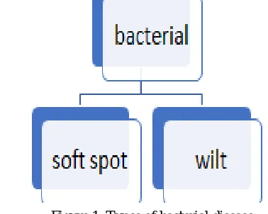
A. Bacterial Disease
The disease caused by bacteria is called bacterial disease. Symptoms of the bacterial disease are as follows wilts, leaf spots, galls, and overgrowth, specks and blight, soft rots as well as scabs and cankers. The bacterial disease can be spread by rain or carried out by the wild birds and insects.
The tissue which has suffered damage through disease appears as dry dead spots. Malady is being categorized as small pale green looking spots which later on become saturated with presence of water over it. Various type of bacterial disease is given in figure 1.
[image:1.612.199.388.571.722.2].
©IJRASET: All Rights are Reserved
447
1) Soft Spot: These maladies are caused by the pathogens that secrets enzymes which can damage the cell wall structures. This [image:2.612.224.402.106.256.2]results in damage of texture of plant tissue. The soft spot shows in figure 2.
Figure 2: Soft Spot
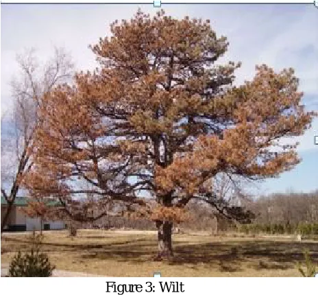
[image:2.612.201.433.295.510.2]2) Wilt: Wilt disease is any number of diseases that influence the vascular system of plants, larges branches or even entire trees. The wilt shows in figure 3.
Figure 3: Wilt B. Fungal Disease
The disease caused by fungus is known as fungal disease. Fungi represent a large number of plant pathogens and are dependable for the various plant diseases, for instance, fungal disease damage plants by killing cells. Types of fungal diseases given in figure 4.
[image:2.612.118.490.559.700.2]©IJRASET: All Rights are Reserved
448
1) Molds: Molds disease is a kind of fungal disease that grows in a humid area. Mold disease caused by fungi in the genus Botrytis, [image:3.612.230.416.117.281.2]generally Cinerea. This disease mainly affects flowers and buds. It can also affect fruits, leaves, and stems. The molds show in figure 5.
Figure 5: Molds
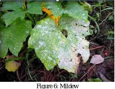
[image:3.612.228.421.334.483.2]2) Mildew: It is also a fungal malady that affects a broad range of plants. It is in white color but the molds are in the shades of black, blue, red and green. The most commonly reported cause is Erysipelas, with podospherexanthii. The mildew shows in figure 6.
Figure 6: Mildew
3) Rots: Rots occur during germination and the growth of the seedlings. It is fungal root disease. The damping off is the most common disease in forest nurseries. Rots contribute about one-third of the losses caused by disease in nurseries. Often seedlings are completely lost. The rots show in figure 7.
[image:3.612.204.444.538.714.2]©IJRASET: All Rights are Reserved
449
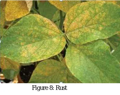
4) Rust: Rusts are plant malady caused by pathogenic fungi of the order Puccini ales a simple variety of rust may be able to affect [image:4.612.225.423.103.260.2]almost two different plant hosts in various stages of its life cycle. The rust shows in figure 8.
Figure 8: Rust
[image:4.612.222.428.306.445.2]5) Canker: Canker is a plant malady caused by variety of fungi that occurs primarily on woody variety. Symptoms contain round to irregular sunken, cracked, etc. Canker disease frequently kills twigs or structurally weakens a plant until the spoiled area breaks free. The canker shows in figure 9.
Figure 9: Canker
C. Viral Disease
The disease caused by the virus is known as the viral disease. A virus that affects plants does not have the ability to replicate without a host. Viral disease is difficult to diagnose. The common carriers of this disease are aphids, hoppers, whiteflies, and cucumber beetles. Types of viral disease are given in figure 10.
Figure 10: Types of viral disease
[image:4.612.169.455.518.694.2]©IJRASET: All Rights are Reserved
450
2) Dwarfing: Dwarfing is a process in which a variety of animals or cultivar of plants is altered to become significantly smaller [image:5.612.214.437.129.288.2]than the standard member of their species. The cause can be induced through human interventions on the human process and can include genetic, nutritional or hormonal means. The dwarfing shows in figure 11.
Figure 12: Dwarfing
3) Distortion: Some of the leaves of plants may become twisted and distorted or the new growth will curl and become deformed.
III.LITERATURE REVIEW
Kulkarni et al. {5] proposed a paper on detecting plant diseases using image processing. The author(s) denotes method for detecting and classifying the diseases. For this texture and colour features are extracted and segmented using Gabor filter. ANN is used as a classifier. This method achieved overall accuracy is 91%. This paper detects the pomegranate diseases like bacterial blight, anthracnose and wilt (dataset 140). The paper shows various stages of image processing which includes a sequential steps of image acquisition, image pre-processing, segmentation followed by feature extraction along with classification. During image acquisition capture the sample and create the database. In image processing suppress undesired distortions or enhance some image features. The next step is a process of segmentation which is applied to differentiate target of interest from background then the extract feature that can be used to establish the meaning of a given sample
We can calculate Gabor filter are omnichannel feature given by using equation (i)
s………(i)
Where, e shows filtered image. The inter-channel characteristics within distinct channels of spectral like i& j along with m& n indicating the filter’s scales is calculated in equation (ii)
………. (ii)
Where, denotes zero offset normalized cross correlation between (x, y) & (x, y)
©IJRASET: All Rights are Reserved
451
background noise then RGB image is changed into other color spaces such as HIS and CIELAB. In segmentation step to find out the infected region and it done by k-means then color, shape and texture feature are extracted. In the final stage identifying a rule according to selected features and conveying each malady to any one the predetermined classes.Kulkarni et al. [13] proposed a leaf identification technique for plant classification through RBPNN and Zernike moments. Researcher discusses a method for detecting the diseases. In this paper we are detecting and recognizing plants using shape vein, color, texture features which are merged Zernike movements. Radial basis probabilistic neural network [14] is used as a classification purpose. RBPNN includes four various layers one input layer, two hidden layer and one output layer. This method achieved overall accuracy is 93.82%.
Patil et al. [15] presented classification of cotton leaf spot diseases through support vector machine. Researcher described a method for detecting diseases. Foliar is the type of malady of cotton and appear in all increasing Indian cotton regions shape. Color, shape and texture feature are extracting using texture and colour feature extraction techniques for the disease spots to recognize diseases. SVM used as a classifier. In this paper image analysis can be useful for the different objectives firstly to identify diseased leaf, stem, fruit secondly measure influence area by diseases third to discover the boundaries of the affected area and the fourth is to establish the colour of the affected area and fifth is to establish size and shape of fruits and last is to determine the object properly. This paper is used several image processing techniques.
Awate et al. [16] described fruit malady detecting using colour, texture analysis and ANN”. Researcher proposed a technique for identify and classify external disease with in fruits. K-means based segmentation is used for extracting the colour, morphology, texture and structural features. In this paper we are detect diseases such as black rot, powdery mildew, downy mildew, apple scab, apple blotch and bacterial blight etc. ANN is used as a classifier. In this paper there are various techniques used such as first image acquisition, second pre-processing, third segmentation, fourth feature extraction and last classification. In image acquisition acquired the image then removes noise. The next step segmentation is to classify and modify rendering of an image then extracting the feature using speed up robust feature (SURF) methods [17] is useful for extracting the features and last step classification is done ANN.
Bagde et al. [18] explained artificial neural network-based plant leaf disease identification”. Researcher discusses a method for detecting diseases such as white powder, ring spot, black spot. K-means based segmentation is used for extract shape feature. ANN is used as a classifier. In this paper describe various type of disease like bacterial, viral and fungal diseases. This paper used in image processing in agricultural demands for following purpose firstly to identify diseased leaf, stem, fruit and second step to quantify impact area by disease third to discover shape of affected area and the last is to identify size and shape of fruits.
Prakash M. Mainkar et al. [19] explained plant leaf disease identification and classification using image processing techniques. Researcher develops a method for recognize and classify plant leaf diseases. Texture feature is extracted using gray level co-occurrence matrix (GLCM) [20] for image segmentation techniques. NN is used as a classifier. In this paper we are discuss image processing technique like first image acquisition, second pre-processing, third segmentation, fourth feature extraction last neural network based classification. In image acquisition acquired the image then device self dependent color space transformation changed the color values in the image to color space specified in the color transformation structure. The next step k-means technique is used for segmentation and texture features are given as input to pretrained NN for automatic classification of diseases.
MS Kiran et al. [21] proposed unhealthy region of citrus leaf recognization using image processing technique. Researcher proposed a method for detecting the bacterial diseases. Texture feature extracting using statistical GLCM and color feature by means of mean values. SVM is used as a classifier. In this we detect disease like citrus canker, anthracnose, citrus greening disease (dataset 200). This paper there is five steps used for the recognition of plant leaf diseases. Firstly, image acquisition acquired leaves using a digital camera. The next step improves the image data that suppress undesired distortion and the image enhancement used for providing better colour images. Third is image segmentation is done through feature based technique after segmentation extract the texture feature. The last step SVM classifiers distinguish citrus leaf malady and it creates hyper plane in a high dimensional space that can be applied for classification purpose.
©IJRASET: All Rights are Reserved
452
the overall accuracy is achieved 96.8%. In this paper image processing techniques is used. In image acquisition captured the image then pre-processing RGB value is converted into gray scale measure. The equation of gray image is given to equation (iii).... (iii)
In feature extraction uses 12 general digital morphological features obtained from 5 features so that a computer can find feature value rapidly. The last step is classification is done using SVM. SVM is a supervised technique.
TABLE I
Comparison of Plant Disease in Different Paper
IV. CONCLUSION
In this field of agriculture the disease detection is very crucial task. These are various techniques, used for detecting diseases in plants. The paper describes the detection of bacterial, fungal and viral disease by using different image processing techniques. Paper includes digest of some existing work of applying disease detection in plants by several image processing techniques individually or co-operatively.
REFERENCES
[1] J. P. Shah, H. B. Prajapati, and V. K. Dabhi, “A survey on detection and classification of rice plant diseases,” 2016 IEEE Int. Conf. Curr. Trends Adv. Comput. ICCTAC 2016, 2016
[2] Sowmya GM,Chandan V and Sampath Kini, “Disease Detection in Pomegranate Leaf Using Image Processing Technique” International Journal of Science, Engineering and Technology Research (IJSETR) Volume 6, Issue 3, March 2017, ISSN: 2278 -7798
[3] S. Vishnu and A. R. Ram, “Plant Disease Detection Using Leaf Pattern: A Review,” vol. 2, no. 6, pp. 774–780, 2015.
[4] B. Mishra, S. Nema, M. Lambert, and S. Nema, “Recent technologies of leaf diseasedetection using image processing approach-A review,” Proc. 2017 Int. Conf. Innov. Information, Embed. Commun. Syst. ICIIECS 2017, vol. 2018–January, pp. 1–5, 2018.
Author Feature Extraction Feature Extraction Technique
Classifier Dataset/ Plant
Anand. Kulkarni, et al Color, Shape, Texture
Gabor Filter ANN 140/ pomegranate
P. Revathi, et al Color, Shape, Texture Power swarm optimization Deep forward neural network 270/ Cotton
Kushal Kairnal, et al Color, shape, Edge Gabor filter canny, sobel edge detector
ANN, SVM Cotton
Dr. H.M. Rai, et al Shape, Vein, Color, Texture
Gray level Co-occurrence matrix
RBPNN -
Prof. Sonal P. Patil et al
Color, Shape Gabor filter, PSO, Gray level Co-occurrence
matrix
SVM Cotton
Ashwani Awate, et al Color, Texture Speed up robust feature (SURF)
ANN Apple,
Pomegranate, grapes ShivkumarBhagde, et
al
Shape, Size Color co-occurrence method, Otsu method
Radial basis function,
ANN
Tomato
Prakash M. Mainkar, et al
Texture Grey level cooccurrence matrix
Radial basis function
Tomato
Ms. Kiran R. Gavhale, et al
Texture Gray level co-occurrence matrix
SVM 200/ Citrus
JyotismitaChaki, et al Shape Moments-invariants, centroid-radii
NN 180
ArunPriya C. et al Vein Digital morphological features
©IJRASET: All Rights are Reserved
453
[5] Anand.H. Kulkarni and Ashwin Patil R. K., “Applying image processing technique to detect plant disease”International Journal of Modern EngineeringResearch (IJMER) Vol.2, Issue.5, Sep-Oct. 2012 pp-3661-3664
[6] P. Revathi and M. Hemalatha, “Identification of cotton diseases based on cross information gain_deep forward neural network classifier with PSO feature selection,” Int. J. Eng. Technol., vol. 5, no. 6, pp. 4637–4642, 2013
[7] D. Bratton and J. Kennedy, “(2007) Defining a Standard for Particle Swarm Optimization.pdf,” no. Sis, pp. 120–127, 2007.
[8] Mikhail Sizintsev, Konstantinos G .Derpanis “HISTOGRAM-BASED SEARCH: A COMPARATIVE STUDY Department of Computer Science and Engineering Toronto, ON, Canada Faculty of Business and Information Technology University of Ontario Institute of Technology Os,” Image (Rochester, N.Y.), 2008.
[9] G. M. H. Amer and A. M. Abushaala, “Edge detection methods,” in 2015 2nd World Symposium on Web Applications and Networking (WSWAN), 2015, pp. 1–7.
[10] R. Roslan and N. Jamil, “Texture feature extraction using 2-D Gabor Filters,” ISCAIE 2012 - 2012 IEEE Symp. Comput. Appl. Ind. Electron., no. Iscaie, pp. 173–178, 2012.
[11] D. Molchanov, A. Ashukha, and D. Vetrov, “Variational Dropout Sparsifies Deep Neural Networks,” vol. 9, pp. 249–256, 2017.
[12] K. Khairnar and R. Dagade, “Disease Detection and Diagnosis on Plant using Image Processing A Review,” Int. J. Comput. Appl., vol. 108, no. 13, pp. 36–38, Dec. 2014.
[13] S. G. Wu, F. S. Bao, E. Y. Xu, Y.-X. Wang, Y.-F. Chang and Q.-L. Xiang, “A Leaf Recognition Algorithm for Plant Classification Using Probabilistic Neural Network,” in 2007 IEEE International Symposium on Signal Processing and Information Technology, 2007, vol. 2, no. 1, pp. 11–16.
[14] H. Deshuang and M. Songde, “New Radial Basis Probabilistic Neural Network Model,” Signal Process. 1996., 3rd Int. Conf. on, Vol. 2, pp. 1449–1452, 1996. [15] P. S. P. Patil and M. R. S. Zambre, “Classification of Cotton Leaf Spot Disease Using Support Vector Machine,” vol. 4, no. 5, pp. 92–97, 2014.
[16] A. Awate, D. Deshmankar, G. Amrutkar, U. Bagul, and S. Sonavane, “Fruit disease detection using color, texture analysis and ANN,” in 2015 International Conference on Green Computing and Internet of Things (ICGCIoT), 2015, pp. 970–975.
[17] E. Oyallon and J. Rabin, “An Analysis of the SURF Method,” Image Process. Line, vol. 5, no. 2004, pp. 176–218, Jul. 2015.
[18] S. Bagde, S. Patil, S. Patil, and P. Patil, “Artificial Neural Network Based Plant Leaf Disease Detection,” Int. J. Comput. Sci. Mob. Comput., vol. 4, no. 4, pp. 900–905, 2015.
[19] Y. M. Oo and N. C. Htun, “Plant Leaf Disease Detection and Classification using Image Processing,” Int. J. Res. Eng., vol. 5, no. 9, pp. 516–523, Nov. 2018. [20] K. R. Gavhale, U. Gawande, and K. O. Hajari, “Unhealthy region of citrus leaf detection using image processing techniques,” in International Conference for
Convergence for Technology-2014, 2014, pp. 1–6.
[21] J. Chaki and R. Parekh, “Plant Leaf Recognition using Shape based Features and Neural Network classifiers,” Int. J. Adv. Comput. Sci. Appl., vol. 2, no. 10, pp. 41–47, 2011.
[22] H. Shu, L. Luo, and J. L. Coatrieux, “Derivation of Moment Invariants,” vol. 1, 2014, pp. 57–90.
[23] D. F. Pachón-Neira and J. C. Figueroa-Garcia, “A comparison between the Centroid and the Yager Index Rank for Type reduction of an Interval Type-2 fuzzy number,” Ingenieria, vol. 21, no. 2, pp. 225–234, 2016.