Scheelite-type NaEr(MoO
4)
2Dan Zhao,a* Feifei Li,aWendan Chengband Hao Zhangb
aDepartment of Physics and Chemistry, Henan Polytechnic University, Jiaozuo,
Henan 454000, People’s Republic of China, andbState Key Laboratory of Structural
Chemistry, Fujian Institute of Research on the Structure of Matter, Chinese Academy of Sciences, Fuzhou, Fujian 350002, People’s Republic of China
Correspondence e-mail: [email protected]
Received 6 March 2010; accepted 5 April 2010
Key indicators: single-crystal X-ray study;T= 173 K; mean(Mo–O) = 0.003 A˚; disorder in main residue;Rfactor = 0.032;wRfactor = 0.089; data-to-parameter ratio = 11.5.
Explorations of the A1+–RE3+–Mo6+–O2 (A1+ is an alkali metal cation, RE3+ is a rare-earth metal cation) quaternary systems prepared by the high-temperature solution growth method led to the title structure, sodium erbium bis(molyb-date), NaEr(MoO4)2. It is isostructural to the scheelite structure (CaWO4) and is composed of [MoO4]2 tetrahedra with 4 symmetry and [(Na/Er)O8]14 polyhedra. The [(Na/ Er)O8]
14
polyhedron is a distorted tetragonal antiprism, also with 4 symmetry, with statistically mixed Na/Er atoms at its centre. There are two sets of Na/Er—O bond lengths [2.420 (4) and 2.435 (3) A˚ ], but just one set of Mo—O bond lengths [1.774 (4) A˚ ].
Related literature
For the structures, properties and applications of the alkali rare-earth tungstates and molybdates with the general formula
A1+RE3+(M6+O4)2 (A 1+
is an alkali metal cation, RE3+ is a rare-earth metal cation,M6+is Mo6+or W6+), see: Huanget al.
(2006); Klevtsova (1975); Klevtsova et al. (1972); Kolitsch (2001); Kuzmichevaet al.(2005); Liet al.(2006); Morozovet al. (2006); Stevens et al. (1991); Zhao et al. (2010). For the scheelite (CaWO4) structure, see: Sillen & Nylander (1943).
Crystal data
NaEr(MoO4)2
Mr= 510.13 Tetragonal,I41=a
a= 5.1816 (8) A˚
c= 11.288 (3) A˚
V= 303.07 (11) A˚3
Z= 2
MoKradiation
= 17.87 mm 1
T= 173 K
0.080.040.04 mm
Data collection
Rigaku Saturn70 CCD diffractometer
Absorption correction: multi-scan (rescaledSADABS; Sheldrick, 1997)
Tmin= 0.263,Tmax= 0.489
520 measured reflections 172 independent reflections 106 reflections withI> 2(I)
Rint= 0.026
Refinement
R[F2> 2(F2)] = 0.032
wR(F2) = 0.089
S= 0.84 172 reflections
15 parameters
max= 1.12 e A˚ 3
min= 1.15 e A˚ 3
Data collection: CrystalClear (Rigaku, 2004); cell refinement: CrystalClear; data reduction:CrystalClear; program(s) used to solve structure:SHELXS97(Sheldrick, 2008) andPLATON(Spek, 2009); program(s) used to refine structure:SHELXL97(Sheldrick, 2008); molecular graphics:DIAMOND(Brandenburg, 2004); software used to prepare material for publication:SHELXTL(Sheldrick, 2008).
Supplementary data and figures for this paper are available from the IUCr electronic archives (Reference: FB2187).
References
Brandenburg, K. (2004).DIAMOND.Crystal Impact GbR, Bonn, Germany. Huang, X. Y., Lin, Z. B., Zhang, L. Z., Chen, J. T. & Wang, G. F. (2006).Cryst.
Growth Des.6, 2271–2274.
Klevtsova, R. F. (1975).Kristallografiya,20, 746–750.
Klevtsova, R. F., Vinokurov, V. A. & Klevtsov, P. V. (1972).Kristallografiya,17, 284–288.
Kolitsch, U. (2001).Z. Kristallogr.216, 449–454.
Kuzmicheva, G. M., Lis, D. A., Subbotin, K. A., Rybakov, V. B. & Zharikov, E. V. (2005).J. Cryst. Growth,275, e1835–e1842.
Li, X. Z., Lin, Z. B., Zhang, L. Z. & Wang, G. F. (2006).J. Cryst. Growth,293, 157–161.
Morozov, V. A., Arakcheeva, A. V., Chapuis, G., Guiblin, N., Rossell, M. D. & Van Tendeloo, G. (2006).Chem. Mater.18, 4075–4082.
Rigaku (2004).CrystalClear.Rigaku Corporation, Tokyo, Japan. Sheldrick, G. M. (1997).SADABS.University of Go¨ttingen, Germany. Sheldrick, G. M. (2008).Acta Cryst.A64, 112–122.
Sillen, L. G. & Nylander, A. L. (1943).Ark. Kemi Mineral. Geol.17, 1–27. Spek, A. L. (2009).Acta Cryst.D65, 148–155.
Stevens, S. B., Morrison, C. A., Allik, T. H., Rheingold, A. L. & Haggerty, B. S. (1991).Phys. Rev. B Condens. Matter.43, 7386–7394.
Zhao, D., Li, F., Cheng, W. & Zhang, H. (2010).Acta Cryst.E66, i2. Structure Reports
Online
supporting information
Acta Cryst. (2010). E66, i36 [https://doi.org/10.1107/S160053681001264X]
Scheelite-type NaEr(MoO
4)
2Dan Zhao, Feifei Li, Wendan Cheng and Hao Zhang
S1. Comment
Alkali rare-earth bis(molybdates) with the general formula A1+RE3+(MO
4)2 (AI is an alkali-metal cation, RE3+ is a
rare-earth metal cation, M is Mo6+ or W6+) have been the subject of interest for many decades, mainly due to their applications
as suitable host materials for fluorescence (Kuzmicheva et al., 2005; Morozov et al., 2006; Li et al., 2006). Some of these
crystals are isostructural to scheelite (CaWO4, I41/a; Sillen & Nylander, 1943), such as NaLa(MoO4)2 (Stevens et al.,
1991), LiNd(MoO4)2 (Kolitsch, 2001), LiNd(WO4)2 (Huang et al., 2006) and LiDy(WO4)2 (Zhao et al., 2010).
In difference to CaWO4 with one cation species only, the cations A1+ and RE3+ are statistically disordered. Within alkali
rare-earth bis(molybdates), different structures from the scheelite type have also been reported, such as LiLa(MoO4)2
(Pbca; Klevtsova, 1975) and CsDy(MoO4)2 (Pccm; Klevtsova et al. 1972).
The X-ray diffraction analysis has shown that the title compound NaEr(MoO4)2 is isostructural with the scheelite. In the
title structure, Na and Er atoms are disordered over the same 4a site while Mo atoms reside on 4b site. The structure of
NaEr(MoO4)2 may be regarded as composed of [MoO4]2- tetrahedra and of [(Na/Er)O8]14- polyhedra (each in the form of a
distorted tetragonal antiprism) that share the oxygens (Fig. 2). Each oxygen of the [MoO4]2- tetrahedron is shared by the
different Na/Er polyhedron and each oxygen of the [(Na/Er)O8]14- polyhedron is shared by the different [MoO4]
2-tetrahedron.
S2. Experimental
Single crystals of NaEr(MoO4)2 have been prepared by the high temperature solution growth (HTSG) method in air. A
powder mixture of Na2CO3 (0.4418 g), Er2O3 (0.2657 g) and MoO3 (2.000 g) at the molar ratio of Na:Er:Mo = 6:1:10 was
first ground in an agate mortar and then transferred to a platinum crucible. The sample was gradually heated in air at 1173
K for 24 h. In this stage, the reagents were completely melted. After that, the intermediate product was slowly cooled to
673 K at the rate of 2 Kh-1. It was kept at 673 for another 10 h and then quenched to room temperature. The obtained
crystals were light-red and of the prismatical shape. The dimensions of the used sample were typical for the grown
crystals in this batch.
S3. Refinement
The Na and Er atoms are in substitutional disorder in the crystal structure. The tentative refinement that included the
corresponding occupancy factors for the disordered Na/Er yielded Na1 : Er1 = 0.501 (2) : 0.499 (2). (The atomic
positional and anisotropic displacement parameters of Na1 and Er1 atoms were constrained to be identical by using
EADP and EXYZ constraint instructions (SHELXL-97; Sheldrick, 2008).) Therefore the ratio of Na and Er was fixed to
1:1 in the final model with the constrained positional and the displacement parameters of na and Er as given above. The
highest peak in the difference electron density map equals to 1.12 e/Å3 at the distance of 0.83 Å from Na1/Er1 site while
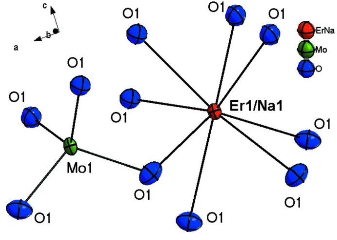
Figure 1
Section of the structure of NaEr(MoO4)2 with the atom labelling scheme. The displacement ellipsoids are drawn at the
Figure 2
View of the crystal structure of NaEr(MoO4)2. The [MoO4]2- tetrahedra are shown in green.
Sodium erbium bis(molybdate)
Crystal data
NaEr(MoO4)2
Mr = 510.13
Tetragonal, I41/a
Hall symbol: -I 4ad
a = 5.1816 (8) Å
c = 11.288 (3) Å
V = 303.07 (11) Å3
Z = 2
F(000) = 454
Dx = 5.590 Mg m−3
Mo Kα radiation, λ = 0.71073 Å Cell parameters from 365 reflections
θ = 4.3–27.3°
µ = 17.87 mm−1
T = 173 K Prism, red
0.08 × 0.04 × 0.04 mm
Data collection
Rigaku Saturn70 CCD diffractometer
Radiation source: fine-focus sealed tube Confocal monochromator
Detector resolution: 28.5714 pixels mm-1
ω scans
520 measured reflections 172 independent reflections 106 reflections with I > 2σ(I)
Rint = 0.026
h = −2→6
k = −5→6
l = −14→14
Refinement
Refinement on F2
Least-squares matrix: full
R[F2 > 2σ(F2)] = 0.032
wR(F2) = 0.089
S = 0.84 172 reflections 15 parameters 0 restraints 9 constraints
Primary atom site location: structure-invariant direct methods
Secondary atom site location: difference Fourier map
w = 1/[σ^2^(Fo^2^) + (0.0639P)^2^]
where P = (Fo^2^ + 2Fc^2^)/3
(Δ/σ)max < 0.001
Δρmax = 1.12 e Å−3
Δρmin = −1.15 e Å−3
Extinction correction: SHELXL97 (Sheldrick, 2008), Fc*=kFc[1+0.001xFc2λ3/sin(2θ)]-1/4
Extinction coefficient: 0.055 (5)
Special details
Geometry. All esds (except the esd in the dihedral angle between two l.s. planes) are estimated using the full covariance matrix. The cell esds are taken into account individually in the estimation of esds in distances, angles and torsion angles; correlations between esds in cell parameters are only used when they are defined by crystal symmetry. An approximate (isotropic) treatment of cell esds is used for estimating esds involving l.s. planes.
Refinement. Refinement of F2 against ALL reflections. The weighted R-factor wR and goodness of fit S are based on F2,
conventional R-factors R are based on F, with F set to zero for negative F2. The threshold expression of F2 > σ(F2) is used
only for calculating R-factors(gt) etc. and is not relevant to the choice of reflections for refinement. R-factors based on F2
are statistically about twice as large as those based on F, and R- factors based on ALL data will be even larger.
Fractional atomic coordinates and isotropic or equivalent isotropic displacement parameters (Å2)
x y z Uiso*/Ueq Occ. (<1)
Er1 0.0000 0.2500 0.1250 0.0081 (5) 0.50
Na1 0.0000 0.2500 0.1250 0.0081 (5) 0.50
Mo1 0.5000 0.7500 0.1250 0.0086 (5)
O1 0.2568 (6) 0.5968 (6) 0.0397 (3) 0.0204 (12)
Atomic displacement parameters (Å2)
U11 U22 U33 U12 U13 U23
Er1 0.0070 (6) 0.0070 (6) 0.0102 (8) 0.000 0.000 0.000
Na1 0.0070 (6) 0.0070 (6) 0.0102 (8) 0.000 0.000 0.000
Mo1 0.0067 (6) 0.0067 (6) 0.0124 (8) 0.000 0.000 0.000
O1 0.025 (2) 0.017 (2) 0.019 (2) 0.0012 (15) −0.0045 (14) −0.0007 (17)
Geometric parameters (Å, º)
Er1—O1i 2.420 (4) Er1—O1vii 2.435 (3)
Er1—O1ii 2.420 (4) Mo1—O1viii 1.774 (4)
Er1—O1iii 2.420 (4) Mo1—O1ix 1.774 (4)
Er1—O1iv 2.420 (4) Mo1—O1 1.774 (4)
Er1—O1vi 2.435 (3) O1—Na1iii 2.420 (4)
Er1—O1 2.435 (3) O1—Er1iii 2.420 (4)
O1i—Er1—O1ii 79.63 (16) O1v—Er1—O1vii 99.01 (7)
O1i—Er1—O1iii 126.16 (10) O1vi—Er1—O1vii 99.01 (7)
O1ii—Er1—O1iii 126.16 (10) O1—Er1—O1vii 133.38 (18)
O1i—Er1—O1iv 126.16 (10) O1i—Er1—Er1xi 38.03 (7)
O1ii—Er1—O1iv 126.16 (10) O1ii—Er1—Er1xi 69.88 (9)
O1iii—Er1—O1iv 79.62 (16) O1iii—Er1—Er1xi 159.67 (8)
O1i—Er1—O1v 75.78 (12) O1iv—Er1—Er1xi 101.19 (8)
O1ii—Er1—O1v 68.76 (7) O1v—Er1—Er1xi 37.75 (8)
O1iii—Er1—O1v 152.76 (16) O1vi—Er1—Er1xi 101.99 (9)
O1iv—Er1—O1v 73.67 (7) O1—Er1—Er1xi 85.52 (9)
O1i—Er1—O1vi 68.76 (7) O1vii—Er1—Er1xi 131.38 (8)
O1ii—Er1—O1vi 75.78 (12) O1i—Er1—Na1xi 38.03 (7)
O1iii—Er1—O1vi 73.67 (7) O1ii—Er1—Na1xi 69.88 (9)
O1iv—Er1—O1vi 152.76 (16) O1iii—Er1—Na1xi 159.67 (8)
O1v—Er1—O1vi 133.38 (18) O1iv—Er1—Na1xi 101.19 (8)
O1i—Er1—O1 73.67 (7) O1v—Er1—Na1xi 37.75 (8)
O1ii—Er1—O1 152.76 (16) O1vi—Er1—Na1xi 101.99 (9)
O1iii—Er1—O1 75.78 (12) O1—Er1—Na1xi 85.52 (9)
O1iv—Er1—O1 68.76 (7) O1vii—Er1—Na1xi 131.38 (8)
O1v—Er1—O1 99.01 (7) O1viii—Mo1—O1ix 114.2 (2)
O1vi—Er1—O1 99.01 (7) O1viii—Mo1—O1 107.15 (11)
O1i—Er1—O1vii 152.76 (16) O1ix—Mo1—O1 107.15 (11)
O1ii—Er1—O1vii 73.67 (7) O1viii—Mo1—O1x 107.15 (11)
O1iii—Er1—O1vii 68.76 (7) O1ix—Mo1—O1x 107.15 (11)
O1iv—Er1—O1vii 75.78 (12) O1—Mo1—O1x 114.2 (2)
![Figure 2View of the crystal structure of NaEr(MoO4)2. The [MoO4]2- tetrahedra are shown in green](https://thumb-us.123doks.com/thumbv2/123dok_us/573256.556871/4.610.134.475.73.454/figure-view-crystal-structure-naer-tetrahedra-shown-green.webp)
