JOURNAL OF FOREST SCIENCE, 53, 2007 (1): 13–19
Mangrove forms thermophilic coastal woody plant communities in tropical and subtropical coastal re-gions and tidal lowland (Lin 1999). They can protect the coast, continental shelf and large areas of land in the estuary and are considered an ecologically essential component in protecting the adjacent land from wave and storm erosion (Lin 1999) while pre-venting terrigenous nutrients from affecting nearby reefs (Dubinsky, Stamber 1996).
The most striking feature of mangrove is its abil-ity to tolerate NaCl in seawater. From physiological aspects, the effect of salinity on photosynthesis of mangroves has been studied to some extent, mostly in relation to transpiration and stomatal conduc-tance. The reduction of stomatal conductance and mesophyll might contribute to the inhibition of photosynthesis under salt stress (Parida et al. 2003). However, Ball and Farquhar (1984a,b) found that
unlike in the salt-sensitive Aegiceras corniculatum, in the most salt tolerant mangrove, Avicennia marina, photosynthesis rates were barely affected by salin-ity, the depression of carbon assimilation that was observed in Aegiceras corniculatum was attributable to a reduction in stomatal opening.
Kandelia candel (L.) Druce is a dominant man-grove species on the eastern coast of southern Fujian, China. In previous experiments, Lin (1999) found that NaCl affected the growth of this plant species. Thus, although K. candel is able to colonize saline habitats, high salinity levels limit its growth. To our knowledge little information is available about physi-ological responses of Kandelia candel (L.) Druce grown in high salinity. This paper presents data concerning leave characteristics and gas exchange as affected by high salinities with the aim to understand their effects on its photosynthesis.
Effects of salinity on leaf characteristics and CO
2
/H
2
O
exchange of Kandelia candel (L.) Druce seedlings
D.-L. QIU
1,2,, P. Lin
1, S. z. Guo
21
School of Life Science, Xiamen University, P.R. China
2
College of Horticulture, Fujian Agriculture and Forestry University, Fuzhou, P.R. China
ABSTRACT: Effects of salinities on leaf characteristics and CO2/H2Oexchange of mangrove species Kandelia candel
seedlings were studied in a pot experiment. The seedlings grown in salinity of 50‰ caused a strong reduction in the rate of growth, but their leaves were black-green, smaller and less expanded or distorted than those of plants in the control and treatment of 25‰. As compared with control plants, leaves of plants treated with salinity of 25‰ were shiny and smooth. Stomatal number and density under the epidermis in leaves were reduced with the increase of salinity. Mesophyll cells in plants grown in salinity of 50‰ were smallest, 25‰ ranked the second and 0‰ were largest. Their arrangement was compact in 50‰, while in the control it was loose. Cells of the upper epidermis in leaves of control plants were loose and the cell wall was thin while that of 50‰ was more compact and the cell wall was thicker than that of 25‰. Chlorophyll (Chl) (a + b) content (μmol/cm2) in plants grown in 50‰ salinity increased significantly compared with that in 0‰, and Chl a/b was also reduced. Carotenoid pigments (Car) increased significantly in different treatments. Photosynthesis (Pn) was significantly inhibited by higher salinity, and the light compensation point of higher salinity leaves increased. Net photosynthetic rate (Pn), stomatal conductance (Cs), and transpiration rate (Tr) were reduced with the increase of salinity while dark respiration (Rd) increased.
Keywords: salinity; mangrove; cell structure; photosynthesis; transpiration
MATERIALS AND METHODS
Plant materials and culture conditions
108 mature propagules of Kandelia candel (L.) Druce were collected at random from 10 plants during April from the mangrove forest at the Jiu-longjiang River Estuary (24°24´N, 117°55´E), Fujian, China. The propagules of similar length (20–25cm) were selected to plant in 0.15cm diameter pots filled with sand at a density of three propagules per pot. Temperatures ranged from 25°C to 32°C during the day and 20°C to 25°C at night. They were raised by irrigating daily with normal tap water in natural day and night for 2 months. Then, plants were grown at the respective salinities of 0.25‰ and 50‰ with each treatment of 6 pots for 20 days. Leaves for measure-ments were young, fully expanded, and selected from the second to sixth nodes. Gas exchange was taken in 15–20 leaves of 12 plants, which later were used for the samples of scanning electron microscopy and the determination of chlorophyll (a + b) and Car.
Scanning electron microscopy
Samples of different treatments from the second nodes were fixed in 2.5% glutaraldehyde solution buffered in 0.1M phosphate buffer solution (PBS), pH 7.0, for 48 h at room temperature. After washing with distilled water, samples were hydrolyzed in a hydro-lytic solution at 20°C. After hydrolysis, samples were washed with distilled water 5 times. The upper and lower epidermis of leaf was peeled off with tweezers, then it was decomposed with osmium tetroxide solu-tion. Ultrasonic treatment was done to eliminate the surface pollutants. Conductive solution (potassium iodide 20 g, redistilled water 100 ml and 0.2 g sugar) was used to soak samples for 8–10h. After washing with distilled water, samples were dehydrated in tert-butanol series, soaked in absolute tert-tert-butanol at 4°C in a refrigerator for 10 min, and finally passed into a vacuum drier for 40–60 min. Dried samples were coated with gold in a JFC-1200 sputter and examined and photographed with a JSM made in Japan.
Determination of chlorophyll and carotenoid pigments
Photosynthetic pigments were extracted in the chilled combination solution including 45% acetone (v/v), 45% ethyl alcohol (v/v) and 10% distilled water (v/v), and were estimated as per procedure of Arnon (1949). However, Car concentrations in these extracts were calculated by the formula given
by Duxbury and Yentsch (1956). Chlorophyll a (Chl a), chlorophyll b (Chl b), total Chl and Car were expressed as μg per unit leaf area (cm2).
Gas exchange
Net photosynthetic rate (Pn), transpiration rate (Tr), stomatal conductance (Cs), and intercellular CO2 concentration (Ci) were measured with an open gas analyzer (CID-301 PS, U.S.A.). The measurement was done under the conditions of photosynthetically active radiation (PAR) 0, 50, 100, 200, 400, 600, 800, 1,000, 1,500, 2,000 µmol/m2/s, respectively, and tem-perature 26–28°C. Dark respiration (Rd) was taken as the Pn at a PAR of 0 μmol/m2/s.
Statistical analysis
The data were analyzed by factor analysis, and with means they were compared with Tukey’s HSD in DPS software. The independent variables are salinities, and the dependent ones are stomatal numbers and density, Chl and Car contents, Pn, Tr, Cs, Ci, Rd.
RESULTS
Leaf characteristics
Kandelia candel seedlings grown in salinity of 50‰ showed a strong reduction in the rate of growth. The leaves were black-green, but small and less expanded or distorted than those of plants in the control and treatment of 25‰. As compared with control plants, leaves treated with salinity of 25‰ were shiny and smooth. Stomatal number and density under the epidermis in leaves of plants were also significantly
0 10 20 30 40 50 60
Chl(a+b) Chl a Chl b Car
Pigments
Co
nt
en
ts
(µ
g/
cm
[image:2.595.307.524.542.725.2]) 0‰ 25‰ 50‰
Fig. 1. The effects of salinities on the contents of photosynthetic pigments in leaves of Kandelia candel seedlings
reduced with the increase of salinity (Fig. 3 A, B, C) (p < 0.05). The reticulate structure of spongy tissue layer and irregular arrangement of mesophyll cells were observed. Mesophyll cells in plants grown in salinity of 50‰ were the smallest, 25‰ salinity ranks the second and in salinity of 0‰ they were the larg-est. Their arrangement was compact in 50‰, loose in the control (Fig. 3 G, H, I). There were also differ-ences between the cells of the upper epidermis in leaves of plants grown in different saline conditions. The cells of the upper epidermis in leaves of control plants were loose and the cell wall was thin while that of 50‰ was compact and the cell wall was thick and that of 25‰ moderate (Fig. 3 D, E, F).
Chl content and Chl a/b
Chl (a + b) content (μg/cm2) in plants grown in 50‰ salinity increased significantly compared with the control (p < 0.05). Chl a/b was reduced, and Car increased significantly in different treatments (p < 0.05) (Fig. 1).
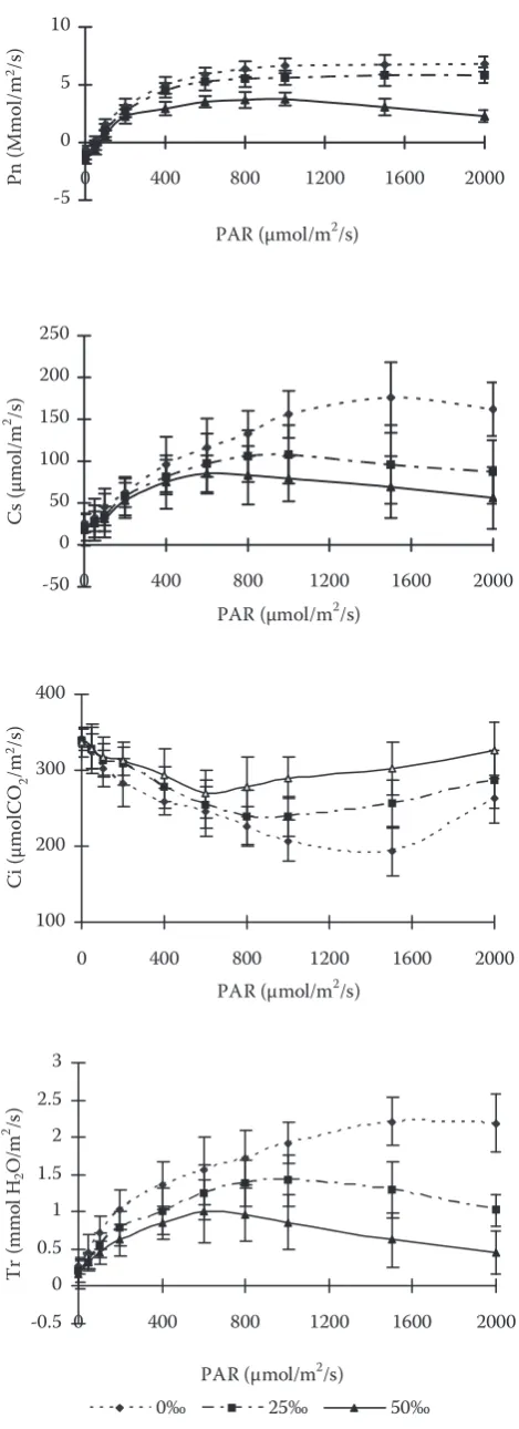
Effects of salinity on CO2/H2O2 exchange of Kandelia candel leaves
Fig. 2 indicates that photosynthesis was sig-nificantly inhibited by salinity (p < 0.05). The light compensatory point of stressed leaves increased while the light saturation point and Pn were strongly reduced by salinity. The decrease scope of Pn in the treatment of 50‰ was larger than that in 25‰. Transpiration (Tr) decreased with the increase of salinity. So did stomatal conductance (Cs). However, intercellular CO2 (Ci) increased. The relative reduc-tion in Cs exceeded that of Pn (Fig. 2). The extent to which the stomatal closure affects photosyn-thetic capacity is still unknown by the increase of Ci (Fig. 2). With increase of salinity from 0‰ to 25‰ and to 50‰, dark respiration (Rd) increased.
DISCUSSION
Chlorophyll is the main pigment of photosynthe-sis in plants. To some extent, the Chl content can reflect the photosynthesis rate of plant. It is strongly influenced by environmental factors. It was interest-ing to find the fact that both Chl and Car content in Kandelia candel seedlings increased significantly with the increase of salinity. This means that the salt stress can promote the synthesis of photosynthetic pigments. The study on the leaves of Bruguiera sex-angula seedlings also proved the fact that chloro-phyll content was accumulated under salt stress
-5 0 5 10
0 400 800 1200 1600 2000
PAR (μmol/m2/s)
Pn (μ m ol /m 2 /s ) -50 0 50 100 150 200 250
0 400 800 1200 1600 2000
PAR (μmol/m2/s)
C
s (
μm
ol
/m
2 /s)
100 200 300 400
0 400 800 1200 1600 2000
PAR (μmol/m2/s)
Ci (μ m ol C O2 / m 2 /s ) -0.5 0 0.5 1 1.5 2 2.5 3
0 400 800 1200 1600 2000
PAR (μmol/m2/s)
Tr (m m ol H2 O /m 2 /s )
[image:3.595.304.539.67.713.2]0‰ 25‰ 50‰
Fig. 2. Effects of salinity on CO2/H2O exchange of Kandelia
candel (L.) Druce seedlings
(Zheng, Lin 1992). The same result was obtained on the Sonneratia apetala Buch-Ham seedlings by Chen et al. (2000).
Because mangrove plants grew at high salt sites, the effects of salinity on photosynthesis provoke extensive interests. Stomata closed under stress con-ditions, and consequently Pn decreased (Taylor, Rowley 1971; Boyer 1976; Farquhar, Sharkey 1982; Martin et al. 1981). The factors causing a decrease in Pn can be grouped into stomatal and non-stomatal ones (Boyer 1976). The closure of stomata results in the shortage of CO2 (Boyer 1976). Non-stomatal factors include:
1. an increase in diffusive resistance to CO2 in the mesophyll;
2. a decrease in the activity of photosystem II, pho-tophosphorylation and ribulose 1,5-bisphosphate carboxylase;
3. a decrease in chlorophyll content and inhibition of electron transport (Boyer 1976; Hay, Walker 1989; Keck, Boyer 1974; Kumer, Gupta 1986; Youngis, Boyer 1979; Vu et al. 1987).
Our results showed the Pn decrease was not re-lated to decline in photosynthetic pigments (Fig. 1). Stomatal number and density under the epidermis in leaves of plants were reduced with the increase of salinity (Fig. 3 A, B, C). It represented the difference in photosynthesis and transpiration between various salinities. The decrease in Pn may result from the restriction of CO2 entrance through stomata. In this aspect, our results agree with the report of a decrease in stomatal conductance in leaves of K. candel seed-lings with the increase of salinity (Yang, Lin 1995) and earlier report of Ball and Farquhar (1984b), who observed that the decline in photosynthetic capacity in the grey mangrove A. marina under
A B C
D E F
G H I
[image:4.595.67.510.54.439.2]Plate Effects of salinities on leaf anatomical characteristic of Kandelia candel Druce seedlings.
Fig. 3. Effects of salinities on leaf anatomical characteristics of Kandelia candel Druce seedlings
A, B, C – the stomatal number and density under the epidermis in leaves of plants treated with salinity of 0‰, 25‰ and to 50‰, respectively; D, E, F – the cell arrangement of the upper epidermis in leaves of plants grown in 0‰, 25‰ and to 50‰ salinity, respectively; G, H, I – the reticulate structure of spongy tissue layer and irregular arrangement of mesophyll cells in plants grown in 0‰, 25‰ and to 50‰ salinity, respectively
A B C
G H I
D E F
A B C
D E F
G H I
Plate Effects of salinities on leaf anatomical characteristic of
Kandelia candel
Druce seedlings.
A B C
D E F
G H I
salinity was accompanied by a decrease in stomatal conductance.
The low photosynthesis observed in salt-stressed glycophytes such as olive was attributed to a reduc-tion of mesophyll conductance (Bongi, Loreto 1989). Reduced mesophyll conductance was also considered as a possible cause for reduction of photosynthesis in cotton leaves grown under salini- ty stress (Brugnoli, Bjorkman 1992). Salt and water stress apparently reduce photosynthesis by a similar mechanism. In fact, it is known that pho-tosynthesis is often limited by low CO2 diffusion conductance under water stress conditions (Kai-ser 1987; Cornic et al. 1992); low CO2 diffusion under salt stress conditions is generally attributed to reduced stomatal conductance and mesophyll conductance (Fig. 3 G, H, I and Fig. 2), which causes the lowering of CO2 concentrations in chlo-roplasts and ultimately the CO2 assimilation rate also decreases (Parida et al. 2003). SEM of leaves showed that NaCl treatment caused a decrease in stomatal number, which might be responsible for decreased stomatal conductance under salt treat-ment. Our results also showed that the dense ar-rangement of spongy cells might therefore explain, at least in part, the reduced conductance to CO2 diffusion. These results were also proved by Lau-teri et al. (1997), Khan et al. (2000), Sobrado and Ball (1999). If stomatal factors are the main ones, Pn and Cs decreased because of the decline in Ci. However, our data showed that Pn, Tr and Cs were declined at the same time while Ci increased with the stress of salinities (Fig. 2). It implied that the reduction of Pn was caused by a combination of stomatal and non-stomatal factors. Despite the salt-induced reduction of Pn and the increase of Ci in our experiments, the extent to which stomatal and non-stomatal factors reduce Pn is not clear. Further study has to be done to specify the ratio of stomatal to non-stomatal factors.
Since plant growth amounts to the balance sheet of photosynthesis gains after the deduction of respiration losses, it is to be expected that what-ever effects salinity has on these processes will be reflected in the growth rate of the plant integrated over time. Any stress exerted on an organism increases maintenance costs, as reflected in its respiration. The available data on the response of mangroves to salinity are no exception. Our results support the idea that high salinity reduces photo-synthesis, transpiration and increases respiration. However, in our experiment, leaves from control plants grown without NaCl addition did not show any symptoms of necrosis, chlorosis, or leaf tissue
damage indicating leaf dysfunction. This is contrary to results for other mangrove species that show anomalous leaf development or poor growth in fresh water (Ball, Pidsley 1995). We concluded that salt treatments promoted the synthesis of pig-ments and induced a reduction of mesophyll and stomatal conductance in leaves of Kandelia candel and this reduction might contribute in part to the inhibition of photosynthesis observed after 20 days of salt stress. The reduction of mesophyll conduct-ance was associated with reduced intercellular spaces in the mesophyll of salt-treated leaves, which may have made the path toward the sites of CO2 fixation more tortuous. However, Pn, Tr and Cs were declined at the same time while Ci increased with the stress of salinities, indicating the reduction of Pn was caused by a combination of stomatal and non-stomatal factors.
The population photosynthetic rate in the man-grove forest is quite different from the single plant, and difficult to carry out. Analysis of the capacity of a species to inhabit a salinity environment is usu-ally based on the performance of the species under laboratory conditions. The findings of the present study showed the photosynthetic properties of the mangrove species Kandelia candel, and implied its wide adaptation to different salinities. The high salinity environment had significant effects on the light compensatory point, which in several other species increased with an increase in the salinity level of growth conditions, and the light satura-tion point decreasing with an increase in salinity. The difference between the natural and laboratory environments mainly lies in the light conditions. It is clear that the understory leaves in the man-grove forest experienced quantum flux densities near their light compensation points much of the day, interrupted periodically by high intensity sun flecks. These leaves might be able to sustain very low rates of assimilation during shaded periods, but it is doubtful that such rates would be sufficient for prolonged survival (Ball, Critchley 1982) while the exposed leaves in the mangrove forest experienced quantum flux densities above their light saturation points, and might be able to appear light inhibition.
Acknowledgments
References
ARNON D.I., 1949. Copper enzyme in isolated chloroplasts, polyphenol oxidase in Beta Vulgaris. Plant Physiology,
130: 267–272.
BALL M.C., CRITCHLEY C., 1982. Photosynthetic responses to irradiance by the grey mangrove, Avicennia marina, grown under different light regimes. Plant Physiology, 70: 1101–1106.
BALL M.C., FARQUHAR C.D., 1984a. Photosynthetic and stomatal responses of two mangrove species, Aegiceras cor-niculatum and Avicennia marina, to long term salinity and humidity conditions. Plant Physiology, 74: 1–6.
BALL M.C., FARQUHAR C.D., 1984b. Photosynthetic and stomatal responses of the grey mangrove Avicennia ma-rina, to transient salinity and humidity conditions. Plant Physiology, 74: 7–11.
BALL M.C., PIDSLEY S.M., 1995. Growth response to sa-linity in relation to distribution of two mangrove species
Sonneratia alba and S. lanceolate in northern Australia. Functional Ecology, 19: 77–85.
BONGI G., LORETO F., 1989. Gas-exchange properties of salt-stressed Olive (Olea europea L.). Plant Physiology, 90: 1408–1416.
BOYER J.S., 1976. Water deficits and photosynthesis. In: KOZLOWSKI T.T. (ed.), Water Deficits and Plant Growth. Volume 4. New York, Academic Press: 153–159.
BRUGNOLI E., BJORKMAN O., 1992. Growth of cotton under continuous salinity stress: influence on allocation pattern, stomatal and non-stomatal components of photosynthesis and dissipation of excess light energy. Planta, 187: 335–347. CHEN C.P., WANG W.Q., LIN P., 2000. Influences of salinity
on the growth and some ecophysiological characteristics of mangrove species Sonneratia apetala seedlings. Chinese Bulletin of Botany, 17: 457–461. (in Chinese)
CORNIC G., GHASGHAIE J., GENTY B., BRIANTAIS J.M., 1992. Leaf photosynthesis is resistant to a mild drought stress. Photosynthetica, 27: 295–309.
DUBINSKY Z., STAMBLER N., 1996. Eutrophication, ma-rine pollution and coral reef. Global Change Biology, 2: 511–526.
DUXBURY A.C., YENTSCH C.S., 1956. Plankton pigment monograph. Journal of Air Pollution Association, 16: 145–150.
FARQUHAR G.D., SHARKEY T.D., 1982. Stomatal conduct-ance and photosynthesis.Annual Review of Plant Physiol-ogy,33: 317–345.
HAY R.K.M., WALKER A.J., 1989. Endogenous and environ-mental factors affecting photosynthesis: water stress. In: HAY R.K.M., WALKER A.J. (ed.), An Introduction to the Physiology of Crop Yield. New York, Longaman Scientific & Technical: 48–80.
KAISER W.M., 1987. Effects of water deficit on photosynthetic capacity. Plant Physiology, 71: 142–149.
KECK R.W., BOYER J.S., 1974. Chloroplast response to low leaf water potential III. Differing inhibition of electronic transport and photophosphorylation. Plant Physiology,
53: 474–479.
KHAN M.A., UNGAR I.A., SHOWALTER A.M., 2000. The effect of salinity on the growth, water status, and ion content of a leaf succulent perennial halophyte Suadea fruticosa (L.) Forssk. Journal of Arid Environments, 45: 73–84.
KUMER P.J., GUPTA P.K., 1986. Influence of different leaf water potential on photosynthetical carbon metabolism in sorghum.Photosynthetica, 20: 391–396.
LAUTERI M., SCARTAZZA A., GUIDO M.C., BRUGNOLI E., 1997. Genetic variation in photosynthetic capacity, carbon isotope discrimination and mesophyll conductance in provenances of Castanea sativa adapted to different environments. Functional Ecology, 11: 657–683.
LIN P., 1999. Mangrove Ecosystem in China. Beijing, New York, Science Press: 8–25.
MARTIN B., ORT D.R., BOYER J.S., 1981. Impairment of photosynthesis by chilling temperatures in tomato. Plant Physiology, 68: 329–334.
PARIDA A.K., DAS A.B., MITTRA B., 2003. Effects of NaCl stress on the structure, pigment complex composition and photosynthetic activity of mangrove Bruguiera parviflora
chloroplasts. Photosynthetica, 41: 191–200.
SOBRADO M.A., BALL M.C., 1999. Light use in relation to carbon gain in the mangrove, Avicennia marina, under hypersaline conditions. Australian Journal of Plant Physiol-ogy, 26: 245–251.
TAYLOR A.O., ROWLEY J.A., 1971. Plants under climatic stress. I. Chilling, high light effects on photosynthesis. Plant Physiology, 47: 713–718.
TERASHIMA I., HIKOSAKA K., 1995. Comparative eco-physiology of leaf and canopy photosynthesis. Plant, Cell and Environment, 18: 1111–1128.
VU C.V., ALLEN J.L., BOWERS G., 1987. Drought stress and elevated CO2 effects on soybean ribulose biphosphate carboxylase activity and canopy photosynthetic rate. Plant Physiology, 83: 573–578.
YANG S.C., LIN P., 1995. A mathematical model of low temperature and exposure time interactions on Kandelia candel leaf cold-sensitivity. Chinese Bulletin of Botany, 7: 164–168. (in Chinese)
YOUNGIS H.M., BOYER J.S., 1979. Conformation and activ-ity of chloroplast coupling factor exposed to low chemical potential water in cells. Biochemica et Biophysica Acta,
548: 328–340.
ZHENG W.J., LIN P., 1992. Effects of salinity on the growth and some eco-physiological characteristic of mangrove
Bruguira sexangula seedlings. Chinese Journal of Applied Ecology, 3: 9–14.
Vliv salinity na vlastnosti listů a CO
2/H
2O
výměnu u
Kandelia candel
(L.)
Druce
ABSTRAKT: V tomto kultivačním experimentu byl hodnocen vliv salinity na vlastnosti listů a CO2/H2Ovýměnu u mangrovového druhu Kandelia candel. Při hodnotě salinity 50 ‰ byla pozorována silná redukce intenzity růstu, listy byly zbarvené černozeleně, byly menší velikosti a méně rozvinuté nebo deformované v porovnání s kontrolní skupinou rostlin, případně s rostlinami pěstovanými při hodnotě salinity 25 ‰. V porovnání s kontrolní skupinou byly listy rostlin pěstovaných při hodnotě salinity 25 ‰ lesklé a hladké. S nárůstem salinity došlo k redukci počtu průduchů v epidermis listů. Buňky mezofilu u rostlin pěstovaných při salinitě 5 0‰ byly nejmenší, větší buňky byly pozorovány při salinitě 25 ‰ a největší v případě kontrolní skupiny. Buňky při salinitě 50 ‰ byly nahloučené, zatímco u kontrolní skupiny byla prostorová vazba volnější. Buňky horní části listové epidermis u kontrolní skupiny měly opět volnější uspořádání s tenkou buněčnou stěnou, zatímco při salinitě 50 ‰ byly buňky nahloučené a buněčná stěna byla tenčí než v případě salinity 25 ‰. Obsah Chl (a + b) (μmol/cm2) u rostlin pěstovaných při salinitě 50 ‰ se významně zvýšil v porovnání s kontrolní skupinou, Chl a/b byl také redukován. Také koncentrace karotenoidových barviv (Car) se významně navýšila při jednotlivých hodnotách salinity. Fotosyntéza (Pn) byla signifikantně inhibována při vyšší salinitě a světelný kompenzační bod byl vyšší. Čistá rychlost fotosyntézy (Pn), stomatální vodivost (Cs) a rychlost transpirace (Tr) byly při nárůstu salinity redukovány, zatímco hodnota temnostní respirace (Rd) se navýšila.
Klíčová slova: salinita; mangrovové lesy; buněčná struktura; fotosyntéza; transpirace
Corresponding author:
Dr. Dong-liang Qiu, Fujian Agriculture and Forestry University, College of Horticulture, Fuzhou 350002, P.R. China